
Seroprevalence and Epidemiology of Toxoplasma gondii in Animals in the Qinghai-Tibetan Plateau Area, China

Abstract

1. Introduction
2. Results
3. Discussion
4. Materials and Methods
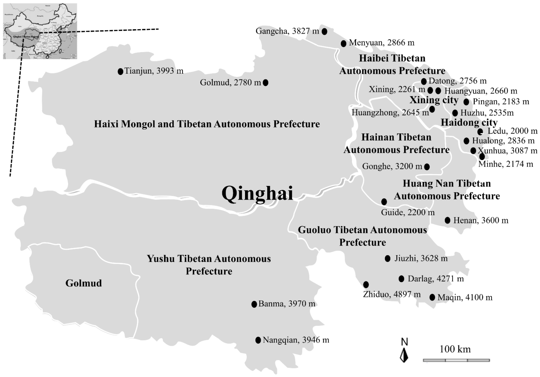
4.1. Sample Collection of Tibetan Sheep (Ovis aries), Yaks (Bos grunniens), Cows, Chickens, Pigs and Horses
4.2. Serum Harvest
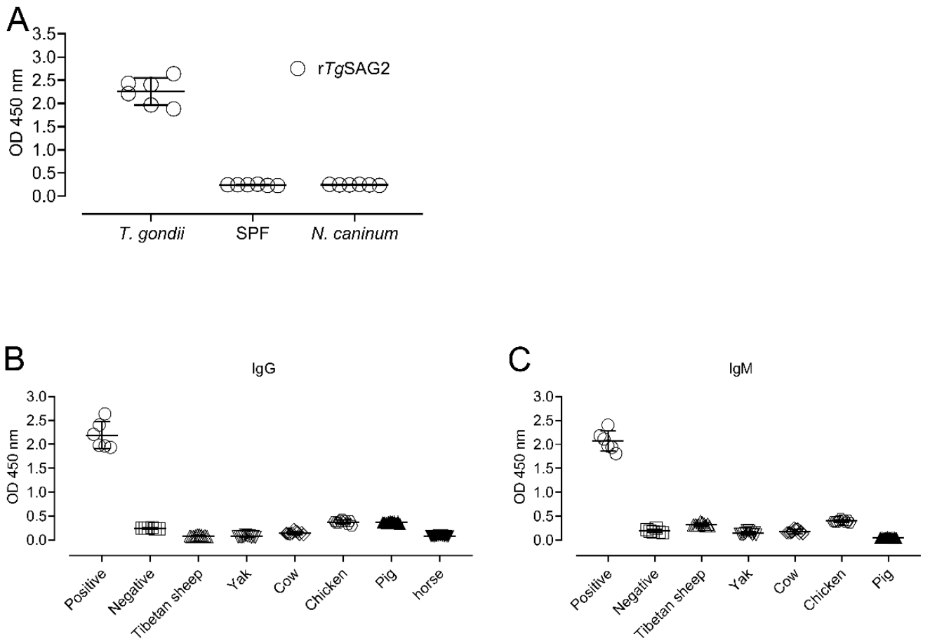
4.3. Recombinant Protein Expression
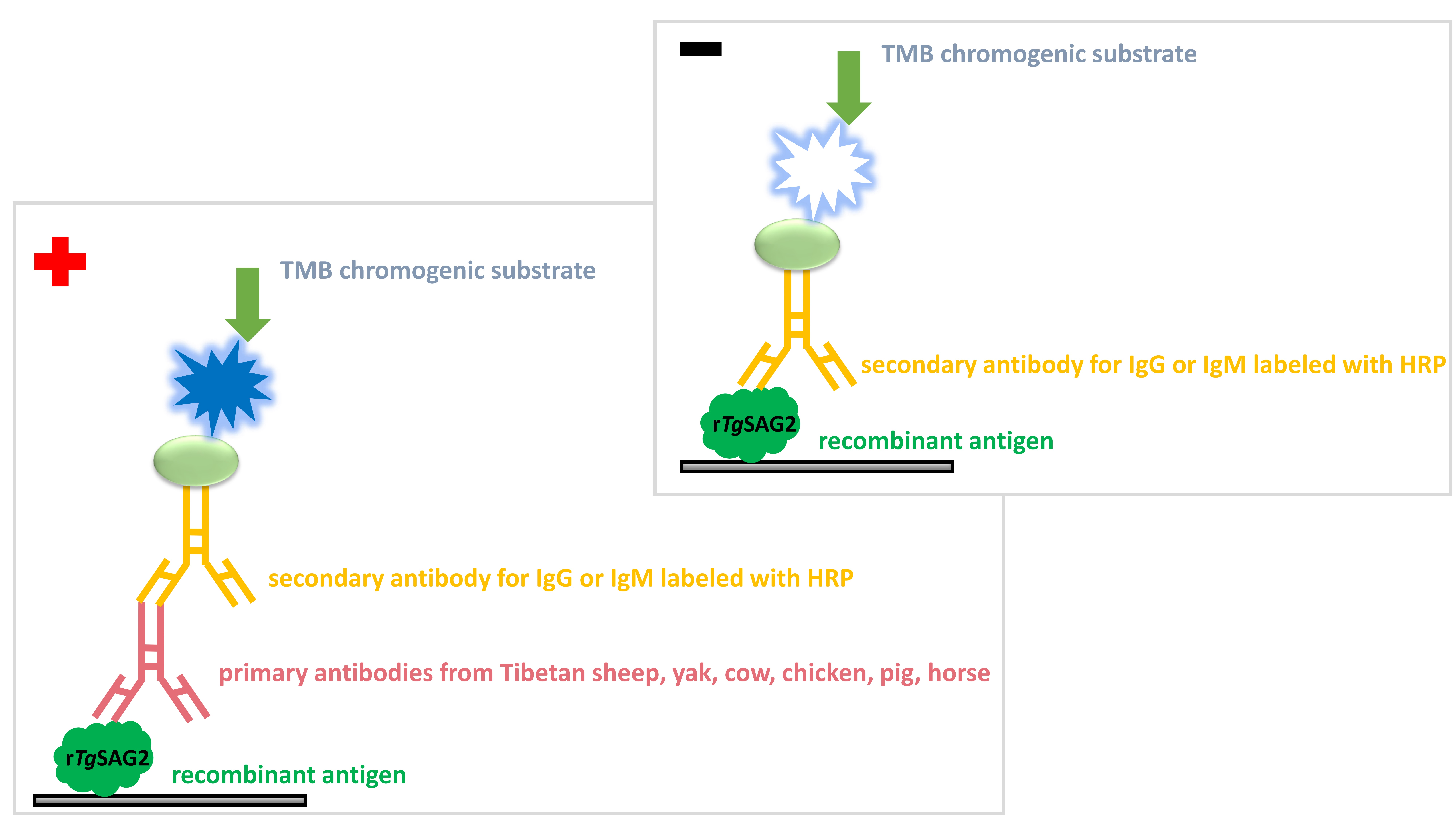
4.4. Indirect ELISA
4.5. Statistical Analysis
5. Conclusions
Author Contributions
Funding
Institutional Review Board Statement
Informed Consent Statement
Data Availability Statement
Acknowledgments
Conflicts of Interest
References
- Tang, L.; Duan, X.; Kong, F.; Zhang, F.; Zheng, Y.; Li, Z.; Mei, Y.; Zhao, Y.; Hu, S. Influences of climate change on area variation of Qinghai Lake on Qinghai-Tibetan Plateau since 1980s. Sci. Rep. 2018, 8, 7331. [Google Scholar] [CrossRef]
- Han, R.; Yang, J.; Niu, Q.; Liu, Z.; Chen, Z.; Kan, W.; Hu, G.; Liu, G.; Luo, J.; Yin, H. Molecular prevalence of spotted fever group rickettsiae in ticks from Qinghai Province, northwestern China. Infect. Genet. Evol. 2018, 57, 1–7. [Google Scholar] [CrossRef] [PubMed]
- Rubel, F.; Brugger, K.; Walter, M.; Vogelgesang, J.R.; Didyk, Y.M.; Fu, S.; Kahl, O. Geographical distribution, climate adaptation and vector competence of the Eurasian hard tick Haemaphysalis concinna. Ticks Tick Borne Dis. 2018, 9, 1080–1089. [Google Scholar] [CrossRef] [PubMed]
- Zhang, Q.; Zhang, Z.; Ai, S.; Wang, X.; Zhang, R.; Duan, Z. Cryptosporidium spp., Enterocytozoon bieneusi, and Giardia duodenalis from animal sources in the Qinghai-Tibetan Plateau Area (QTPA) in China. Comp. Immunol. Microbiol. Infect. Dis. 2019, 67, 101346. [Google Scholar] [CrossRef]
- Jin, Y.; Fei, J.; Cai, J.; Wang, X.; Li, N.; Guo, Y.; Feng, Y.; Xiao, L. Multilocus genotyping of Giardia duodenalis in Tibetan sheep and yaks in Qinghai, China. Vet. Parasitol. 2017, 247, 70–76. [Google Scholar] [CrossRef] [PubMed]
- Gong, X.; Liu, L.; Zheng, F.; Chen, Q.; Li, Z.; Cao, X.; Yin, H.; Zhou, J.; Cai, X. Molecular investigation of bovine viral diarrhea virus infection in yaks (Bos gruniens) from Qinghai, China. Virol. J. 2014, 11, 29. [Google Scholar] [CrossRef]
- Li, J.; Jian, Y.; Jia, L.; Galon, E.M.; Benedicto, B.; Wang, G.; Cai, Q.; Liu, M.; Li, Y.; Ji, S.; et al. Molecular characterization of tick-borne bacteria and protozoans in yaks (Bos grunniens), Tibetan sheep (Ovis aries) and Bactrian camels (Camelus bactrianus) in the Qinghai-Tibetan Plateau Area, China. Ticks Tick Borne Dis. 2020, 11, 101466. [Google Scholar] [CrossRef] [PubMed]
- Dubey, J.P. Advances in the life cycle of Toxoplasma gondii. Int. J. Parasitol. 1998, 28, 1019–1024. [Google Scholar] [CrossRef]
- Dubey, J.P. Toxoplasmosis in pigs—The last 20 years. Vet. Parasitol. 2009, 164, 89–103. [Google Scholar] [CrossRef]
- Dubey, J.P. History of the discovery of the life cycle of Toxoplasma gondii. Int. J. Parasitol. 2009, 39, 877–882. [Google Scholar] [CrossRef]
- Elmore, S.A.; Jones, J.L.; Conrad, P.A.; Patton, S.; Lindsay, D.S.; Dubey, J.P. Toxoplasma gondii: Epidemiology, feline clinical aspects, and prevention. Trends. Parasitol. 2010, 26, 190–196. [Google Scholar] [CrossRef]
- Montoya, J.G.; Liesenfeld, O. Toxoplasmosis. Lancet 2004, 363, 1965–1976. [Google Scholar] [CrossRef]
- Weiss, L.M.; Dubey, J.P. Toxoplasmosis: A history of clinical observations. Int. J. Parasitol. 2009, 39, 895–901. [Google Scholar] [CrossRef]
- Saadatnia, G.; Golkar, M. A review on human toxoplasmosis. Scand. J. Infect. Dis. 2012, 44, 805–814. [Google Scholar] [CrossRef] [PubMed]
- Tirosh-Levy, S.; Steinman, A.; Minderigiu, A.; Arieli, O.; Savitski, I.; Fleiderovitz, L.; Edery, N.; Schvartz, G.; Mazuz, M.L. High exposure to Toxoplasma gondii and Neospora Spp. in donkeys in Israel: Serological survey and case reports. Animals 2020, 10, 1921. [Google Scholar] [CrossRef] [PubMed]
- Li, J.; Guo, H.; Galon, E.M.; Gao, Y.; Lee, S.H.; Liu, M.; Li, Y.; Ji, S.; Jia, H.; Xuan, X. Hydroxylamine and carboxymethoxylamine can inhibit Toxoplasma gondii growth through an aspartate aminotransferase-independent pathway. Antimicrob. Agents Chemother. 2020, 64, e01889-19. [Google Scholar] [CrossRef] [PubMed]
- Dubey, J.P. Toxoplasmosis of Animals and Humans, 2nd ed.; CRC Press: Boca Raton, FL, USA, 2010; pp. 1–313. [Google Scholar]
- Sudan, V.; Tewari, A.K.; Singh, H. Detection of antibodies against Toxoplasma gondii in Indian cattle by recombinant SAG2 enzyme-linked immunosorbent assay. Acta Parasitol. 2019, 64, 148–151. [Google Scholar] [CrossRef] [PubMed]
- Gao, X.J.; Zhao, Z.J.; He, Z.H.; Wang, T.; Yang, T.B.; Chen, X.G.; Shen, J.L.; Wang, Y.; Lv, F.L.; Hide, G.; et al. Toxoplasma gondii infection in pregnant women in China. Parasitology 2012, 139, 139–147. [Google Scholar] [CrossRef]
- Pan, M.; Lyu, C.; Zhao, J.; Shen, B. Sixty years (1957–2017) of research on toxoplasmosis in China-an overview. Front. Microbiol. 2017, 8, 1825. [Google Scholar] [CrossRef]
- Dong, H.; Su, R.; Lu, Y.; Wang, M.; Liu, J.; Jian, F.; Yang, Y. Prevalence, risk factors, and genotypes of Toxoplasma gondii in food animals and humans (2000–2017) from China. Front. Microbiol. 2018, 9, 2108. [Google Scholar] [CrossRef]
- Li, K.; Shahzad, M.; Zhang, H.; Jiang, X.; Mehmood, K.; Zhao, X.; Li, J. Socio-economic burden of parasitic infections in yaks from 1984 to 2017 on Qinghai Tibetan Plateau of China-A review. Acta Trop. 2018, 183, 103–109. [Google Scholar] [CrossRef] [PubMed]
- Liu, Q.; Ma, R.; Zhao, Q.; Shang, L.; Cai, J.; Wang, X.; Li, J.; Hu, G.; Jin, H.; Gao, H. Seroprevalence of Toxoplasma gondii infection in Tibetan sheep in northwestern China. J. Parasitol. 2010, 96, 1222–1223. [Google Scholar] [CrossRef]
- Liu, Z.K.; Li, J.Y.; Pan, H. Seroprevalence and risk factors of Toxoplasma gondii and Neospora caninum infections in small ruminants in China. Prev. Vet. Med. 2015, 118, 488–492. [Google Scholar] [CrossRef]
- Lv, Q.Y.; Quan, M.X.; Tang, H.L.; Wu, X.T.; Liu, G.H.; Li, F.; Hu, S.F. Seroprevalence, risk factors, and genotypes of Toxoplasma gondii in free-range chickens intended for human consumption in China. Foodborne Pathog. Dis. 2020, 10, 1089. [Google Scholar]
- Wu, S.M.; Danba, C.; Huang, S.Y.; Zhang, D.L.; Chen, J.; Gong, G.; Xu, M.J.; Yuan, Z.G.; Zhu, X.Q. Seroprevalence of Toxoplasma gondii infection in Tibetan sheep in Tibet, China. J. Parasitol. 2011, 97, 1188–1189. [Google Scholar] [CrossRef]
- Zhou, M.; Cao, S.; Sevinc, F.; Sevinc, M.; Ceylan, O.; Liu, M.; Wang, G.; Moumouni, P.F.; Jirapattharasate, C.; Suzuki, H.; et al. Enzyme-linked immunosorbent assays using recombinant TgSAG2 and NcSAG1 to detect Toxoplasma gondii and Neospora caninum-specific antibodies in domestic animals in Turkey. J. Vet. Med. Sci. 2017, 78, 1877–1881. [Google Scholar] [CrossRef]
- Khanaliha, K.; Motazedian, M.H.; Kazemi, B.; Shahriari, B.; Bandehpour, M.; Sharifniya, Z. Evaluation of recombinant SAG1, SAG2, and SAG3 antigens for serodiagnosis of toxoplasmosis. Korean J. Parasitol. 2014, 52, 137–142. [Google Scholar] [CrossRef] [PubMed]
- Yin, M.Y.; Wang, J.L.; Huang, S.Y.; Qin, S.Y.; Zhou, D.H.; Liu, G.X.; Tan, Q.D.; Zhu, X.Q. Seroprevalence and risk factors of Toxoplasma gondii in Tibetan Sheep in Gansu province, Northwestern China. BMC Vet. Res. 2015, 11, 41. [Google Scholar] [CrossRef] [PubMed]
- Liu, Q.; Cai, J.; Zhao, Q.; Shang, L.; Ma, R.; Wang, X.; Li, J.; Hu, G.; Jin, H.; Gao, H. Seroprevalence of Toxoplasma gondii infection in yaks (Bos grunniens) in northwestern China. Trop. Anim. Health Prod. 2011, 43, 741–743. [Google Scholar] [CrossRef]
- Liu, J.; Cai, J.Z.; Zhang, W.; Liu, Q.; Chen, D.; Han, J.P.; Liu, Q.R. Seroepidemiology of Neospora caninum and Toxoplasma gondii infection in yaks (Bos grunniens) in Qinghai, China. Vet. Parasitol. 2008, 152, 330–332. [Google Scholar] [CrossRef]
- Wang, M.; Wang, Y.H.; Ye, Q.; Meng, P.; Yin, H.; Zhang, D.L. Serological survey of Toxoplasma gondii in Tibetan mastiffs (Canis lupus familiaris) and yaks (Bos grunniens) in Qinghai, China. Parasites Vectors 2012, 5, 35. [Google Scholar] [CrossRef]
- Li, K.; Gao, J.F.; Shahzad, M.; Han, Z.Q.; Nabi, F.; Liu, M.Y.; Zhang, D.; Li, J.K. Seroprevalence of Toxoplasma gondii infection in yaks (Bos grunniens) on the Qinghai-Tibetan Plateau of China, 2014. Vet. Parasitol. 2014, 205, 354–356. [Google Scholar] [CrossRef] [PubMed]
- Wu, F.; Wang, Y.L.; Yang, Z.; Li, X.L.; Li, Z.R.; Lin, Q. Seroprevalence and Risk Factors of Toxoplasma gondii in Slaughter Pigs in Shaanxi Province, Northwestern China. Vector Borne Zoonotic Dis. 2017, 17, 517–519. [Google Scholar] [CrossRef] [PubMed]
- Li, X.; Ni, H.B.; Ren, W.X.; Jiang, J.; Gong, Q.L.; Zhang, X.X. Seroprevalence of Toxoplasma gondii in horses: A global systematic review and meta-analysis. Acta Trop. 2020, 201, 105222. [Google Scholar] [CrossRef] [PubMed]
- Xu, P.; Cai, Y.N.; Leng, X.; Wang, J.; Ma, W.; Mu, G.D.; Jiang, J.; Liu, X.Y.; Wang, Z.D.; Zhao, Q.; et al. Seroprevalence of Toxoplasma gondii infection in pigs in Jilin Province, Northeastern China. Trop. Biomed. 2015, 32, 116–120. [Google Scholar] [PubMed]
- Wu, D.; Lv, R.; Sun, X.; Shu, F.; Zhou, Z.; Nie, K.; Duan, G.; Zou, F. Seroprevalence of Toxoplasma gondii antibodies from slaughter pigs in Chongqing, China. Trop. Anim. Health Prod. 2012, 44, 685–687. [Google Scholar] [CrossRef] [PubMed]
- Wu, S.M.; Ciren, D.; Huang, S.Y.; Xu, M.J.; Ga, G.; Yan, C.; Mahmoud, M.S.; Zou, F.C.; Zhu, X.Q. First report of Toxoplasma gondii prevalence in Tibetan pigs in Tibet, China. Vector Borne Zoonotic Dis. 2012, 12, 654–656. [Google Scholar] [CrossRef]
- Li, X.; Wang, Y.; Yu, F.; Li, T.; Zhang, D. An outbreak of lethal toxoplasmosis in pigs in the Gansu province of China. J. Vet. Diagn. Investig. 2010, 22, 442–444. [Google Scholar] [CrossRef]
- Feng, Y.; Lu, Y.; Wang, Y.; Liu, J.; Zhang, L.X.; Yang, Y.R. Toxoplasma gondii and Neospora caninum in free-range chickens in Henan Province of China. BioMed Res. Int. 2016, 2016, 8290536. [Google Scholar] [CrossRef]


| Prefectures | Sampling Sites | No. of Serum Samples * | ||||||
|---|---|---|---|---|---|---|---|---|
| Tibetan Sheep | Yak | Cow | Chicken | Pig | Horse | Total | ||
| Haibei | Gangca | 75 | 0 | 0 | 0 | 0 | 0 | 75 |
| Menyuan | 60 | 20 | 0 | 0 | 0 | 0 | 80 | |
| Hainan | Gonghe | 60 | 20 | 0 | 0 | 0 | 0 | 80 |
| Guide | 32 | 0 | 0 | 0 | 0 | 0 | 32 | |
| Haixi | Golmud | 60 | 20 | 0 | 0 | 20 | 0 | 100 |
| Tianjun | 30 | 0 | 0 | 0 | 0 | 0 | 30 | |
| Yushu | Zhiduo | 60 | 20 | 0 | 0 | 0 | 0 | 80 |
| Nangqian | 52 | 0 | 0 | 0 | 0 | 0 | 52 | |
| Guoluo | Jiuzhi | 45 | 66 | 0 | 0 | 0 | 0 | 111 |
| Maqin | 152 | 110 | 0 | 0 | 0 | 0 | 262 | |
| Darlag | 141 | 103 | 0 | 0 | 0 | 0 | 244 | |
| Banma | 0 | 132 | 0 | 0 | 0 | 0 | 132 | |
| Xining | Xining | 0 | 0 | 0 | 50 | 50 | 0 | 100 |
| Datong | 40 | 119 | 0 | 50 | 0 | 0 | 209 | |
| Huangyuan | 0 | 0 | 0 | 50 | 47 | 0 | 97 | |
| Huangzhong | 0 | 0 | 0 | 50 | 48 | 0 | 98 | |
| Haidong | Ledu | 60 | 20 | 0 | 50 | 72 | 0 | 202 |
| Pingan | 0 | 0 | 0 | 50 | 50 | 0 | 100 | |
| Huzhu | 0 | 0 | 0 | 50 | 50 | 0 | 100 | |
| Minhe | 0 | 0 | 205 | 50 | 0 | 0 | 255 | |
| Hualong | 0 | 0 | 0 | 50 | 0 | 0 | 50 | |
| Xunhua | 0 | 0 | 0 | 50 | 0 | 0 | 50 | |
| Huangnan | Henan | 40 | 33 | 0 | 0 | 0 | 61 | 134 |
| Total | 907 | 663 | 205 | 500 | 337 | 61 | 2673 | |
| Animals | Prefectures | No. of Tested * | Total IgG-Seropositive | Total IgM-Seropositive | ||
|---|---|---|---|---|---|---|
| Frequency | Prevalence (95% CI #) | Frequency | Prevalence (95% CI #) | |||
| Tibetan sheep | Haibei | 135 | 49 | 36.3 (28.2–44.4) | 11 | 8.1 (3.5–12.8) |
| Hainan | 92 | 56 | 60.9 (50.9–70.8) | 31 | 33.7 (24.0–43.4) | |
| Haixi | 90 | 28 | 31.1 (21.5–40.7) | 10 | 11.1 (4.6–17.6) | |
| Yushu | 112 | 30 | 26.8 (18.6–35.0) | 16 | 14.3 (7.8–20.8) | |
| Guoluo | 338 | 220 | 65.1 (60.0–70.2) | 92 | 27.2 (22.5–32.0) | |
| Xining | 40 | 14 | 35.0 (20.2–49.8) | 10 | 25.0 (11.6–38.4) | |
| Haidong | 60 | 51 | 85.0 (76.0–94.0) | 15 | 25.0 (14.0–36.0) | |
| Huangnan | 40 | 12 | 30.0 (15.8–44.2) | 6 | 15.0 (3.9–26.1) | |
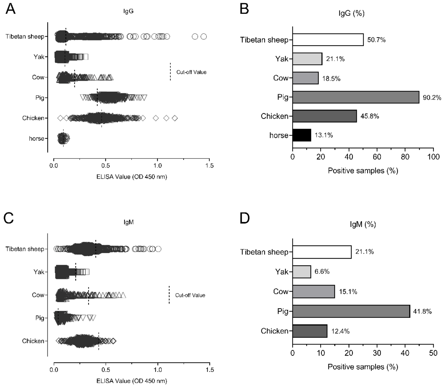
| Total | 907 | 460 | 50.7 (47.5–54.0) | 191 | 21.1 (18.4–23.7) | |
| Yak | Haibei | 20 | 4 | 20.0 (2.5–37.5) | 4 | 20.0 (2.5–37.5) |
| Hainan | 20 | 2 | 10.0 (3.1–23.1) | 0 | 0 | |
| Haixi | 20 | 3 | 15.0 (0.6–30.6) | 0 | 0 | |
| Yushu | 20 | 4 | 20.0 (2.5–37.5) | 1 | 5.0 (4.6–14.6) | |
| Guoluo | 411 | 105 | 25.5 (21.3–29.8) | 36 | 8.8 (6.0–11.5) | |
| Xining | 119 | 14 | 11.8 (6.0–17.6) | 3 | 2.5 (0.3–5.3) | |
| Haidong | 20 | 1 | 5.0 (4.6–14.6) | 0 | 0 | |
| Huangnan | 33 | 7 | 21.2 (7.3–35.2) | 0 | 0 | |
| Total | 663 | 140 | 21.1 (18.0–24.2) | 44 | 6.6 (4.7–8.5) | |
| Cow | Haidong | 205 | 38 | 18.5 (13.2–23.9) | 31 | 15.1 (10.2–20.0) |
| Chicken | Xining | 200 | 67 | 33.5 (27.0–40.0) | 12 | 6.0 (2.7–9.3) |
| Haidong | 300 | 162 | 54.0 (48.4–59.6) | 50 | 16.7 (12.4–20.9) | |
| Total | 500 | 229 | 45.8 (41.4–50.2) | 62 | 12.4 (9.5–15.3) | |
| Pig | Xining | 145 | 128 | 88.3 (83.0–93.5) | 59 | 40.7 (32.7–48.7) |
| Haidong | 172 | 156 | 90.7 (86.4–95.0) | 71 | 41.3 (33.9–48.6) | |
| Haixi | 20 | 20 | 100.0 (100.0–100.0) | 11 | 55.0 (33.2–76.8) | |
| Total | 337 | 304 | 90.2 (86.4–92.9) | 141 | 41.8 (36.6–47.1) | |
| Horse | Huangnan | 61 | 8 | 13.1 (4.6–21.6) | - | - |
| Total | 2673 | 1179 | 44.1 (42.2–46.0) | 469 | 18.0 (16.5–19.4) | |
| Animals | Prefectures | No. of Tested * | No. of Positive Samples (%) * | Both IgG and IgM Seropositive | Single-IgG-Seropositive | Single-IgM-Seropositive | |||
|---|---|---|---|---|---|---|---|---|---|
| Frequency | Prevalence (95% CI #) | Frequency | Prevalence (95% CI #) | Frequency | Prevalence (95% CI #) | ||||
| Tibetan sheep | Haibei | 135 | 50 (37.0) | 10 | 7.4 (3.0–11.8) | 39 | 28.9 (21.2–36.5) | 1 | 0.7 (0.7–2.2) |
| Hainan | 92 | 64 (69.6) | 23 | 25.0 (16.2–33.8) | 33 | 35.9 (26.1–45.7) | 8 | 8.7 (2.9–14.5) | |
| Haixi | 90 | 29 (32.2) | 9 | 10.0 (3.8–16.2) | 19 | 21.1 (12.7–29.5) | 1 | 1.1 (1.1–3.3) | |
| Yushu | 112 | 36 (32.1) | 10 | 8.9 (3.6–14.2) | 20 | 17.9 (10.8–25.0) | 6 | 5.4 (1.2–9.5) | |
| Guoluo | 338 | 228 (67.5) | 84 | 24.9 (20.2–29.5) | 136 | 40.2 (35.0–45.5) | 8 | 2.4 (0.7–4.0) | |
| Xining | 40 | 19 (47.5) | 5 | 12.5 (2.3–22.7) | 9 | 22.5 (9.6–35.4) | 5 | 12.5 (2.3–22.7) | |
| Haidong | 60 | 52 (86.7) | 14 | 23.3 (12.6–34.0) | 37 | 61.7 (49.4–74.0) | 1 | 1.7 (1.6–4.9) | |
| Huangnan | 40 | 16 (40.0) | 2 | 5.0 (1.8–11.8) | 10 | 25.0 (11.6–38.4) | 4 | 10.0 (0.7–19.3) | |
| Total | 907 | 494 (54.5) | 157 | 17.3 (14.8–19.8) | 303 | 33.4 (30.3–36.5) | 34 | 3.7 (2.5–5.0) | |
| Yak | Haibei | 20 | 5 (25.0) | 3 | 15.0 (0.6–30.6) | 1 | 5.0 (4.6–14.6) | 1 | 5.0 (4.6–14.6) |
| Hainan | 20 | 2 (10.0) | 0 | 0 | 2 | 10.0 (3.1–23.1) | 0 | 0 | |
| Haixi | 20 | 3 (15.0) | 0 | 0 | 3 | 15.0 (0.6–30.6) | 0 | 0 | |
| Yushu | 20 | 4 (20.0) | 1 | 5.0 (4.6–14.6) | 3 | 15.0 (0.6–30.6) | 0 | 0 | |
| Guoluo | 411 | 117 (28.5) | 24 | 5.8 (3.6–8.1) | 81 | 19.7 (15.9–23.6) | 12 | 2.9 (1.3–4.5) | |
| Xining | 119 | 15 (12.6) | 2 | 1.7 (0.6–4.0) | 12 | 10.1 (4.7–15.5) | 1 | 0.8 (0.8–2.5) | |
| Haidong | 20 | 1 (5.0) | 0 | 0 | 1 | 5.0 (4.6–14.6) | 0 | 0 | |
| Huangnan | 33 | 7 (21.0) | 0 | 0 | 7 | 21.2 (7.3–35.2) | 0 | 0 | |
| Total | 663 | 154 (23.2) | 30 | 4.5 (2.9–6.1) | 110 | 16.6 (13.8–19.4) | 14 | 2.1 (1.0–3.2) | |
| Cow | Haidong | 205 | 56 (27.3) | 13 | 6.3 (3.0–9.7) | 25 | 12.2 (7.7–16.7) | 18 | 8.8 (4.9–12.7) |
| Chicken | Xining | 200 | 67 (33.5) | 12 | 6.0 (2.7–9.3) | 55 | 27.5 (21.3–33.7) | 0 | 0 |
| Haidong | 300 | 165 (55.0) | 47 | 15.7 (11.6–19.8) | 115 | 38.3 (32.8–43.8) | 3 | 1.0 (0.1–2.1) | |
| Total | 500 | 232 (46.4) | 59 | 11.8 (9.0–14.6) | 170 | 34.0 (29.8–38.2) | 3 | 0.6 (0.1–1.3) | |
| Pig | Xining | 145 | 134 (92.4) | 52 | 35.9 (28.1–43.7) | 75 | 51.7 (43.6–59.9) | 7 | 4.8 (1.3–8.3) |
| Haidong | 172 | 160 (93.0) | 67 | 39.0 (31.7–46.2) | 89 | 51.7 (44.3–59.2) | 4 | 2.3 (0.1–4.6) | |
| Haixi | 20 | 20 (100.0) | 11 | 55.0 (33.2–76.8) | 9 | 45.0 (23.2–66.8) | 0 | 0 | |
| Total | 337 | 314 (93.2) | 130 | 38.6 (33.4–43.8) | 173 | 51.3 (46.0–56.7) | 11 | 3.3 (1.4–5.2) | |
| Horse | Huangnan | 61 | 8 (13.1) | - | - | 8 | 13.1 (4.6–21.6) | - | - |
| Total | 2673 | 1258 (47.1) | 389 | 14.9 (13.5–16.3) | 789 | 30.2 (28.4–32.0) | 80 | 3.0 (2.4–3.7) | |
| Animal\Altitude (m) | 2000–3000 | 3000–4000 | 4000–5000 | p-Value | |
|---|---|---|---|---|---|
| Tibetan sheep | No. of tested | 252 | 302 | 353 | |
| No. of IgG positive (%) | 145 (57.5) | 107 (35.4) | 208 (58.9) | 0.0012 | |
| No. of IgM positive (%) | 52 (20.6) | 46 (15.2) | 93 (26.3) | 0.0005 | |
| Yak | No. of tested | 179 | 251 | 233 | |
| No. of IgG positive (%) | 22 (12.3) | 62 (24.7) | 56 (24.0) | 0.0019 | |
| No. of IgM positive (%) | 7 (3.9) | 8 (3.2) | 29 (12.4) | 0.0038 | |
| Chicken | No. of tested | 450 | 50 | 0 | |
| No. of IgG positive (%) | 199 (44.2) | 30 (60.0) | - | 0.2138 | |
| No. of IgM positive (%) | 40 (8.9) | 22 (44.0) | - | <0.0001 | |
| Pig | No. of tested | 337 | 0 | 0 | |
| No. of IgG positive (%) | 304 (90.2) | - | - | - | |
| No. of IgM positive (%) | 141 (41.8) | - | - | - | |
Publisher’s Note: MDPI stays neutral with regard to jurisdictional claims in published maps and institutional affiliations. |
© 2021 by the authors. Licensee MDPI, Basel, Switzerland. This article is an open access article distributed under the terms and conditions of the Creative Commons Attribution (CC BY) license (https://creativecommons.org/licenses/by/4.0/).
Share and Cite
Li, G.; Zheng, W.; Yang, J.; Qi, T.; He, Y.; Chen, W.; Ma, H.; Sun, Y.; Li, Y.; Kang, M.; et al. Seroprevalence and Epidemiology of Toxoplasma gondii in Animals in the Qinghai-Tibetan Plateau Area, China. Pathogens 2021, 10, 432. https://doi.org/10.3390/pathogens10040432
Li G, Zheng W, Yang J, Qi T, He Y, Chen W, Ma H, Sun Y, Li Y, Kang M, et al. Seroprevalence and Epidemiology of Toxoplasma gondii in Animals in the Qinghai-Tibetan Plateau Area, China. Pathogens. 2021; 10(4):432. https://doi.org/10.3390/pathogens10040432
Chicago/Turabian StyleLi, Guojing, Wangli Zheng, Jinfang Yang, Tongsheng Qi, Yongcai He, Wangkai Chen, Hejia Ma, Yali Sun, Ying Li, Ming Kang, and et al. 2021. "Seroprevalence and Epidemiology of Toxoplasma gondii in Animals in the Qinghai-Tibetan Plateau Area, China" Pathogens 10, no. 4: 432. https://doi.org/10.3390/pathogens10040432
APA StyleLi, G., Zheng, W., Yang, J., Qi, T., He, Y., Chen, W., Ma, H., Sun, Y., Li, Y., Kang, M., & Li, J. (2021). Seroprevalence and Epidemiology of Toxoplasma gondii in Animals in the Qinghai-Tibetan Plateau Area, China. Pathogens, 10(4), 432. https://doi.org/10.3390/pathogens10040432