
Genetic Evidence of the Black Death in the Abbey of San Leonardo (Apulia Region, Italy): Tracing the Cause of Death in Two Individuals Buried with Coins

 ,
,  ,
,
Abstract
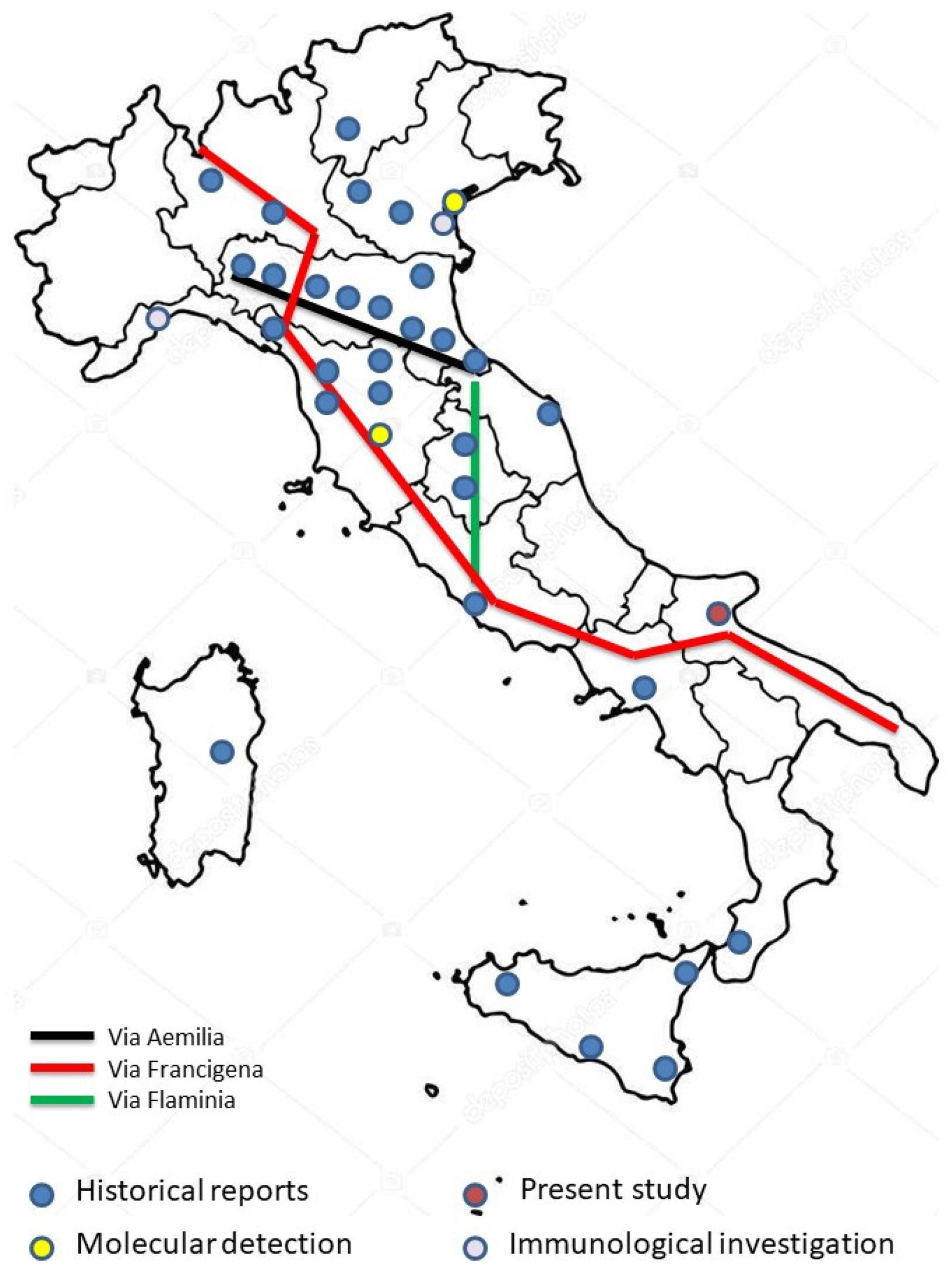
:1. Introduction
2. Materials and Methods
2.1. The Graves: Bones, Biological Profiles and Macroscopic Paleopathology
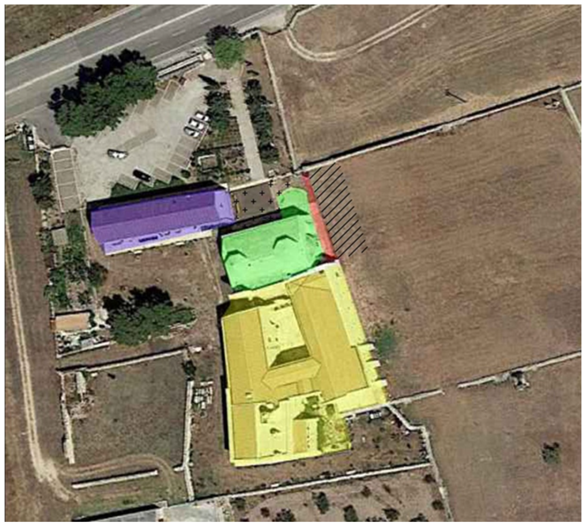
2.2. The Graves: Archaeological, Taphonomic and Numismatic Data
2.3. Ancient DNA (aDNA): Extraction, Procedures and Analysis
3. Results
3.1. Anthropological and Paleopathological Analysis
3.2. Archaeological, Taphonomic and Numismatic Data
- One on the right hemithorax near the sternum: 39 Deniers;
- One right above the clavicle near the right shoulder: 14 Deniers;
- One under the humerus and close to the right scapula: 43 Deniers and the Gigliato.
3.3. Molecular Analysis
4. Discussion
5. Conclusions
Supplementary Materials
Author Contributions
Funding
Institutional Review Board Statement
Informed Consent Statement
Data Availability Statement
Acknowledgments
Conflicts of Interest
References
- Camobreco, F. Regesto di S. Leonardo di Siponto. In Regesta Chartarum Italiae; E. Loescher (W. Regenberg): Roma, Italy, 1913. [Google Scholar]
- Mastrobuoni, S. San Leonardo di Siponto. In Storia di un Antico Monastero della Puglia; Studio Editoriale Dauno: Foggia, Italy, 1960. [Google Scholar]
- Ventura, A. Il patrimonio dell’Abbazia di San Leonardo di Siponto. Illustrazione e trascrizione del manoscritto di una “visita pastorale” di fine secolo XVII conservato nella Biblioteca di Foggia. Foggia Fondi Della Bibl. Prov. 1978, 3. Available online: https://ixtheo.de/Record/1120330041 (accessed on 13 August 2021).
- Mazzoleni, J. Le Carte del Monastero di S. Leonardo della Matina in Siponto (1090–1771). In Codice Diplomatico Pugliese; Società Storia Patria Bari: Napoli, Italy, 1991; Volume XXXI. [Google Scholar]
- Pensato, G. Il Cabreo di San Leonardo di Siponto 1634–1799; Edizioni Scientifiche Italiane: Foggia, Italy, 2000. [Google Scholar]
- Houben, H. (Ed.) San Leonardo di Siponto. In Cella Monastica, Canonica, Domus Theutonicorum, Atti del Convegno Internazionale, Manfredonia, Italy, 18–19 March 2005; Congedo Editore: Galatina, Italy, 2006. [Google Scholar]
- Carbone, A. Hospital assistance in Apulia: Institutions, archives and sources (14th–17th centuries). RiMe 2019, 4, 39–62. [Google Scholar] [CrossRef]
- Enrico, L.; Caragnano, D.; Scarascia Mugnozza, G. The Medieval Pilgrims Routes in the Apulian Cave Settlements and their relationship with Rome and Santiago de Compostela. In The European Pilgrimage Routes for Promoting Sustainable and Quality Tourism in Rural Areas, International Conference Proceedings, Firenze, Italy, 4–6 December 2014; Firenze University Press: Florence, Italy, 2015; pp. 715–736. [Google Scholar]
- Peyer, H.C. Viaggiare nel Medioevo—Dall’ospitalità alla Locanda; Laterza: Bari, Italy, 1997; pp. 125–147. [Google Scholar]
- Pellegrino, L. L’Abbazia di San Leonardo di Siponto nel XIX Secolo; Nuovo Centro di Documentazione Storica: Manfredonia, Italy, 2004. [Google Scholar]
- Pellegrino, L. Hospitale Sancti Michaelis di Monte Sant’Angelo; Edizioni del Golfo: Manfredonia, Italy, 2001; pp. 23–29. [Google Scholar]
- D’Ardes, A. Nota intorno alle vicende architettoniche del complesso abbaziale di San Leonardo in “Lama Volara” presso Siponto. In Il Cabreo di San Leonardo di Siponto 1634–1799; Ventura, A., Ed.; Guido Pensato. Edizioni Scientifiche Italiane: Manfredonia, Italy, 1999; pp. 39–58. [Google Scholar]
- Sarcinelli, G.; Tunzi, A.M.; Panzarino, G. Coins in a medieval grave in Siponto (Apulia): Intentional or unintentional deposition? J. Archaeol. Numis. 2019, 9, 283–292. [Google Scholar]
- Martin, R.; Saller, K. Lehrbuch der Anthropologie; G. Fischer: Stuttgart, Germany, 1956–1959. [Google Scholar]
- Sjøvold, T. Estimation of Stature from Long Bones Utilizing the Line of Organic Correlation. Hum. Evol. 1990, 5, 431–447. [Google Scholar] [CrossRef]
- Trotter, M.; Gleser, G.C. Corrigenda to “Estimation of stature from long bones of American whites and negroes”. Am. J. Phys. Anthropol. 1977, 47, 355–356. [Google Scholar] [CrossRef]
- Acsádi, G.; Nemeskéri, J. History of Human Life, Span and Mortality; Akadémiai Kiadó: Budapest, Hungary, 1970. [Google Scholar]
- Brooks, S.; Suchey, J.M. Skeletal age determination based on the os pubis: A comparison of the Acsadi–Nemeskeri and Suchey–Brooks methods. Hum. Evol. 1990, 5, 227–238. [Google Scholar] [CrossRef]
- Brothwell, D.R. Digging up Bones: The Excavation, Treatment and Study of Human Skeletal Remains; British Museum (Natural History): London, UK, 1981. [Google Scholar]
- Bruzek, J. A Method for Visual Determination of Sex, Using the Human Hip Bone. Am. J. Phys. Anthropol. 2002, 117, 157–168. [Google Scholar] [CrossRef]
- Ferembach, D.; Schwidetzky, I.; Stloukal, M. Recommendation for Age and Sex Diagnoses of Skeletons. J. Hum. Evol. 1980, 9, 517–549. [Google Scholar]
- Lovejoy, C.O.; Meindl, R.S.; Pryzbeck, T.R.; Mensforth, R.P. Chronological Metamorfosis of the Auricular Surface of the Ilium: A New Method for the Determination of Adult Skeletal Age at Death. Am. J. Phys. Anthropol. 1985, 68, 15–28. [Google Scholar] [CrossRef]
- Meindl, R.; Lovejoy, C.O. Ectocranial Suture Closure: A Revised Method for the Determination of Age at Death Based on the Lateral-Anterior Sutures. Am. J. Phys. Anthropol. 1985, 68, 57–66. [Google Scholar] [CrossRef]
- Stloukal, M.; Hanakova, H. Die länge der Längsknochen altslawischer Bevölkerungen—Unter besonderer Berücksichtigung von Wachstumsfragen. Homo 1978, 29, 53–69. [Google Scholar]
- Kelley, M.A.; Larsen, C.S. Advances in Dental Anthropology; Wiley-Liss: New York, NY, USA, 1991. [Google Scholar]
- Capasso, L.; Kennedy, K.A.R.; Wilczak, C.A. Atlas of Occupational Markers on Human Remains; Edigrafital s.p.a.: Teramo, Italy, 1999. [Google Scholar]
- Rogers, J.; Waldron, T.; Dieppe, P.; Watt, I. Arthropaties in paleopatology. The basis of the classification according to most probable cause. J. Archaeol. Sci. 1987, 14, 179–193. [Google Scholar] [CrossRef]
- Kennedy, K.A.R. Skeletal markers of occupational stress. In Reconstruction of Life from the Skeleton; Iscan, M.J., Kennedy, K.A.R., Eds.; Alan, R. Liss: New York, NY, USA, 1989; pp. 129–160. [Google Scholar]
- Mariotti, V.; Facchini, F.; Belcastro, M.G. The study of Entheses: Proposal of Standardised Scoring Method for Twenty-Three Entheses of the Postcranial Skeleton. Coll. Antropol. 2007, 31, 291–313. [Google Scholar] [PubMed]
- Ortner, D.J.; Putschar, W.J. Identification of Pathological Conditions in Human Skeletal Remains; Smithsonian Contributions to Anthropology: Washington, DC, USA, 1985. [Google Scholar]
- Fornaciari, G.; Giuffra, V. Lezioni di Paleopatologia; Ecig: Pisa, Italy, 2009. [Google Scholar]
- Rubini, M. Elementi di Paleopatologia. In Atlante; CISU: Roma, Italy, 2008. [Google Scholar]
- Gordon, G.; Buikstra, J.E. Soil PH, Bone Preservation and Sampling Bias at Mortuary Sites. Am. Antiq. 1981, 46, 566–571. [Google Scholar] [CrossRef]
- Junkins, E.N.; Carter, D.O. The Depositional Environment. In Taphonomy of Human Remains. Forensic Analysis of the Dead and the Depositional Environment; Schotsmans, E.M.J., Márquez-Grant, N., Forbes, S.L., Eds.; Wiley: Chichester, UK, 2017; pp. 143–154. [Google Scholar]
- Ubelaker, D.H. Human Skeletal Remains; Taraxacum: Washington, DC, USA, 1989. [Google Scholar]
- Antoine, D.; Taylor, E. Collection Care: Handling, Storing and Transporting Human Remains. In Regarding the Dead: Human Remains in the British Museum; Fletcher, A., Antoine, D., Hill, J.D., Eds.; The British Museum: London, UK, 2014; pp. 43–48. [Google Scholar]
- Cassman, V.; Odegaard, N.; Powell, J. Human Remains: Guide for Museums and Academic Institutions; AltaMira Press: Oxford, UK, 2007. [Google Scholar]
- Roberts, C. Archaeological human remains and laboratories: Attaining acceptable standards for curating skeletal remains for teaching and research. In Curating Human Remains—Caring for the Dead in the United Kingdom; Giesen, M., Ed.; Boydell & Brewer: Woodbridge, CT, USA, 2013; pp. 123–134. [Google Scholar]
- Wills, B.; Ward, C.; Sáiz Gómez, V. Conservation of Human Remains from Archaeological Contexts. In Regarding the Dead: Human Remains in the British Museum; Fletcher, A., Antoine, D., Hill, J.D., Eds.; The British Museum: London, UK, 2014; pp. 49–74. [Google Scholar]
- Duday, H. Lezioni di Archeotanatologia. In Archeologia Funeraria e Antropologia sul Campo; Soprintendenza Archeologica: Roma, Italy, 2005. [Google Scholar]
- Duday, H.; Courtaud, P.; Crubézy, E.; Sellier, P.; Tillier, A.M. L’anthropologie de «terrain». Reconnaissance et interprétation des gestes funéraires. Bull. Mémoires Société D’anthropologie Paris 1990, 2, 29–49. [Google Scholar] [CrossRef]
- Biaggi, E. Monete e Zecche Medievali Italiane: Dal sec. VIII al Sec. XV; Royal Numismatic Society: London, UK, 1995. [Google Scholar]
- Metcalf, D.M. Coinage of the Crusades and the Latin East; Royal Numismatic Society: London, UK, 1995. [Google Scholar]
- Baker, J. Coinage and Money in the Medieval Greece 1200–1430—Volume I; Brill: Leiden, The Netherland; Boston, MA, USA, 2020. [Google Scholar]
- Cook, B.; Locatelli, S.; Sarcinelli, G.; Travaini, L. The Italian Coins in the British Museum—Volume I—South Italy, Sicily, Sardinia; Edizioni D’Andrea: Roseto degli Abruzzi, Italy, 2020. [Google Scholar]
- Grierson, P.H.; Travaini, L. Medieval European Coinage: With a Catalogue of the Coins in the Fitzwilliam Museum, Cambridge, 14. Italy (III), (South Italy, Sicily, Sardinia); Elina Screen: Cambridge, UK, 1998. [Google Scholar]
- Parsons, T.J.; Weedn, V.W. Preservation and Recovery of DNA in Postmortem Specimens and Trace Samples. In Forensic Taphonomy: The Postmortem Fate of Human Remains; Haglund, W.D., Sorg, M.H., Eds.; CRC-Press: New York, NY, USA, 1996. [Google Scholar]
- Drancourt, M.; Signoli, M.; La Vu Dang, B.B.; Roux, V.; Tzortzis, S.; Raoult, D. Yersinia pestis Orientalis in remains of ancient plague patients. Emerg. Infect. Dis. 2007, 13, 332–333. [Google Scholar] [CrossRef]
- Raoult, D.; Aboudharam, G.; Crubézy, E.; Larrouy, G.; Ludes, B.; Drancourt, M. Molecular identification by “suicide PCR” of Yersinia pestis as the agent of medieval black death. Proc. Natl. Acad. Sci. USA 2000, 97, 12800–12803. [Google Scholar] [CrossRef] [Green Version]
- Hinić, V.; Brodard, I.; Thomann, A.; Cvetnić, Z.; Makaya, P.V.; Frey, J.; Abril, C. Novel identification and differentiation of Brucella melitensis, B. abortus, B. suis, B. ovis, B. canis, and B. neotomae suitable for both conventional and real-time PCR systems. J. Microbiol. Methods 2008, 75, 375–378. [Google Scholar] [CrossRef]
- Mongkol, N.; Suputtamongkol, Y.; Taweethavonsawat, P.; Foongladda, S. Molecular Evidence of Rickettsia in Human and Dog Blood in Bangkok. Vector Borne Zoonotic Dis. 2018, 18, 297–302. [Google Scholar] [CrossRef]
- Lorente-Leal, V.; Liandris, E.; Castellanos, E.; Bezos, J.; Domínguez, L.; de Juan, L.; Romero, B. Validation of a Real-Time PCR for the Detection of Mycobacterium tuberculosis Complex Members in Bovine Tissue Samples. Front. Vet. Sci. 2019, 6, 61. [Google Scholar] [CrossRef]
- Varagnol, M.; Parola, P.; Jouan, R.; Beaucournu, J.C.; Rolain, J.M.; Raoult, D. First detection of Rickettsia felis and Bartonella clarridgeiae in fleas from Laos. Clin. Microbiol. Infect. 2009, 15 (Suppl. S2), 334–335. [Google Scholar] [CrossRef] [Green Version]
- Stewart, A.; Satterfield, B.; Cohen, M.; O’Neill, K.; Robison, R. A quadruplex real-time PCR assay for the detection of Yersinia pestis and its plasmids. J. Med. Microbiol. 2008, 57 Pt 3, 324–331. [Google Scholar] [CrossRef] [Green Version]
- Laroche, M.; Almeras, L.; Pecchi, E.; Bechah, Y.; Raoult, D.; Viola, A.; Parola, P. MALDI-TOF MS as an innovative tool for detection of Plasmodium parasites in Anopheles mosquitoes. Malar. J. 2017, 16, 5. [Google Scholar] [CrossRef] [PubMed]
- Greer, C.E.; Peterson, S.L.; Kiviat, N.B.; Manos, M.M. PCR amplification from paraffin-embedded tissues. Effects of fixative and fixation time. Am. J. Clin. Pathol. 1991, 95, 117–124. [Google Scholar] [CrossRef] [PubMed]
- Huang, X.; Madan, A. CAP3: A DNA sequence assembly program. Genome Res. 1999, 9, 868–877. [Google Scholar] [CrossRef] [PubMed] [Green Version]
- D’Ardes, A. L’antico ospedale di San Leonardo in Lama Volara tra fondazione, riedificazione e abbandono. In San Leonardo di Siponto. Cella Monastica, Canonica, Domus Theutonicorum, Atti del Convegno Internazionale, Manfredonia, Italy, 18–19 March 2005; Houben, H., Ed.; Congedo Editore: Galatina, Italy, 2006; pp. 269–299. [Google Scholar]
- Cohn, S.K., Jr. Epidemiology of the Black Death and successive waves of plague. Med. Hist. Suppl. 2008, 27, 74–100. [Google Scholar] [CrossRef] [Green Version]
- Ayyadurai, S.; Sebbane, F.; Raoult, D.; Drancourt, M. Body lice, Yersinia pestis orientalis, and black death. Emerg. Infect. Dis. 2010, 16, 892–893. [Google Scholar] [CrossRef]
- Piarroux, R.; Abedi, A.A.; Shako, J.C.; Kebela, B.; Karhemere, S.; Diatta, G.; Davoust, B.; Raoult, D.; Drancourt, M. Plague epidemics and lice, Democratic Republic of the Congo. Emerg. Infect. Dis. 2013, 19, 505–506. [Google Scholar] [CrossRef] [PubMed]
- Mead, P.S. 231—Yersinia Species (Including Plague). In Mandell, Douglas, and Bennett’s Principles and Practice of Infectious Diseases, 8th ed.; Bennett, J.E., Dolin, R., Blaser, M.J., Eds.; W.B. Saunders: Philadelphia, PA, USA, 2015; pp. 2607–2618.e2. ISBN 9781455748013. [Google Scholar] [CrossRef]
- Jullien, S.; Lakshitha de Silva, N.; Garner, P. Risk of plague transmission from human cadavers: A systematic review. medRxiv 2021. [Google Scholar] [CrossRef]
- Pechous, R.D.; Sivaraman, V.; Stasulli, N.M.; Goldman, W.E. Pneumonic Plague: The Darker Side of Yersinia pestis. Trends Microbiol. 2016, 24, 190–197. [Google Scholar] [CrossRef]
- Kim-Farley, R. Principles of infectious disease control. In Oxford Textbook of Global Public Health; Oxford University Press: Oxford, UK, 2015; Available online: https://oxfordmedicine.com/view/10.1093/med/9780199661756.001.0001/med-9780199661756-chapter-238 (accessed on 9 August 2021).
- Bitam, I.; Baziz, B.; Rolain, J.M.; Belkaid, M.; Raoult, D. Zoonotic focus of plague, Algeria. Emerg. Infect. Dis. 2016, 12, 1975–1977. [Google Scholar] [CrossRef]
- Ditchburn, J.-L.; Ryan, H. Yersinia pestis, a problem of the past and a re-emerging threat. Biosaf. Health 2019, 1, 65–70. [Google Scholar] [CrossRef]
- Barbieri, R.; Drancourt, M.; Raoult, D. Plague, camels, and lice. Proc. Natl. Acad. Sci. USA 2019, 116, 7620–7621. [Google Scholar] [CrossRef] [PubMed] [Green Version]
- Wang, Y.; Zhou, L.; Fan, M.; Wang, Q.; Li, J.; Li, Q.; Feng, Z.; Gao, G.F.; Xu, C.; Chen, L. Isolated cases of plague-Inner Mongolia-Beijing, 2019. China CDC Wkly. 2019, 1, 13–16. [Google Scholar] [CrossRef] [PubMed]
- DeWitte, S.N. Mortality risk and survival in the aftermath of the medieval Black Death. PLoS ONE 2014, 9, e96513. [Google Scholar] [CrossRef]
- Benedictow, O.J. The Black Death: A Complete History; Boydell & Brewer: Woodbridge, UK, 2017. [Google Scholar]
- Da Piazza, M. Cronaca (1336–1361). In Annali delle Epidemie Occorse in Italia dalle Prime Memorie Fino al 1850; Corradi, A., Ed.; Tipi Gamberini e Parmeggiani: Bologna, Italy, 1865; Volume 1, pp. 193–198. [Google Scholar]
- De Cornazano, J. Chronica abreviata. In Chronica Parmensia a Sec. XI. ad Exitum Sec. XIV; Barbieri, L., Ed.; Ex offic. P. Ficcadorii: Parma, Italy, 1858. [Google Scholar]
- Zietz, B.P.; Dunkelberg, H. The history of the plague and the research on the causative agent Yersinia pestis. Int. J. Hyg. Environ. Health 2004, 207, 165–178. [Google Scholar] [CrossRef]
- Gambaro, L.; Rigeade, C.; De Piero, M.; Ardagna, Y.; Gobbo, V.; Buchet, L.; Fozzati, L.; Drusini, A.; Signoli, M. La fouille de l’île du Lazzareto Vecchio de Venise: Premières données. In Peste: Entre Épidémies et Sociétés; Signoli, M., Chevé, D., Adalian, P., Boëtsch, G., Dutour, O., Eds.; Firenze University Press: Firenze, Italy, 2007; pp. 137–146. [Google Scholar]
- Cambi, F.; Dallai, L. Archeologia di un monastero: Gli scavi a San Salvatore al monte Amiata. Archeol. Mediev. 2000, 27, 193. [Google Scholar]
- Cerutti, N.; Marin, A.; Rabino Massa, E. Plague in ancient remains: An immunological approach. In Plague: Epidemics and Societies; Signoli, M., Chevé, D., Adalian, P., Boëtsch, G., Dutour, O., Eds.; Florence University Press: Florence, Italy, 2007; pp. 238–241. [Google Scholar]
- Cesana, D.; Benedictow, O.; Bianucci, R. The origin and early spread of the Black Death in Italy: First evidence of plague victims from 14th-century Liguria (northern Italy). Anthropol. Sci. 2017, 125, 15–24. [Google Scholar] [CrossRef] [Green Version]
- Dean, K.R.; Krauer, F.; Walløe, L.; Lingjærde, O.C.; Bramanti, B.; Stenseth, N.C.; Schmid, B.V. Human ectoparasites and spread of plague in Europe. Proc. Natl. Acad. Sci. USA 2018, 115, 1304–1309. [Google Scholar] [CrossRef] [Green Version]
- Namouchi, A.; Guellil, M.; Kersten, O.; Hänsch, S.; Ottoni, C.; Schmid, B.V.; Pacciani, E.; Quaglia, L.; Vermunt, M.; Bauer, E.L.; et al. Integrative approach using Yersinia pestis genomes to revisit the historical landscape of plague during the Medieval Period. Proc. Natl. Acad. Sci. USA 2018, 115, E11790–E11797. [Google Scholar] [CrossRef] [Green Version]
- Tran, T.N.N.; Signoli, M.; Fozzati, L.; Aboudharam, G.; Raoult, D.; Drancourt, M. High Throughput, Multiplexed Pathogen Detection Authenticates Plague Waves in Medieval Venice, Italy. PLoS ONE 2011, 6, e16735. [Google Scholar] [CrossRef]
- Mazzei, M. Siponto Antica; Claudio Grenzi Editore: Foggia, Italy, 1999. [Google Scholar]
- Laganara, C. Siponto: Archeologia di una Città Abbandonata nel Medioevo; Claudio Grenzi Editore: Manfredonia, Italy, 2011. [Google Scholar]
- Martin, J.M. La città di Siponto nei secoli XI-XIII. In San Leonardo di Siponto. Cella monastica, Canonica, Domus Theutonicorum, Atti del Convegno Internazionale, Manfredonia, Italy, 18–19 March 2005; Houben, H., Ed.; Congedo Editore: Galatina, Italy, 2006; pp. 15–32. [Google Scholar]
- Ognissanti, P. Il porto di Siponto e di Manfredonia. La Capit. 1984–1985, XLII, 9–51. [Google Scholar]
- Ognissanti, P. Contributo alla conoscenza della società sipontina nell’Alto Medioevo. La Capit. 1984–1985, I, 63–74. [Google Scholar]
- Violante, F. Organizzazione del territorio e strutture produttive tra XI e XVI secolo. In Storia di Manfredonia; Licinio, R., Ed.; Edipuglia: Bari, Italy, 2008; Volume I, pp. 101–123. [Google Scholar]
- Licinio, R. Aspetti della gestione economica di San Leonardo di Siponto all’epoca dei Teutonici. In San Leonardo di Siponto: Cella Monastica, Canonica, Domus Theutonicorum, Atti del Convegno Internazionale, Manfredonia, Italy, 18–19 March 2005; Houben, H., Ed.; Congedo Editore: Galatina, Italy, 2006; pp. 153–166. [Google Scholar]
- Sarnelli, P. Cronologia de’ vescovi e arcivescovi sipontini. In Manfredonia MDCLXXX; Rosselli: Napoli, Italy, 1986. [Google Scholar]
- Baker, J. Repertorio. In Tornesi Gigliati e Pierreali in un Tesoretto Rinvenuto a Muro Leccese; Libero Mangieri: Spoleto, Italy, 2010; pp. 13–17. [Google Scholar]
- Aglietti, S.; Altamura, F.; Cerino, P. Un gruzzolo di monete da un contesto funerario in località Montecrescenzio, Marino (Rm). In Proceedings of the 6th International Numismatic Congress in Croatia, University of Zadar, Zadar, Croatia, 26–29 September 2010; Dobrinić & Dobrinić: Zadar, Opatija, Croatia, 2011; pp. 1–10. [Google Scholar]
- Travaini, L. Saints and Sinners: Coins in Medieval Italian Graves. NC 2004, 164, 159–181.52. [Google Scholar]
- Esposito, A.M.; Saccocci, A.; Gori, S.; Salvadori, P. L’attività della Soprintendenza per i Beni Archeologici della Toscana a Volterra: Lo scavo nella chiesa di San Michele in Foro. In Peccioli e la Valdera dal Medioevo all’Ottocento. Itinerari Archeologici fra Pisa e Volterra, Atti della Giornata di Studi del 18 Aprile 2009, Peccioli—Centro Polivalente; Ciampoltrini, G., Ed.; Fondazione Peccioliper: Peccioli, Italy, 2010; pp. 145–156. [Google Scholar]
- Degasperi, A. Monete nelle tombe basso e postmedievali della Toscana centro-settentrionale: Rito o casualità? In Monete Antiche. Usi e Flussi Monetali in Valdera e nella Toscana Nord-Occidentale dall’Età Romana al Medioevo; Alberti, A., Baldassarri, M., Eds.; La grafica Pisana: Bientina, Italy, 2013; pp. 101–123. [Google Scholar]
- Saccocci, A. Il gruzzolo di una tomba nella chiesa di San Bartolomeo in Silice e la moneta di Lucca nel Trecento. In Il Passo di Gentucca. Il San Francesco di Lucca nel Medioevo: Un Itinerario Archeologico; Ciampoltrini, G., Spataro, C., Eds.; PubliEd editore di Lucca: Lucca, Italy, 2014; pp. 127–142. [Google Scholar]
- Chiaravalle, M. Pila monetale di epoca viscontea da una tomba nella chiesa dei Ss. Antonio da Padova e Eusebio da Vercelli di Azzio (VA). In La Chiesa e il Convento di Azzio. Studi e Aggiornamenti, Monografie della Società Storica Varesina; Società Storica Varesina: Varese, Italy, 2017; Volume 14, pp. 39–49. [Google Scholar]
- Grainger, I.; Hawkins, D.; Cowal, L.; Mikulski, R. The Black Death Cemetery, East Smithfield London; Museum of London Archaeology Svc: London, UK, 2008. [Google Scholar]
- Emmerig, H. Die Geldbörse beim Leichnam, in Leben mit dem Tod. der Umgang mit Sterblichkeit. Mittelalt. Und Neuzeit. Beiträge Zur Mittelalt. Osterr. 2019, 35, 187–208. [Google Scholar]
- Jullien, S.; de Silva, N.; Garner, P. Plague Transmission from Corpses and Carcasses. Emerg. Infect. Dis. 2021, 27, 2033–2041. [Google Scholar] [CrossRef] [PubMed]
- Pigozzo, F. La moneta cucita: I nascondigli pper il denaro alla fine del medioevo. Boll. Mus. Civ. Padova 2004, 94, 155–158. [Google Scholar]
- Aufderheide, A.C.; Rodríguez-Martín, C. The Cambridge Encyclopedia of Human Paleopathology; Cambridge University Press: Cambridge, UK, 2006. [Google Scholar]
- Roberts, C.; Manchester, A.K. The Archaeology of Disease; The History Press: Ithaca, NY, USA, 2010. [Google Scholar]
- Scasciamacchia, S.; Serrecchia, L.; Giangrossi, L.; Garofolo, G.; Balestrucci, A.; Sammartino, G.; Fasanella, A. Plague epidemic in the Kingdom of Naples, 1656–1658. Emerg. Infect. Dis. 2012, 18, 186–188. [Google Scholar] [CrossRef] [PubMed]
- Anfossi, E. Dominicus (de Gravina). In Chronicon de Rebus in Apulia Gestis; Harvard University: Cambridge, MA, USA, 1899. [Google Scholar]


| Target Organism | Target Gene | Expected Amplicon Size | Reference |
|---|---|---|---|
| Brucella spp. | IS711 | 63 bp | Hinic et al., 2008 [50] |
| Rickettsia spp. | glta | 167 bp | Mongkol et al., 2018 [51] |
| Mycobacterium tubercolosis complex | mpb70 | 133 bp | Lorente-Leal et al., 2019 [52] |
| Bartonella spp. | 16S/23S | 126 bp | Varagnol et al., 2009 [53] |
| Yersinia pestis | pla | 144 bp | Stewart et al., 2008 [54] |
| glpD | 288 bp | Drancourt et al., 2007 [48] | |
| Plasmodium spp. | 18S rRNA | 189 bp | Laroche et al., 2017 [55] |
| Human genome | β-Globulin | 110 bp | Greer et al., 1991 [56] |
Publisher’s Note: MDPI stays neutral with regard to jurisdictional claims in published maps and institutional affiliations. |
© 2021 by the authors. Licensee MDPI, Basel, Switzerland. This article is an open access article distributed under the terms and conditions of the Creative Commons Attribution (CC BY) license (https://creativecommons.org/licenses/by/4.0/).
Share and Cite
Raele, D.A.; Panzarino, G.; Sarcinelli, G.; Cafiero, M.A.; Maria Tunzi, A.; Dellù, E. Genetic Evidence of the Black Death in the Abbey of San Leonardo (Apulia Region, Italy): Tracing the Cause of Death in Two Individuals Buried with Coins. Pathogens 2021, 10, 1354. https://doi.org/10.3390/pathogens10111354
Raele DA, Panzarino G, Sarcinelli G, Cafiero MA, Maria Tunzi A, Dellù E. Genetic Evidence of the Black Death in the Abbey of San Leonardo (Apulia Region, Italy): Tracing the Cause of Death in Two Individuals Buried with Coins. Pathogens. 2021; 10(11):1354. https://doi.org/10.3390/pathogens10111354
Chicago/Turabian StyleRaele, Donato Antonio, Ginevra Panzarino, Giuseppe Sarcinelli, Maria Assunta Cafiero, Anna Maria Tunzi, and Elena Dellù. 2021. "Genetic Evidence of the Black Death in the Abbey of San Leonardo (Apulia Region, Italy): Tracing the Cause of Death in Two Individuals Buried with Coins" Pathogens 10, no. 11: 1354. https://doi.org/10.3390/pathogens10111354
APA StyleRaele, D. A., Panzarino, G., Sarcinelli, G., Cafiero, M. A., Maria Tunzi, A., & Dellù, E. (2021). Genetic Evidence of the Black Death in the Abbey of San Leonardo (Apulia Region, Italy): Tracing the Cause of Death in Two Individuals Buried with Coins. Pathogens, 10(11), 1354. https://doi.org/10.3390/pathogens10111354