
Detection of Anti-LipL32 Antibodies in Serum Samples from Horses with Chronic Intraocular Infection with Leptospira spp.


1
Clinic for Horses, University of Veterinary Medicine Hannover, 30559 Hanover, Germany
2
Equine Clinic, Clinical Department, Ludwig Maximilians University, 80539 Munich, Germany
*
Author to whom correspondence should be addressed.
Pathogens 2021, 10(10), 1325; https://doi.org/10.3390/pathogens10101325
Submission received: 20 September 2021
/
Revised: 2 October 2021
/
Accepted: 12 October 2021
/
Published: 14 October 2021
(This article belongs to the Section Bacterial Pathogens)
Abstract
:Equine recurrent uveitis (ERU) is typically caused by chronic intraocular leptospiral infection in warm-blooded horses in central Europe. The most effective therapy for leptospiral-induced ERU is the surgical removal of diseased vitreous (vitrectomy). Since vitrectomy is a highly specialized and invasive surgery, the indication must be determined very carefully. In order to obtain evidence of intraocular leptospiral infection by laboratory diagnostics in questionable leptospiral ERU-cases, sampling of aqueous humor is required, because serum tests using microscopic agglutination test (MAT) are too unspecific. The SNAP Lepto is a cross-species rapid test for the detection of anti-Lipl32 antibodies that has a high sensitivity (0.97) and specificity (1.00) for the detection of anti-leptospiral antibodies using aqueous humor or vitreous samples, which is comparable to MAT. To evaluate sensitivity and specificity of SNAP Lepto using serum, serum samples from 90 horses with confirmed leptospiral ERU and from 103 ocularly healthy horses were tested by both MAT and SNAP Lepto. Sensitivity was similar for both tests (0.82 vs. 0.79), but specificity was lower for MAT (0.52 vs. 0.95). Sensitivity and specificity are therefore lower in serum samples compared to intraocular samples, however, the SNAP Lepto is far superior to MAT and suitable as a screening method using equine serum.
1. Introduction
In central Europe, equine recurrent uveitis (ERU) with its classic symptoms is typically caused by a chronic intraocular leptospiral infection [1,2,3,4,5,6,7,8,9,10,11]. In the following, the term "ERU" will therefore be used for leptospiral induced recurrent uveitis in warm-blooded horses with painful episodes, and which has been demonstrated to be a chronic intraocular infection. Recently, it has been demonstrated that infectious leptospiral uveitis is accompanied by biofilm formation [12,13].
ERU is a late sequela of systemic leptospirosis and becomes clinically apparent from about 6 months to several years after systemic leptospirosis [6,14,15,16,17,18,19,20,21,22,23,24,25,26,27]. The most effective therapy for ERU is vitrectomy of the diseased eyes [11,28,29,30,31,32,33,34,35,36,37,38,39,40]. Vitrectomy is used to eliminate the intraocular leptospiral infection so that no further ERU attacks occur in more than 95% of operated eyes [11,33,35]. If the surgical course is uncomplicated and if vitrectomy is performed before irreversible damage to the lens and/or retina has occurred due to ERU, vision can be preserved [11,33]. The most frequently detected serovar in ERU is Grippotyphosa (Supplementary 1, Figures S1 and S2).
In most cases, both the history and the ophthalmologic findings are conclusive in an ERU, so that the indication for vitrectomy can be reliably made [11,33,35]. In other cases, where the history is suggestive of ERU but the ophthalmologic findings are questionable, the indication for vitrectomy cannot be reliably established by ophthalmologic examination. Examination of serum by micro agglutination test (MAT) unfortunately does not allow a reliable statement about a local leptospiral infection in the eye, because too many healthy horses in Europe [5,6,10,41,42,43,44,45,46,47,48] as well as in other parts of the world [24,27,49,50,51,52,53,54,55,56,57,58,59,60,61] have agglutinating antibodies in the serum.
Therefore, an antibody titer in a serum sample determined with MAT has no significance for the diagnosis of ERU in an individual horse [1,3,4,5,6,10,62,63,64,65]. Consequently, serum tests using MAT do not allow a careful decision on the indication for surgery. However, since vitrectomy is a highly specialized and demanding ophthalmosurgical invention and complications can lead to blindness of the eye and even can make enucleation necessary. Thus, the correct indication is crucial.
For this reason, aqueous humor testing is indicated preoperatively in questionable ERU cases [5,6,11,65,66,67,68,69]. If either anti-Leptospira antibodies are detectable in the aqueous humor and/or the LipL32 gene of pathogenic Leptospira spp. can be detected by PCR, there is an indication for irrigation of the vitreous cavity. To avoid the relative invasive aqueous humor sampling for preoperative laboratory tests, laboratory methods for testing serum samples, which are less complicated to obtain than aqueous humor samples, need to be improved.
The SNAP Lepto, a rapid ELISA test has been commercially available for a few years. It is not species-specific and detects antibodies of different immunoglobulin classes directed against LipL32. LipL32 is a lipoprotein which is expressed at high levels by pathogenic Leptospira spp. [70]. Anti-LipL32 antibodies have been shown to be highly specific for the detection of infection with pathogenic Leptospira spp. [70]. In addition to its strong immunogenicity, LipL32 is also present in all pathogenic Leptospira spp. [71].
This quick ELISA test has proven to be very reliable for the examination of intraocular samples (aqueous humor and vitreous material) [69]. When testing intraocular specimens, the sensitivity and specificity of this rapid test are equivalent to those of the MAT [68], with a kappa value of 0.735 for MAT and SNAP tests [69]. The purpose of the present study was to examine the results of the SNAP Lepto test when serum samples were used and to compare the results of the MAT and SNAP Lepto test. The hypothesis was that using serum samples, the results of the SNAP Lepto test would be similar to those of the MAT and, thus, would not provide any additional information regarding local intraocular leptospiral infection in an individual horse.
2. Materials and Methods
2.1. Preliminary Examination and Classification of Equine Patients
Samples from 207 horses were used for the study. One hundred and three of the 207 horses in which neither the history nor the ophthalmologic examination revealed hints for ERU were considered to be ocularly healthy. Ninety horses had been diagnosed with ERU, and 14 horses with a leopard coat pattern (Appaloosas and Knabstruppers) had a history of chronic insidious uveitis without previous painful episodes of inflammation.
In the ocularly healthy horses, no intraocular samples but only serum samples were examined.
In the horses suffering from ERU, aqueous humor had either been collected during preoperative diagnostics in order to confirm the indication for vitrectomy on the basis of laboratory tests, or vitrectomy had been performed on the basis of the history and ophthalmologic findings without first collecting aqueous humor. If vitrectomy has been performed without previous aqueous humor testing, vitreous material obtained during therapeutically indicated surgery was used for laboratory diagnosis. The vitreous samples were collected at the beginning of each operation to avoid dilution of the samples by the irrigation fluid as far as possible. Horses were assigned to the ERU group if either in the aqueous humor samples or in the vitreous samples an antibody titer against a leptospiral serovar of 1:100 or higher was detectable by MAT, if the SNAP Lepto test was positive, or if the LipL32 gene was detectable in the intraocular samples by real-time PCR (target LipL32 gene, positive if Ct ≤ 40) (Supplementary 2).
In each case, aqueous humor was collected from the leopard coat pattern horses to determine a possible indication for vitrectomy. Although leopard coat pattern uveitis is not Leptospira-induced in most cases [72], meaning the eyes usually do not benefit from vitrectomy, intraocular leptospiral infection may be present in these horses in individual cases. Therefore, it was indicated to take aqueous fluid
SNAP Lepto was performed on-site as previously described [68]. MAT and PCR were performed at an external laboratory (IVD GmbH, Society for Innovative Veterinary Diagnostics, 30926 Seelze-Letter, German accreditation authority DAkkS, DIN EN ISO/IEC 17025, D-PL-18303-02-00; Reg.-Nr.: SAL-BY-L20-04-03). For MAT, the serovars Australis (Serogroup Australis), Bratislava (Serogroup Australis), Autumnalis, Canicola, Grippotyphosa, Copenhageni (Serogroup Icterohaemorrhagiae), Icterohaemorrhagiae (Serogroup Icterohaemorrhagiae), Pomona (Serogroup Pomona), Altodouro (Serogroup Pomona), Hardjo (Serogroup Sejroe), Saxkoebing (Serogroup Sejroe) and Tarassovi were used. A MAT titer of ≥ 1:100 was considered “positive” [73,74,75]. (Supplementary 3, Figures S3 and S4).
2.2. Collection of the Serum Samples
Serum samples were available from all 207 horses. The serum either came from blood samples whose collection had been indicated preoperatively for other reasons, or blood was collected when the venous catheter was inserted for anesthesia (blood always drips off when the catheter is advanced so that the correct position can be checked). Thus, no vein was punctured specifically for obtaining the serum samples. The serum was allowed to stand for about one hour, then centrifuged (5 min, 2500× g) and decanted. Approximately 1 mL of serum from each horse was used for the present study. Few samples were frozen at −28 °C for a few days, most samples were directly examined and sent to the external reference laboratory immediately afterwards.
2.3. Examination of Serum Samples
2.3.1. Preliminary Examination for the Use of SNAP Lepto
Preliminarily, 141 serum samples obtained for previous other studies (from horses not included in this study) and stored at −30 °C were examined with the goal to establish a baseline for agreement between MAT and SNAP Lepto test. For these samples, no consideration was given to the history of the horses. The serum samples were all from equine surgical patients and were tested for the presence of anti-LipL32 antibodies using the SNAP Lepto test (IDEXX company, Ludwigsburg, Germany) which was performed as previously described [68]. Any blue coloration of the sample spot, even a very slight one, was considered “positive”. The same serum samples were then sent to the IVD laboratory (see above) for MAT and tested for antibodies against the mentioned serovars. The result of the MAT was considered "positive" if the antibody titer against one serovar or more serovars was at least 1:100.
2.3.2. Examination of the 207 Horse Sera of this Study
After obtaining serum, the SNAP Lepto test was first performed on-site. Subsequently, the remaining serum was shipped to the IVD-laboratory. The SNAP Lepto test and MAT were performed exactly as for the other serum samples.
2.4. Statistical Analysis
The data from the preliminary examination (141 serum samples) and the results from the 207 horse sera of this study were coded in Microsoft Excel 2011 and then analyzed in SPSS 25. Data collection took place between 2017 and 2019. Pearson’s chi-square test was used to determine dependencies and statistical correlations. The null hypothesis was defined as the independence of two variables of the four-field table. The significance level in this case was p = 0.05. The laboratory tests (MAT, SNAP Lepto and PCR) were evaluated for statistical agreement using the kappa value.
3. Results
3.1. Preliminary Examination: Use of MAT and SNAP Lepto for Equine Serum Samples
In 104 of the 141 serum samples (74%), a titer of 1:100 or higher was measured. In the sera in which no anti-Leptospira antibodies were detectable by MAT (titer < 1:100), no anti-LipL32 antibodies were detectable by SNAP Lepto either in most cases (32/37 sera, 86.5%) (Table 1). The agreement of both tests for a negative response was significant (Pearson’s chi-square test, p < 0.001). A MAT titer of ≥1:100 against one serovar or multiple serovars was present in 104 of the 141 sera (74%). In contrast, among the sera that reacted positively in MAT, antibodies directed against LipL32 could be detected by SNAP Lepto in only 59 of these 104 sera (56.7%). In 45 of the 104 sera positive in MAT (43.3%), no antibodies directed against LipL32 were detectable by SNAP Lepto. Thus, the agreement regarding a positive result in MAT and SNAP Lepto test was low (kappa value 0.34).
3.2. Examination of the 207 Horse Sera of This Study
Comparing the results of MAT and SNAP Lepto, it can be seen that while MAT is positive in over 80% of horses with ERU, it is also positive in almost half of the ocularly healthy horses (Table 2). The SNAP Lepto, on the other hand, is negative in over 90% of ocularly healthy horses and positive in nearly 80% of horses with ERU. The difference between the ocularly healthy horses and horses suffering from ERU when examined by the SNAP Lepto test is significant (Pearson’s chi-square test, p < 0.001). All 14 sera from horses with leopard coat patterns had a negative SNAP Lepto result when the serum samples were tested.
For the calculation of sensitivity, specificity, positive and negative predictive values, the ocular healthy horses and the horses with leopard coat pattern were combined in one group (“no ERU”) (Supplementary 4, Tables S1–S3). The sensitivity of serum tests with respect to detecting intraocular leptospiral infection was similar for MAT and SNAP Lepto (0.82 and 0.79, respectively). However, the specificity when using the SNAP Lepto was significantly higher at 0.95 compared to the MAT, which had a specificity of only 0.52. A similar difference was found for the positive predictive value, which was 0.92 for SNAP Lepto and 0.57 for MAT. The difference was less for the negative predictive value (MAT 0.79; SNAP Lepto 0.85). Statistical agreement for SNAP Lepto using serum and ERU was high (kappa value 0.76).
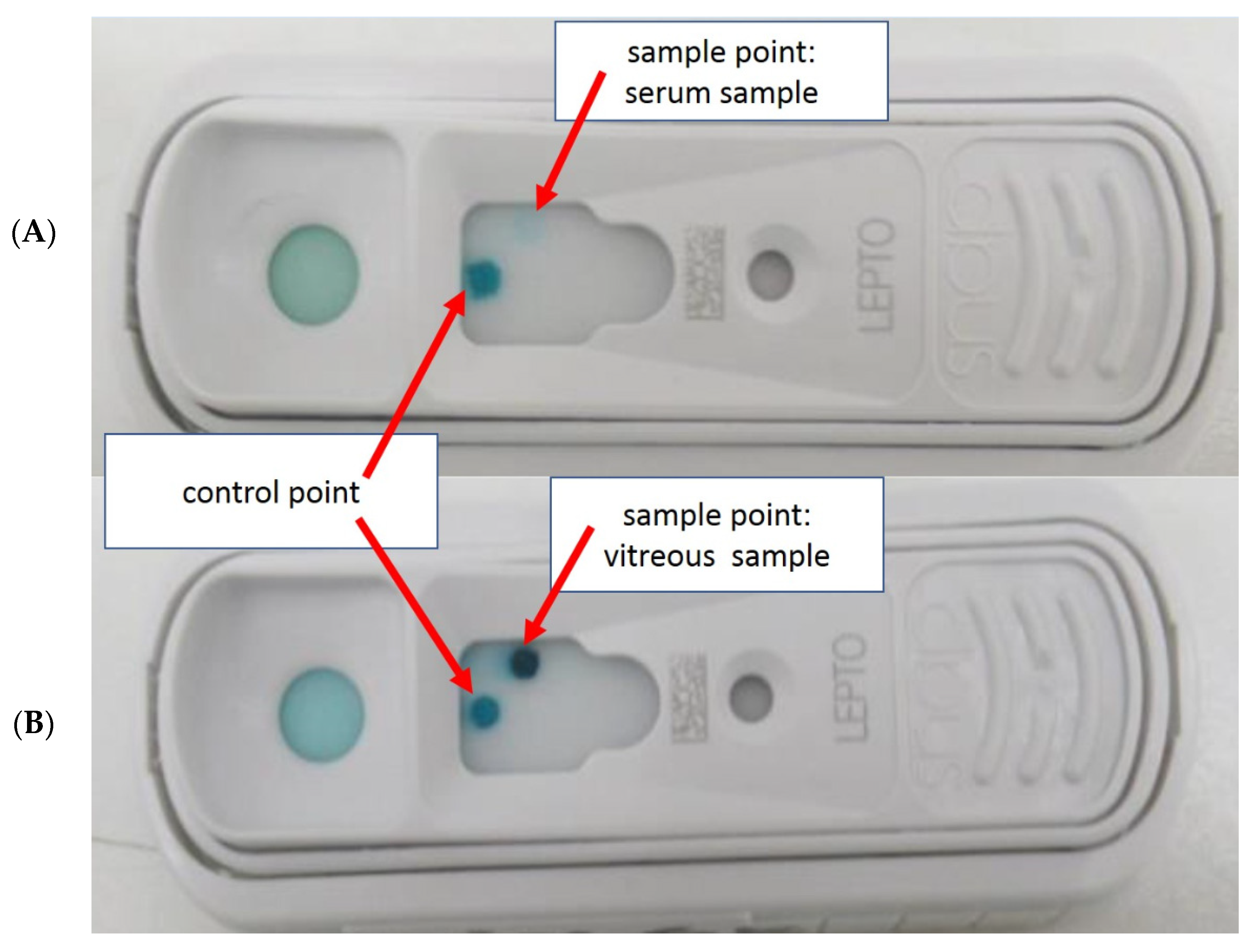
Noticeably, the blue coloration of the sample spot of SNAP Lepto was usually less intense in the serum samples than in the corresponding intraocular samples (Figure 1).
4. Discussion
Once again, as expected, MAT with serum samples was found to be too unspecific for the diagnosis of intraocular leptospiral infection in this study. Eighty-five percent of sera from horses affected with ERU reacted with a titer ≥1:100 in MAT, but MAT titers ≥1:100 were also detectable in approximately half of the samples in the sera from the ocularly healthy horses. Thus, the specificity of MAT with equine serum samples is too low (0.52) to reliably predict the presence of intraocular leptospiral infection in individual horses.
Serum tests for anti-Leptospira antibodies have been frequently performed in ocular healthy horses and horses suffering from ERU [5,41,48,61,64]. Most of these studies used MAT, which has long been considered the gold standard in serum diagnostics [70,76,77], is cited in almost all studies as the reference method for the humoral immune response to leptospiral infection, and is still listed by WHO as the only reference method for screening tests [75,78]. The MAT is a challenging test, requiring some professional experience to perform and numerous serovars to keep available [70,74,75,76,77].
The fact that the examination of numerous horse sera in some studies has shown a difference between the MAT results of the group of ocularly healthy horses compared with those affected with ERU [21,41,49,79,80] does not change the fact that the examination of serum samples by MAT is not an appropriate method to diagnose Leptospira-induced uveitis in an individual horse. MAT lacks specificity because the background level of exposure in the equine population is too high in most studies [4,6,10,48,62,64].
Regardless of whether equine serologic studies were purely epidemiologic or focused on an association between Leptospira spp. and ERU, the results of the studies available in the literature vary considerably. The percentage of horses that had anti-Leptospira antibodies in serum using MAT was 1.5% in one study [44], but up to over 80% in other studies [6,53,57]. In numerous other studies, the frequency of seropositive horses is in between [43,46,48,58]. Using a specific ELISA test for detecting anti-Leptospira antibodies, even in up to 98% positive-reacting sera, has been detected in horses affected with ERU [63].
The different results in publications on serological testing of horse sera for anti-Leptospira antibodies can be explained, on one hand, by the fact that horses were exposed to different infection burdens, having lived in different geographic regions, countries, climatic zones, and environmental conditions and having been tested in different years. However, the MAT titer, which is considered "positive", also plays an essential role, being ≥1:50 [53,58], ≥1:100 [6,9,10,42,43,49,51,54,57,62,81,82,83], ≥1:400 [80,84,85,86] and even ≥1:800 [87] in different studies. Furthermore, different serovars were also used for MAT in different surveys. Although reference laboratories usually have serovars that are relevant to human medicine for the region in question, other species may harbor other serovars that were not tested for. The considerably varying number of serovars used in each case may also have influenced the different results. For example, the MAT included five serovars [58] in one study and 28 serovars [51] in another one. In the present study, the cut-off titer for MAT (1:100) was consistent with most recent studies and the current recommendations [88]. The number of serovars used is average and includes the serogroups relevant in Germany and neighboring countries.
In contrast to MAT, the ELISA rapid test (SNAP Lepto) provided an unexpected result in the present study. Anti-LipL32 antibodies were detected in the serum of 71 of the 90 (79%) horses affected with ERU, whereas in only 6% (6/103) of serum samples from horses with clinically healthy eyes anti-LipL32 antibodies were detected. Thus, the SNAP Lepto was particularly useful in detecting those horses that did not have a chronic leptospiral infection in the eye but had some history of recurrent eye disease. The SNAP Lepto is much more specific (specificity 0.95) than the MAT (specificity 0.52). Thus, the hypothesis that the use of the SNAP Lepto does not offer any advantage over the MAT to diagnose chronic intraocular leptospiral infection from serum samples was not confirmed.
The number of sera from horses affected with ERU examined in this study is, on average, similar to the numbers of other studies [10,21,64,79,89]. However, an expansion of the sample contingent would be beneficial for further substantiation of the present results, but is not achievable within an acceptable time frame in equine medicine.
The 6% of horses with clinically healthy eyes in which anti-LipL32 antibodies were detectable in the present study correspond approximately to the incidence of horses developing ERU in Germany [90]. In other studies, positive PCR results (detection of the LipL32 gene) had been described in 5% [83,91], 3% [92], and 0.4% [68] of intraocular specimens from clinically healthy eyes. These PCR-positive intraocular specimens suggest asymptomatic leptospiral infection of the eye. It is not possible to determine whether the horses with clinically healthy eyes from which the SNAP Lepto positive serum samples were obtained would have developed clinically recognizable symptoms in terms of ERU over the following months or years. Thus, the sera from ocularly healthy horses in this study that reacted "false-positive" in SNAP Lepto could have come from horses that had an intraocular leptospiral infection that had not (yet) resulted in clinical signs. The examination of intraocular samples from the clinically healthy horses was not possible in the present study for ethical reasons.
In few previous studies with serum samples from horses, different ELISA tests had already been used [55,63,65,93]. In horses, the detection of serovar-specific IgA antibodies using an in-house ELISA assay had been shown to be highly sensitive and highly specific for the diagnosis of intraocular leptospiral infection when intraocular samples were evaluated [63,65]. However, when serum samples are examined, IgA antibodies are also very often detectable in healthy horses [63,69]. Only in a species- and immunoglobulin-specific in-house ELISA test it was found that the simultaneous detection of antibodies of different immunoglobulin classes (IgM + IgA + IgG) was quite predominantly possible in horses suffering from ERU and hardly in horses with ophthalmologically healthy eyes [63]. However, the number of horses in the study was relatively small, so this relationship would need further investigation.
The humoral response to leptospiral infections has been thoroughly investigated, particularly in human medicine. In addition to agglutinating antibodies, antibodies against various dominant immunoreactive protein antigens are also produced in leptospiral infections, e.g., against the outer membrane lipoprotein LipL32 or the heat shock proteins GroEL and DnaK [70,94]. Although the detection of antibodies directed against LipL32 does not provide any information about the serovar causing the infection, this is irrelevant for the therapy. Antibodies against LipL32 are also detectable when the infection is caused by a serovar that is not available for MAT [76].
Different dynamics of agglutinating and other antibodies have been repeatedly described for humans and animals [70,74,94,95,96,97,98]. Especially in cases where the MAT is negative against clinical expectation, supplementary serological tests are of importance [75,76,77]. Various ELISA tests, among others, have been described as serological tests complementary to MAT [70,74,75,76,94,99]. Most commercially available ELISA tests detect immunoglobulin class M antibodies, which are critical for early diagnosis of acute infection and are often detectable before MAT becomes positive [75,76,78,98,100,101].
Not only in the very early stages of leptospirosis, but also in chronic leptospiral infections, MAT is less reliable than ELISA tests [76,78]. The current OIE manual indicates that MAT is an imperfect test in some chronic infections, having a sensitivity of less than 50% [88]. In horses, it has been shown that in 9.5% of horses in which Leptospira spp. were cultured in vitreous samples, the corresponding serum samples reacted negatively in MAT [6]. In hedgehogs and rodents in which PCR or culture had yielded a positive result when urine was examined, MAT was often negative when the corresponding serum samples were examined [102,103]. Here, it would be interesting to know whether anti-LipL32 antibodies would have been detectable.
In horses suffering from ERU, infections with different serovars from different serogroups could be detected by culture and MAT in previous studies when examining intraocular sample material [6,104,105], but this is therapeutically irrelevant. The advantage of the immunoglobulin-nonspecific SNAP Lepto used in the present study is that basically all antibodies directed against LipL32 (having at least 2 binding sites) are detectable, which increases the sensitivity compared with other immunoglobulin-specific or even serovar-specific ELISA tests, provided that the humoral immune response has led to the formation of anti-LipL32 antibodies.
The reason for the different results of MAT and SNAP Lepto in the present study may be that in horses, too, the agglutinating antibodies detectable by MAT have different dynamics in the course of leptospiral infections in horses than the antibodies directed against LipL32. The result of this study suggests that chronic local infection with Leptospira spp. in the eye persistently leads to the production of anti-LipL32 antibodies in most horses, which are almost exclusively detectable in the serum of horses affected with ERU and hardly detectable in horses with clinically healthy eyes. It is possible that the formation of antibodies directed against LipL32 declines more rapidly than agglutinating antibodies after a systemic Leptospira spp. infection that has not resulted in chronic local infection. The antibodies detectable in serum by MAT may also be due to intraocular leptospiral infection in horses suffering from ERU, but it is also possible that they represent a residual titer after previous leptospiral infection, completely independent of ERU. Thus, for MAT results it seems to be irrelevant whether the systemic leptospirosis has resulted in intraocular leptospiral infection or not.
The problems of diagnosing chronic intraocular infection with Leptospira spp. by serum testing in horses may be due not only to the immune privileged site in the eye and lack of immune responses, but also to biofilm formation of the bacteria inside the eye [13]. Uveitis caused by Leptospira spp. as a late consequence of systemic leptospirosis is also known in human medicine [106,107,108,109]. Here, too, diagnosis by serological tests is unreliable and the diagnosis of leptospiral uveitis can be challenging [110,111]. Thus, biofilm formation accompanying chronic leptospiral infection of the vitreous cavity might also be present in humans.
Detection of specific antibodies is also challenging in other biofilm-associated local infections, such as cystic fibrosis, and improvement of serological diagnostics is suggested [112,113,114]. It is interesting to note that the lower the immune response and the fewer antibodies are produced, the better the prognosis is often for people suffering from cystic fibrosis, as the inflammation in the tissues associated with the immune responses causes more significant damage than the infectious agents themselves [115,116,117]. This seems to be similar in ERU: higher intraocular anti-Leptospira antibody titers correlate with more severe intraocular inflammatory changes and, on the other hand, Leptospira spp. could be detected in culture even in specimens from ophthalmoscopically apparently healthy eyes in which no anti-Leptospira antibodies were detectable [5].
In chronic bacterial infections and especially in infections associated with biofilm formation, antibodies of the classes IgG and particularly IgA are of great diagnostic value [115,117,118,119]. IgA had also proven to be particularly sensitive and specific in the examination of intraocular samples from horses [65]. However, to the authors’ knowledge, IgA-specific ELISA tests for leptospiral serodiagnosis are currently hardly offered and are only available in the form of in-house tests with antigen preparations. In addition, they are time-consuming and expensive.
The results of the present study suggest that SNAP Lepto is a very good screening method for ERU when equine serum is examined. Particularly in questionable ERU cases, examination of serum by SNAP test may help to decide on the further course of action. If the SNAP Lepto with the serum gives a negative result, long transports of the horses to a clinic can be avoided. If the result of the SNAP Lepto is positive with serum, it is advisable to transport the horse to a clinic specialized in ophthalmology. There, on the basis of further ophthalmologic examinations (or, if necessary, an aqueous humor analysis), a decision can be made as to whether or not vitrectomy is indicated. Thus, the SNAP test can provide important information to veterinarians with little ophthalmologic experience that can help decide on further steps.
5. Conclusions
Results of the available samples have shown that serum testing using SNAP Lepto does not have the same high informative value as testing intraocular samples. Nevertheless, SNAP Lepto is significantly more informative than MAT when using serum samples. The results of this study indicate that the detection of anti-LipL32 antibodies in equine serum is very reliably indicative of chronic local (intraocular) infection with pathogenic Leptospira spp. (specificity 0.95) at an acceptable sensitivity (0.79). The positive predictive value for the detection of intraocular leptospiral infection was 0.92, and the negative predictive value was 0.85. Consequently, the SNAP test has proven to be a very good screening method for the practice to obtain evidence of ERU in otherwise questionable cases. Rapid tests detecting anti-LipL32 antibodies of different immunoglobulin classes could also prove valuable in other species.
Supplementary Materials
The following are available online at https://www.mdpi.com/article/10.3390/pathogens10101325/s1, Supplementary 1: Short information about common serovars horses with ERU in Germany and neighboring countries, Figure S1: Comparing culture results of vitreous material (cultures from 187 eyes) and MAT results of serum samples, Figure S2: Culture results (n = 189) and MAT results with vitreous samples went together pretty well, Supplementary 2: Short explanation of uveitis in horses with leopard coat pattern, Supplementary 3: Laboratory results for the classification of patients, Figure S3: Preliminary examination, classification of patients: serovar distribution in intraocular samples using MAT, titers 1:100 or higher, Figure S4: Preliminary examination, results of MAT, PCR and SNAP Lepto in intraocular samples of ERU eyes, Supplementary 4: Results of serum examinations (Section 3.2.), Table S1: Results of MAT using equine Serum, Table S2: Results of SNAP Lepto using equine Serum, Table S3: Calculation of sensitivity, specificity, positive and negative predictive values (ppv and npv respectively) of MAT and SNAP Lepto using equine serum. References [120,121,122,123,124,125] are cited in the Supplementary Materials.
Author Contributions
Conceptualization, T.G. and B.W.; methodology, T.G. and B.W.; validation, T.G.; formal analysis, T.G.; investigation, T.G.; resources, H.G.; data curation, T.G.; writing—original draft preparation, T.G. and B.W.; writing—review and editing, T.G., B.W. and H.G.; supervision, B.W.; project administration, T.G. and B.W. All authors have read and agreed to the published version of the manuscript.
Funding
This research received no external funding. IDEXX neither initiated nor sponsored this study.
Institutional Review Board Statement
Chair of Animal Welfare, Ethology, Animal Hygiene, and Animal Husbandry Ethical review and approval were waived for this study because all samples used had been taken during diagnostic or therapeutic indicated procedures. No invasive procedure was required for the present study.
Conflicts of Interest
The authors declare no conflict of interest.
References
- Wollanke, B.; Gerhards, H.; Brem, S.; Kopp, H.; Meyer, P. Intraocular and serum antibody titers to Leptospira in 150 horses with equine recurrent uveitis (ERU) subjected to vitrectomy. Berl. Munch. Tierarztl. Wochenschr. 1998, 111, 134–139. [Google Scholar] [PubMed]
- Brem, S.; Gerhards, H.; Wollanke, B.; Meyer, P.; Kopp, H. 35 Leptospirenisolationen aus Glaskörpern von 32 Pferden mit rezidivierender Uveitis (ERU). Berl. Munch. Tierarztl. Wochenschr. 1999, 112, 390–393. [Google Scholar] [PubMed]
- Wollanke, B.; Gerhards, H.; Brem, S.; Wolf, E.; Kopp, H.; Meyer, P. Zur Leptospirenaetiologie der equinen rezidivierenden Uveitis (ERU): Ergebnisse der Untersuchungen von Serum- und Glaskörperproben. Tieraerztl. Prax. 2000, 28, 153–158. [Google Scholar]
- Wollanke, B.; Rohrbach, B.W.; Gerhards, H. Serum and vitreous humor antibody titers in and isolation of Leptospira interrogans from horses with recurrent uveitis. J. Am. Vet. Med. Assoc. 2001, 219, 795–800. [Google Scholar] [CrossRef]
- Wollanke, B. Die Equine Rezidivierende Uveitis (ERU) Als Intraokulare Leptospirose. Habilitation Thesis, Ludwig-Maximilians-University, Munich, Germany, 2002. [Google Scholar]
- Wollanke, B.; Gerhards, H.; Brem, S.; Meyer, P.; Kopp, H. Aetiologie der equinen rezidivierenden Uveitis (ERU): Autoimmunkrankheit oder intraokulare Leptospireninfektion? Pferdeheilkunde 2004, 20, 327–340. [Google Scholar] [CrossRef] [Green Version]
- Von Borstel, M.V.; Oey, L.; Strutzberg-Minder, K.; Boevé, M.H.; Ohnesorge, B. Direct and indirect detection of leptospires in vitreal samples of horses with ERU. Pferdeheilkunde 2010, 26, 219–225. [Google Scholar] [CrossRef]
- Wiehen, L.E. Retrospektive Analyse zum Vorkommen der Equinen Rezidivierenden Uveitis—Unter Beruecksichtigung der Leptospireninfektion—An der LMU München von 01/2005 bis 06/2010. Ph.D. Thesis, Ludwig-Maximilians-University, Munich, Germany, 2012. Ludwig-Maximilians-University Library, Electronic Publications. Available online: https://edoc.ub.uni-muenchen.de/14095/1/Wiehen_Lina_Elisabeth.pdf (accessed on 17 September 2021).
- Baake, E.; von Borstel, M.; Rohn, K.; Ohnesorge, B. Detection of intraocular leptospiral DNA, antibodies and Leptospira spp. in horses with equine recurrent uveitis in different laboratories. Pferdeheilkunde 2016, 32, 346–356. [Google Scholar] [CrossRef] [Green Version]
- Dorrego-Keiter, E.; Tóth, J.; Dikker, L.; Sielhorst, J.; Schusser, G.F. Detection of leptospira by culture of vitreous humor and detection of antibodies against leptospira in vitreous humor and serum of 225 horses with equine recurrent uveitis. Berl. Munch. Tierarztl. Wochenschr. 2016, 129, 209–215. [Google Scholar] [CrossRef]
- Wollanke, B.; Gerhards, H.; Schinagl, C. Results of 654 trans-pars plana vitrectomies of equine eyes with recurrent uveitis-follow-up until 18 years after surgery. Pferdeheilkunde Equine Med. 2021, 37, 204–214. [Google Scholar] [CrossRef]
- Geißler, P.; Wollanke, B. Biofilm formation in persistent infections and its role in the pathogenesis of equine recurrent uveitis (ERU)—A literature review. Pferdeheilkunde Equine Med. 2021, 37, 225–233. [Google Scholar] [CrossRef]
- Ackermann, K.; Kenngott, R.; Settles, M.; Gerhards, H.; Maierl, J.; Wollanke, B. In Vivo Biofilm Formation of Pathogenic Leptospira spp. in the Vitreous Humor of Horses with Recurrent Uveitis. Microorganisms 2021, 9, 1915. [Google Scholar] [CrossRef]
- Gsell, O.; Rehsteiner, K.; Verrey, F. Iridocyclitis as a late consequence of Leptospirosis Pomona (porter’s disease): Agglutinin and lymphocytosis in the aqueous humor. Ophthalmologica 1946, 112, 320–334. [Google Scholar] [CrossRef] [PubMed]
- Heusser, H. Zur Aetiologie der periodischen Augenentzündung. Schweiz. Arch. Tierheilk. 1952, 94, 296–306. [Google Scholar]
- Hartwigk, H. Die periodische Augenentzündung des Pferdes als Spaetsymptom der Leptospirose. Tierarztl. Umsch. 1953, 8, 154–157. [Google Scholar]
- Bryans, J.T. Studies on equine leptospirosis. Cornell Veter. 1955, 45, 16–50. [Google Scholar]
- Kathe, J. Die Leptospirosen und ihre Verbreitung in der Deutschen Demokratischen Republik. Z. Hyg. 1955, 1, 39–62. [Google Scholar]
- Bolte, H.F. Uveitis, a Sequela to Experimentally Induced Leptospira Pomona Infection in the Shetland Pony. Master’s Thesis, Faculty of Purdue University, West Lafayette, IN, USA, 1966. [Google Scholar]
- Zwierzchowski, J. Klinik und Therapie der Leptospirosen der Haus- und Nutztiere. In Leptospiren und Leptospirosen, Teil I.; Kathe, J., Mochmann, H., Eds.; Gustav Fischer Verlag: Jena, Germany, 1967; pp. 79–137. [Google Scholar]
- Dwyer, A.E.; Crockett, R.; Kalsow, C.M. Association of leptospiral seroreactivity and breed with uveitis and blindness in horses: 372 cases (1986–1993). J. Am. Vet. Med. Assoc. 1995, 207, 1327–1331. [Google Scholar] [PubMed]
- Williams, R.D. The Presence and Duration of Persistence of Leptospira Pomona in Equine Ocular Tissues Following Experimentally Induced Systemic Infection. Master’s Thesis, Purdue University, West Lafayette, IN, USA, 1968. [Google Scholar]
- Williams, R.D.; Morter, R.L.; Freeman, M.J.; Lavignette, A.M. Experimental chronic uveitis. Ophthalmic signs following equine leptospirosis. Investig. Ophthalmol. 1971, 10, 948–954. [Google Scholar]
- Verma, A.; Stevenson, B.; Adler, B. Leptospirosis in horses. Vet. Microbiol. 2013, 167, 61–66. [Google Scholar] [CrossRef]
- Ellis, W.A. 4. Disease in Animals. 4.4 Horses and Donkeys. In Leptospira and Leptospirosis, Current Topics in Microbiology and Immunology; Adler, B., Ed.; Springer: Berlin/Heidelberg, Germany, 2015; pp. 109–110. ISBN 978-3-662. [Google Scholar] [CrossRef]
- Malalana, F.; Stylianides, A.; McGowan, C. Equine recurrent uveitis: Human and equine perspectives. Vet. J. 2015, 206, 22–29. [Google Scholar] [CrossRef]
- Divers, T.J.; Schang, Y.F.; Irby, N.L.; Smith, J.L.; Carter, C.N. Leptospirosis: An important infectious disease in North American horses. Equine Vet. J. 2019, 51, 287–292. [Google Scholar] [CrossRef] [PubMed]
- Werry, H.; Gerhards, H. Technique and indications for surgical treatment of equine recurrent uveitis. Pferdeheilkunde 1991, 7, 321. [Google Scholar] [CrossRef]
- Werry, H.; Gerhards, H. Surgical treatment of equine recurrent uveitis: A preliminary report. Tierarztl. Prax. 1992, 20, 178–186. [Google Scholar] [PubMed]
- Winterberg, A.; Gerhards, H. Longterm-results of pars-plana-vitrectomy in equine recurrent uveitis. Pferdeheilkunde 1997, 13, 377–383. [Google Scholar] [CrossRef] [Green Version]
- Frühauf, B.; Ohnesorge, B.; Deegen, E.; Boevé, M. Surgical management of equine recurrent uveitis with single port pars plana vitrectomy. Vet. Ophthalmol. 1998, 1, 137–151. [Google Scholar] [CrossRef] [PubMed]
- Gerhards, H.; Wollanke, B.; Winterberg, A.; Werry, H. Technique for and results with vitrectomy in horses with recurrent uveitis. In Proceedings of the 29th Annual ACVO-Meeting, Seattle, WA, USA, 21–24 October 1998; p. 30. [Google Scholar]
- Gerhards, H.; Wollanke, B.; Brem, S. Vitrectomy as a diagnostic and therapeutic approach for equine recurrent uveitis (ERU). In Proceedings of the 45th Annual Convention AAEP, Albuquerque, NM, USA, 8 December 1999; Volume 45, pp. 89–93. [Google Scholar]
- Gerhards, H.; Wollanke, B. Uveitis bei Pferden-Diagnose und Therapie. Pferdeheilkunde 2001, 20, 319–329. [Google Scholar] [CrossRef] [Green Version]
- Gerhards, H.; Wollanke, B. Surgical treatment of equine recurrent uveitis: Trans-pars-plana vitrectomy in horses. In Equine Ophthalmology, 1st ed.; Gilger, B.C., Ed.; Elsevier Saunders: Philadelphia, PA, USA, 2005; pp. 314–319. [Google Scholar]
- Von Borstel, M.; Von Oppen, T.; Glitz, F.; Frühauf, B.; Deegen, E.; Boevé, M.H.; Ohnesorge, B. Long-term results of pars-plana (double-port) vitrectomy in equine recurrent uveitis. Pferdeheilkunde 2005, 21, 13–18. [Google Scholar] [CrossRef] [Green Version]
- Tömördy, E.; Hässig, M.; Spiess, B.M. The outcome of pars plana vitrectomy in horses with equine recurrent uveitis with regard to the presence or absence of intravitreal antibodies against various serovars of Leptospira interrogans. Pferdeheilkunde 2010, 26, 251–254. [Google Scholar] [CrossRef] [Green Version]
- Dorrego-Keiter, E.; Tóth, J.; Dikker, L.; Sielhorst, J.; Schusser, G.F. Long-term results of pars plana vitrectomy in relationship to leptospiral antibody detection in vitreous humor in 118 horses with equine recurrent uveitis (ERU). Pferdeheilkunde Equine Med. 2017, 33, 112–118. [Google Scholar] [CrossRef] [Green Version]
- Baake, E.I.A.; von Borstel, M.; Rohn, K.; Boevé, M.H.; Ohnesorge, B. Long-term ophthalmologic examinations of eyes with equine recurrent uveitis after pars plana vitrectomy. Pferdeheilkunde 2019, 35, 220–233. [Google Scholar] [CrossRef]
- Voelter, K.; Vial, Z.; Pot, A.S.; Spiess, B.M. Leptospiral antibody prevalence and surgical treatment outcome in horses with Equine Recurrent Uveitis (ERU) in Switzerland. Vet. Ophthalmol. 2020, 23, 648–658. [Google Scholar] [CrossRef]
- Wollanke, B. Untersuchungen zur Aetiologie der Equinen Rezidivierenden Uveitis (ERU). Doctoral Thesis, Ludwig-Maximilians-University, Munich, Germany, 1995. [Google Scholar]
- Båverud, V.; Gunnarsson, A.; Engvall, E.O.; Franzén, P.; Egenvall, A. Leptospira seroprevalence and associations between seropositivity, clinical disease and host factors in horses. Acta Vet. Scand. 2009, 51, 15. [Google Scholar] [CrossRef] [PubMed] [Green Version]
- Blatti, S.; Overesch, G.; Gerber, V.; Frey, J.; Hussy, D. Seroprevalence of Leptospira spp. in clinically healthy horses in Switzerland. Schweiz. Arch. Tierh. 2011, 153, 449–456. [Google Scholar] [CrossRef] [PubMed] [Green Version]
- Ebani, V.V.; Bertelloni, F.; Pinzauti, P.; Cerri, D. Seroprevalence of Leptospira spp. and Borrelia burgdorferi sensu lato in Italian horses. Ann. Agric. Environ. Med. 2012, 19, 237–240. [Google Scholar] [PubMed]
- Arent, Z.J.; Kedzierska-Mieszkowska, S. Seroprevalence study of leptospirosis in horses in northern Poland. Vet. Rec. 2013, 127, 269. [Google Scholar] [CrossRef] [PubMed]
- Habus, J.; Persic, Z.; Spicic, S.; Vince, S.; Stritof, Z.; Milas, Z.; Cvetnic, Z.; Perharic, M.; Turk, N. New trends in human and animal leptospirosis in Croatia, 2009–2014. Acta Trop. 2017, 168, 1–8. [Google Scholar] [CrossRef]
- Vera, E.; Taddei, S.; Cavirani, S.; Schiavi, J.; Angelone, M.; Cabassi, C.S.; Schiano, E.; Quintavalla, F. Leptospira Seroprevalence in Bardigiano Horses in Northern Italy 2019. Animals 2020, 10, 23. [Google Scholar] [CrossRef] [Green Version]
- Wasinski, B.; Paschalis-Trela, K.; Trela, J.; Czopowicz, M.; Kita, J.; Zychska, M.; Cywinska, A.; Markowska-Daniel, I.; Carter, C.; Witkowski, L. Serological Survey of Leptospira Infection in Arabian Horses in Poland. Pathogens 2021, 10, 688. [Google Scholar] [CrossRef]
- Hathaway, S.; Little, T.; Finch, S.; Stevens, A. Leptospiral infection in horses in England: A serological study. Vet. Rec. 1981, 108, 396–398. [Google Scholar] [CrossRef]
- Adler, B.; de la Peña Moctezuma, A. Leptospira and leptospirosis. Vet. Microbiol. 2010, 140, 287–296. [Google Scholar] [CrossRef]
- Hamond, C.; Martins, G.; Lawson-Ferreira, R.; Medeiros, M.A.; Lilenbaum, W. The role of horses in the transmission of leptospirosis in an urban tropical area. Epidemiol. Infect. 2013, 141, 33–35. [Google Scholar] [CrossRef]
- Loureiro, A.P.; Hamond, C.; Lilenbaum, W. Leptospirosis in horses. Vet. Rec. 2013, 172, 479–480. [Google Scholar] [CrossRef]
- Finger, M.A.; Filho, I.R.D.B.; Leutenegger, C.; Estrada, M.; Ullmann, L.S.; Langoni, H.; Kikuti, M.; Dornbush, P.T.; Deconto, I.; Biondo, A.W. Serological and molecular survey of Leptospira spp. among cart horses from an endemic area of human leptospirosis in Curitiba, Southern Brazil. Rev. Inst. Med. Trop. Sao Paulo 2014, 56, 473–476. [Google Scholar] [CrossRef] [Green Version]
- Tsegay, K.; Potts, A.D.; Aklilu, N.; Loetter, C.; Gummow, B. Circulating serovars of Leptospira in cart horses of central and southern Ethiopia and associated risk factors. Prev. Vet. Med. 2016, 125, 106–115. [Google Scholar] [CrossRef] [PubMed] [Green Version]
- Sohail, M.; Khan, M.; Ijaz, M.; Fatima, Z.; Naseer, O.; Ahamad, W.; Ahmad, A. Seroprevalence of Leptospira spp. In Horses of Distinct Climatic Regions of Punjab, Pakistan. In Proceedings of the 10th International Leptospirosis Society Conferecnce (ILS 2017), Palmerston North, New Zealand, 27 November–1 December 2017; p. 237. [Google Scholar]
- Ribeiro, T.M.P.; Correia, L.; Spohr, K.A.H.; Aguiar, D.M.; Martins, G.; de Sa Jayme, V. Risk Factors Associated with Seroreactivity Against Leptospira sp. in Horses from Brazilian Amazon. J. Equine Vet. Sci. 2018, 68, 59–62. [Google Scholar] [CrossRef]
- Calderon, J.C.; Astudillo, M.; Romero, M.H. Epidemiological characterization of Leptospira spp. infection in working horses and in an occupationally exposed population in six Colombian police stations. Biomedica 2019, 39, 19–34. [Google Scholar] [CrossRef]
- Bolwell, C.F.; Rogers, C.W.; Benschop, J.; Collins-Emerson, J.M.; Adams, B.; Scarfe, K.R.; Gee, E.K. Seroprevalence of Leptospira in Racehorses and Broodmares in New Zealand. Animals 2020, 10, 1952. [Google Scholar] [CrossRef]
- Da Silva, A.S.; Jaguezeski, A.M.; Laber, I.F.; von Laer, A.E.; Lovato, L.T.; da Silva, M.O.; de Moura, A.B. Leptospira spp. in horses in southern Brazil: Seroprevalence, infection risk factors, and influence on reproduction. Comp. Immunol. Microbiol. Infect. Dis. 2020, 73, 101551. [Google Scholar] [CrossRef] [PubMed]
- Fagre, A.C.; Mayo, C.E.; Pabilonia, K.L.; Landolt, G.A. Seroprevalence of Leptospira spp. in Colorado equids and association with clinical disease. J. Vet. Diagn. Investig. 2020, 32, 718–721. [Google Scholar] [CrossRef] [PubMed]
- Tirosh-Levy, S.; Baum, M.; Schvartz, G.; Kalir, B.; Pe’er, O.; Shnaiderman-Torban, A.; Bernstein, M.; Blum, S.E.; Steinman, A. Seroprevalence of Leptospira spp. in horses in Israel. Pathogens 2021, 10, 408. [Google Scholar] [CrossRef] [PubMed]
- Gilger, B.C.; Salmon, J.H.; Yi, N.Y.; Barden, C.A.; Chandler, H.L.; Wendt, J.A.; Colitz, C.M.H. Role of bacteria in the pathogenesis of recurrent uveitis in horses from the southeastern United States. Am. J. Vet. Res. 2008, 69, 1329–1335. [Google Scholar] [CrossRef] [PubMed]
- Loibl, J. Immunologische und Mikrobiologische Untersuchungen zur Intraokular Persistierenden Leptospireninfektion bei Pferden mit Rezidivierender Uveitis. (Dissertation), LMU München. 2009. Ludwig-Maximilians-University Library, Electronic Publications. Available online: https://edoc.ub.uni-muenchen.de/10508/1/Loibl_Julia.pdf (accessed on 17 September 2021).
- Malalana, F.; Blundell, R.J.; Pinchbeck, G.L.; McGowan, C.M. The role of Leptospira spp. in horses affected with recurrent uveitis in the UK. Equine Vet. J. 2017, 49, 706–709. [Google Scholar] [CrossRef] [PubMed] [Green Version]
- Loibl, J.K.; Gerhards, H.; Brem, S.; Wollanke, B. Improving the laboratory diagnosis of leptospiral uveitis in horses by using an indirect ELISA for the detection of antibodies against Leptospira spp. in intraocular samples. Pferdeheilkunde Equine Med. 2018, 34, 267–277. [Google Scholar] [CrossRef] [Green Version]
- Wollanke, B.; Gerhards, H. Differential diagnosis of equine recurrent uveitis: The importance of a paracentesis of the anterior chamber and aqueous analysis. In Proceedings of the 30th WSAVA Congress, International Society of Veterinary Ophthalmology (IVO) Pre-Congress Programme, Mexico City, Mexico, 9–10 May 2005. [Google Scholar]
- Gesell, S.; Wollanke, B.; Brem, S.; Gerhards, H. Vergleich der Antikoerpertiter gegen Leptospiren in Kammerwasser- und Glaskoerperproben bei Pferden mit rezidivierender Uveitis. In Proceedings of the 19th DVG-Fachtagung Pferdekrankheiten, Hanover, Germany, 10–11 February 2006; 2006; pp. 239–241. [Google Scholar]
- Wollanke, B.; Geiger, T.; Gerhards, H. Evaluation of “SNAP® Lepto”-ELISA and comparison with MAT and PCR results for diagnosis of leptospiral uveitis in horses using intraocular samples. Pferdeheilkunde Equine Med. 2018, 34, 508–516. [Google Scholar] [CrossRef]
- Geiger, T. Evaluierung eines ELISA-Schnelltests (SNAP-Lepto®) für den Nachweis von Antikörpern gegen LipL32 in Serum und Intraokularem Probenmaterial von Pferden als Diagnostikum der Equinen Rezidivierenden Uveitis. (Dissertation), LMU, München. 2019. Ludwig-Maximilians-University Library, Electronic Publications. Available online: https://edoc.ub.uni-muenchen.de/24938/1/Geiger_Tobias_Xaver.pdf (accessed on 18 September 2021).
- Guerreiro, H.; Croda, J.; Flannery, B.; Mazel, M.; Matsunaga, J.; Reis, M.G.; Levett, P.N.; Ko, A.I.; Haake, D.A. Leptospiral proteins recognized during the humoral immune response to leptospirosis in humans. Infect. Immun. 2001, 69, 4958–4968. [Google Scholar] [CrossRef] [PubMed] [Green Version]
- Haake, D.A.; Chao, G.; Zuerner, R.L.; Barnett, J.K.; Barnett, D.; Mazel, M.; Matsunaga, J.; Levett, P.N.; Bolin, C.A. The leptospiral major outer membrane protein LipL32 is a lipoprotein expressed during mammalian infection. Infect. Immun. 2000, 68, 2276–2285. [Google Scholar] [CrossRef] [PubMed] [Green Version]
- Baumgart, A.; Gerhards, H. Besonderheiten der Tigerschecken-Uveitis und möglicher Cyclosporin A-Einsatz in deren Therapie in Deutschland. Pferdeheilkunde 2014, 30, 626–632. [Google Scholar] [CrossRef] [Green Version]
- Faine, S. Guidelines for the Control of Leptospirosis; World Health Organization: Geneva, Switzerland, 1982. [Google Scholar]
- Levett, P.N. Leptospirosis. Clin. Microbiol. Rev. 2001, 14, 296–326. [Google Scholar] [CrossRef] [Green Version]
- Haake, D.A.; Levett, P.N. Leptospirosis in humans. Curr. Top. Microbiol. Immunol. 2015, 387, 65–97. [Google Scholar] [CrossRef] [Green Version]
- Day, N. Leptospirosis: Epidemiology, Microbiology, Clinical Manifestations, and Diagnosis. UpToDate® Home Page, Topic 5527, Version 27.0. Available online: https://www.uptodate.com/contents/leptospirosis-epidemiology-microbiology-clinical-manifestations-and-diagnosis (accessed on 14 September 2021).
- Limmathurotsakul, D.; Turner, E.L.; Wuthiekanun, V.; Thaipadungpanit, J.; Suputtamongkol, Y.; Chierakul, W.; Smythe, L.D.; Day, N.P.; Cooper, B.; Peacock, S.J. Fool’s gold: Why imperfect reference tests are undermining the evaluation of novel diagnostics: A reevaluation of 5 diagnostic tests for leptospirosis. Clin. Infect. Dis. 2012, 55, 322–331. [Google Scholar] [CrossRef]
- World Health Organization and International Leptospirosis Society. Annex 10, Serological Techniques (MAT and ELISA). In Human Leptospirosis: Guidance for Diagnosis, Surveillance and Control; World Health Organisation, Ed.; WHO Library Cataloguing-in-Publication Data: Geneva, Switzerland, 2003; pp. 63–75. ISBN 92 4 154589 5. Available online: https://www.who.int/publications/i/item/human-leptospirosis-guidance-for-diagnosis-surveillance-and-control (accessed on 15 September 2021).
- Halliwell, R.E.; Brim, T.A.; Hines, M.A.; Wolf, D.; White, F.H. Studies on equine recurrent uveitis. II: The role of infection with Leptospira interrogans serovar pomona. Curr. Eye Res. 1985, 4, 1033–1040. [Google Scholar] [CrossRef]
- Alexander, C.; Keller, H. Aetiology and occurrence of periodic ophthalmia among horses in Berlin. Tierarztl. Prax. 1990, 18, 623–627. [Google Scholar] [PubMed]
- Jung, B.Y.; Lee, K.W.; Ha, T.Y. Seroprevalence of Leptospira spp. in clinically healthy racing horses in Korea. J. Vet. Med. Sci. 2010, 72, 197–201. [Google Scholar] [CrossRef] [Green Version]
- Pikalo, J.; Sattler, T.; Eichinger, M.; Loitsch, A.; Sun, H.; Schmoll, F.; Schusser, G.F. Occurrance of antibodies against Leptospira in horses in Middle Germany. Berl. Munch. Tierarztl. Wochenschr. 2016, 129, 202–208. [Google Scholar] [PubMed]
- Gesell, S. Gibt es eine Asymptomatische Intraokulare Leptospireninfektion beim Pferd? Ph.D. Thesis, LMU, Munich, Germany, 2004. Ludwig-Maximilians-University Library, Electronic Publications. Available online: https://edoc.ub.uni-muenchen.de/2527/1/Gesell_Stefan.pdf (accessed on 17 September 2021).
- Heusser, H. Die periodische Augenentzündung, eine Lepospirose? Schweiz. Arch. Tierheilk. 1948, 90, 287–312. [Google Scholar]
- Hupka, E.; Behrens, H. Untersuchungen über die Leptospirose des Pferdes. Dtsch. Tierarztl. Wochenschr. 1951, 58, 245–248. [Google Scholar]
- Kalisch, J. Leptospirose und periodische Augenentzündung. Berl. Munch. Tierarztl. Wschr. 1952, 65, 5–9. [Google Scholar]
- Zaharija, J.; Maralt, J.; Cermak, K.; Andrasic, N.; Sanovic, I. Leptospirose und periodische Augenentzündung beim Pferd. Schweiz. Arch. Tierheilk. 1960, 102, 400–408. [Google Scholar]
- World Organization of Animal Health—OIE. Chapter 3.1.12 Leptospirosis. Manual of Diagnostic Tests and Vaccines for Terrestrial Animals OIE Terrestrial manual 2021; Manual of Diagnostic Tests and Vaccines for Terrestrial Animals. OIE Terrestrial manual 2021: Paris, France OIE Home Page, most recent updates adopted 2021. Available online: https://www.oie.int/fileadmin/Home/eng/Health_standards/tahm/3.01.12_LEPTO.pdf (accessed on 15 September 2021).
- Himebaugh, N.E.; Gilger, B.C. Role of Leptospira spp. testing and ocular examination in horses with equine recurrent uveitis: A retrospective study of 63 horses. Equine Vet. Educ. 2021. [Google Scholar] [CrossRef]
- Szemes, P.A.; Gerhards, H. Untersuchungen zur Prävalenz der equinen rezidivierenden Uveitis im Großraum Köln-Bonn. Prakt. Tierarzt 2000, 81, 408–420. [Google Scholar]
- Gesell-May, S.; Brem, S.; Wollanke, B.; Gerhards, H. Untersuchung gesunder Pferdeaugen auf eine intraokulare Leptospireninfektion. Pferdeheilkunde Equine Med. 2021, 37, 215–224. [Google Scholar] [CrossRef]
- Wollanke, B.; Gerhards, H.; Kaufmann, S. Investigations on the Borrelia-etiology in equine recurrent uveitis (ERU). Pferdeheilkunde Equine Med. 2017, 33, 447–451. [Google Scholar] [CrossRef] [Green Version]
- Khurana, S.; Malik, P.; Nandal, A.; Srivastava, S. Seroprevalence of Leptospirosis in Equines in India. Indian J. Comp. Microbiol. Immunol. Infect. Dis. 2003, 24, 93–95. [Google Scholar]
- Pissawong, T.; Maneewatchararangsri, S.; Ritthisunthorn, N.; Soonthornworasiri, N.; Reamtong, O.; Adisakwattana, P.; Kalambaheti, T.; Chaisri, U.; Doungchawee, G. Immunodominance of LipL3293–272 peptides revealed by leptospirosis sera and therapeutic monoclonal antibodies. J. Microbiol. Immunol. Infect. 2020, 53, 11–22. [Google Scholar] [CrossRef]
- Adler, B.; Murphy, A.; Locarnini, S.; Faine, S. Detection of specific anti-leptospiral immunoglobulins M and G in human serum by solid-phase enzyme-linked immunosorbent assay. J. Clin. Microbiol. 1980, 11, 452–457. [Google Scholar] [CrossRef] [PubMed] [Green Version]
- Leonard, F.; Quinn, P.; Ellis, W.; O’Farrell, K. Association between cessation of leptospiruria in cattle and urinary antibody levels. Res. Vet. Sci. 1993, 55, 195–202. [Google Scholar] [CrossRef]
- Da Silva, M.V.; Nakamura, P.M.; Camargo, E.D.; Batista, L.; Vaz, A.J.; Romero, E.C.; Brandao, A.P. Immunodiagnosis of human leptospirosis by dot-ELISA for the detection of IgM, IgG, and IgA antibodies. Am. J. Trop. Med. Hyg. 1997, 56, 650–655. [Google Scholar] [CrossRef]
- Ismail, Z.B.; Abutarbush, S.M.; Al-Majali, A.; Gharaibeh, M.H.; Al-Khateeb, B. Seroprevalence and risk factors of Leptospira serovar Pomona and Leptospira serovar Hardjo infection in dairy cows in Jordan. J. Infect. Dev. Ctries. 2019, 13, 473–479. [Google Scholar] [CrossRef]
- Goris, M.G.A.; Leeflang, M.M.G.; Loden, M.; Wagenaar, J.F.P.; Klatser, P.R.; Hartskeerl, R.A.; Boer, K.R. Prospective Evaluation of Three Rapid Diagnostic Tests for Diagnosis of Human Leptospirosis. PLoS Negl. Trop. Dis. 2013, 7, e2290. [Google Scholar] [CrossRef] [Green Version]
- Goris, M.G.A.; Leeflang, M.M.G.; Boer, K.R.; Goeijenbier, M.; van Gorp, E.C.M.; Wagenaar, J.F.P.; Hartskeerl, R.A. Establishment of Valid Laboratory Case Definition for Human Leptospirosis. J. Bacteriol. Parasitol. 2012, 3, 132. [Google Scholar] [CrossRef] [Green Version]
- Tan, X.T.; Amran, F.; Cheong, K.C.; Ahmad, N. In-house ELISA screening using a locallyisolated Leptospira in Malaysia: Determination of its cut-off points. BMC Infect. Dis. 2014, 14, 563. [Google Scholar] [CrossRef] [Green Version]
- Brockie, R.E.; Till, D.G. Leptospira ballum isolated from hedgehogs. New Zealand Vet. J. 1977, 25, 28–30. [Google Scholar] [CrossRef]
- Hathaway, S.; Blackmore, D.K. Ecological aspects of the epidemiology of infection with leptospires of the Ballum serogroup in the black rat (Rattus rattus) and the brown rat (Rattus norvegicus) in New Zealand. J. Hyg. 1981, 87, 427–436. [Google Scholar] [CrossRef] [Green Version]
- Hartskeerl, R.A.; Goris, M.G.A.; Brem, S.; Meyer, P.; Kopp, H.; Gerhards, H.; Wollanke, B.J. Classification of Leptospira from the Eyes of Horses Suffering from Recurrent Uveitis. Vet. Med. B Infect. Dis. Vet. Public Health 2004, 51, 110–115. [Google Scholar] [CrossRef] [PubMed]
- Wollanke, B.; Gerhards, H.; Brem, S.; Geiger, T.; Wiehen, L. Leptospira serovars in Germany and neighbouring countries in horses suffering from recurrent uveitis looking at intraocular and serum samples. In Proceedings of the 3rd ELS Scientific Meeting on Leptospirosis and Other Rodent Borne Haemorrhagic Fevers, Alghero, Sardinia, Italy, 24–26 May 2018; p. 51. [Google Scholar]
- Rathinam, S.R. Manifestations of ocular leptospirosis. J. Postgrad. Med. 2005, 51, 189–194. Available online: https://www.jpgmonline.com/article.asp?issn=0022-3859;year=2005;volume=51;issue=3;spage=189;epage=194;aulast=Rathinam (accessed on 17 September 2021). [PubMed]
- Shukla, D.; Rathinam, S.R.; Cunningham, E.T. Leptospiral uveitis in the developing world. Int. Ophthalmol. Clin. 2010, 50, 113–124. [Google Scholar] [CrossRef] [PubMed]
- Kalogeropoulos, D.; Asproudis, I.; Stefaniotou, M.; Moschos, M.; Gartzonika, C.; Bassukas, I.; Konitsiotis, S.; Milionis, H.; Gaitanis, G.; Malamos, K.; et al. Spirochetal uveitis: Spectrum of clinical manifestations, diagnostic and therapeutic approach, final outcome and epidemiological data. Int. Ophthalmol. 2021. Available online: https://link.springer.com/article/10.1007%2Fs10792-021-01984-x (accessed on 17 September 2021). [CrossRef] [PubMed]
- Ng, H.R.; Cheong, M.Y.; Mustapha, M. Ocular leptospirosis in four patients: A diagnostic dilemma. Med. J. Malaysia 2021, 76, 569–572. Available online: http://www.e-mjm.org/2021/v76n4/ocular-leptospirosis.pdf (accessed on 17 September 2021). [PubMed]
- Verma, A.; Stevenson, B. Leptospiral Uveitis—There Is More to It Than Meets the Eye! Zoonoses Public Health 2012, 59 (Suppl. 2), 132–141. [Google Scholar] [CrossRef]
- Sivakolundu, S.; Sivakumar, R.R.; Chidambaranathan, G.P.; Sritharan, M. Serological diagnosis of leptospiral uveitis by HbpA IgG ELISA. J. Med. Microbiol. 2012, 61, 1681–1687. [Google Scholar] [CrossRef] [Green Version]
- Bjarnsholt, T. The Role of Bacterial Biofilms in Chronic Infections. Acta Pathol. Microbiol. Scand. 2013, 121 (Suppl. 136), 1–51. [Google Scholar] [CrossRef] [PubMed]
- Høiby, N.; Bjarnsholt, T.; Moser, C.; Jensen, P.Ø.; Kolpen, M.; Qvist, T.; Aanaes, K.; Pressler, T.; Skov, M.; Ciofu, O. Diagnosis of biofilm infections in cystic fibrosis patients. Acta Pathol. Microbiol. Scand. 2017, 125, 339–343. [Google Scholar] [CrossRef] [PubMed] [Green Version]
- Moser, C.; Pedersen, H.T.; Lerche, C.J.; Kolpen, M.; Line, L.; Thomsen, K.; Høiby, N.; Jensen, P.Ø. Biofilms and host response—Helpful or harmful. Acta Pathol. Microbiol. Scand. 2017, 125, 320–338. [Google Scholar] [CrossRef] [PubMed] [Green Version]
- Bjarnsholt, T.; Jensen, P.; Fiandaca, M.J.; Pedersen, J.; Hansen, C.R.; Andersen, C.B.; Pressler, T.; Givskov, M.; Høiby, N. Pseudomonas aeruginosa biofilms in the respiratory tract of cystic fibrosis patients. Pediatr. Pulmonol. 2009, 44, 547–558. [Google Scholar] [CrossRef]
- Høiby, N. Pseudomonas aeruginosa infection in cystic fibrosis. Diagnostic and prognostic significance of pseudomonas aeruginosa precipitins determined by means of crossed immunoelectrophoresis. A survey. Acta Pathol. Microbiol. Scand. 1977, 262 (Suppl. C), 3–96. [Google Scholar] [PubMed]
- Høiby, N.; Bjarnsholt, T.; Moser, C.; Bassi, G.L.; Coenye, T.; Donelli, G.; Hall-Stoodley, L.; Holá, V.; Imbert, C.; Kirketerp-Møller, K.; et al. ESCMID guideline for the diagnosis and treatment of biofilm infections 2014. Clin. Microbiol. Infect. 2015, 21 (Suppl. 1), S1–S25. [Google Scholar] [CrossRef] [Green Version]
- Pedersen, S.S.; Espersen, F.; Høiby, N.; Jensen, T. Immunoglobulin A and immunoglobulin G antibody responses to alginates from Pseudomonas aeruginosa in patients with cystic fibrosis. J. Clin. Microbiol. 1990, 28, 747–755. [Google Scholar] [CrossRef] [Green Version]
- Aanaes, K.; Johansen, H.K.; Poulsen, S.S.; Pressler, T.; Buchwald, C.; Høiby, N. Secretory IgA as a diagnostic tool for Pseudomonas aeruginosa respiratory colonization. J. Cyst. Fibros. 2013, 12, 81–87. [Google Scholar] [CrossRef] [Green Version]
- Cibulski, S.; Wollanke, B. Investigations of wild small mammals and water samples from horse farms for DNA of pathogenic leptospires by real-time PCR. Pferdeheilkunde 2016, 32, 635–641. [Google Scholar] [CrossRef] [Green Version]
- Fritz, K.L.; Kaese, H.J.; Valberg, S.J.; Hendrickson, J.A.; Rendahl, A.K.; Bellone, R.R.; Dynes, K.M.; Wagner, M.L.; Lucio, M.A.; Cuomo, F.M.; et al. Genetic risk factors for insidious equine recurrent uveitis in Appaloosa horses. Anim. Genet. 2014, 45, 392–399. [Google Scholar] [CrossRef]
- Gilger, B.C. Chapter 8 Diseases of the uvea, uveitis, and recurrent uveitis. In Equine Ophthalmology, 3rd ed.; Gilger, B.C., Ed.; Wiley Blackwell: Ames, IA, USA, 2017; pp. 369–406. [Google Scholar]
- Rockwell, H.; Mack, M.; Famula, T.; Sandmeyer, L.; Bauer, B.; Dwyer, A.; Lassaline, M.; Beeson, S.; Archer, S.; McCue, M.; et al. Genetic investigation of equine recurrent uveitis in Appaloosa horses. Anim. Genet. 2020, 51, 111–116. [Google Scholar] [CrossRef] [PubMed]
- Sandmeyer, L.S.; Bauer, B.S.; Feng, C.X.; Grahn, B.H. Equine recurrent uveitis in western Canadian prairie provinces: A retrospective study (2002–2015). Can. Vet. J. 2017, 58, 717–722. [Google Scholar] [PubMed]
- Sandmeyer, L.S.; Kingsley, N.B.; Walder, C.; Archer, S.; Leis, M.L.; Bellone, R.R.; Bauer, B.S. Risk factors for equine recurrent uveitis in a population of Appaloosa horses in western Canada. Vet. Ophthalmol. 2020, 23, 515–525. [Google Scholar] [CrossRef] [Green Version]
Figure 1.
Faint blue sample point (A) of the serum sample and dark blue sample point (B) when examining the corresponding vitreous sample from the same horse.
Figure 1.
Faint blue sample point (A) of the serum sample and dark blue sample point (B) when examining the corresponding vitreous sample from the same horse.

Table 1.
Results of the analysis of 141 horse sera using MAT and SNAP Lepto.
| SNAP Lepto | |||
|---|---|---|---|
| Negative | Positive | ||
| MAT 1 | negative | 86.5% | 13.5% |
| (titer < 1:100) | (32/37) | (5/37) | |
| positive | 43.3% | 56.7% | |
| (titer ≥ 1:100) | (45/104) | (59/104) | |
1 Serovars: Australis (Serogroup Australis), Bratislava (Serogroup Australis), Autumnalis, Canicola, Grippotyphosa, Copenhageni (Serogroup Icterohaemorrhagiae), Icterohaemorrhagiae (Serogroup Icterohaemorrhagiae), Pomona (Serogroup Pomona), Altodouro (Serogroup Pomona), Hardjo (Serogroup Sejroe), Saxkoebing (Serogroup Sejroe) and Tarassovi.
Table 2.
Results of serum sample testing using SNAP Lepto in ocular healthy horses, in horses suffering from ERU and in horses with leopard coat pattern uveitis (ERU: intraocular samples of these horses MAT positive and/or PCR positive).
Table 2.
Results of serum sample testing using SNAP Lepto in ocular healthy horses, in horses suffering from ERU and in horses with leopard coat pattern uveitis (ERU: intraocular samples of these horses MAT positive and/or PCR positive).
| MAT Using Serum Samples | SNAP Lepto Using Serum Samples | |||
|---|---|---|---|---|
| Negative | Positive | Negative | Positive | |
| no signs of ERU | 51.5% | 48.5% | 92.2% | 5.8% |
| (n = 103) | (53/103) | (50/103) | (97/103) | (6/103) |
| ERU | 17.8% | 82.2% | 21.1% | 78.9% |
| (n = 90) | (16/90) | (74/90) | (19/90) | (71/90) |
| uveitis in horses with leopard coat pattern | 57% | 43% | 100% | 0% |
| (n = 14) | (8/14) | (6/14) | (14/14) | (0/14) |
Publisher’s Note: MDPI stays neutral with regard to jurisdictional claims in published maps and institutional affiliations. |
© 2021 by the authors. Licensee MDPI, Basel, Switzerland. This article is an open access article distributed under the terms and conditions of the Creative Commons Attribution (CC BY) license (https://creativecommons.org/licenses/by/4.0/).
Share and Cite
MDPI and ACS Style
Geiger, T.; Gerhards, H.; Wollanke, B. Detection of Anti-LipL32 Antibodies in Serum Samples from Horses with Chronic Intraocular Infection with Leptospira spp. Pathogens 2021, 10, 1325. https://doi.org/10.3390/pathogens10101325
AMA Style
Geiger T, Gerhards H, Wollanke B. Detection of Anti-LipL32 Antibodies in Serum Samples from Horses with Chronic Intraocular Infection with Leptospira spp. Pathogens. 2021; 10(10):1325. https://doi.org/10.3390/pathogens10101325
Chicago/Turabian StyleGeiger, Tobias, Hartmut Gerhards, and Bettina Wollanke. 2021. "Detection of Anti-LipL32 Antibodies in Serum Samples from Horses with Chronic Intraocular Infection with Leptospira spp." Pathogens 10, no. 10: 1325. https://doi.org/10.3390/pathogens10101325
Note that from the first issue of 2016, this journal uses article numbers instead of page numbers. See further details here.