Article

Assessment of Biogenic Healing Capability, Mechanical Properties, and Freeze–Thaw Durability of Bacterial-Based Concrete Using Bacillus subtilis, Bacillus sphaericus, and Bacillus megaterium

1
Department of Civil and Environmental Engineering, Center for the Built Environment and Infrastructure Studies (CBEIS), Morgan State University, Baltimore, MD 21251, USA
2
Department of Biology, School of Computer, Mathematical & Natural Sciences (SCMNS), Morgan State University, Baltimore, MD 21251, USA
*
Author to whom correspondence should be addressed.
Buildings 2025, 15(6), 943; https://doi.org/10.3390/buildings15060943
Submission received: 17 February 2025 / Revised: 14 March 2025 / Accepted: 14 March 2025 / Published: 17 March 2025

Abstract

Microbial-induced carbonate precipitation technology allows concrete to detect and diagnose cracks autonomously. However, the concrete’s compact structure and alkaline environment necessitate the adoption of a proper carrier material to safeguard microorganisms. In this study, various bacterial strains, including Bacillus subtilis, Bacillus sphaericus, and Bacillus megaterium, were immobilized in lightweight expanded clay aggregates (LECA) to investigate their effect on the self-healing performance, mechanical strength, and freeze–thaw durability. Self-healing concrete specimens were prepared using immobilized LECA, directly added bacterial spores, polyvinyl acetate (PVA) fibers, and air-entraining admixture (AEA). The pre-cracked prisms were monitored for 224 days to assess self-healing efficiency through ultrasonic pulse velocity (UPV) and surface crack analysis methods. A compressive strength restoration test was conducted by pre-loading the cube specimens with 60% of the failure load and re-testing them after 28 days for strength regain. Additionally, X-ray diffraction and scanning electron microscopy (SEM) were conducted to analyze the precipitate material. The findings revealed that self-healing efficiency improved with the biomineralization activity over the healing period demonstrated by the bacterial strains. Compression and flexural strengths decreased for the bacterial specimens attributed to porous LECA. However, restoration in compression strength and freeze–thaw durability significantly improved for the bacterial mixes compared to control and reference mixes. XRD and SEM analyses confirmed the formation of calcite as a self-healing precipitate. Overall, results indicated the superior performance of Bacillus megaterium followed by Bacillus sphaericus and Bacillus subtilis. The findings of the current study provide important insights for the construction industry, showcasing the potential of bacteria to mitigate the degradation of concrete structures and advocating for a sustainable solution that reduces reliance on manual repairs, especially in inaccessible areas of the structures.

1. Introduction

The cracking phenomenon is inherent in concrete attributed to shrinkage including autogenous, drying and chemical shrinkages, carbonation, and corrosion cracking [1]. The formation of cracks compromises structural stiffness and integrity, reducing the service life of concrete structures. This is because these cracks allow aggressive ions including sulfate ions [2] and chloride ions [3] from the ambient environment causing the expansion of pore cracks and exacerbating the deterioration of structures [4]. Identifying these cracks demands substantial labor and capital, especially in high-rise and large-volume concrete structures including underwater structures, bridges, and dams. Also, traditional repair methods are often limited by short-term effectiveness, time consumption, high cost, environmental concerns, and debonding issues [5]. In addition, such methods are unable to remediate the internal fractures in concrete structures [6], urging the need for eco-friendly, economical, and efficient solutions capable of self-diagnosing and self-healing cracks in concrete.
Bacterial-based self-healing concrete is gaining interest due to its self-healing properties. This is achieved by microbial-induced carbonate precipitation (MICP) which allows microorganisms such as bacteria to carry out microbial activities in the presence of calcium precursors, forming mineralization products and facilitating crack closure. Bio-based mineralization for crack healing in concrete provides numerous advantages, such as conserving natural resources and lowering costs [7]. Self-healing concrete significantly reduces repair and maintenance needs, potentially saving up to 50% of the lifetime cost of concrete. Furthermore, bacteria-based self-healing concrete decreases demolition waste and enhances the durability of structures [8]. By extending the service life of concrete structures, this technology also helps mitigate carbon emissions, thereby lowering the construction industry’s environmental impact [9].
In bacterial-based self-healing concrete, the selection of proper bacteria plays a significant role in the success of the MICP. In this regard, 84% of the previous work is performed using the bacillus genus due to their unique biomineralization characteristics, longer survivability, and non-harmfulness [10,11]. Commonly used bacillus bacteria in the bacterial self-healing concrete include Bacillus subtilis [12,13,14], Bacillus sphaericus [15,16,17], Bacillus cohnii [18,19], Bacillus megaterium [20,21,22], Bacillus pasteurii [15,23,24], Bacillus pseudofirmus [19,25], and Bacillus alkalinitrilicus [26]. Each type of bacteria requires proper calcium precursors provided in the form of nutrients for their germination and carrying out MICP. Many studies have indicated the positive influence of Bacillus bacteria on mechanical strength such as Feng et al. [27] and Elmahdy et al. [28] who observed that the overall concrete strength can be enhanced using Bacillus subtilis. Jonkers et al. [29] and Zhang et al. [30] reported no impact on the mechanical behavior of self-healing concrete incorporating Bacillus cohnii. Mostofinejad et al. [31] demonstrated that by employing Bacillus sphaericus, the tensile strength of self-healing concrete was improved by 72% cured in seawater. Ahmed et al. [32] investigated the mechanical and physical properties of mortar mixes with Bacillus megaterium and reported a 21.4% increase in 28-day compressive strength and a 12.4% reduction in water permeability after 180 days. Chaerun et al. [33] used Lysinibacillus sphaericus and reported a decrease in porosity and water permeability. In literature, most of the previous studies have primarily focused on investigating the self-healing properties of individual bacterial strains, making it challenging to directly compare and identify the most suitable candidate for an efficient self-healing system. Therefore, further study is required to investigate comparative studies involving multiple bacterial strains under consistent experimental conditions to understand their effectiveness and suitability for self-healing applications.
In bacterial self-healing concrete, the incorporation of bacteria can be directly added to concrete or through bacterial carriers. In the case of direct addition, the viability of bacteria and the effectiveness of self-healing concrete can be decreased with time due to high alkalinity and reduced pore size in concrete [34]. Therefore, bacterial spores are combined with carrier materials forming self-healing agents to improve the effectiveness of self-healing concrete for a longer time. Various carrier materials have been explored to safeguard bacteria during the mixing process and protect them from the highly alkaline environment inside the concrete. Zhang et al. [35] used capsules as a bacterial carrier incorporating them as a partial replacement of fine aggregate and found that the compressive strength was not affected and the microorganisms continued their metabolic activities for 203 days, beneficial for later age crack healing. Wang et al. [36] reported an enhancement of 48–80% crack-healing performance of self-healing concrete reinforced with melamine-based healing agents encapsulated with Pseudomonas spores. Wang et al. [37,38] extensively investigated the use of modified and chitosan-based hydrogels and revealed that the moisture retained in the hydrogels supported microorganism viability and enabled the healing of cracks up to 0.5 mm, achieving an average reduction in water permeability of 80%. Palin et al. [39] proposed a feasibility study on using calcium alginate-carrying spores for self-healing purposes in underwater concrete structures. Their findings revealed that 30 alginate beads having 1 mm diameter precipitated 1 mm3 of calcite. Shahid et al. [40] incorporated 2–3% of the bacillus strain (of concrete weight) in alginate beads which enhanced the self-healing performance and compressive strength of concrete. Other carrier materials such as polyurethane (PU) and silica gel were used as encapsulation agents by Wang et al. [17] and revealed that concrete reinforced with PU exhibited enhanced self-healing performance in comparison to silica gel. The reason was because of the porous nature of PU foam which offered nucleation sites for bacteria during immobilization, thereby accelerating the rate of calcite deposition. In literature, despite the effectiveness of various carrier materials mentioned above, the high cost of manufacturing them and their immobilization techniques limit their practicality for large-scale implementation. Hence, there is a need to adopt more cost-effective and feasible carrier material and encapsulation methods for bacteria-based self-healing concrete. According to Yan et al. [41], the carrier material must be porous to accommodate microorganisms and inert to cementitious materials, ensuring it does not interfere with cement hydration. In addition, the carrier material should withstand shear stresses during the concrete mixing operation and break upon cracking of the concrete, allowing air or water to activate the embedded bacteria, thereby initiating biomineralization. In this regard, lightweight expanded clay aggregates (LECAs) have been used by different researchers [12,42,43], satisfying the aforementioned principles as a carrier material by effectively carrying, protecting, and controlling the release of bacteria.
In addition to the bacterial type and carrier material selection, ambient environmental conditions also play a significant role in affecting the biomineralization process of encapsulated microorganisms in concrete. For example, a common durability problem in concrete is freeze–thaw damage which occurs due to service ambient temperatures. In cold areas, freeze–thaw damage occurs when the water inside the pores freezes and exerts pressure, deteriorating the pore structure inside the concrete [44]. Therefore, the application of bacterial self-healing concrete in real-world engineering must face freeze–thaw damage in colder regions. Yan et al. [4] studied the effect of freeze–thaw cycles on the self-healing performance of bioconcrete embedded with immobilized expanded perlite and revealed that the microbial self-healing performance decreases with the increase in the number of freeze–thaw cycles. This is because the immobilized microorganisms inside the carrier were damaged due to freeze–thaw action, thereby reducing the microbial self-healing efficiency. However, Liu et al. [45] exposed bacterial-treated recycled aggregate concrete to freeze–thaw cycles and found that the freeze–thaw damage was reduced by bacteria precipitating calcite deposits within the microstructure of the concrete matrix. From the study of the literature, it was deduced that most of the studies were conducted on the crack-healing performance of bacterial self-healing concrete. However, moving from the laboratory to real-world applications, complex environmental factors including freeze–thaw durability limit the self-healing performance of microorganisms.
Despite extensive research on bacterial-based self-healing concrete, most studies have focused on the self-healing performance of individual bacterial strains under controlled laboratory conditions, making direct comparisons difficult and limiting the identification of the most effective strain for practical applications. Additionally, while various carrier materials have been explored to enhance bacterial viability, their high manufacturing costs and complex immobilization techniques hinder large-scale implementation. Moreover, the impact of freeze–thaw cycles on microbial self-healing efficiency remains inadequately studied, raising concerns about the durability of self-healing concrete in cold regions. To bridge these gaps, the present study systematically investigates and compares the self-healing performance of three bacterial strains such as Bacillus subtilis, Bacillus sphaericus, and Bacillus megaterium immobilized in LECAs using vacuum impregnation. This study evaluates long-term self-healing efficiency over 224 days through visual assessment, ultrasonic pulse velocity (UPV) testing, and surface crack analysis, alongside mechanical strength, freeze–thaw durability, and microstructural analyses (XRD and SEM-EDS). By providing a comparative assessment of multiple bacterial strains under consistent conditions and assessing their resilience to freeze–thaw cycles, this research offers valuable insights into optimizing bacterial selection and carrier material design for enhanced self-healing performance in real-world applications.

2. Materials and Testing Methods

2.1. Concrete Materials

ASTM Type I cement was used with a relative density of 3.15 according to ASTM C 150 [46]. Natural sand was used as a fine aggregate with the fineness modulus, water absorption, and specific gravity as 2.80, 0.7%, and 2.64. Similarly, a well-graded coarse aggregate with 9.25 mm as a maximum size was used, with the moisture content, water absorption, unit weight, and specific gravity as 0.34%, 0.5%, 1635 kg/m3, and 2.9, respectively. LECAs were used as a carrier for bacteria as they can sustain the mixing and casting process of concrete. The reason for using LECAs as a bacterial carrier was due to their internal porous structure which helped shelter the bacterial spores. LECAs used in the current study were 4–8 mm in size, having the water absorption and specific gravity of 25.3% and 1.54 by following ASTM 127 [47], respectively. The amount of LECA was adopted as 20% by weight of the natural coarse aggregate. Before incorporating LECA into the concrete, various proportions such as 15%, 20%, and 25% of natural coarse aggregate (by weight) were tested to ensure uniform distribution and optimize the compressive strength of concrete cylinders. Among these three proportions, 20% LECA was chosen as the final proportion for the mix design because of the optimum cylinder compression strength whereas their uniform distribution across the specimen was ensured from the visual observations of the cracked faces of cylinders obtained from the cylinder splitting test. AEA (MaterAir 111), obtained from Sika Maryland, Baltimore, MD, USA, was used as an admixture for better workability. The dosage of AEA was used as 3 mL per kg of cement mass based on the manufacturer’s recommendation, in the current study. Besides the workability improvement of concrete, another purpose of adding AEA was to encase the directly added bacterial spores and isolate them from mixing with the nutrients to prevent premature triggers of the spores. PVA fibers were used to strengthen the mechanical properties of bioconcrete incorporated with LECAs. PVA fibers were added as 1% of the volume fraction of the concrete batch. Besides PVA fibers’ function to improve the mechanical properties, another reason was to provide attachment sites for the directly added bacterial spores in the concrete.

2.2. Bacterial Microorganisms

Bacillus subtilis, Bacillus sphaericus, and Bacillus megaterium were used as self-healing microorganisms in the current study. Strains of these microorganisms were sourced as lyophilized pellets from Fisher Scientific, Waltham, MA, USA. These microorganisms have the capability to make spores, which help in their survival [48]. Besides spore formation, these microorganisms have the ability to induce calcite precipitation [49] which helps in crack healing in concrete, and therefore Bacillus bacteria is mostly adopted in self-healing concrete as compared to other bacterial classes [50].
Subcultures of each bacterium were prepared by growing their vegetative cells in Tryptic Soy Broth (TSB), followed by incubation for 196 h. TSB was composed of Agar (15 g/L), sodium chloride (5 g/L), Papaic digestion of Soybean (5 g/L) and Pancreatic digestion of Casein (15 g/L). After the incubation period, the subcultures were added with minimal basal salt to prompt the vegetative cells into spores. After 24 h, the cultures were checked, and the number of spores was counted manually using hemocytometers and cell counters. Once it was confirmed that 90% of the cells had been transformed into the spore state, then they were extracted from the growth media. In doing so, the spore solution was centrifuged at 4 °C and 7000 rpm for 7 min. The process was marked successful when a cluster of spores was indicated at the bottom of the tube. A sterile physiological saline solution replaced the excess media from the tube. A spore suspension having 109 cells per milliliter was confirmed by comparing it with the McFarland equivalence turbidity solution of standard 4.0. The spore solution was then pasteurized at 80 °C for 20 min, followed by cooling in an ice bath for 5 min. The spore solution was stored in the refrigerator before incorporation into the concrete.

2.3. Nutrient Media

Calcium lactate and yeast extract were used as a nutrient for bacteria to induce calcite precipitation in self-healing concrete. Calcium lactate served as a source for providing calcium precursors during calcite precipitation whereas yeast extract served as a nutritional source for bacterial growth, containing vitamins, carbohydrates, and amino acids. A nutrient medium was prepared by dissolving calcium lactate (80 gm) in distilled water (1 L), followed by the addition of yeast extract (1 g). The medium was stirred well until a homogenous solution was formed and was used for impregnation in LECA. Calcium lactate was selected as the calcium source in this study due to its high bioavailability and proven effectiveness in promoting bacterial-induced calcite precipitation as verified in different studies [51,52,53]. However, alternative calcium sources such as calcium acetate, calcium nitrate, and calcium chloride could influence bacterial growth and MICCP differently due to variations in solubility, ion release rates, and effects on pH values. According to Xu et al. [54], kinetic analysis findings revealed that the rate of calcite precipitation was two times for using calcium lactate as a calcium source instead of calcium nitrate, suggesting the suitability of calcium lactate for cell activity.

2.4. Spores Immobilization

Before spore immobilization procedures, LECAs were examined for water absorption through two techniques such as water immersion and vacuum impregnation. In the first technique, LECAs were oven-dried for 24 h at 45 °C followed by immersion in water for 24 h. LECAs were surface-dried and then weighed for water absorption. In the second technique, after the oven drying, LECAs were vacuum-impregnated with water for one hour. It was found that the absorption capacity of LECAs increased from 16% during 24 h water immersion to 25.3% with vacuum impregnation. For effective self-healing of cracks in concrete, the nutrients need to be readily available for the bacteria. Therefore, nutrient media was prepared and mixed with LECAs in a vacuum impregnation container. Vacuum impregnation was conducted at −0.7 bar pressure for one hour, followed by the filtration of the excess media. The saturated surface-dried LECA was further oven-dried at 37 °C for 5 days. The aforementioned two steps, i.e., nutrient media preparation and vacuum impregnation, were repeated. Thereafter, the nutrient-impregnated LECAs were autoclaved to remove the possible existence of other microorganisms such as viruses, fungi, or bacteria. The autoclaved LECAs were then immobilized with the spores by mixing the LECAs with the spore solution in a vacuum chamber and the impregnation was carried out at a pressure of −0.7 bar for one hour. At the end of the vacuum impregnation, the LECAs were once again oven-dried at 37 °C for 5 days to prevent the preterm reaction of spores with the nutrient. It is worth noting that each round of impregnation (nutrient and spore) was accompanied by a leftover solution from the calculated amount. Therefore, the leftover solution was directly incorporated into the concrete mix during the mixing process to ensure that every individual mix received the designed dosage of nutrients and spores.
To prevent the leaching of the nutrients or spores out of the impregnated LECAs during the concrete mixing process, the impregnated LECAs were surface-coated with the polyvinyl alcohol polymer solution. The coating process was carried out using a bottle sprayer containing polyvinyl alcohol polymer solution, followed by a drying operation using a hot air gun, as shown in Figure 1. At the end of the drying operation, the liquid solution on the surfaces of LECAs converted into a glassy coating, completing the manufacturing of the healing agent. The coated LECAs were then stored in an airtight container until incorporated into the concrete.

2.5. Specimen Preparation

A total of five mixes were cast, including a control mix (CM) containing 100% natural coarse aggregate with directly added spore solution, one reference mix (R1) containing 20% LECAs (by weight of natural coarse aggregate), and three bacterial mixes: one containing 20% LECAs immobilized with Bacillus subtilis (MSb), Bacillus sphaericus (MSp), and Bacillus megaterium (MMe), as shown in Table 1. Concrete prisms (40 mm × 40 mm × 160 mm) and cubes (50 mm × 50 mm) were prepared by first mixing the dry materials, such as cement, fine aggregate, and natural coarse aggregate, for 2 min. This was followed by adding LECAs and mixing for one more minute, then adding water and leftover solution (nutrient media and spore solution) and mixing for two more minutes before casting. A constant water-to-cement ratio of 0.45 was maintained for all the mixes. Before pouring concrete into the molds, a steel rod was placed in the middle of the prism molds for pre-cracking purposes. After casting, the specimens were demolded after 24 h and then immersed in water until testing.

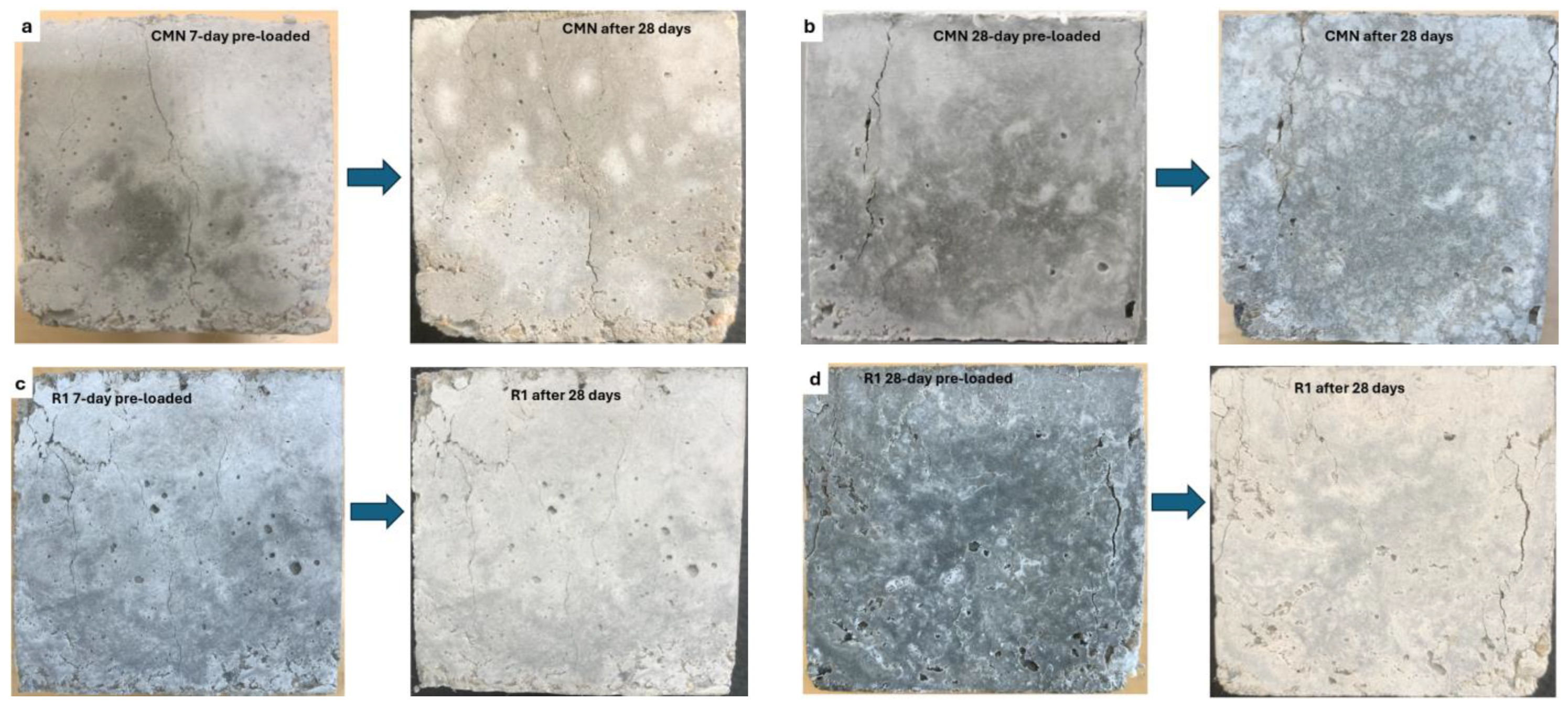
2.6. Specimens Pre-Cracking

The specimens were pre-cracked by employing an Instron hydraulic universal testing machine (UTM) in the material laboratory at Morgan State University as shown in Figure 2. Before pre-cracking, the specimens were dried in an indoor environment for one hour at the end of the 7-day curing period. Pre-cracking was facilitated by the embedded steel rod within the prisms. Both ends of the steel rod were anchored within the tension setup of the UTM. The tensile load was gradually applied until multiple visible cracks appeared within the concrete specimen. The crack width was controlled using the elastic yielding of the steel rod as an indicator, meaning that the test was terminated once the elastic yielding in the steel rod was converted to plastic yielding. With the permanent deformation in the steel rod, the induced crack width remained unaffected, avoiding elastic closure of the crack when the load was removed. While some elastic recovery of the steel rod was expected upon unloading, the extent of plastic deformation ensured that the residual crack width remained within the targeted range (0.1–0.45 mm). Crack widths were measured after unloading to confirm their stability, minimizing concerns regarding elastic closure.
Specimens without PVA fibers exhibited wider and more localized cracks due to the brittle nature of the concrete matrix, which led to sudden crack propagation upon loading, whereas specimens with PVA fibers showed finer, more distributed cracks as the fibers acted as crack arrestors, resisting crack widening and maintaining a more controlled crack opening. The target crack width range (0.1–0.45 mm) was achieved in both cases by adjusting the applied load; however, fiber-reinforced specimens generally required higher loads to induce visible cracks due to the fiber-bridging effect. Upon unloading, PVA fiber-reinforced specimens retained tighter cracks compared to unreinforced specimens, as the fibers reduced plastic deformation and prevented further crack widening. The pre-cracked specimens from various mixes were stored to heal in separate water containers to keep track of the healing process in the individual mix. The pre-cracked specimens were taken out after every 28 days for the UPV testing.

2.7. Self-Healing Evaluation

In the context of the current study, self-healing refers to the ability of pre-cracked concrete to heal the crack either through an autogenous mechanism or through MICP. Self-healing efficiency of microbial concrete, on the other hand, is the quantitative measure of the restoration of functional performance through MICP. The terms self-healing efficiency and healing efficiency are often used interchangeably; however, they have slightly different meanings depending on the context. Healing efficiency is a more general term that encompasses both self-healing (autonomous) and externally assisted healing (e.g., repair agents, external treatments, or manual interventions). In the context of bacterial-based self-healing concrete, the word self-healing efficiency is preferable as it typically quantifies the extent to which cracks or damage are repaired due to MICP or other self-healing mechanisms. In the current study, self-healing efficiency was measured using the visual assessment method to trace the healing of the crack size and the UPV testing to assess the internal healing of the concrete specimens.

2.7.1. Visual Assessment

In this method, the specimen surfaces with cracks were photographed every 28 days after the pre-cracking stage. The photographs were analyzed through ImageJ software (1.54d) to monitor the healing progress of the crack area [55]. In this study, for each mixture, three specimens were cast, cured, pre-cracked, and monitored for self-healing efficiency using the surface crack analysis method. Since crack formation varied among mixtures, we analyzed all visible surface cracks rather than selecting a single crack per specimen. Each specimen typically exhibited multiple cracks, and photographs of all cracked surfaces were taken before and after the healing period. A total of 12 images per mixture (4 per specimen) were documented and processed using ImageJ software to quantify crack closure. The self-healing efficiency of each mixture was determined based on the average crack area reduction from these images. ImageJ is an open-source software used for the analysis of images. The steps involved the conversion of the raw images into 8-bit images, followed by the scaling, black-and-white (B&W) threshold adjustments, and removal of unnecessary pixels through the binary-erode function. The B&W threshold could be readjusted, if necessary, to achieve the fine crack area profile. Finally, the crack area was subtracted from the total image area. Equation (1) describes self-healing efficiency based on the binary image processing method [12,55].
S e l f h e a l i n g   e f f i c i e n c y % = C 7 C n C 7 × 100
where C7 = crack area after pre-cracking at 7 days before healing, and Cn = crack area measured after pre-cracking on the nth day, such as 28 days, 56 days, 84 days, 112 days, etc.

2.7.2. UPV Assessment

UPV testing was useful in the assessment of self-healing caused internally within the concrete matrix without damaging the specimen. Through this method, the mechanical pulses pass through the specimen length from the transmitting transducer placed at one end surface of the specimen, received by the receiving transducer at the other end surface of the specimen. During ultrasonic pulse transmission, internal cracks disrupt the wave propagation, causing a longer transmission time to the receiver. It is important to note that while UPV effectively assesses the overall integrity of the concrete and tracks self-healing progress, it does not explicitly distinguish between multiple small cracks and a single large crack. Instead, it provides a cumulative assessment of the disruptions in wave propagation caused by internal voids and cracks. The measured UPV values represent the overall degree of damage or healing within the concrete matrix rather than the specific geometry or number of cracks. When the cracks are sealed with calcite precipitation, the medium becomes more homogeneous, allowing the pulse to travel faster and reducing the transmission time compared to the cracked state. Therefore, this method was adopted to trace the progress of internal crack healing through MICP and determine the efficiency of different bacteria in self-healing. In this method, enhancement in the UPV values for individual specimens was the indication of improvement in self-healing efficiency and was measured using Equation (2).
S e l f h e a l i n g   e f f i c i e n c y % = U P V 7 U P V n U P V 7 × 100
where UPV7 = UPV measured 7 days after pre-cracking and before healing, and UPVn = UPV measured after pre-cracking on the nth day, such as 28 days, 56 days, 84 days, 112 days, etc.

2.8. Mechanical and Freeze–Thaw Durability Tests

Compression and flexural tests were carried out to determine the influence of bacterial self-healing agents on the mechanical strength of the concrete using six mixes as shown in Table 1. The three mixes (CMN, R1, and R2) were made with no bacteria whereas the other three mixes were made with different bacteria (MSb, MSp, and MMe). CMN represents a control mix with 100% natural coarse aggregates and bacteria. R2 was the same as R1 except with the addition of AEA and PVA fibers to evaluate the effect of PVA fibers and AEA on the mechanical strength and freeze–thaw durability of concrete and compare it with the bacterial mixes.
For the compression strength test, 50 mm × 50 mm cubes were prepared for each mix and cured for 7 and 28 days. The small size of the specimens was chosen because of the costly bacterial spores. A uniaxial compression load was applied to the cube specimen at a speed of 0.1 MPa per second until failure [56].
For the flexural strength test, prisms having dimensions of 40 mm × 40 mm × 160 mm were prepared. This test was performed on 7-day and 28-day cured prismatic specimens by using a four-point bending setup. The specimen was applied at a loading rate of 0.1 mm/min until failure [57,58]. A set of three specimens was tested for each test in the UTM and the mean value was reported as a result.
A compression strength regain test was carried out to evaluate the compression strength restoration capabilities of various bacteria adopted in the current study. For this purpose, the cube specimens were pre-loaded with 60% of the ultimate load (failure load recorded during the compression strength test for each mix) at 7 and 28 days. The pre-loaded cube specimens from each mix were put back in water for healing purposes and were tested after 28 days for compression strength regain.
Freeze–thaw test was carried out to assess the influence of bacteria on the durability of concrete following ASTM C666 [59]. This test was performed on prisms having dimensions of 40 mm × 40 mm × 160 mm. According to this test, the prisms were cured in water for 14 days after casting and demolding. After 14 days, the prisms were shifted to the freeze–thaw cabinet. The cabinet was set to run for 36 cycles completing a total of 300 cycles. Each cycle consisted of raising the temperature from 0 to 40 °F and then lowering the temperature from 40 to 0 °F. After every 36 cycles, the prisms were measured for mass loss and relative dynamic elastic modulus (RDEM). The test terminates in cases when the mass loss ratio reaches 5% or when the RDEM decreases to 60% of the original value. If neither of these conditions were met, the test was concluded after 300 cycles. During mass measurement, the prisms were covered with a damp towel to prevent loss of moisture. Mass loss ratio, dynamic modulus of elasticity, and RDEM were calculated using Equations (3) [60], (4) [61], and (5) [62], respectively.
m t = m 0 m t m t × 100 %
where Δmt shows the mass loss rate after t number of cycles, mt shows the mass after t number of cycles, and m0 shows the original mass before the freeze–thaw exposure.
E d = ρ × V 2
where Ed represents the dynamic modulus, ρ represents the density of prism, and V represents the ultrasonic pulse velocity.
R D E M = E n E o × 100 %
where Eo and En indicate the dynamic moduli at 0 and after n number of freeze–thaw cycles, respectively.

2.9. Microstructural Characterization Tests

Microstructural tests such as SEM-EDS and XRD tests were conducted to describe the elemental composition, morphology, and mineralogy of the healing precipitate, respectively. For SEM analysis, a sample from the healed crack of each bacterial specimen was scratched and tested under high-resolution SEM (Hitachi S-5500, Hitachi, Tokyo, Japan) using a metal stub and carbon tape. Similarly, the scratched sample from the healed crack of each bacterial specimen was collected and tested under an X-ray diffraction spectrometer (Rigaku MiniFlex, Rigaku, Tokyo, Japan). The specifications involved in the test were Cu Kα radiation of 40 kV and 40 mA, a scanning rate of 5°/min, and a range from 10° to 80° (2θ).

3. Results and Discussion

3.1. Evaluation of Self-Healing Efficiency

Self-healing efficiency was evaluated using visual assessment aided by binary image processing and UPV analysis. The results obtained from the two methods are detailed in the following sections.

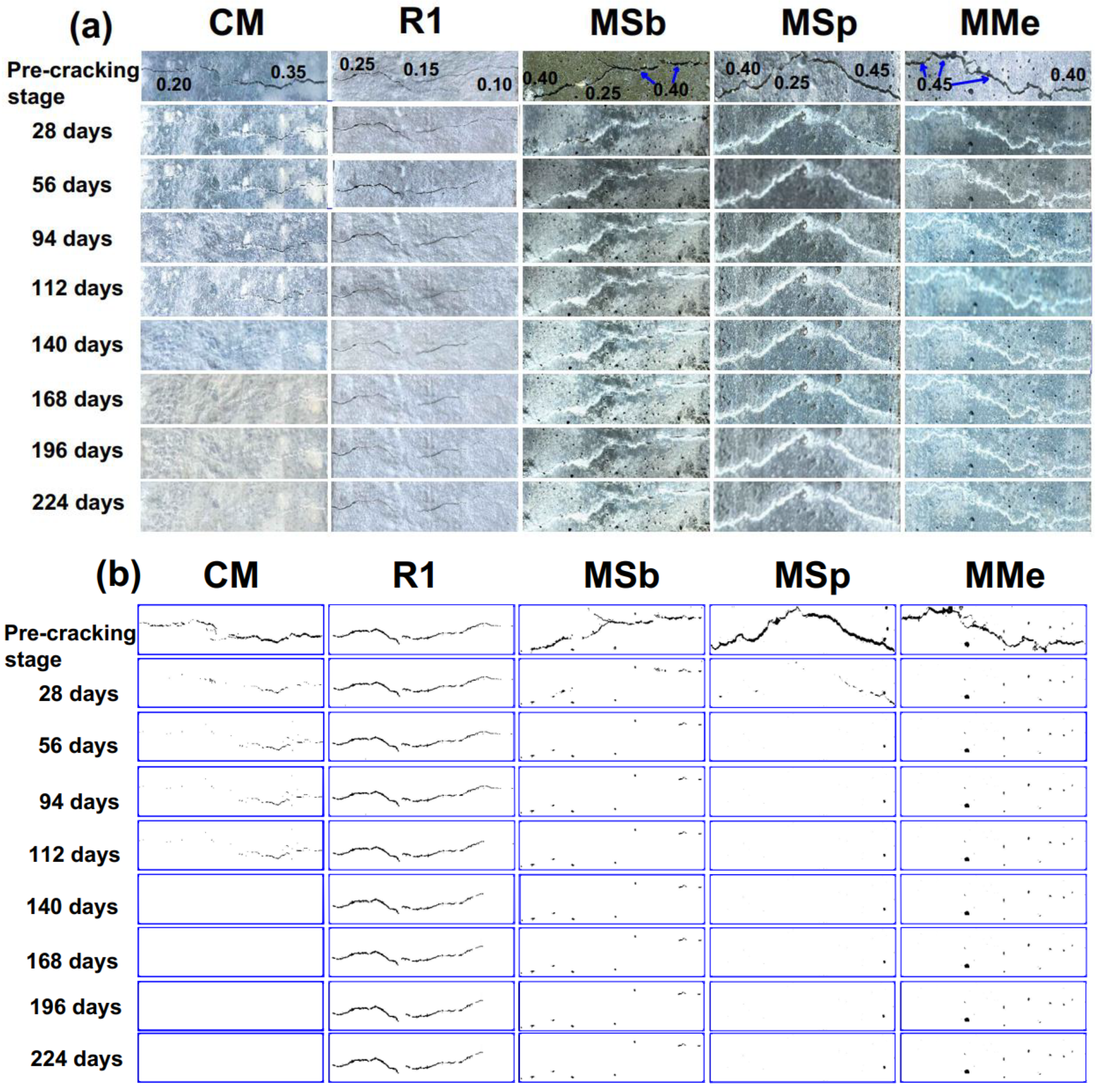
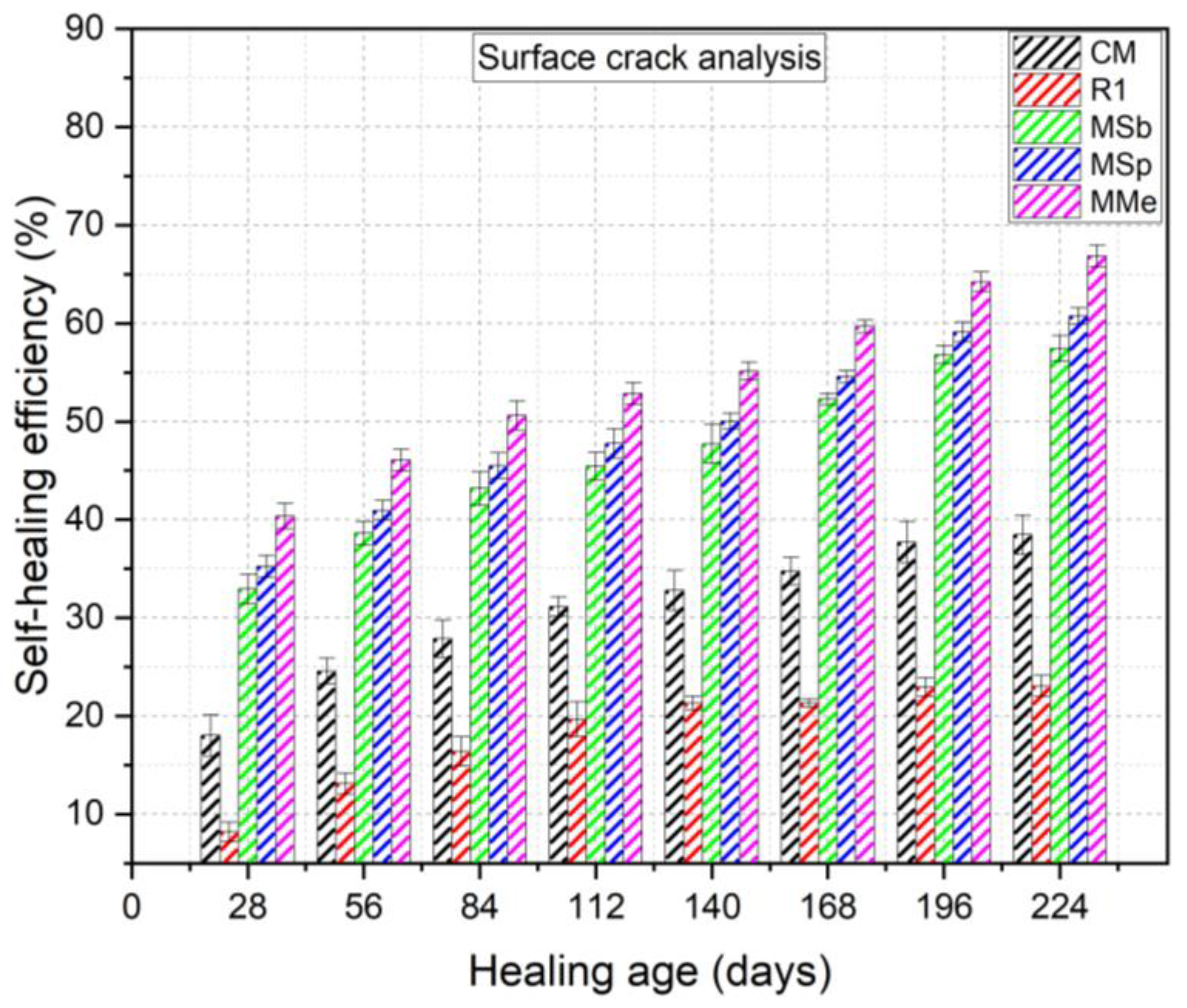
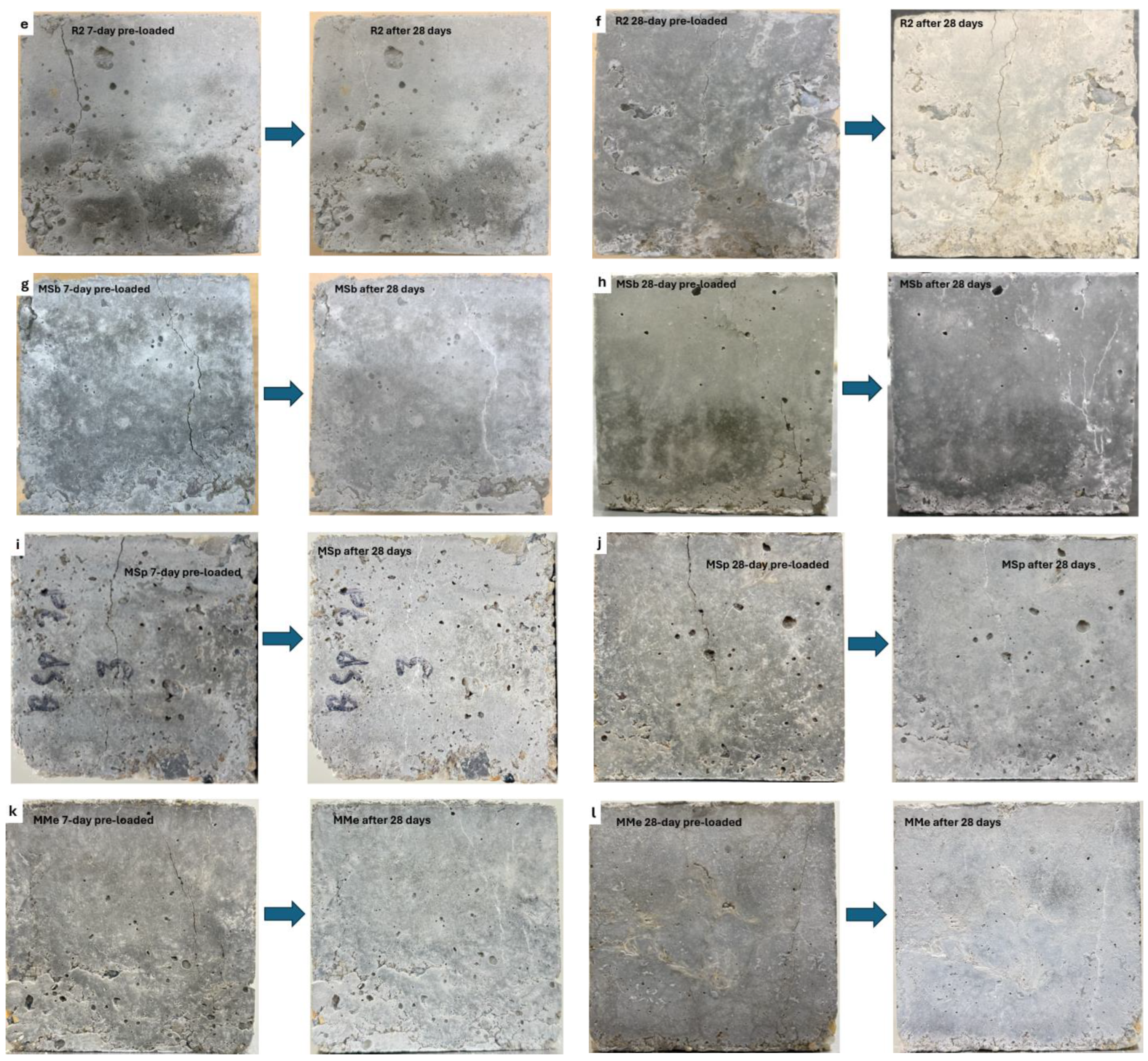
3.1.1. Visual Assessment and Analysis of Surface Cracks

The control, reference, and bioconcrete mixes, as mentioned in Table 1, were monitored for 224 days for visual assessment. The typical cracks in the prisms in which self-healing was observed visually are shown in Figure 3a. Visually it appeared that the cracks healed faster in mixes containing bacteria as compared to the reference RM specimens. The RM specimens with no bacteria showed no signs of healing over 28 days. However, a crack width of 0.10 mm appeared partially and fully healed autogenously at 56 and 94 days, respectively. The CM specimens that contained directly added bacteria exhibited partial healing with a crack width of 0.20 mm in the first 28 days. The bacterial mixes such as MSb, MSp, and MMe exhibited significant healing within the first 28 days with crack widths of 0.35 mm, 0.40 mm, and 0.45 mm, respectively. However, among the three bacteria, MMe specimens showed faster and complete healing of a crack width of 0.45 mm in the first 28 days, followed by MSp specimens with complete healing of a crack width of 0.40 mm and then MSb specimens with complete healing of 0.35 mm crack width and partial healing of 0.40 mm crack width. The dominant repair capability of Bacillus megaterium may be regarded due to a number of factors. First, Bacillus megaterium possesses an exosporium [63,64] in addition to its endospore [65], which provides an extra layer of protection against harsh environments, including the high alkalinity of cementitious matrices. The exosporium is the outermost surface layer covering the spore and helps to shield the bacteria from its harsh environment. This exosporium, which is not present in Bacillus subtilis and is structurally different in Bacillus sphaericus [64], enhances its survival rate and ability to withstand extreme pH conditions. Additionally, Bacillus megaterium exhibits a broader pH tolerance range [66] than Bacillus sphaericus, allowing it to thrive in both mild and highly alkaline conditions. This suggests that its effectiveness in self-healing concrete is not solely due to alkaline tolerance but also its inherent metabolic adaptability, likely driven by specific enzyme (called protease)-regulated responses to extreme environments [66].
Second, Bacillus megaterium’s larger cell size (3 to 5 μm in length) [67] compared to Bacillus subtilis (0.8 to 1.2 μm in length) [68] and Bacillus sphaericus (1 to 2 μm in length) [69] provides additional attachment sites for calcite precipitation. Since carbonate crystallization is heavily influenced by bacterial cell surfaces and extracellular polymeric substances (EPS) [70], Bacillus megaterium likely promotes superior self-healing due to increased nucleation sites for calcium carbonate formation. Findings in Lian et al. [71] indicate that biomineralization in Bacillus megaterium is a biologically induced process where bacterial cell walls and metabolic products facilitate nucleation and growth of calcite crystals, further enhancing crack-healing efficiency.
Together, these factors suggest that Bacillus megaterium’s superior repair mechanisms stem from a combination of its exosporium-mediated resilience, broad pH adaptability, and enhanced biomineralization capacity. This interplay between structural protection, metabolic activity, and crystallization dynamics likely contributes to its dominance in the effective self-healing of concrete cracks.
The crack-healing progress was also documented with photographs and processed in the ImageJ program. In this process, the four surfaces of the prisms having cracks were processed as described in Section 2.7.1, and the self-healing efficiency was calculated using Equation (1). Binary images of the typical cracks in representative specimens from each mix are shown in Figure 3b. In Figure 4, self-healing efficiency is presented as the average values of all the crack surfaces for the individual mix at a specified age. It was found that the maximum self-healing efficiency was exhibited by MMe (66.87%), followed by MSp (60.75%), MSb (57.45%), CM (38.45%), and then R1 (23.05%) after 224 days. It was indicated that crack healing was superior in bacterial mixes containing immobilized LECAs than in CM, though both mixes contained the same dosage of bacterial spores and nutrient media. The results indicated that the immobilization of bacterial spores in LECAs had superior performance rather than the direct addition of spores in the mix. From our previous study [12], it was identified that the PVA fibers and AEA assisted in the calcite precipitation by providing the attachment sites for the bacterial spores and carrying them through encapsulation, respectively. PVA fibers helped the spores in carrying and keeping them dormant until exposed to air or moisture upon crack formation and thus contributing towards calcite precipitation.

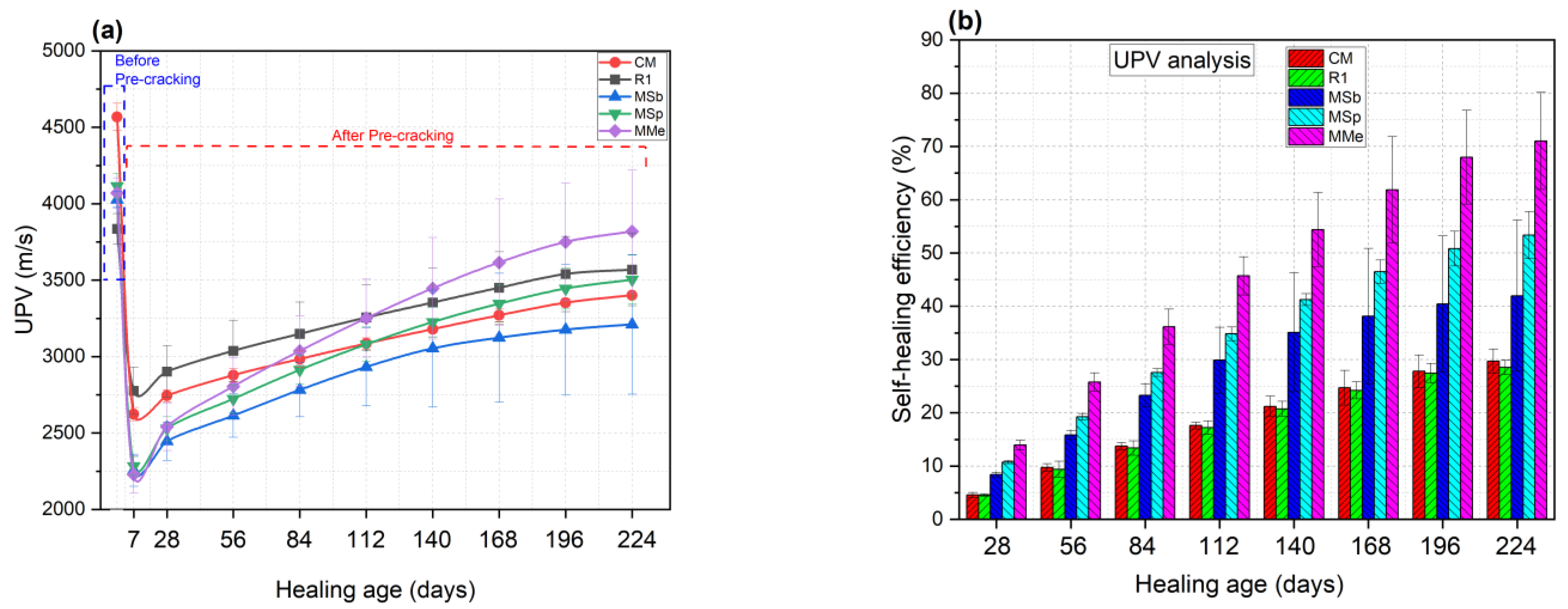
3.1.2. Self-Healing Effectiveness Based on UPV Analysis

The results of the UPV testing on the concrete prisms are shown in Figure 5a. UPV measurements were taken before and after the pre-cracking of the prisms. In the current study, UPV was used as an indicator of the internal self-healing ability of the various mixes. The UPV values recorded for the five mixes before pre-cracking varied because of the differences in their ingredients and distribution of the healing agents, such as CM which showed the highest UPV values than the rest of the mixes because of no LECA which was a part of the other mixes. Similarly, among the LECA-contained mixes, R1 exhibited the lowest UPV values because of the non-impregnated LECAs. However, in MSb, MSp, and MMe, the pores in LECAs were occupied by the nutrient and spores. After pre-cracking at 7 days, the UPV values dropped due to the presence of multiple cracks. Since the UPV propagates faster in hardened concrete than in water or air, when a pulse comes across a fissure, it will travel around the fissure and will result in lower UPV values. However, with the healing of cracks, the pulse velocity steadily increased with the healing period. The results of self-healing efficiency obtained from UPV analysis followed the same trend as evaluated by the surface crack analysis method. However, the resulting self-healing efficiency values in both methods were different. The self-healing efficiency values obtained using the UPV analysis method were lower than those from the surface crack analysis method. This may likely arise because surface cracks have more direct exposure to environmental conditions (moisture, CO₂, and oxygen), which facilitate microbial activity and calcite precipitation. In contrast, in UPV analysis, internal cracks may not be fully healed due to the uneven distribution or limited availability of healing agents. On the contrary, the surface crack analysis method dealt only with the surface cracks including both shallow and deep cracks. The cracking methodology adopted in this study, along with the UPV results, indicated that more internal cracks were generated rather than surface cracks. Future studies could further explore this aspect by employing advanced non-destructive techniques such as X-ray computed tomography (CT) or 3D digital image correlation (DIC) to quantitatively assess repair uniformity at different crack depths.
It was found that the maximum self-healing efficiency was exhibited in MMe (71.03%), followed by the MSp (53.38%), MSb (41.96%), CM (29.70%), and then R1 (28.53%) at the age of 224 days as shown in Figure 5b. These results indicated that bacterial mixes possessed higher self-healing efficiency than R1 at the age of 224 days, which means that the self-healing in the bacterial mixes was due to calcite precipitation by bacteria. Similarly, the higher self-healing efficiency of bacterial mixes than CM revealed that the spores in CM were added directly and may have compromised the viability of bacteria due to no protection in a carrier media. The difference in the self-healing performances of Bacillus subtilis, sphaericus, and megaterium was contingent on calcite-forming abilities, spore concentrations, and compatibility with the nutrients [56]. The bio-deposition rate varied among various bacterial species, attributed to their varying genetic composition and metabolic activities. Therefore, the superior self-healing performance of Bacillus megaterium was attributed to its fast growth and higher calcite formation capability which sealed the cracks and pores thus compacting the microstructure and allowing the easy propagation of UPV.
Self-healing efficiency increased with the healing age of the bacterial mixes. However, the rate of increase slowed down with time. In the first 28 days, self-healing was faster than the rest of the period because of the rapid cement hydration, which resulted in the formation of hydration products that aided bacterial calcite precipitation at an early age. Another reason was the abundant availability of nutrients at an early age, which were gradually consumed by the bacteria for calcite precipitation, reducing the rate of healing. For example, self-healing efficiencies for MSb, MSp, and MMe reduced from 8.41, 10.75, and 13.98% at 28 days to 1.51, 2.56, and 3.08% at 224 days, respectively.

3.2. Compressive Strength

The results of compressive strength for control, reference, and bacterial mixes are depicted in Figure 6a. It was noticed that the compression strengths of CMN outclassed the other mixes both at 7 and 28 days. The reason was because of the presence of 100% natural coarse aggregates. However, the other mixes including R1, R2, MSb, MSp, and MMe contained 20% LECAs as a partial replacement for natural coarse aggregates. Due to the porous nature of the LECAs, the 7-day compression strengths were reduced by 25.66, 5.61, 12.47, 11.68, and 8.60% for R1, R2, MSb, MSp, and MMe in comparison to CMN, respectively. Similarly, the 28-day compression strengths were reduced by 27.79, 18.09, 21.05, 20.81, and 19.23% for R1, R2, MSb, MSp, and MMe in comparison to CMN, respectively. Shivanshi et al. [43] obtained similar results stating that the 28-day compression strength of bacterial concrete containing immobilized LECAs with Bacillus coagulans and Lysinibacillus sphaericus decreased as compared to the non-LECA concrete. Tziviloglou et al. [72] also concluded that the replacement of sand with LECAs substantially decreased the bulk density of the concrete specimens and also the compression and flexural strengths.
The above results indicated that the inclusion of PVA fibers and bacterial spores reduced the losses in compressive strengths of both reference and bacterial mixes compared to CMN. The 7-day and 28-day compressive strengths were enhanced by 21.25 and 11.84%, respectively, for R2 as compared to R1. This was because of the bridging action of PVA fibers which held concrete intact as the cube specimens did not burst upon failure when subjected to ultimate load. The compressive strengths decreased by 3.61%, 3.30%, and 1.38% for MSB, MSp, and MMe as compared to R2 because of the calcium lactate. The addition of calcium lactate had a negative effect on the compressive strength of concrete when more than 0.5% of the cement mass is added to the concrete mix [73,74], whereas the dosage of calcium lactate employed in the current study was 5%. This may be due to high concentrations of calcium lactate which can interfere with cement hydration by adsorbing onto cement particles and inhibiting the formation of hydration products like C-S-H gel or impairing the interfacial bonding between the hydration products and the aggregates [75]. It was also noticed that the reduction in the compressive strength for MMe was lower than the MSp and MSb. For example, the 28-day compressive strengths decreased by 3.61%, 3.31%, and 1.38% for MSb, MSp, and MMe, respectively. These results showed the superior self-healing performance of Bacillus megaterium as compared to Bacillus subtilis and Bacillus sphaericus, confirming the results of self-healing efficiency. These results are in agreement with the previous study by Elmenshawy et al. [76]; their findings showed that the 28-day compression strength of concrete with Bacillus megaterium was 9.45% higher than the concrete with Bacillus sphaericus.

3.3. Restoration in Compression Strength

For compression strength restoration, the pre-loaded specimens experienced surface spalling and cracks. The percentage restored compressive strength for all the mixes is depicted in Figure 6b. The results indicated that compression strength was regained successfully for all the mixes after 28 days of healing, but the rate of regain in the non-bacterial mixes was not significant in comparison to the bacterial mixes. During the 28-day healing period, the specimens made with immobilized bacteria such as MSb, MSp, and MMe exhibited signs of calcite precipitation whereas the non-bacterial specimens showed no visible signs of self-healing as shown in Figure 7. Therefore, the regain in the non-bacterial specimens may be attributed to autogenous self-healing taking place internally. The 7-day preloaded compressive strengths were restored by 0.27%, 0.33%, 0.39%, 3.71%, 3.77%, and 3.84% for CMN, R1, R2, MSb, MSp, and MMe, respectively. Similarly, the 28-day preloaded compressive strength was restored by 0.21%, 0.31%, 0.18%, 2.43%, 2.30%, and 2.71% for CMN, R1, R2, MSb, MSp, and MMe, respectively. It was noticed that the specimens pre-loaded at 7 days exhibited higher percentages of restored compressive strength compared to the specimens pre-loaded at 28 days. This may be attributed to the ongoing cement hydration at early ages, which contributed to efficient compressive strength restoration of 7-day pre-loaded specimens in comparison to the 28-day pre-loaded specimens. Furthermore, the higher percentage of compressive strength restoration for bacterial specimens is evidence of calcite precipitation by the selected bacteria. In other words, the immobilized bacteria were able to utilize the nutrient media and carried out the metabolic activities, leading to the healing of the cracks induced by the pre-loading. The calcite precipitated within the healing zones, and densified the composite matrix which eventually led to the enhanced bonding strength of the composite. Furthermore, the superior performance of Bacillus megaterium can be attributed to the early activation of Bacillus megaterium, which initiated the biomineralization of calcium-based compounds. This process led to pore filling within the specimens through effective adhesion followed by crystallization [22].

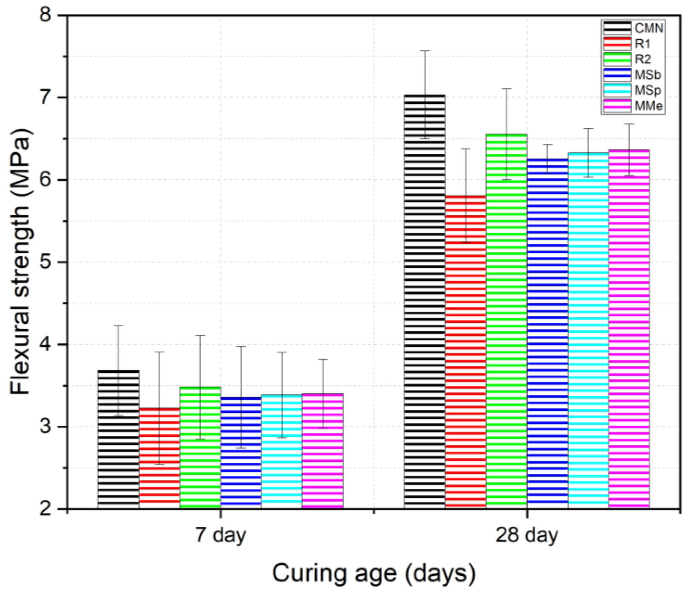
3.4. Flexural Strength

The flexural strength results for the control, reference, and bacterial mixes are shown in Figure 8. The flexural strength results followed the same trend as the compression strength. CMN exhibited the highest flexural strengths both at 7 and 28 days, followed by R2, bacterial mixes, and then R1. The losses in the flexural strengths for reference and bacterial mixes were due to the presence of 20% LECAs. However, the inclusion of PVA fibers as bacterial spores helped neutralize the negative impact of the LECAs on flexural strength. At the ultimate load, the CMN and R1 specimens failed into halves whereas the R2, MSb, MSp, and MMe specimens maintained their physical shape and avoided the brittle failure because of the presence of PVA fibers. The PVA fibers absorbed the tensile stresses during flexural loading, preventing the sample from splitting into two pieces. It was worth noting that the crack that appeared at the bottom of the specimens was large enough (ranging from 1 to 2 mm) to be healed by the bacteria. The flexural strength of the bacterial mixes is reported in the literature studies [32,77] to increase as compared to the non-bacterial mixes. However, in the current study, the loss in flexural strength for the bacterial mixes was attributed to the effect of calcium lactate on the strength performance of the concrete. Therefore, the current study suggests further research on the optimum dosage of calcium lactate while ensuring the long-term self-healing capabilities of bioconcrete. It was also noticed that the reduction in the flexural strength for MMe was lower than the MSp and MSb. For example, the 28-day flexural strengths decreased by 4.53%, 3.48%, and 2.89%, for MSb, MSp, and MMe, respectively. These results showed the superior self-healing performance of Bacillus megaterium followed by the Bacillus sphaericus and then Bacillus subtilis. The results of the current study are in agreement with the previous studies such as Rauf et al. [56] who reported that Bacillus sphaericus exhibited higher calcite precipitation ability as compared to Bacillus subtilis and Bacillus cohnii. Similarly, another study by Wani et al. [22] reported the superior self-healing ability of Bacillus megaterium over Bacillus sphaericus.

3.5. Freeze–Thaw Durability

In the current study, the freeze–thaw durability of various concrete mixes was evaluated using mass loss and RDEM indicators. The rate of mass loss of concrete indicates the extent of surface delamination and spalling caused by freeze–thaw cycles whereas the variation in the RDEM serves as an indicator of internal damage in concrete caused by freeze–thaw action. The specimens exposed to freeze–thaw cycles were evaluated for mass loss and RDEM measurements after every 36 cycles according to ASTM C666 [59]. Representative specimens from each mix after the completion of 300 freeze–thaw cycles are shown in Figure 9. Mass loss measurements of the bacterial and non-bacterial mixes are shown in Figure 10a. The mass losses measured at the end of 300 freeze–thaw cycles were 1.44%, 1.62%, 1.38%, 1.29%, 1.20%, and 1.16% for CMN, R1, R2, MSb, MSp, and MMe, respectively. These results showed that the highest mass loss was experienced by R1 followed by CMN, R2, MSb, MSp, and then MMe. During the freezing cycle, the existing water inside the concrete pores freezes and exerts an outward pressure contributing to surface deterioration or crack formation depending on the location of the pores in the concrete matrix. Before transferring to the freeze–thaw cabinet, the specimens were submerged in the water, making the pores saturated. In R1 specimens, the pores within the LECAs and in the concrete matrix were fully saturated with water. At low temperatures, the freezing of the pore water generated freezing pressure, leading to the peeling of the surface mortar. This caused small internal voids to interconnect, thereby exacerbating the freeze–thaw damage within the concrete [41]. The cracking of LECAs located near the surface of the specimen due to freeze–thaw action is evident in the R1 and R2 specimens as shown in Figure 9. The mass loss for CMN was relatively smaller than R1, likely because of the absence of LECAs, which may potentially increase overall pore content in R1. Furthermore, the mass loss was reduced in R2 in comparison to R1 and CMN due to the presence of PVA fibers and AEA. Due to freeze–thaw action, the degradation in concrete and surface peeling were mitigated by the PVA fibers efficiently holding the concrete intact, reducing the mass loss. In support of this, Nam et al. [78] reported a reduction in the mass loss after 300 freeze–thaw cycles for the cementitious composites reinforced with the PVA fibers. In addition, the presence of AEA facilitated the release of pressure caused by the freezing water and reduced the stress concentration in the concrete matrix [79]. At the same bacterial dosage, the lowest rate of mass loss was exhibited by MMe followed by the MSp and MSb, proving the superior calcite-forming capability of Bacillus megaterium validating the results of self-healing efficiency and mechanical strength. The reduced mass loss in bacterial specimens was attributed to the microbial activity carried out by directly added bacterial spores and the nutrient media. Yan et al. [4] also reported similar results that the frost resistance of concrete mix significantly improved with bacteria embedded in expanded perlite with a mass loss of 1.18% at 300 freeze–thaw cycles. During the thawing phase in a freeze–thaw cycle, with the rise in the ambient temperature, the bacteria germinate, perform microbial activity, and attract Ca2+ ions from the decomposition of calcium lactate, depositing calcite within the microcracks generated due to freeze–thaw action. The deposited calcite impedes the ingress of water, thus enhancing the freeze–thaw resistance of self-healing concrete. Cappellesso et al. [80] also concluded that after exposing cracked bacterial concrete to freeze–thaw cycles, the resistance to scaling was increased because of calcite deposition in the pores causing the densification in concrete.
The RDEM results for the bacterial and non-bacterial mixes are shown in Figure 10b. The RDEM values recorded at the end of 300 freeze–thaw cycles were 84.87%, 83.84%, 86.53%, 89.53%, 91.46%, and 93.87% for CMN, R1, R2, MSb, MSp, and MMe, respectively. These results indicate that the RDEM decreased by 15.13%, 16.66%, 13.47%, 10.47%, 8.54%, and 6.13% for CMN, R1, R2, MSb, MSp, and MMe, respectively. The RDEM results were consistent with the mass loss results. It was deduced that during freeze–thaw exposure, the initial spalling of mortar from the concrete surface was followed by gradual damage penetration into the interior, resulting in a decline in RDEM with the increase in the number of freeze–thaw cycles. The lower RDEM values observed in the bacterial mixes suggest that incorporating directly added spores with media and immobilized LECA enhanced resistance to freeze–thaw conditions. This improvement may also be attributed to two factors below. (1) The first factor is germination and subsequent calcite precipitation during the thawing and maintenance phase as reported in [4] that during thawing and maintenance, the embedded microorganisms are activated by moisture and oxygen, prompting them to germinate and interact with the self-healing components. This process helps mitigate freeze–thaw damage by repairing interconnected pores. (2) The second factor is polymer coating on the LECAs, which densified the interfacial transition zones (ITZs) between the LECAs and mortar, and is a critical zone affected by freeze–thaw action, as reported in [81]. In contrast, the CMN mix experienced internal damage caused by osmotic pressure, progressing from interconnected pores to the less dense ITZ. This facilitated damage propagation throughout the concrete matrix as the number of freeze–thaw cycles increased. The greatest loss in RDEM was observed in R1, indicating severe internal damage, likely due to the higher number of ITZs. Additionally, the round shape of LECAs in R1 may have contributed to a weaker (non-densified) ITZ and poor freeze–thaw resistance compared to natural coarse aggregates. However, R2 showed a lower decrease in RDEM compared to R1 and CMN, attributed to the addition of PVA fibers and AEA, which enhanced resistance to freeze–thaw damage. Overall, the results indicated that despite challenges like the high alkaline environment inside the concrete and freezing temperature limiting microbial activities, the bacteria can still slowly carry out the mineralization process. The pores in the concrete are cemented with the resulting mineralization products resulting in a densified composite and enhancing the freeze–thaw durability. A similar conclusion was also deduced by Ivaske et al. [82] that bacterial spores embedded in concrete can withstand low temperatures and freeze–thaw cycles, maintaining approximately 50% viability, enabling bacterial self-healing concrete that can be adopted in cold areas.

3.6. Microstructural Analysis

Microstructural analyses such as SEM-EDS and XRD tests were carried out to determine the morphology, chemical composition, and polymorphs of the self-healing precipitate in the bacterial mixes. SEM analysis showed that the rhombohedral crystals corresponding to calcite had formed in the cracks of all three bacterial mixes. Similar crystal morphologies have been reported in previous studies [15,83,84]. Calcite crystals observed in SEM analysis are marked by red arrows in Figure 11a,c,e. Examination of the scratched healing precipitate in MSb also showed minor contents of ettringite from the hydration of cement. The EDS analysis shown in Figure 11b,d,f for the healing precipitate scratched from the three bacterial mixes indicated the presence of calcium, carbon, and oxygen as abundant elements, confirming the formation of calcite. The EDS spectrum of the healing precipitate obtained from MMe showed a maximum content of calcium (39.08%) element, which was comparatively more than observed in MSb and MSp. These results indicated that a larger amount of calcium lactate was consumed by the Bacillus megaterium effectively producing more calcite.
XRD diffractograms for the three bacterial mixes MSb, MSp, and MMe are shown in Figure 12. The results of XRD analysis exhibited the calcite peaks and comparatively lower portlandite peaks for the three mixes, indicating the formation of calcite as a major crack-healing precipitate, as reported in previous studies [12,22,48]. The relative intensity peak difference among the bacterial mixes exhibits the highest calcite peak at 28 days for MMe followed by MSp and then MSb. These results indicate that calcite precipitation was higher for MMe as compared to MSp and MSb, showing consistency with the EDS results.

4. Conclusions

In the current study, a comparative analysis of the self-healing performance of three different bacteria including Bacillus subtilis, Bacillus sphaericus, and Bacillus megaterium immobilized in LECA was carried out. Their influence on the mechanical strength and freeze–thaw durability was investigated. Furthermore, a microstructural study was also conducted to describe the morphological shape and chemical composition of the self-healing precipitate. This study distinguished itself from prior work by integrating LECA as a bacterial carrier in self-healing concrete while specifically focusing on its performance under freeze–thaw conditions, an area that has received limited attention. Unlike previous studies that typically investigated single bacterial strains, this research provided a comparative analysis of multiple strains such as Bacillus megaterium, Bacillus sphaericus, and Bacillus subtilis, identifying the most effective strain for both self-healing efficiency and freeze–thaw durability. Additionally, the incorporation of PVA fibers, alongside bacteria and LECA, offered a unique synergy that improved crack control and mechanical performance. A comprehensive multi-scale characterization, including UPV analysis, strength recovery, freeze–thaw durability assessment, and microstructural evaluations through SEM, EDS, and XRD, further set this study apart by providing an in-depth understanding of the biomineralization process. The main findings are shown as follows.
  • Crack healing results exhibited that LECA is a promising carrier material for microorganisms, sheltering them for up to 224 days due to its internal porous structure, which accommodated both the microorganisms and nutrients while shielding them from the highly alkaline environment within the concrete.
  • Assessment of self-healing performance through UPV analysis demonstrated that MMe achieved the highest self-healing efficiency, with a value of 71.03%, followed by MSp (53.38%), MSb (41.96%), CM (29.70%), and R1 (28.53%) after 224 days. Additionally, the maximum crack width completely healed by MMe was 0.45 mm within the first 28 days, while MSp and MSb achieved complete healing of crack widths of 0.45 mm and 0.40 mm within 56 days, respectively. These results indicated the rapid germination and sequential calcite precipitation capability of Bacillus megaterium as compared to Bacillus subtilis and Bacillus sphaericus.
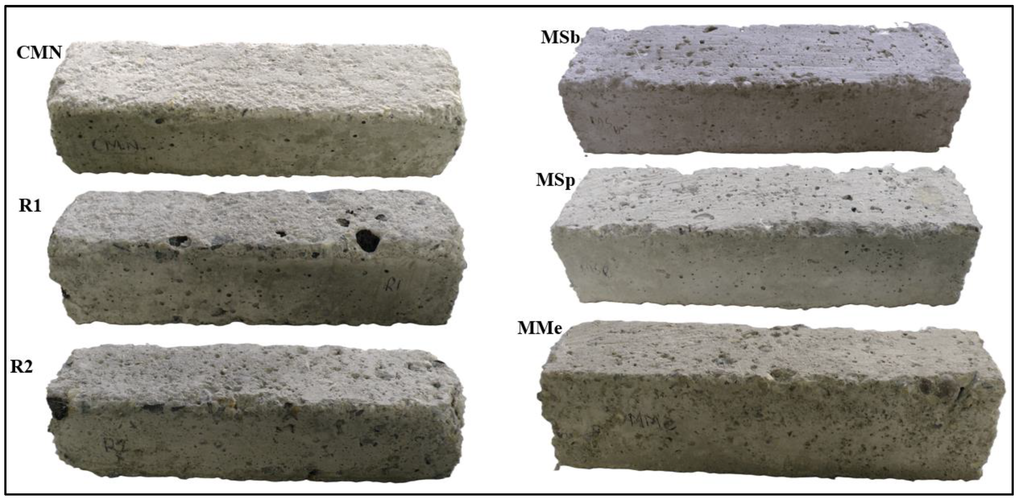
  • Compression and flexural strengths were reduced by the partial replacement of natural coarse aggregates with LECAs. However, this negative impact on strength behavior was slightly reduced with the inclusion of PVA fibers and bacteria, resulting in strength values higher than R1 but still lower than CMN. Furthermore, the slightly reduced strength behavior of bacterial mixes as compared to R2 was attributed to the impact of higher concentrations of calcium lactate.
  • Bacterial mixes successfully restored both 7-day and 28-day compression strengths after 28 days of healing, underscoring the effectiveness of biomineralization in improving the bond strength and densifying the matrix.
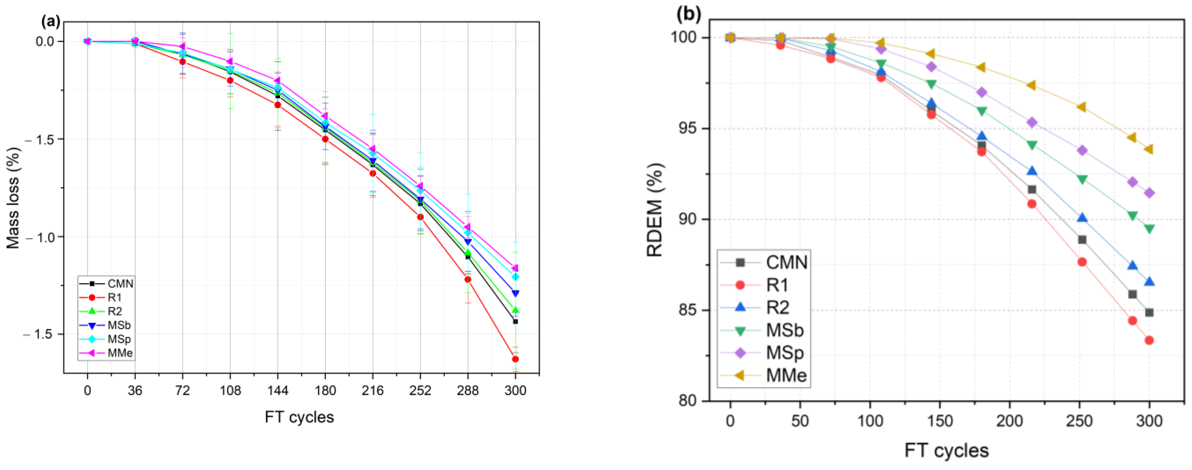
  • Freeze–thaw durability performance of bacterial mixes, particularly MMe, exhibited reduced mass loss and higher RDEM compared to control and reference mixes after 300 freeze–thaw cycles. The mass losses recorded at the end of 300 freeze–thaw cycles were 1.44%, 1.62%, 1.38%, 1.29%, 1.20%, and 1.16% for CMN, R1, R2, MSb, MSp, and MMe, respectively. Similarly, the RDEM values recorded at the end of 300 freeze–thaw cycles were 84.87%, 83.84%, 86.53%, 89.53%, 91.46%, and 93.87% for CMN, R1, R2, MSb, MSp, and MMe, respectively. These results indicated the contribution of calcite precipitation in the densification of the critical zones such as ITZs, impeding water ingress and thus reducing the freeze–thaw damage.
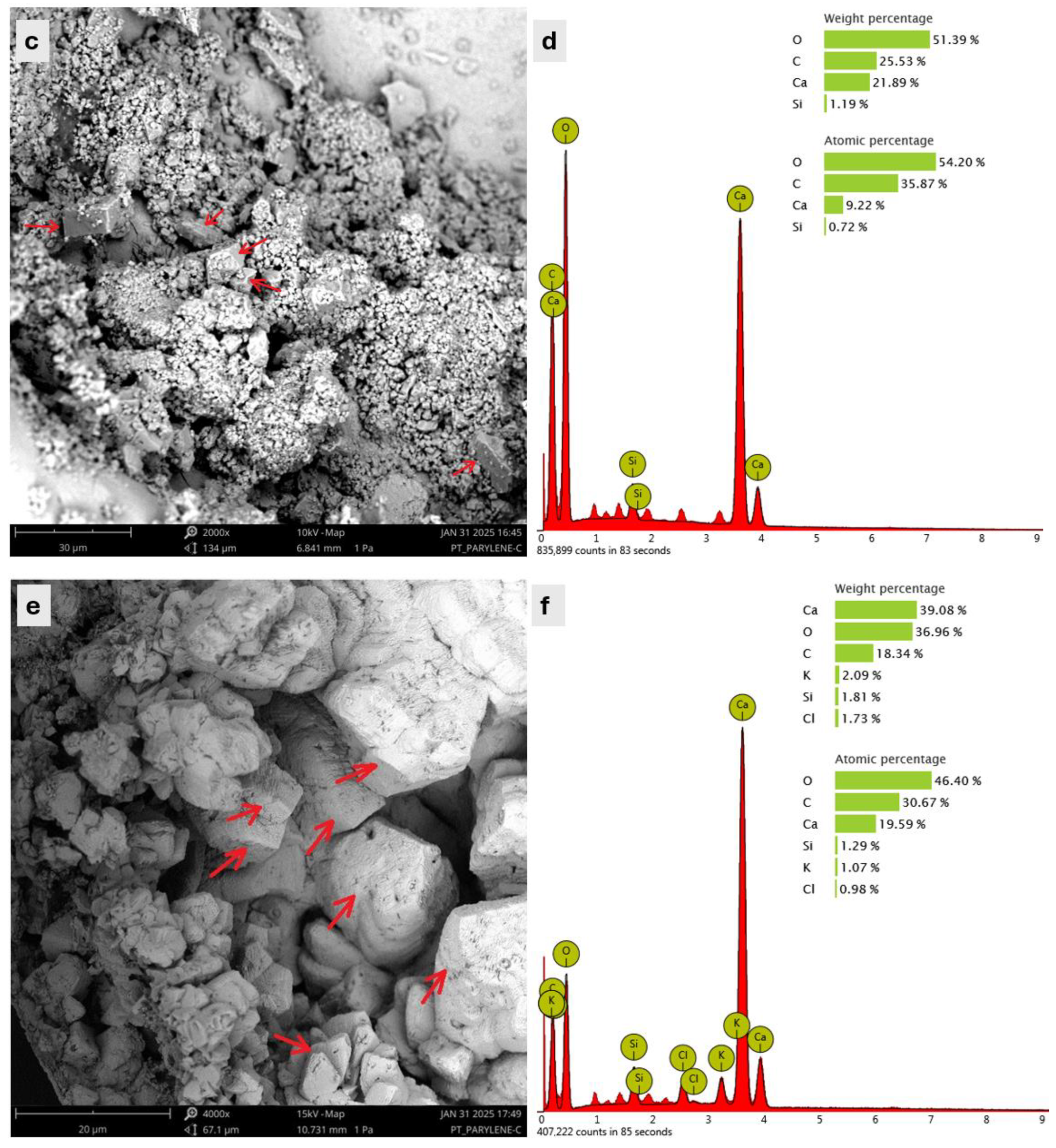
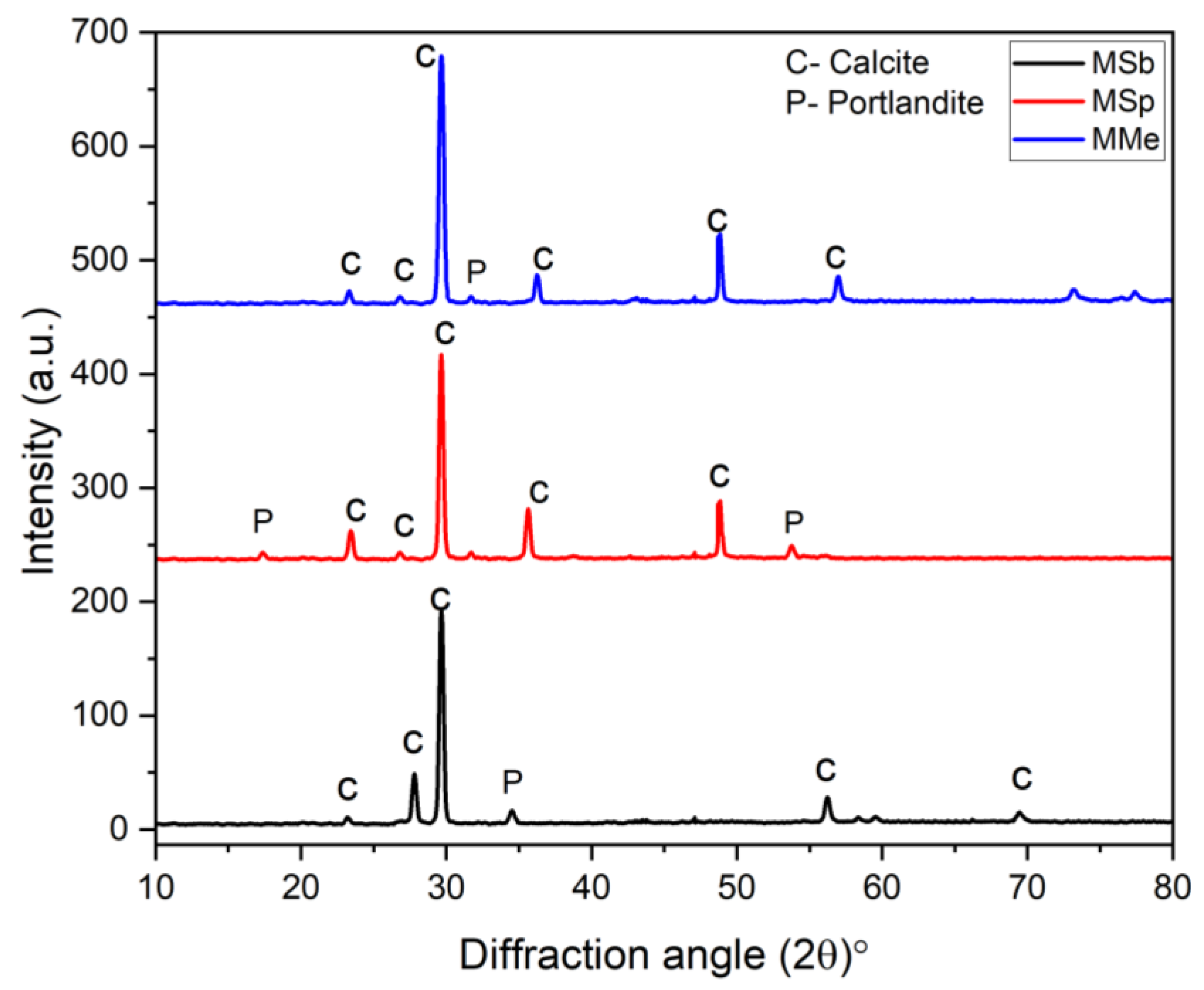
  • SEM analysis verified the formation of rhombohedral crystals composed of calcium, carbon, and oxygen elements corresponding to the precipitation of calcite as a healing product. Furthermore, the EDS and XRD results demonstrated a higher content of calcite formation in the case of MMe as compared to MSp and MSb at the healing age of 28 days, proving the fast rate of biomineralization capability of Bacillus megaterium.
The findings of this study can be adopted as a scientific foundation for the practical application of bacterial-based self-healing concrete in areas subjected to freeze–thaw environments. Future studies should focus on investigating the long-term self-healing performance of bacterial concrete in hot environments by adopting the same healing agents.

Author Contributions

Conceptualization, I.A., M.S., D.O. and M.J.; methodology, I.A.; software, I.A.; formal analysis, M.J. and G.L.M.; investigation, I.A. and D.O.; resources, M.S.; writing—original draft preparation, I.A.; writing—review and editing, I.A. and M.S.; visualization, M.J. and G.L.M.; supervision, M.S and G.L.M.; project administration, M.S.; funding acquisition, M.S. All authors have read and agreed to the published version of the manuscript.

Funding

This research received funding from the U.S. Department of Transportation’s University Transportation Centers Program through the Center for Integrated Asset Management for Multimodal Transportation Infrastructure Systems (CIAMTIS) (Grant No. 69A3551847103).

Data Availability Statement

The data presented in this study are available upon request from the corresponding author.

Conflicts of Interest

The authors declare no conflicts of interest.

References

  1. Ahmad, I.; Shen, D.; Khan, K.A.; Jan, A.; Khubaib, M.; Ahmad, T.; Nasir, H. Effectiveness of limestone powder in controlling the shrinkage behavior of cement based system: A review. Silicon 2021, 14, 359–371. [Google Scholar] [CrossRef]
  2. Liu, Y.; Li, W.; Guan, J.; Zhou, X.; Guo, L. Fully coupled peridynamic model for analyzing the chemo-diffusion-mechanical behavior of sulfate attack in concrete. Constr. Build. Mater. 2023, 409, 133874. [Google Scholar] [CrossRef]
  3. Chen, J.; Jia, J.; Zhu, M. Development of admixtures on seawater sea sand concrete: A critical review on concrete hardening, chloride ion penetration and steel corrosion. Constr. Build. Mater. 2024, 411, 134219. [Google Scholar] [CrossRef]
  4. Yan, Y.; Liu, W.; Li, Z.; Jia, G.; Zhang, Y.; Ma, G.; Gao, Y. Mechanical properties and frost resistance of self-healing concrete based on expanded perlite with different particle sizes as microbial carrier. Constr. Build. Mater. 2024, 422, 135450. [Google Scholar] [CrossRef]
  5. Mignon, A.; Snoeck, D.; Dubruel, P.; Van Vlierberghe, S.; De Belie, N. Crack mitigation in concrete: Superabsorbent polymers as key to success? Materials 2017, 10, 237. [Google Scholar] [CrossRef]
  6. Verma, D.; Sharma, M.; Goh, K.-L.; Jain, S.; Sharma, H. Sustainable Biopolymer Composites: Biocompatibility, Self-Healing, Modeling, Repair and Recyclability; Woodhead Publishing: Sawston, UK, 2021. [Google Scholar]
  7. Zhao, J.; Csetenyi, L.; Gadd, G.M. Fungal-induced CaCO3 and SrCO3 precipitation: A potential strategy for bioprotection of concrete. Sci. Total Environ. 2022, 816, 151501. [Google Scholar] [CrossRef]
  8. Khushnood, R.A.; Qureshi, Z.A.; Shaheen, N.; Ali, S. Bio-mineralized self-healing recycled aggregate concrete for sustainable infrastructure. Sci. Total Environ. 2020, 703, 135007. [Google Scholar] [CrossRef]
  9. Song, Y.; Chetty, K.; Garbe, U.; Wei, J.; Bu, H.; O’moore, L.; Li, X.; Yuan, Z.; McCarthy, T.; Jiang, G. A novel granular sludge-based and highly corrosion-resistant bio-concrete in sewers. Sci. Total Environ. 2021, 791, 148270. [Google Scholar] [CrossRef]
  10. Meera, C.; Subha, V. Strength and durability assessment of bacteria based self-healing concrete. IOSR J. Mech. Civ. Eng. 2016, 3, 1–7. [Google Scholar]
  11. Ahmad, I.; Shokouhian, M.; Jenkins, M.; McLemore, G.L. Factors influencing bacterial-based precipitation, assessment of crack inducing, durability and characterization methods: A comprehensive review. Innov. Infrastruct. Solut. 2025, 10, 107. [Google Scholar] [CrossRef]
  12. Ahmad, I.; Shokouhian, M.; Jenkins, M.; McLemore, G.L. Quantifying the self-healing efficiency of bioconcrete using Bacillus subtilis immobilized in polymer-coated lightweight expanded clay aggregates. Buildings 2024, 14, 3916. [Google Scholar] [CrossRef]
  13. Huynh, N.N.T.; Imamoto, K.-I.; Kiyohara, C. A study on biomineralization using Bacillus subtilis natto for repeatability of self-Healing concrete and strength improvement. J. Adv. Concr. Technol. 2019, 17, 700–714. [Google Scholar] [CrossRef]
  14. Ferral-Pérez, H.; Galicia-García, M. Bioprecipitation of calcium carbonate by Bacillus subtilis and its potential to self-healing in cement-based materials. J. Appl. Res. Technol. 2020, 18, 245–258. [Google Scholar]
  15. Nasser, A.A.; Sorour, N.M.; Saafan, M.A.; Abbas, R.N. Microbially-Induced-Calcite-Precipitation (MICP): A biotechnological approach to enhance the durability of concrete using Bacillus pasteurii and Bacillus sphaericus. Heliyon 2022, 8, e09879. [Google Scholar] [CrossRef]
  16. Wang, J.-Y.; De Belie, N.; Verstraete, W. Diatomaceous earth as a protective vehicle for bacteria applied for self-healing concrete. J. Ind. Microbiol. Biotechnol. 2012, 39, 567–577. [Google Scholar] [CrossRef]
  17. Wang, J.; Van Tittelboom, K.; De Belie, N.; Verstraete, W. Use of silica gel or polyurethane immobilized bacteria for self-healing concrete. Constr. Build. Mater. 2012, 26, 532–540. [Google Scholar] [CrossRef]
  18. Justo-Reinoso, I.; Reeksting, B.J.; Hamley-Bennett, C.; Heath, A.; Gebhard, S.; Paine, K. Air-entraining admixtures as a protection method for bacterial spores in self-healing cementitious composites: Healing evaluation of early and later-age cracks. Constr. Build. Mater. 2022, 327, 126877. [Google Scholar] [CrossRef]
  19. Ivaškė, A.; Gribniak, V.; Jakubovskis, R.; Urbonavičius, J. Bacterial viability in self-healing concrete: A case study of non-ureolytic Bacillus species. Microorganisms 2023, 11, 2402. [Google Scholar] [CrossRef]
  20. Bifathima, S.; Matcha, B.N. Self healing concrete by adding Bacillus megaterium MTCC with glass & steel fibers. Civ. Environ. Eng. 2020, 16, 184–197. [Google Scholar]
  21. Su, Y.; Zheng, T.; Qian, C. Application potential of Bacillus megaterium encapsulated by low alkaline sulphoaluminate cement in self-healing concrete. Constr. Build. Mater. 2021, 273, 121740. [Google Scholar] [CrossRef]
  22. Wani, S.; Gęca, M.J.; Selvaraj, T.; Priya, T.S. Assessing the influence of Bacillus megaterium and Bacillus sphaericus in cementitious materials: Promoting sustainability towards strength, durability and crack repair. Ain Shams Eng. J. 2024, 15, 102748. [Google Scholar] [CrossRef]
  23. Bang, S.S.; Galinat, J.K.; Ramakrishnan, V. Calcite precipitation induced by polyurethane-immobilized Bacillus pasteurii. Enzym. Microb. Technol. 2001, 28, 404–409. [Google Scholar] [CrossRef]
  24. Sarda, D.; Choonia, H.S.; Sarode, D.; Lele, S. Biocalcification by Bacillus pasteurii urease: A novel application. J. Ind. Microbiol. Biotechnol. 2009, 36, 1111–1115. [Google Scholar] [CrossRef]
  25. Omar, O.; Noorvand, H.; Hassan, M.; Subedi, S.; Milla, J.; Rupnow, T. Effect of using calcium lactate with Bacillus pseudofirmus bacteria on self-healing efficiency of bacterial concrete. Tran-SET 2022, 2022, 67–76. [Google Scholar]
  26. Elgendy, I.M.; Elkaliny, N.E.; Saleh, H.M.; Darwish, G.O.; Almostafa, M.M.; Metwally, K.; Yahya, G.; Mahmoud, Y.A. Bacteria-powered self-healing concrete: Breakthroughs, challenges, and future prospects. J. Ind. Microbiol. Biotechnol. 2024, 52, kuae051. [Google Scholar] [CrossRef] [PubMed]
  27. Feng, J.; Chen, B.; Sun, W.; Wang, Y. Microbial induced calcium carbonate precipitation study using Bacillus subtilis with application to self-healing concrete preparation and characterization. Constr. Build. Mater. 2021, 280, 122460. [Google Scholar] [CrossRef]
  28. Elmahdy, M.A.; ELShami, A.; Yousry, E.-S.M.; Ahmad, S.S. Self-healing mortar using different types, content, and concentrations of bacteria to repair cracks. Frat. Integrita Strutt. 2022, 59, 486–513. [Google Scholar]
  29. Jonkers, H.M.; Thijssen, A.; Muyzer, G.; Copuroglu, O.; Schlangen, E. Application of bacteria as self-healing agent for the development of sustainable concrete. Ecol. Eng. 2010, 36, 230–235. [Google Scholar] [CrossRef]
  30. Zhang, J.; Liu, Y.; Feng, T.; Zhou, M.; Zhao, L.; Zhou, A.; Li, Z. Immobilizing bacteria in expanded perlite for the crack self-healing in concrete. Constr. Build. Mater. 2017, 148, 610–617. [Google Scholar] [CrossRef]
  31. Mostofinejad, D.; Nosouhian, F.; Tayebani, B. Evaluation of mechanical properties of bacteria-containing mortar in seawater environment. ACI Mater. J. 2021, 118, 35–43. [Google Scholar]
  32. Ahmed, S.O.; Nasser, A.A.; Abbas, R.N.; Kamal, M.M.; Zahran, M.A.; Sorour, N.M. Production of bioconcrete with improved durability properties using Alkaliphilic Egyptian bacteria. 3 Biotech 2021, 11, 231. [Google Scholar] [CrossRef] [PubMed]
  33. Chaerun, S.K.; Syarif, R.; Wattimena, R.K. Bacteria incorporated with calcium lactate pentahydrate to improve the mortar properties and self-healing occurrence. Sci. Rep. 2020, 10, 17873. [Google Scholar] [CrossRef]
  34. Achal, V.; Pan, X.; Özyurt, N. Improved strength and durability of fly ash-amended concrete by microbial calcite precipitation. Ecol. Eng. 2011, 37, 554–559. [Google Scholar] [CrossRef]
  35. Zhang, X.; Qian, C. A new type capsule-based healing agent for concrete and its protective function of spores. Smart Mater. Struct. 2020, 29, 105035. [Google Scholar] [CrossRef]
  36. Wang, J.; Soens, H.; Verstraete, W.; De Belie, N. Self-healing concrete by use of microencapsulated bacterial spores. Cem. Concr. Res. 2014, 56, 139–152. [Google Scholar] [CrossRef]
  37. Wang, J.; Snoeck, D.; Van Vlierberghe, S.; Verstraete, W.; De Belie, N. Application of hydrogel encapsulated carbonate precipitating bacteria for approaching a realistic self-healing in concrete. Constr. Build. Mater. 2014, 68, 110–119. [Google Scholar] [CrossRef]
  38. Wang, J.; Mignon, A.; Trenson, G.; Van Vlierberghe, S.; Boon, N.; De Belie, N. A chitosan based pH-responsive hydrogel for encapsulation of bacteria for self-sealing concrete. Cem. Concr. Compos. 2018, 93, 309–322. [Google Scholar] [CrossRef]
  39. Palin, D.; Wiktor, V.; Jonkers, H. A bacteria-based bead for possible self-healing marine concrete applications. Smart Mater. Struct. 2016, 25, 084008. [Google Scholar] [CrossRef]
  40. Shahid, S.; Aslam, M.A.; Ali, S.; Zameer, M.; Faisal, M. Self-healing of cracks in concrete using Bacillus strains encapsulated in sodium alginate beads. ChemistrySelect 2020, 5, 312–323. [Google Scholar] [CrossRef]
  41. Yan, Y.; Jia, G.; Zhang, Y.; Gao, Y.; Li, Z. The influence of expanded perlite as a bio-carrier on the freeze–thaw properties of self-healing concrete. Constr. Build. Mater. 2023, 409, 133891. [Google Scholar] [CrossRef]
  42. Prasad, P.; Neeraja, D.; Ravitheja, A. Effect of LECA on mechanical properties of self-curing concrete. Mater. Today Proc. 2019, 19, 484–488. [Google Scholar]
  43. Shivanshi, S.; Chakraborti, G.; Upadhyaya, K.S.; Kannan, N. A study on bacterial self-healing concrete encapsulated in lightweight expanded clay aggregates. Mater. Today Proc. 2023. [Google Scholar] [CrossRef]
  44. Yan, Y.; Tian, H.; Liu, W.; Jia, G.; Li, Z.; Gao, Y.; Zhang, Y.; Ma, G. Study on frost resistance and life prediction of microbial self-healing concrete based on expanded perlite as carrier. J. Build. Eng. 2024, 91, 109693. [Google Scholar] [CrossRef]
  45. Liu, Z.; Chin, C.S.; Xia, J. Novel method for enhancing freeze–thaw resistance of recycled coarse aggregate concrete via two-stage introduction of denitrifying bacteria. J. Clean. Prod. 2022, 346, 131159. [Google Scholar] [CrossRef]
  46. ASTM C150/C150M–19a; Standard Specification for Portland Cement. American Society for Testing and Materials (ASTM) International: West Conshohocken, PA, USA, 2019.
  47. ASTM C127–15; Standard Test Method for Relative Density (Specific Gravity) and Absorption of Coarse Aggregate. ASTM International: West Conshohocken, PA, USA, 2015.
  48. Khaliq, W.; Ehsan, M.B. Crack healing in concrete using various bio influenced self-healing techniques. Constr. Build. Mater. 2016, 102, 349–357. [Google Scholar] [CrossRef]
  49. Pachaivannan, P.; Hariharasudhan, C.; Mohanasundram, M.; Bhavani, M.A. Experimental anaylsis of self healing properties of bacterial concrete. Mater. Today Proc. 2020, 33, 3148–3154. [Google Scholar] [CrossRef]
  50. Hossain, M.R.; Sultana, R.; Patwary, M.M.; Khunga, N.; Sharma, P.; Shaker, S.J. Self-healing concrete for sustainable buildings. A review. Environ. Chem. Lett. 2022, 20, 1265–1273. [Google Scholar] [CrossRef]
  51. Ducasse-Lapeyrusse, J.; Gagné, R.; Lors, C.; Damidot, D. Effect of calcium gluconate, calcium lactate, and urea on the kinetics of self-healing in mortars. Constr. Build. Mater. 2017, 157, 489–497. [Google Scholar] [CrossRef]
  52. Abo Sabah, S.H.; Anneza, L.H.; Juki, M.I.; Alabduljabbar, H.; Othman, N.; Al-Gheethi, A.A.; Al-Shalif, A.F. The use of calcium lactate to enhance the durability and engineering properties of bioconcrete. Sustainability 2021, 13, 9269. [Google Scholar] [CrossRef]
  53. Lucas, S.S.; Moxham, C.; Tziviloglou, E.; Jonkers, H. Study of self-healing properties in concrete with bacteria encapsulated in expanded clay. Sci. Technol. Mater. 2018, 30, 93–98. [Google Scholar] [CrossRef]
  54. Xu, J.; Du, Y.; Jiang, Z.; She, A. Effects of calcium source on biochemical properties of microbial CaCO3 precipitation. Front. Microbiol. 2015, 6, 1366. [Google Scholar] [CrossRef] [PubMed]
  55. Rong, H.; Wei, G.; Ma, G.; Zhang, Y.; Zheng, X.; Zhang, L.; Xu, R. Influence of bacterial concentration on crack self-healing of cement-based materials. Constr. Build. Mater. 2020, 244, 118372. [Google Scholar] [CrossRef]
  56. Rauf, M.; Khaliq, W.; Khushnood, R.A.; Ahmed, I. Comparative performance of different bacteria immobilized in natural fibers for self-healing in concrete. Constr. Build. Mater. 2020, 258, 119578. [Google Scholar] [CrossRef]
  57. Alghamri, R.; Kanellopoulos, A.; Al-Tabbaa, A. Impregnation and encapsulation of lightweight aggregates for self-healing concrete. Constr. Build. Mater. 2016, 124, 910–921. [Google Scholar] [CrossRef]
  58. Kanellopoulos, A.; Qureshi, T.; Al-Tabbaa, A. Glass encapsulated minerals for self-healing in cement based composites. Constr. Build. Mater. 2015, 98, 780–791. [Google Scholar] [CrossRef]
  59. ASTM C666; Standard Test Method for Resistance of Concrete to Rapid Freezing and Thawing. ASTM International: West Conshohocken, PA, USA, 2015.
  60. Zou, D.; Wang, Z.; Shen, M.; Liu, T.; Zhou, A. Improvement in freeze-thaw durability of recycled aggregate permeable concrete with silane modification. Constr. Build. Mater. 2021, 268, 121097. [Google Scholar] [CrossRef]
  61. Li, Q.; Zheng, J.; Fu, M. Effect of molding methods on free-thaw durability of pervious concrete. Proc. J. Phys. Conf. Ser. 2023, 2424, 012015. [Google Scholar] [CrossRef]
  62. Chen, S.; Ren, J.; Ren, X.; Li, Y. Deterioration laws of concrete durability under the coupling action of salt erosion and drying–wetting cycles. Front. Mater. 2022, 9, 1003945. [Google Scholar] [CrossRef]
  63. Lanzilli, M.; Donadio, G.; Addevico, R.; Saggese, A.; Cangiano, G.; Baccigalupi, L.; Christie, G.; Ricca, E.; Isticato, R. The exosporium of Bacillus megaterium QM B1551 is permeable to the red fluorescence protein of the coral Discosoma sp. Front. Microbiol. 2016, 7, 1752. [Google Scholar] [CrossRef]
  64. Stewart, G.C. The exosporium layer of bacterial spores: A connection to the environment and the infected host. Microbiol. Mol. Biol. Rev. 2015, 79, 437–457. [Google Scholar] [CrossRef]
  65. Checinska, A.; Paszczynski, A.; Burbank, M. Bacillus and other spore-forming genera: Variations in responses and mechanisms for survival. Annu. Rev. Food Sci. Technol. 2015, 6, 351–369. [Google Scholar] [CrossRef] [PubMed]
  66. Uttatree, S.; Kobtrakool, K.; Ketsuk, A.; Kaenngam, W.; Thakolprajak, P.; Charoenpanich, J. A novel metal-tolerant, solvent and surfactant stable protease from a new strain of Bacillus megaterium. Biocatal. Agric. Biotechnol. 2017, 12, 228–235. [Google Scholar] [CrossRef]
  67. Grage, K.; McDermott, P.; Rehm, B.H. Engineering Bacillus megaterium for production of functional intracellular materials. Microb. Cell Factories 2017, 16, 211. [Google Scholar] [CrossRef] [PubMed]
  68. Zhang, X.; Al-Dossary, A.; Hussain, M.; Setlow, P.; Li, J. Applications of Bacillus subtilis spores in biotechnology and advanced materials. Appl. Environ. Microbiol. 2020, 86, e01096-20. [Google Scholar] [CrossRef]
  69. Obidi, O.; Wenner, S.; Halverson, L. Molecular-based analysis of the bacterial community structure of spoilt latex paints in Nigeria. Egypt. Soc. Exp. Biol. 2017, 13, 145–149. [Google Scholar] [CrossRef]
  70. Enyedi, N.T.; Makk, J.; Kótai, L.; Berényi, B.; Klébert, S.; Sebestyén, Z.; Molnár, Z.; Borsodi, A.K.; Leél-Őssy, S.; Demény, A. Cave bacteria-induced amorphous calcium carbonate formation. Sci. Rep. 2020, 10, 8696. [Google Scholar] [CrossRef]
  71. Lian, B.; Hu, Q.; Chen, J.; Ji, J.; Teng, H.H. Carbonate biomineralization induced by soil bacterium Bacillus megaterium. Geochim. Cosmochim. Acta 2006, 70, 5522–5535. [Google Scholar] [CrossRef]
  72. Tziviloglou, E.; Wiktor, V.; Jonkers, H.; Schlangen, E. Bacteria-based self-healing concrete to increase liquid tightness of cracks. Constr. Build. Mater. 2016, 122, 118–125. [Google Scholar] [CrossRef]
  73. Vijay, K.; Murmu, M. Effect of calcium lactate on compressive strength and self-healing of cracks in microbial concrete. Front. Struct. Civ. Eng. 2019, 13, 515–525. [Google Scholar] [CrossRef]
  74. Bashaveni, B.; Pannem, R.M.R. Development of self-healing property in self compacting concrete. Case Stud. Constr. Mater. 2024, 20, e02942. [Google Scholar] [CrossRef]
  75. Wang, X.; Xu, J.; Wang, Z.; Yao, W. Use of recycled concrete aggregates as carriers for self-healing of concrete cracks by bacteria with high urease activity. Constr. Build. Mater. 2022, 337, 127581. [Google Scholar] [CrossRef]
  76. Elmenshawy, Y.; Elmahdy, M.A.; Moawad, M.; Elshami, A.A.; Ahmad, S.S.; Nagai, K. Investigating the bacterial sustainable self-healing capabilities of cracks in structural concrete at different temperatures. Case Stud. Constr. Mater. 2024, 20, e03188. [Google Scholar] [CrossRef]
  77. Gupta, S.; Kua, H.W.; Pang, S.D. Healing cement mortar by immobilization of bacteria in biochar: An integrated approach of self-healing and carbon sequestration. Cem. Concr. Compos. 2018, 86, 238–254. [Google Scholar] [CrossRef]
  78. Nam, J.; Kim, G.; Lee, B.; Hasegawa, R.; Hama, Y. Frost resistance of polyvinyl alcohol fiber and polypropylene fiber reinforced cementitious composites under freeze thaw cycling. Compos. Part B Eng. 2016, 90, 241–250. [Google Scholar] [CrossRef]
  79. He, R.; Lu, N.L. Air void system and freezing-thawing resistance of concrete composite with the incorporation of thermo-expansive polymeric microspheres. Constr. Build. Mater. 2024, 419, 135535. [Google Scholar] [CrossRef]
  80. Cappellesso, V.G.; Van Mullem, T.; Gruyaert, E.; Van Tittelboom, K.; De Belie, N. Bacteria-based self-healing concrete exposed to frost salt scaling. Cem. Concr. Compos. 2023, 139, 105016. [Google Scholar] [CrossRef]
  81. Ahmad, I.; Shokouhian, M.; Cheng, H.; Radlińska, A. Enhancement of mechanical properties and freeze–thaw durability of recycled aggregate concrete using aggregate pretreatment. Iran. J. Sci. Technol. Trans. Civ. Eng. 2024, 1–25. [Google Scholar] [CrossRef]
  82. Ivaškė, A.; Jakubovskis, R.; Boris, R.; Urbonavičius, J. Effects of low temperature, freeze–thaw cycles, and healing conditions on viability of non-ureolytic bacteria in biological self-healing concrete. Materials 2024, 17, 5797. [Google Scholar] [CrossRef]
  83. Seifan, M.; Sarmah, A.K.; Ebrahiminezhad, A.; Ghasemi, Y.; Samani, A.K.; Berenjian, A. Bio-reinforced self-healing concrete using magnetic iron oxide nanoparticles. Appl. Microbiol. Biotechnol. 2018, 102, 2167–2178. [Google Scholar] [CrossRef]
  84. Wiktor, V.; Jonkers, H.M. Quantification of crack-healing in novel bacteria-based self-healing concrete. Cem. Concr. Compos. 2011, 33, 763–770. [Google Scholar] [CrossRef]
Figure 1. Flowchart of the immobilization process of LECAs.
Buildings 15 00943 g001
Figure 2. Pre-cracking of concrete specimens.
Buildings 15 00943 g002
Figure 3. Crack healing in control, reference, and bacterial concrete mixes: (a) visual assessment; (b) binary image analysis.
Buildings 15 00943 g003
Figure 4. Self-healing efficiency based on surface crack analysis for bacterial and non-bacterial concrete mixes.
Buildings 15 00943 g004
Figure 5. UPV analysis for bacterial and non-bacterial concrete mixes: (a) UPV values from testing; (b) self-healing efficiency based on UPV testing.
Buildings 15 00943 g005
Figure 6. Compression test of concrete mixes: (a) compressive strength; (b) restored compression strength.
Buildings 15 00943 g006
Figure 7. Pre-loaded cube specimens till re-loading: (a) CMN at 7 days; (b) CMN at 28 days; (c) R1 at 7 days; (d) R1 at 28 days; (e) R2 at 7 days; (f) R2 at 28 days; (g) MSb at 28 days; (h) MSb at 28 days; (i) MSp at 7 days; (j) MSp at 28 days; (k) MMe at 7 days; (l) MMe at 28 days.
Buildings 15 00943 g007aBuildings 15 00943 g007b
Figure 8. Flexural strengths of concrete mixes.
Buildings 15 00943 g008
Figure 9. Prism specimens after 300 freeze–thaw cycles.
Buildings 15 00943 g009
Figure 10. Freeze–thaw resistance indicators for control, reference, and bacterial mixes: (a) rate of mass loss; (b) RDEM.
Buildings 15 00943 g010
Figure 11. SEM with EDX analysis of the crack-healing precipitate in mixes, respectively, red arrows indicate the calcite crystals (a,b) MSb, (c,d) MSp, and (e,f) MMe.
Buildings 15 00943 g011aBuildings 15 00943 g011b
Figure 12. XRD patterns of healing products in various bacterial mixes.
Buildings 15 00943 g012
Table 1. Mix proportioning for self-healing concrete mixes.
Materials (per m3 Concrete)UnitsCMCMNR1R2MSbMSpMMe
Cementkg483483483483483483483
Natural coarse aggregatekg755755604604604604604
Fine aggregatekg933933933933933933933
LECAkg00151151151151151
w/c ratio-0.450.450.450.450.450.450.45
Amount of spores (per gram of LECAs)µL20---202020
Air-entraining admixtureL---1.451.451.451.45
PVA fibers%---1111
Disclaimer/Publisher’s Note: The statements, opinions and data contained in all publications are solely those of the individual author(s) and contributor(s) and not of MDPI and/or the editor(s). MDPI and/or the editor(s) disclaim responsibility for any injury to people or property resulting from any ideas, methods, instructions or products referred to in the content.

