
Nanocellulose Application for Metal Adsorption and Its Effect on Nanofiber Thermal Behavior

 , , and
Abstract
1. Introduction
2. Materials and Methods
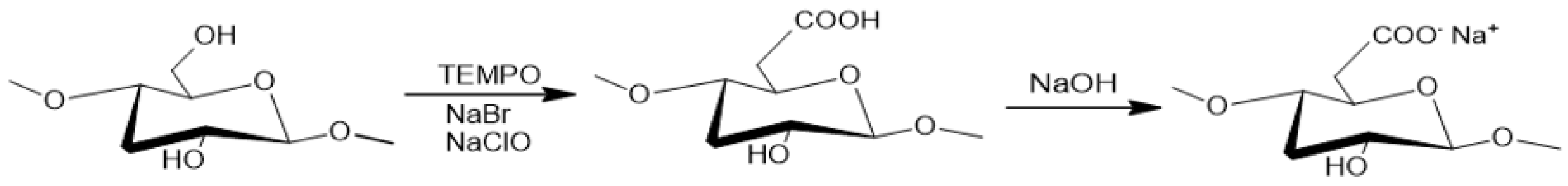
2.1. Synthesis of SCNC and TCNF
2.2. Adsorption Tests
2.3. Fourier Transform Infrared Spectroscopy (FTIR)
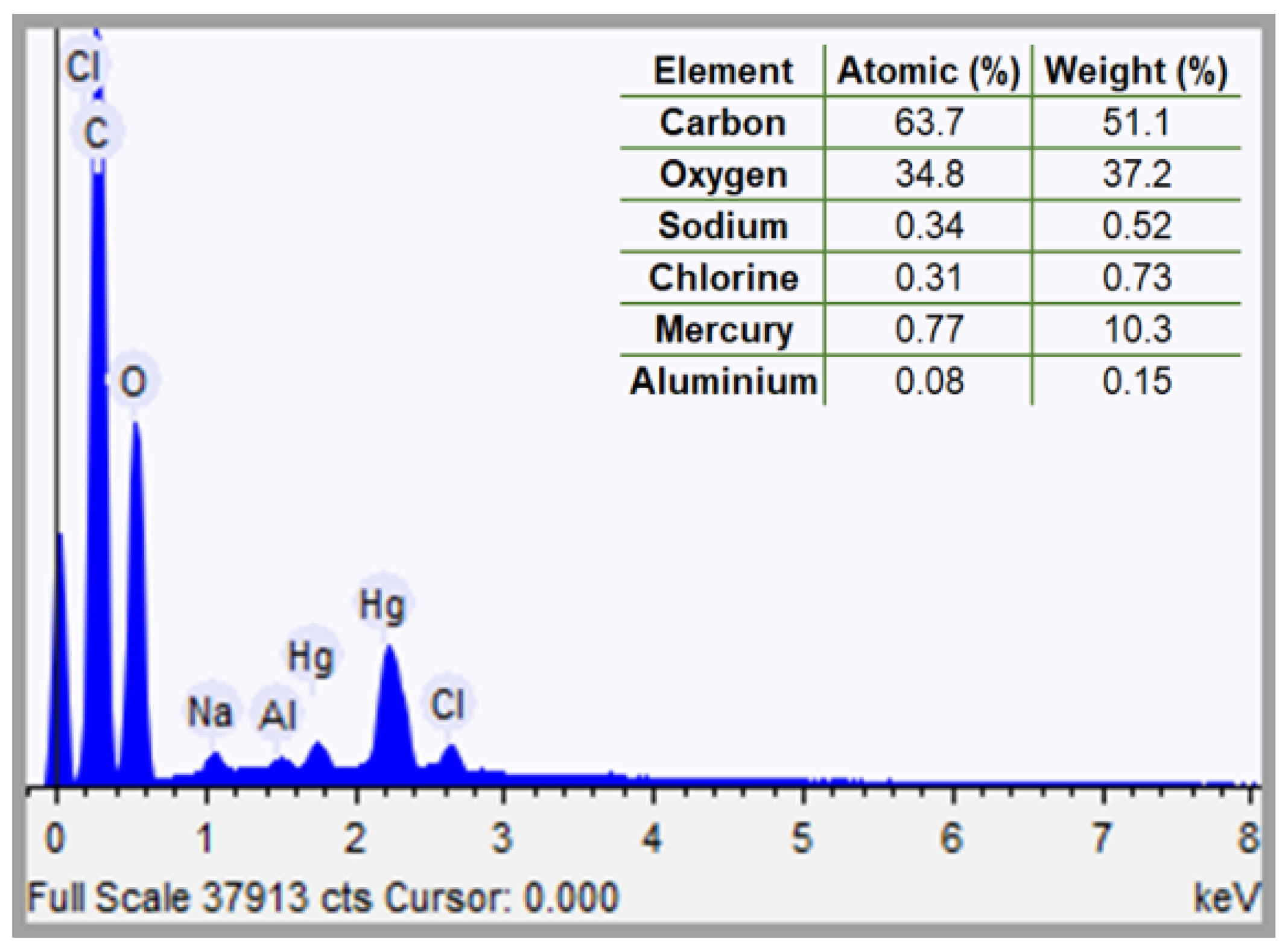
2.4. Scanning Electron Microscopy with Energy Dispersive Spectroscopy (SEM-EDS)
2.5. Thermogravimetric Analysis (TGA)
3. Results and Discussion
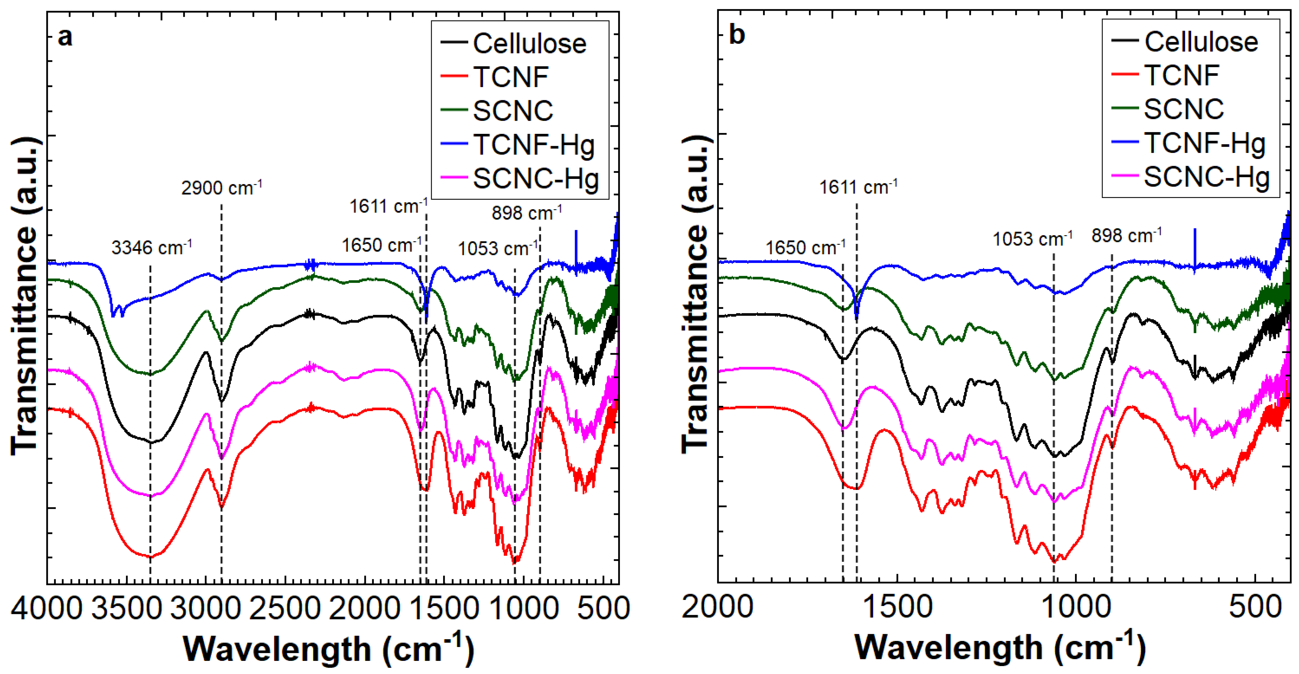
3.1. FTIR
3.2. Adsorption Tests
3.3. SEM-EDS
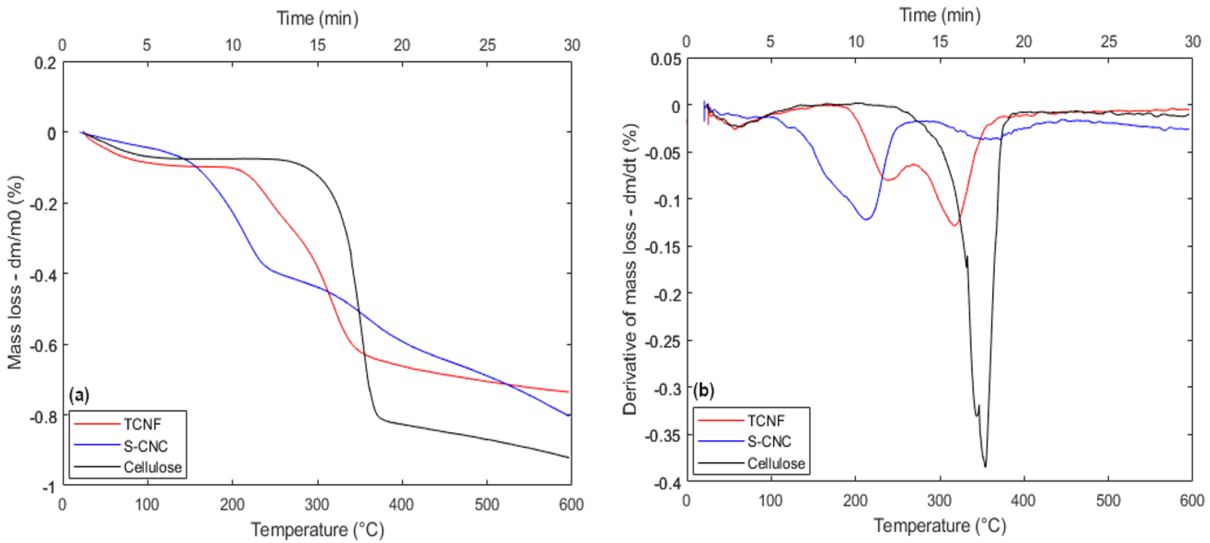
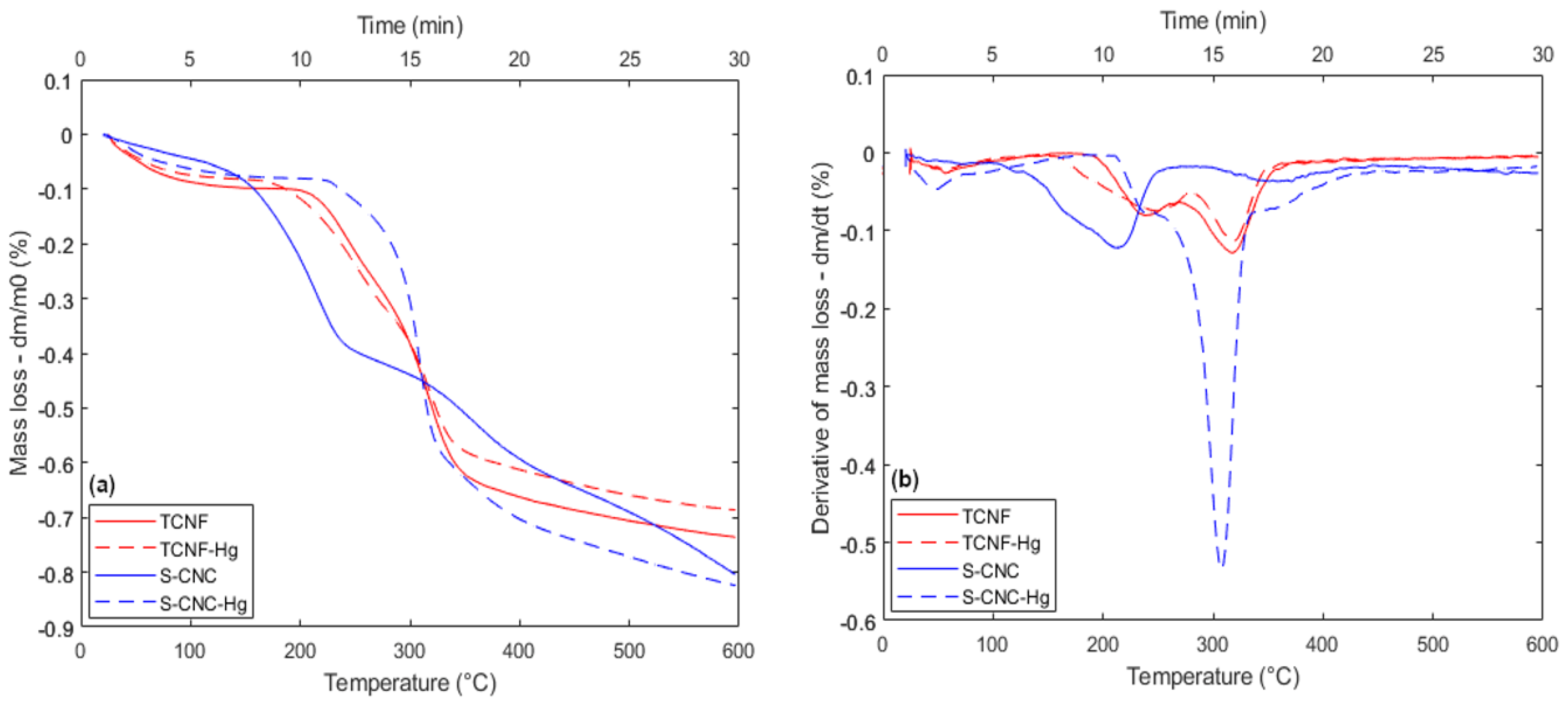
3.4. TGA
4. Conclusions
Author Contributions
Funding
Data Availability Statement
Acknowledgments
Conflicts of Interest
Abbreviations
| TCNF | TEMPO-oxidized Cellulose Nanofiber |
| SCNC | Sulfonated Cellulose Nanocrystals |
| TEMPO | 2,2,6,6-Tetramethylpiperidine-1-oxyl |
| FTIR | Fourier Transform Infrared Spectroscopy |
| TGA | Thermogravimetric Analysis |
| DTG | Derivative Thermogravimetry |
| SEM-EDS | Scanning Electron Microscopy with Energy Dispersive Spectroscopy |
| ICP-OES | Inductively Coupled Plasma - Optical Emission Spectrometry |
References
- Sahu, P. A comprehensive review of saline effluent disposal and treatment: Conventional practices, emerging technologies, and future potential. Water Reuse 2021, 11, 33–65. [Google Scholar] [CrossRef]
- Obrist, D.; Kirk, J.L.; Zhang, L.; Sunderland, E.M.; Jiskra, M.; Selin, N.E. A review of global environmental mercury processes in response to human and natural perturbations: Changes of emissions, climate, and land use. Ambio 2018, 47, 116–140. [Google Scholar] [CrossRef] [PubMed]
- Wagner-Döbler, I.; Von Canstein, H.; Li, Y.; Timmis, K.N.; Deckwer, W.D. Removal of mercury from chemical wastewater by microoganisms in technical scale. Environ. Sci. Technol. 2000, 34, 4628–4634. [Google Scholar] [CrossRef]
- Panagopoulos, A. Study and evaluation of the characteristics of saline wastewater (brine) produced by desalination and industrial plants. Environ. Sci. Pollut. Res. 2022, 29, 23736–23749. [Google Scholar] [CrossRef] [PubMed]
- Appiah-Brempong, M.; Essandoh, H.M.K.; Asiedu, N.Y.; Dadzie, S.K.; Momade, F.W.Y. Artisanal tannery wastewater: Quantity and characteristics. Heliyon 2022, 8, e08680. [Google Scholar] [CrossRef] [PubMed]
- Sharkh, B.A.; Al-Amoudi, A.A.; Farooque, M.; Fellows, C.M.; Ihm, S.; Lee, S.; Li, S.; Voutchkov, N. Seawater desalination concentrate—a new frontier for sustainable mining of valuable minerals. npj Clean Water 2022, 5, 9. [Google Scholar] [CrossRef]
- Quddoos, A.; Muhmood, K.; Naz, I.; Aslam, R.W.; Usman, S.Y. Geospatial insights into groundwater contamination from urban and industrial effluents in Faisalabad. Discov. Water 2024, 4, 50. [Google Scholar] [CrossRef]
- Forsido, T.T.; McCrindle, R.I.; Maree, J.; Monyatsi, L. Removal of Al, Ba and Mg from industrial wastewater using EAFDS and lime. Appl. Water Sci. 2020, 10, 157. [Google Scholar] [CrossRef]
- Le, V.G.; Vo, D.V.N.; Tran, H.T.; Duy Dat, N.; Luu, S.D.; Rahman, M.M.; Huang, Y.H.; Vu, C.T. Recovery of magnesium from industrial effluent and its implication on carbon capture and storage. ACS Sustain. Chem. Eng. 2021, 9, 6732–6740. [Google Scholar] [CrossRef]
- Barbosa, F.F.; Paulista, A.P.F.; Torres, M.A.M.; Braga, T.P. Synthesis of the Fe–Co alloy from hybrid spheres using carboxymethylcellulose as template and its application in catalysis. Mater. Chem. Phys. 2020, 242, 122550. [Google Scholar] [CrossRef]
- Ataee-Esfahani, H.; Nemoto, Y.; Imura, M.; Yamauchi, Y. Facile synthesis of nanoporous Pt-Ru alloy spheres with various compositions toward highly active electrocatalysts. Chem.-An Asian J. 2012, 7, 876–880. [Google Scholar] [CrossRef] [PubMed]
- Sivaprakash, G.; Mohanrasu, K.; Obeth, J.; Bora, A.; Yuvakkumar, R.; Mahmoud, A.H.; El-Abedein, A.I.Z.; Saravanan, S.; Arun, A. Zinc based iron mixed oxide catalyst for biodiesel production from Entermorpha intestinalis, Caulerpa racemosa and Hypnea musicoformisis and antibiofilm analysis using leftover catalyst after transesterification. J. King Saud-Univ.-Sci. 2020, 32, 1604–1611. [Google Scholar] [CrossRef]
- Baskar, G.; Soumiya, S. Production of biodiesel from castor oil using iron (II) doped zinc oxide nanocatalyst. Renew. Energy 2016, 98, 101–107. [Google Scholar] [CrossRef]
- Meneghetti, S.M.P.; Meneghetti, M.R.; Wolf, C.R.; Silva, E.C.; Lima, G.E.; de Lira Silva, L.; Serra, T.M.; Cauduro, F.; de Oliveira, L.G. Biodiesel from castor oil: A comparison of ethanolysis versus methanolysis. Energy Fuels 2006, 20, 2262–2265. [Google Scholar] [CrossRef]
- Sreeprasanth, P.; Srivastava, R.; Srinivas, D.; Ratnasamy, P. Hydrophobic, solid acid catalysts for production of biofuels and lubricants. Appl. Catal. A Gen. 2006, 314, 148–159. [Google Scholar] [CrossRef]
- Ahemed, J.; Rao D, V.; Pasha, J.; Kore, R.; Gade, R.; Bhongiri, Y.; Chetti, P.; Pola, S. Synthesis of new Zn (II) complexes for photo decomposition of organic dye pollutants, industrial wastewater and photo-oxidation of methyl arenes under visible-light. J. Photochem. Photobiol. A Chem. 2021, 419, 113455. [Google Scholar] [CrossRef]
- Tan, G.; Guo, Y.Q.; Zuo, L.Y.; Zhang, K.; Zhang, Y.M.; Zhang, L.L.; Yu, J.J.; Feng, X.; Li, B.; Wang, L.Y. Synthesis of zinc-based metal–organic framework as highly efficient photocatalyst for decomposition of organic dyes in aqueous solution. Rare Met. 2023, 42, 1205–1213. [Google Scholar] [CrossRef]
- Norfarhana, A.; Ilyas, R.; Ngadi, N. A review of nanocellulose adsorptive membrane as multifunctional wastewater treatment. Carbohydr. Polym. 2022, 291, 119563. [Google Scholar] [CrossRef] [PubMed]
- Mahfoudhi, N.; Boufi, S. Nanocellulose as a novel nanostructured adsorbent for environmental remediation: A review. Cellulose 2017, 24, 1171–1197. [Google Scholar] [CrossRef]
- Sarma, G.K.; Sen Gupta, S.; Bhattacharyya, K.G. Nanomaterials as versatile adsorbents for heavy metal ions in water: A review. Environ. Sci. Pollut. Res. 2019, 26, 6245–6278. [Google Scholar] [CrossRef] [PubMed]
- Ali, I. New generation adsorbents for water treatment. Chem. Rev. 2012, 112, 5073–5091. [Google Scholar] [CrossRef] [PubMed]
- Sehaqui, H.; Zhou, Q.; Berglund, L.A. High-porosity aerogels of high specific surface area prepared from nanofibrillated cellulose (NFC). Compos. Sci. Technol. 2011, 71, 1593–1599. [Google Scholar] [CrossRef]
- Aggarwal, R.; Saini, D.; Sonkar, S.; Sonker, A.; Westman, G. Sunlight Promoted Removal of Toxic Hexavalent Chromium by Cellulose Derived Photocative Carbon Dots. Chemosphere 2022, 287, 132287. [Google Scholar] [CrossRef] [PubMed]
- Jiang, H.; Wu, S.; Zhou, J. Preparation and Modification of Nanocellulose and Its Application to Heavy Metal Adsorption: A Review. Int. J. Biol. Macromol. 2023, 236, 123916. [Google Scholar] [CrossRef] [PubMed]
- Reshmy, R.; Philip, E.; Madhavan, A.; Pugazhendhi, A.; Sindhu, R.; Sirohi, R.; Awasthi, M.; Pandey, A.; Binod, P. Nanocellulose as Green Material for Remediation of Hazardous Heavy Metal Contaminants. J. Hazard. Mater. 2022, 424. [Google Scholar] [CrossRef] [PubMed]
- de Borja Oiembarrena, F.; Hullebusch, E.; Marsac, R.; Merayo, N.; Blanco, A.; Negro, C. Selective Recovery of Co(II), Mn(II), Cu(II), and Ni(II) by Multiple Step Batch Treatments with Nanocellulose Products. Environ. Sci. Pollut. Res. 2024, 31, 66725–66741. [Google Scholar] [CrossRef] [PubMed]
- Gobi, M.; Kumar, A.; Singh, J.; Singh, S.; Ramamurthy, P. Nanocellulose-Based Adsorption for the Removal of Heavy Metal from Wastewater—A Review. Water Conserv. Sci. Eng. 2024, 9, 24. [Google Scholar] [CrossRef]
- Sejje, F.; Matshwele, J.; Obuseng, V.; Nareetsile, F. Nanocellulose Chemical Treatment Reactions and Their Influence on the Adsorption of Aqueous Transition Metal Ions—A Review. Chem. Rev. Lett. 2023, 6, 245–255. [Google Scholar]
- Kardam, A.; Raj, K.R.; Srivastava, S.; Srivastava, M.M. Nanocellulose fibers for biosorption of cadmium, nickel, and lead ions from aqueous solution. Clean Technol. Environ. Policy 2014, 16, 385–393. [Google Scholar] [CrossRef]
- Liu, P.; Borrell, P.F.; Božič, M.; Kokol, V.; Oksman, K.; Mathew, A.P. Nanocelluloses and their phosphorylated derivatives for selective adsorption of Ag+, Cu2+ and Fe3+ from industrial effluents. J. Hazard. Mater. 2015, 294, 177–185. [Google Scholar] [CrossRef] [PubMed]
- Singh, K.; Arora, J.K.; Sinha, T.J.M.; Srivastava, S. Functionalization of nanocrystalline cellulose for decontamination of Cr(III) and Cr(VI) from aqueous system: Computational modeling approach. Clean Technol. Environ. Policy 2014, 16, 1179–1191. [Google Scholar] [CrossRef]
- Teixeira, L.T.; Braz, W.F.; de Siqueira, R.N.C.; Pandoli, O.G.; Geraldes, M.C. Sulfated and carboxylated nanocellulose for Co+2 adsorption. J. Mater. Res. Technol. 2021, 15, 434–447. [Google Scholar] [CrossRef]
- Sehaqui, H.; De Larraya, U.P.; Liu, P.; Pfenninger, N.; Mathew, A.P.; Zimmermann, T.; Tingaut, P. Enhancing adsorption of heavy metal ions onto biobased nanofibers from waste pulp residues for application in wastewater treatment. Cellulose 2014, 21, 2831–2844. [Google Scholar] [CrossRef]
- Ma, H.; Hsiao, B.S.; Chu, B. Ultrafine cellulose nanofibers as efficient adsorbents for removal of UO22+ in water. ACS Macro Lett. 2012, 1, 213–216. [Google Scholar] [CrossRef] [PubMed]
- Miranda, M.; Bica, C.; Nachtigall, S.; Rehman, N.; Rosa, S. Kinetical thermal degradation study of maize straw and soybean hull celluloses by simultaneous DSC–TGA and MDSC techniques. Thermochim. Acta 2013, 565, 65–71. [Google Scholar] [CrossRef]
- Fukuzumi, H.; Saito, T.; Okita, Y.; Isogai, A. Thermal stabilization of TEMPO-oxidized cellulose. Polym. Degrad. Stab. 2010, 95, 1502–1508. [Google Scholar] [CrossRef]
- Roman, M.; Winter, W.T. Effect of Sulfate Groups from Sulfuric Acid Hydrolysis on the Thermal Degradation Behavior of Bacterial Cellulose. Biomacromolecules 2004, 5, 1671–1677. [Google Scholar] [CrossRef] [PubMed]
- Jebali, Z.; Granados, A.; Nabili, A.; Boufi, S.; do Rego, A.M.B.; Majdoub, H.; Vallribera, A. Cationic cellulose nanofibrils as a green support of palladium nanoparticles: Catalyst evaluation in Suzuki reactions. Cellulose 2018, 25, 6963–6975. [Google Scholar] [CrossRef]
- Cirtiu, C.M.; Dunlop-Briere, A.F.; Moores, A. Cellulose nanocrystallites as an efficient support for nanoparticles of palladium: Application for catalytic hydrogenation and Heck coupling under mild conditions. Green Chem. 2011, 13, 288–291. [Google Scholar] [CrossRef]
- Eisa, W.H.; Abdelgawad, A.M.; Rojas, O.J. Solid-state synthesis of metal nanoparticles supported on cellulose nanocrystals and their catalytic activity. ACS Sustain. Chem. Eng. 2018, 6, 3974–3983. [Google Scholar] [CrossRef]
- Madivoli, E.S.; Kareru, P.G.; Gachanja, A.N.; Mugo, S.; Murigi, M.K.; Kairigo, P.; Kipyegon, C.; Mutembei, J.K.; Njonge, F.K. Adsorption of selected heavy metals on modified nano cellulose. Int. Res. J. Pure Appl. Chem. 2016, 12, 1–9. [Google Scholar] [CrossRef]
- Chattopadhyay, D.; Patel, B.H. Synthesis, Characterization and Application of Nano Cellulose for Enhanced Performance of Textiles. J. Text. Sci. Eng. 2016, 6, 8. [Google Scholar] [CrossRef]
- Jiang, F.; Hsieh, Y.L. Chemically and mechanically isolated nanocellulose and their self-assembled structures. Carbohydr. Polym. 2013, 95, 32–40. [Google Scholar] [CrossRef] [PubMed]
- Coates, J. Interpretation of Infrared Spectra, A Practical Approach. In Encyclopedia of Analytical Chemistry; Meyers, R.A., Ed.; John Wiley & Sons: Chichester, UK, 2000; pp. 10815–10837. [Google Scholar]
- Paul, H.R.; Bera, M.K.; Macke, N.; Rowan, S.J.; Tirrell, M.V. Quantitative Determination of Metal Ion Adsorption on Cellulose Nanocrystals Surfaces. ACS Nano 2024, 18, 1921–1930. [Google Scholar] [CrossRef] [PubMed]
- Bansal, M.; Ram, B.; Chauhan, G.S.; Kaushik, A. L-Cysteine functionalized bagasse cellulose nanofibers for mercury(II) ions adsorption. Int. J. Biol. Macromol. 2018, 112, 728–736. [Google Scholar] [CrossRef] [PubMed]
- Lei, X.; Li, H.; Luo, Y.; Sun, X.; Guo, X.; Hu, Y.; Wen, R. Novel fluorescent nanocellulose hydrogel based on gold nanoclusters for the effective adsorption and sensitive detection of mercury ions. J. Taiwan Inst. Chem. Eng. 2021, 123, 79–86. [Google Scholar] [CrossRef]
- Kandile, N.G.; Mohamed, H.M. Chitosan nanoparticle hydrogel based sebacoyl moiety with remarkable capability for metal ion removal from aqueous systems. Int. J. Biol. Macromol. 2019, 122, 578–586. [Google Scholar] [CrossRef] [PubMed]
- Khozemy, E.E.; Nasef, S.M.; Mohamed, T.M. Radiation synthesis of superabsorbent hydrogel (wheat flour/acrylamide) for removal of mercury and lead ions from waste solutions. J. Inorg. Organomet. Polym. Mater. 2020, 30, 1669–1685. [Google Scholar] [CrossRef]
- Isogai, A.; Saito, T.; Fukuzumi, H. TEMPO-oxidized cellulose nanofibers. Nanoscale 2011, 3, 71–85. [Google Scholar] [CrossRef] [PubMed]
- De M. Teixeira, E.; De Oliveira, C.; Mattoso, L.; Corrêa, A.; Paladin, P. Cotton nanofibers obtained by different hydrolytic acid conditions | Nanofibras de algodão obtidas sob diferentes condições de hidrólise ácida. Polimeros 2010, 20, 264–268. [Google Scholar] [CrossRef]
- Habibi, Y.; Lucia, L.A.; Rojas, O.J. Cellulose nanocrystals: Chemistry, self-assembly, and applications. Chem. Rev. 2010, 110, 3479–3500. [Google Scholar] [CrossRef] [PubMed]
- Peng, F.; Dhar, N.; Liu, H.; Tam, K.C. Chemistry and applications of nanocrystalline cellulose and its derivatives: A nanotechnology perspective. Can. J. Chem. Eng. 2018, 96, 282–296. [Google Scholar] [CrossRef]
- Saito, T.; Kimura, S.; Nishiyama, Y.; Isogai, A. Cellulose nanofibers prepared by TEMPO-mediated oxidation of native cellulose. Biomacromolecules 2007, 8, 2485–2491. [Google Scholar] [CrossRef] [PubMed]


| Adsorbent | Adsorbent Mass (g) | Adsorbate | Cationic Precursor | Metal Concentration (mg·L−1) |
|---|---|---|---|---|
| SCNC | 1 | Na+ | NaNO3 | 1000 |
| Hg2+ | HgCl2 | 500 | ||
| TCNF | 0.5 | Hg2+ | HgCl2 | 1000 |
| Mg2+ | Mg(OH)2 | 2.4 |
| Material | Band Wave Number [cm−1] | Functional Group | Reference |
|---|---|---|---|
| Cellulose | 3346 | O-H stretching from C2 and C3 | |
| 2900 | Stretching of aliphatic C-H bonds | [41] | |
| 1053 | Ring vibration of C-O-C bonds | ||
| 898 | Stretching of glycosidic bonds (-1,4 C-O-C) | [42] | |
| 1650 | O-H stretching from adsorbed water molecules | [36] | |
| CNF | 1611 | Asymmetric stretching of C=O in carboxylate COO− | [43] |
| Material | Adsorbate | Metal Recovery (%) | Final Metal Concentration (mg·L−1) | Adsorp. Capacity (mg·g−1) |
|---|---|---|---|---|
| SCNC | Na+ | 63.3 | 367 ± 1.84 | 63.3 ± 0.95 |
| Hg2+ | 35.2 | 324 ± 1.62 | 17.6 ± 0.25 | |
| TCNF | Hg2+ | 89.3 | 107 ± 0.54 | 178.6 ± 2.69 |
| Mg2+ | 100 | 0 | 0.48 ± 0.07 |
| Study/Reference | Ion | Adsorbent | Conditions | Ads. Capacity (mg·g−1) | Equilibrium Time |
|---|---|---|---|---|---|
| Bansal et al. [46] | Hg2+ | TCNF | pH 6.0, 3 h | 80 | 3 h |
| Leiat et al. [47] | Hg2+ | TCNF + Au nanoclusters | pH 6.0 | 55 | 240 min |
| Kandle & Mohamed [48] | Hg2+ | Chitosan hydrogel | pH 5.0 | 35 | - |
| Kozemy et al. [49] | Hg2+ | Wheat flour hydrogel | pH 6.0, 30 °C | 13 | 600 min |
| Previous work [32] | Co2+ | TCNF and SCNF | pH 6.0 | 91 | 30 min |
Disclaimer/Publisher’s Note: The statements, opinions and data contained in all publications are solely those of the individual author(s) and contributor(s) and not of MDPI and/or the editor(s). MDPI and/or the editor(s) disclaim responsibility for any injury to people or property resulting from any ideas, methods, instructions or products referred to in the content. |
© 2025 by the authors. Licensee MDPI, Basel, Switzerland. This article is an open access article distributed under the terms and conditions of the Creative Commons Attribution (CC BY) license (https://creativecommons.org/licenses/by/4.0/).
Share and Cite
Braz, W.F.; Teixeira, L.T.; Navarro, R.; Pandoli, O.G. Nanocellulose Application for Metal Adsorption and Its Effect on Nanofiber Thermal Behavior. Metals 2025, 15, 832. https://doi.org/10.3390/met15080832
Braz WF, Teixeira LT, Navarro R, Pandoli OG. Nanocellulose Application for Metal Adsorption and Its Effect on Nanofiber Thermal Behavior. Metals. 2025; 15(8):832. https://doi.org/10.3390/met15080832
Chicago/Turabian StyleBraz, Wanderson Ferreira, Lucas Tonetti Teixeira, Rogério Navarro, and Omar Ginoble Pandoli. 2025. "Nanocellulose Application for Metal Adsorption and Its Effect on Nanofiber Thermal Behavior" Metals 15, no. 8: 832. https://doi.org/10.3390/met15080832
APA StyleBraz, W. F., Teixeira, L. T., Navarro, R., & Pandoli, O. G. (2025). Nanocellulose Application for Metal Adsorption and Its Effect on Nanofiber Thermal Behavior. Metals, 15(8), 832. https://doi.org/10.3390/met15080832