Article

Investigation of Surface Oxidation of Cast Austenitic 304 Stainless Steel at High Temperatures

1
Department of Experimental Physics, Faculty of Science, Palacký University, 17. listopadu 12, 77900 Olomouc, Czech Republic
2
SIGMA GROUP a.s., Jana Sigmunda 313, 78349 Lutín, Czech Republic
3
Department of Mechanical Technology, Faculty of Mechanical Engineering, Technical University of Ostrava, 17. listopadu 15, 70800 Ostrava, Czech Republic
*
Authors to whom correspondence should be addressed.
Metals 2025, 15(7), 748; https://doi.org/10.3390/met15070748
Submission received: 23 May 2025 / Revised: 27 June 2025 / Accepted: 29 June 2025 / Published: 2 July 2025

Abstract

The microstructure and surface behavior of iron-based 304 stainless steel after temperature exposure was studied by Mössbauer spectroscopy, powder X-ray diffraction, scanning electron microscopy, energy dispersive analysis and positron annihilation. The tested specimens were in the form of cylinders produced by the casting process. The samples were annealed in air in the 600–1000 °C temperature range for 36 h. Under the influence of temperature, cast 304 stainless steel underwent austenitic–ferritic transformation and tended to form an oxide layer on the surface. The oxides were mainly found in the thin surface layer (0.3 μm) and consisted of Fe oxides and oxides of alloying elements (Cr and Mn) in the form of corundum, while, in the bulk region (10 μm), the phase transformation of austenite to ferrite occurred. Surface phase inhomogeneity was studied by Mössbauer spectroscopy. The method of positron annihilation was used to study defects and the effect of annealing on the formation and removal of a defect structure.

1. Introduction

Stainless steel is widely used in industry due to its exceptional properties [1,2]. Of all the different types of stainless steel, 304 stainless steel is the most widely used and well known for its sufficient resistance to corrosion, which is achieved by the formation of a protective layer of chromium oxide under the influence of oxygen. The corrosion resistance of 304 stainless steel at different temperatures, atmospheres and solutions can be improved by adding elements (boron, TiO2) to the 304 stainless steel [3,4,5].
Temperature annealing, as part of post-processing treatment, is necessary for 304 austenitic stainless steel to relieve residual stresses and improve mechanical and microstructural properties [6,7]. A number of papers have been devoted to the study of the effect of temperature on the corrosion properties of 304 stainless steel using Mössbauer spectroscopy [8,9,10,11,12,13,14]. Mössbauer spectroscopy (MS) has been used to identify phase changes during the heat treatment of 304 steel [15,16,17]. Positron electron annihilation spectroscopy (PAS) was also used to study corrosion processes and phase transformations in 304 steel. Additionally, Doppler broadening spectroscopy (DBS) [18,19,20,21,22,23,24,25] and positron annihilation lifetime spectroscopy (PALS) [25,26] can be applied. Nonetheless, the combination of Mössbauer spectroscopy and positron annihilation has not yet been utilized to study the corrosion and phase changes of 304 steel.
MS and PAS can be excellent tools for studying the microstructural properties of metallic materials. MS provides qualitative and quantitative phase analysis and leads to information about the structure, phases and magnetic properties of the studied material. MS [27] is a non-destructive research method that can analyze surface and bulk by recording the free path of conversion electrons and the penetration depth of X-rays. This method is well suited to iron alloys due to the measurable effect of the resonant absorption of a gamma-ray isotope 57Fe. Moreover, a high resolution of MS can provide information about local features of the atomic environment of an alloy or compound by recording changes in the energy of nuclear transitions near the resonant nucleus. PAS is also a non-destructive testing method that allows for the analysis of a defective structure and the concentration of vacancies. This technique can detect vacancy-type defects in a crystal structure that are beyond the resolving power of a scanning electron microscope [28]. In 304 stainless steel, PAS was also used to study fatigue [28,29], irradiation effects [30], hydrogen-induced defects [31] and surface damage after sandblasting [32]. The principle of positron annihilation is the capture of a positron implanted into a sample by structural defects. There is an increase in the lifetime of the positron and a narrowing of the pulse distribution of the positron ray (Doppler broadening).
This study is devoted to the investigation of the surface behavior of cast austenitic 304 stainless steel after exposure to high temperatures. The temperature effect consisted of annealing in air at a temperature of 600–1000 °C for 36 h. It is well known that austenitic stainless steels are annealed in various atmospheres and temperatures to relieve internal stresses. After exposure to a temperature in air, both oxidation and the ‘austenite ferrite’ phase transformations occur on the surface of cast austenitic 304 stainless steel. These transformations proceed in accordance with the Fe-Cr-Ni binary phase diagram for 304 austenitic stainless steel [33] (Figure 4). The effect of heat treatment on the microstructure and mechanical properties of 304 stainless steel was studied in various publications [33,34,35,36], in which the authors identified ‘austenite–ferrite’ phase transformations and chromium carbide formation. The oxidation products of the stainless steel surface after exposure to temperature were also studied in these publications [37,38,39]. The morphology of the oxide layer and the distribution of alloying elements after temperature exposure were studied using scanning electron microscopy (SEM) and energy-dispersive spectrometry (EDS), respectively. Conversion electron Mössbauer spectroscopy (CEMS) was used to study phase transformations in the thin surface layer (0.3 μm) under the influence of temperature. Conversion X-ray Mössbauer spectroscopy (CXMS) was used to study the phase transformations that occurred in the bulk (10 μm) of the material. In addition, X-ray diffraction (XRD) was used for phase composition as well as for the purpose of comparison with Mössbauer data. The formation of a defect structure after exposure to temperature in cast austenitic 304 stainless steel was investigated by positron annihilation spectroscopy.

2. Materials and Methods

The test samples were made of cast 304 stainless steel, the chemical composition of which is given in Table 1. The samples studied were cut from a cast 304 austenitic stainless steel cylinder. The samples were annealed for 36 h in air at a temperature range of 600–1000 °C. Temperature annealing was carried out in an LE05/11 laboratory furnace (LAC, Židlochovice, Czech Republic). Thermal treatment consisted of 1 h ramp-up to the required temperature, dwell time of 36 h and spontaneous cooling to RT. The sample was then left in the furnace until it had completely cooled. The samples showed visible changes in color depending on the annealing temperature (Figure 1).
The backscattering 57Fe Mössbauer spectra were measured by a backscattering Mössbauer spectrometer at room temperature. The Mössbauer spectrometer operated in constant acceleration mode and was equipped with a 57Co(Rh) source. The spectra were processed and accumulated using MS96 Mössbauer spectrometer software (V.3.6.3, Department of Experimental Physics, Palacký University in Olomouc, Olomouc, Czech Republic) [40]. The spectra were recorded on 512 channels. Two types of detectors were used to record the CXMS spectra and CEMS spectra. An air scintillation detector was used to register CEMS spectra, and for registration of the CXMS spectra, a proportional gas detector registering 6.4 keV X-rays was used [41]. MossWinn 4.0 software was used to analyze the Mössbauer spectra with least-squares fitting of the lines [42,43]. The centroid of the α-Fe foil (thickness 30 μm) spectrum was used as the standard for determining the value of the isomer shift values at room temperature.
The samples were studied by means of positron annihilation spectroscopy, specifically by Doppler broadening of the annihilation spectral line (DBS) measurements. The values of the Doppler broadening ∆E were units of keV, so a spectrometer with an energy resolution of 1–2 keV was needed to measure them. An HPGe detector (BE2825, Canberra, Atlanta, GA, USA) with a resolution (FWHM) of 1.9 keV for 1332.5 keV was used for DBS measurements. 22Na with an activity of about 1 MBq was used as a positron source. The broadening of the annihilation spectral line was characterized by means of the standard parameters S and W, which were defined according to Figure 2. The S parameter was the integral of the central part of the annihilation line (511 keV) normalized to the total area of the line. The S parameter contained information on the size of defects and their concentration [44]. The W parameter was an integral of the wing-shaped part of the annihilation line at 511 keV and provided information about the chemical environment of the vacancy defects [45]. The evolution of the S and W positron annihilation parameters could best be monitored by changes in the so-called S-W correlation plots. In these plots, different slopes in the data sets correlated with the vacancy defects at different locations [46].
The phase composition of cast 304 stainless steel after temperature exposure was studied by the X-ray diffraction technique. All measurements were performed on a Bruker Advance D8 X-ray diffractometer (Bruker, Billerica, MA, USA) with a Co Kα X-ray source (wavelength 1.79026 Å, voltage 35 kV, current 40 mA). The diffractometer operated with Bregg–Brentano parafocusing geometry and was equipped with a LYNXEYE position-sensitive detector. On the primary beam path, the instrument was equipped with a 0.6 mm divergence slit and 2.5° axial Soller slits. A 20 μm Fe Kβ filter and 2.5° axial Soller slits were used on the secondary beam path.
To obtain information about the surface morphology and elemental composition, a scanning electron microscope, VEGA3 LMU (TESCAN, Brno, Czech Republic), was used. The SEM system was equipped with an Everhart–Thornley secondary electron detector (TESCAN, Brno, Czech Republic) and an XFlash 410-M silicon drift detector (Bruker Nano GmbH, Berlin, Germany).
A detailed description of the methods used in this research can be found in the Supplementary Materials.

3. Results and Discussion

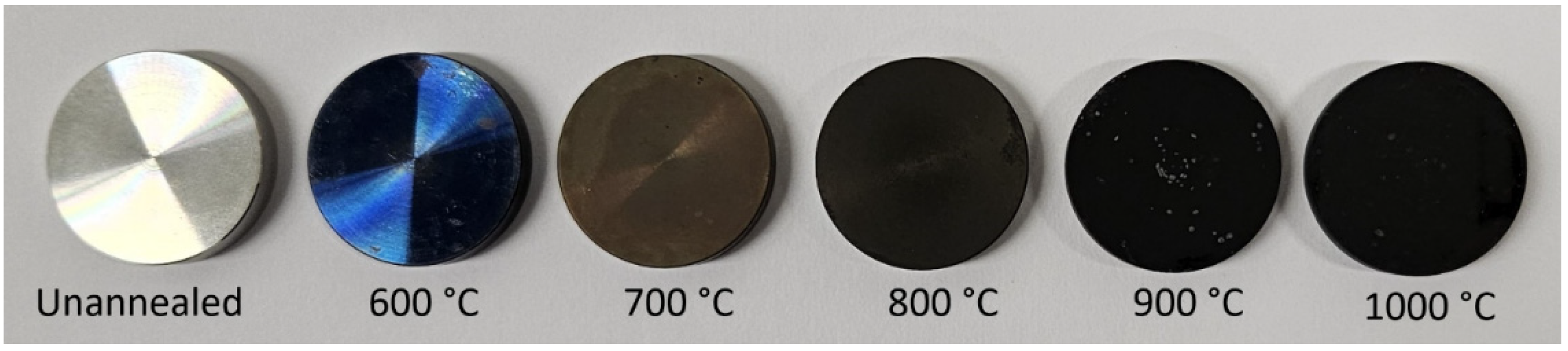
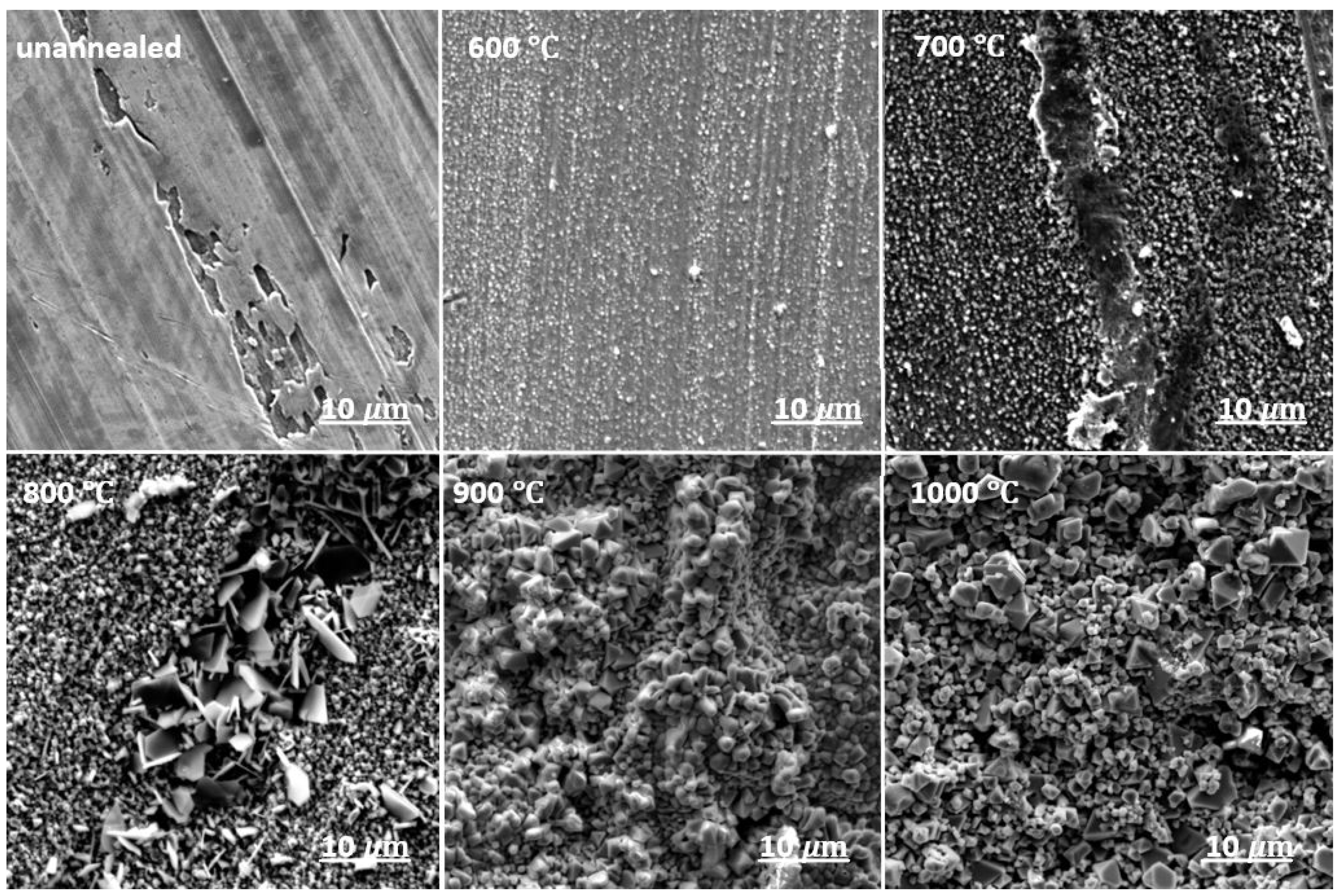
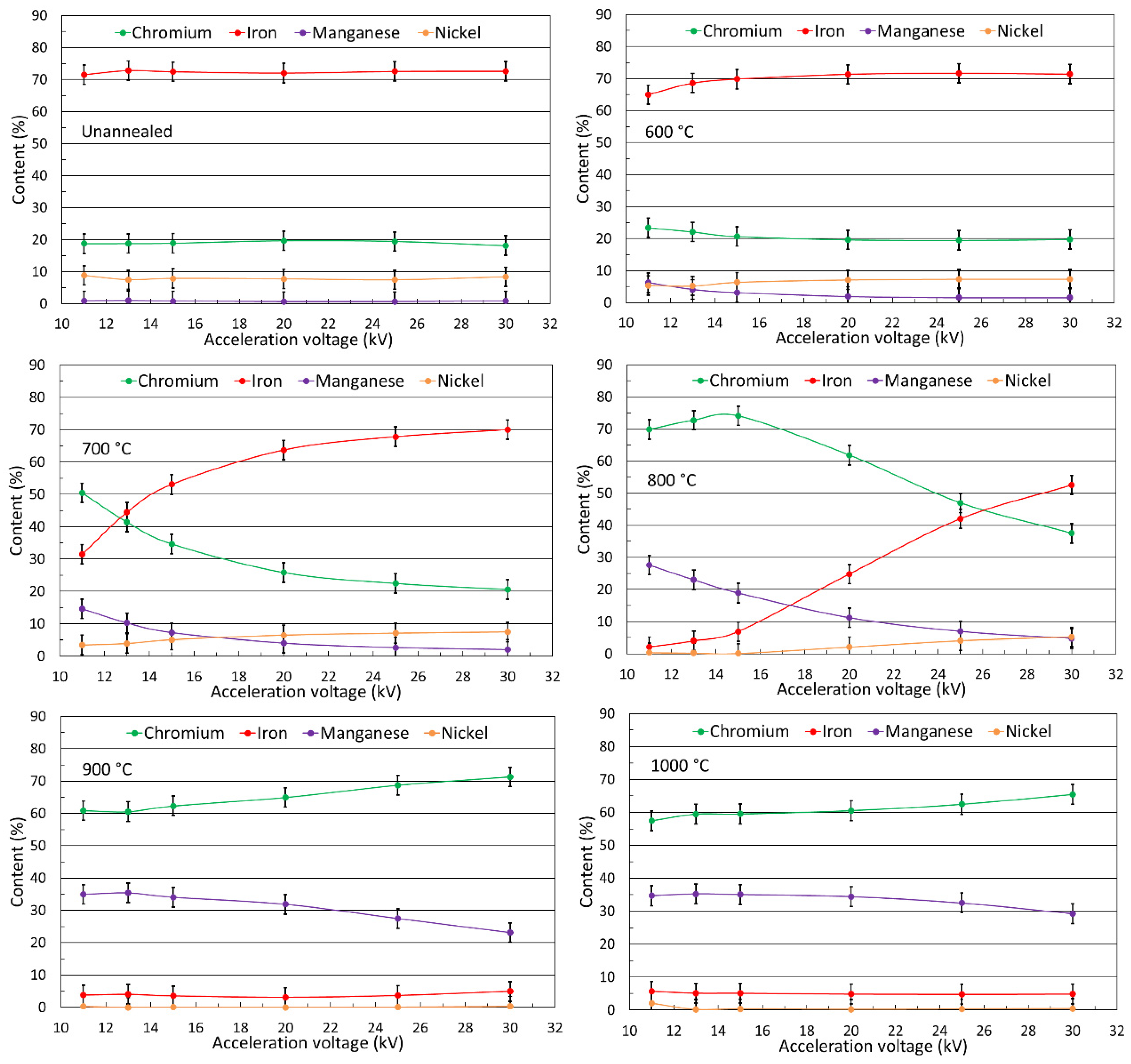
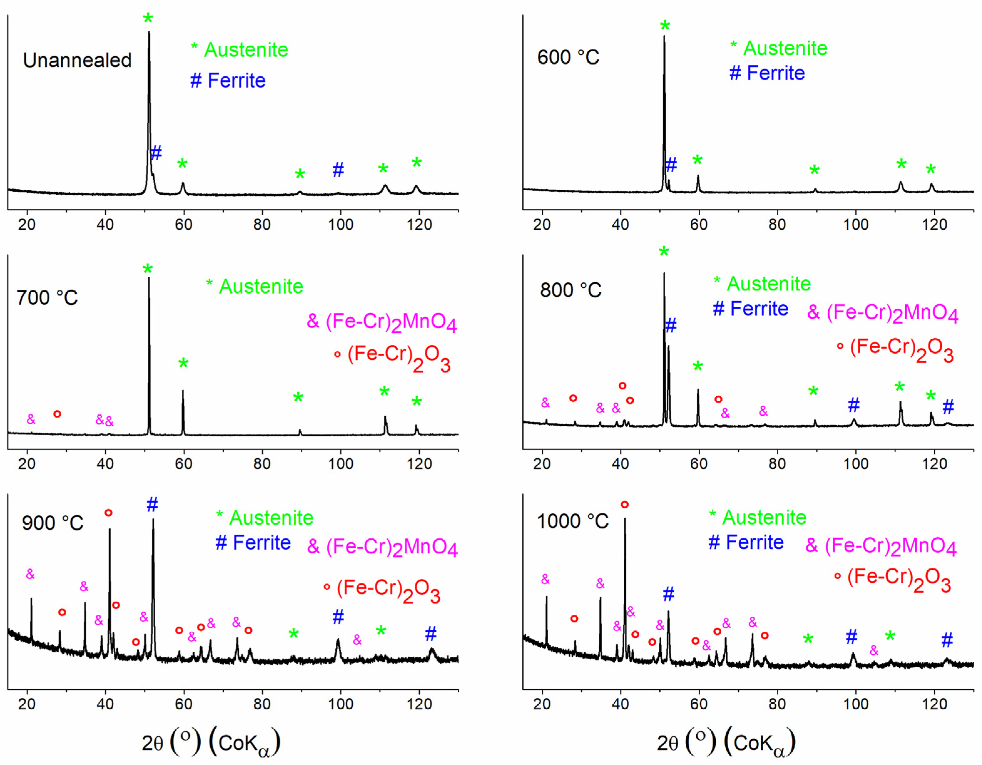
3.1. Oxide Morphology and Element Distribution in the Surface Layer

The surface morphology and oxide scales formed on the cast 304 stainless steel surface under high temperatures are shown in Figure 3 and Figure 4. SEM images were obtained at different magnifications. As can be seen in both figures, under the influence of temperature and atmosphere, the surface was covered with a broad oxide layer. Globular oxide particles started to grow at 600 °C; the same growth of globular particles was observed in [47]. The tendency to increase the oxide layer also increased with higher annealing temperatures. At higher magnification (Figure 4), sheet-shaped crystals were observed at an annealing temperature of 800 °C. Additionally, it can be seen that the surface of the sample at temperatures of 900 °C and 1000 °C was fully covered with globular and octahedral particles. In [48], the oxidation behavior of additive-made 316L stainless steel after annealing in an air atmosphere was investigated. The formation of octahedral particles was observed under the temperature impact of 900 °C and 1000 °C for both 304 [47] and 316L [48] stainless steel grades. In the case of 316L stainless steel, this surface diversity was explained by the formation of oxides containing chromium and manganese. The octahedral particles at the top of the surface were identified as manganese-containing oxides on additive-manufactured 316L stainless steel.
In [49], the authors studied the behavior of 304 austenitic stainless steel under the influence of temperature and atmosphere. The 304 stainless steel was annealed in an air/CH4 atmosphere at a temperature of 1373 K for 20 min. After annealing and atmospheric impact, as shown in [49], spherical crystals were formed on the surface of the material. The surface oxide layer was shown to be a layer of iron oxide and a thin layer of chromium oxide. Thus, spherical-shaped crystals belonged to the Fe-Cr mixed oxide. In [36,49], it was mentioned that chromium oxide can evaporate under the influence of temperature (annealing). As a result, the protective layer of chromium oxide is destroyed and the surface of 304 stainless steel is covered with a porous layer of iron oxide. Thus, at an annealing temperature of 600 °C in air, intensive oxidation of the surface began, and at an annealing temperature of 600 °C–800 °C, the surface of 304 stainless steel was covered with Fe-Cr oxide particles. As the exposure temperature increased (900–1000 °C), particles of Mn-containing oxide also appeared on the surface.
According to Table 1, the main alloying elements in the chemical composition of 304 stainless steel were Cr, Ni and Mn. These elements were selected for elemental analysis using the energy-dispersive spectroscopy (EDS) method. Additionally, these alloying elements were present in all stainless steel grades and were also investigated in [48] additive-manufactured 316L stainless steel. In 304 stainless steel, the effect of temperature caused a redistribution of the main alloying elements in the surface layer [50]. The EDS technique is a tool for the determination of the concentration of elements at different depths and their redistribution under the influence of temperature and atmosphere. Cast 304 stainless steel specimens were examined at accelerating voltages of 11 to 30 keV, which corresponded to penetration depths of 0.3 to 2.6 μm according to Castaing’s formula [5]. The elemental distribution in the surface layer of cast 304 stainless steel after annealing for 36 h in an air atmosphere is shown in Figure 5. As can be seen, at the initial exposure temperature of 600 °C, only at a surface depth of 0.3 μm was there a slight increase in chromium concentration with a decrease in iron concentration. This phenomenon can be related to the affinity of chromium atoms for oxygen and the ability of stainless steel to form a corrosion-protective chromium oxide layer. At the temperature impact of 700 °C and 800 °C in the bulk region (2.6 μm), iron was still the predominant element in the chemical composition. At a temperature of 800 °C, a significant increase in the content of chromium and manganese with a proportional decrease in iron could also be observed in the thin surface layer. With annealing at 900 °C and 1000 °C, there were noticeable changes in the distribution of the main alloying elements. At this point, cast 304 stainless steel behaved differently than additive-manufactured 316L stainless steel in [48]. In Figure 5, it can be seen that the concentration of iron content decreased and stabilized at annealing at 900 °C and 1000 °C. In addition, the chromium content increased threefold throughout the depths examined in this study compared to the original composition. There was also a stabilization of the concentration at depth and an almost tenfold increase in the concentration of manganese. The surface manganese content was different for cast 304 stainless steel and additively manufactured 316L stainless steel [48], where only a twofold increase in manganese concentration was observed after annealing. In addition, nickel was not susceptible to concentration fluctuations under the influence of temperature. The results obtained by the elemental analysis correlated with the results of the SEM. Based on this, it can be assumed that the surface of the cast 304 stainless steel was coated with chromium and manganese oxides under the influence of temperature.

3.2. Surface Phase Transformation Analysis

One of the methods used to study the composition of the oxide phases was XRD at room temperature. Furthermore, the application of Mössbauer spectroscopy to the analysis of phases with iron content is shown in the Section 3.3. The XRD patterns of the cast 304 stainless steel samples annealed in air for 36 h in the temperature range of 600–1000 °C are shown in Figure 6.
The austenitic stainless steel family has the predominant phase as a face-centered cubic (FCC) structure at room temperature. In addition, Ni is known to be an FCC stabilizing element. Regarding the chemical composition of 304 stainless steel, Ni accounts for 8–10 wt %, which is sufficient to stabilize the FCC phase. Meanwhile, Cr (17.5–19.5 wt %) is known as a body-centered cubic (BCC) stabilizer [52,53]. Austenitic stainless steels undergo phase transformations as a result of exposure to temperature and plastic deformation. In the case of plastic deformation, an austenite–martensitic transformation occurs (γα′) [54,55].
As can be seen, the original sample annealed at a temperature of 600 °C contained a dominant austenitic phase (γ) and a ferritic phase (α). Surface oxidation started at a temperature of 700 °C. According to the XRD data, the austenitic–ferrite transformation (γα) took place at a temperature of 800–1000 °C. Austenite–ferritic transformations were observed for all annealing temperatures except 700 °C, where the cast 304 stainless steel retained only the austenitic phase. As the exposure temperature increased, the surface was actively oxidized, and, as can be seen with annealing at 1000 °C, the sample surface consisted mainly of an oxide phase. The oxide phases were identified as (Fe-Cr)2O3 and (Fe-Cr)2MnO4.
The formation of chromium oxide on the surface can occur due to the high diffusion of chromium under the influence of temperature [56]. Cr2O3 and Fe2O3 oxides can form on the surface of stainless steel. On the XRD patterns, these oxides overlap and it is difficult to distinguish them due to similarity in crystallographic parameters and crystal lattice type (trigonal). In addition, a Mn-containing oxide ((Fe-Cr)2MnO4) was found in the XRD pattern. The formation of this oxide was correlated with the high diffusion rate of Mn under the temperature influence [57] and the octahedral particles observed on the surface by the SEM method.

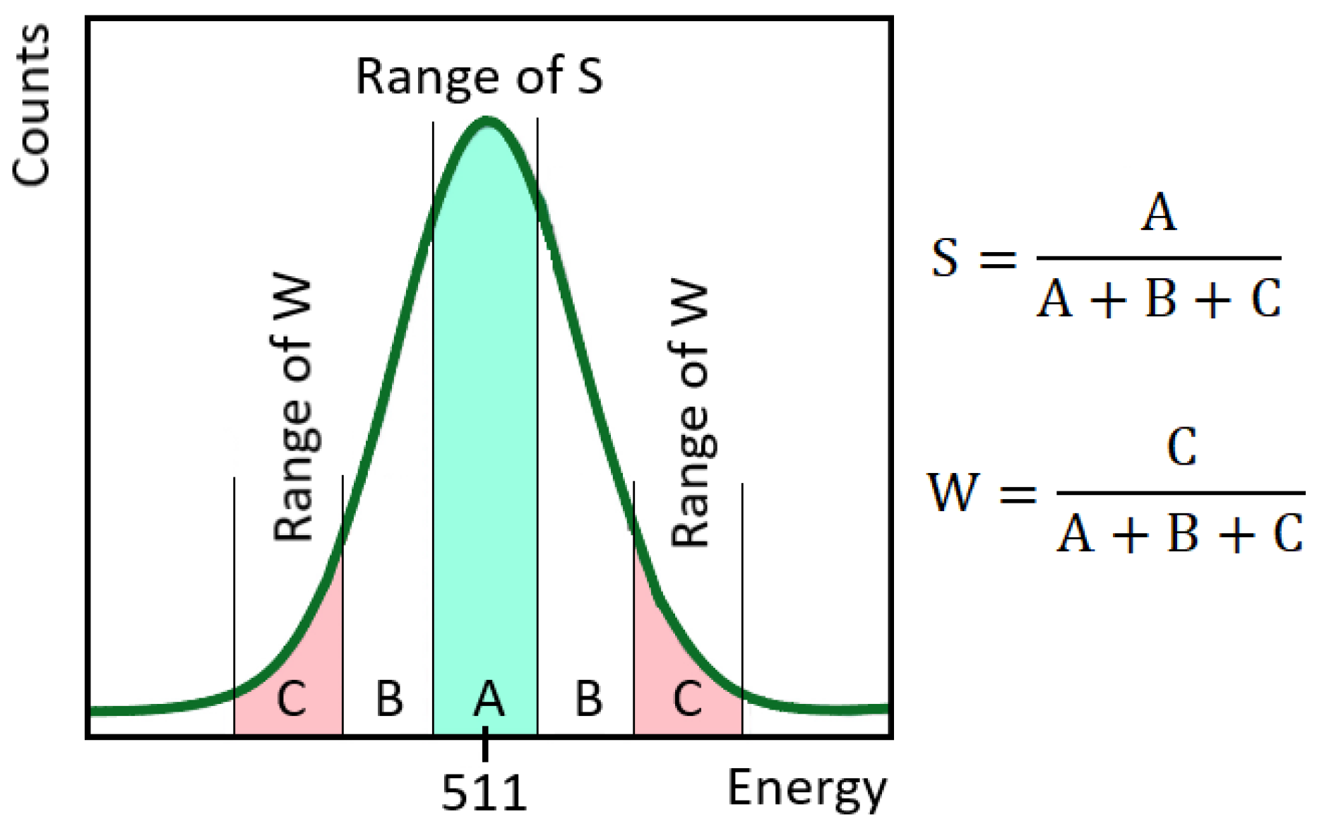
3.3. Application of Mössbauer Spectroscopy for Phase Transformation Analysis

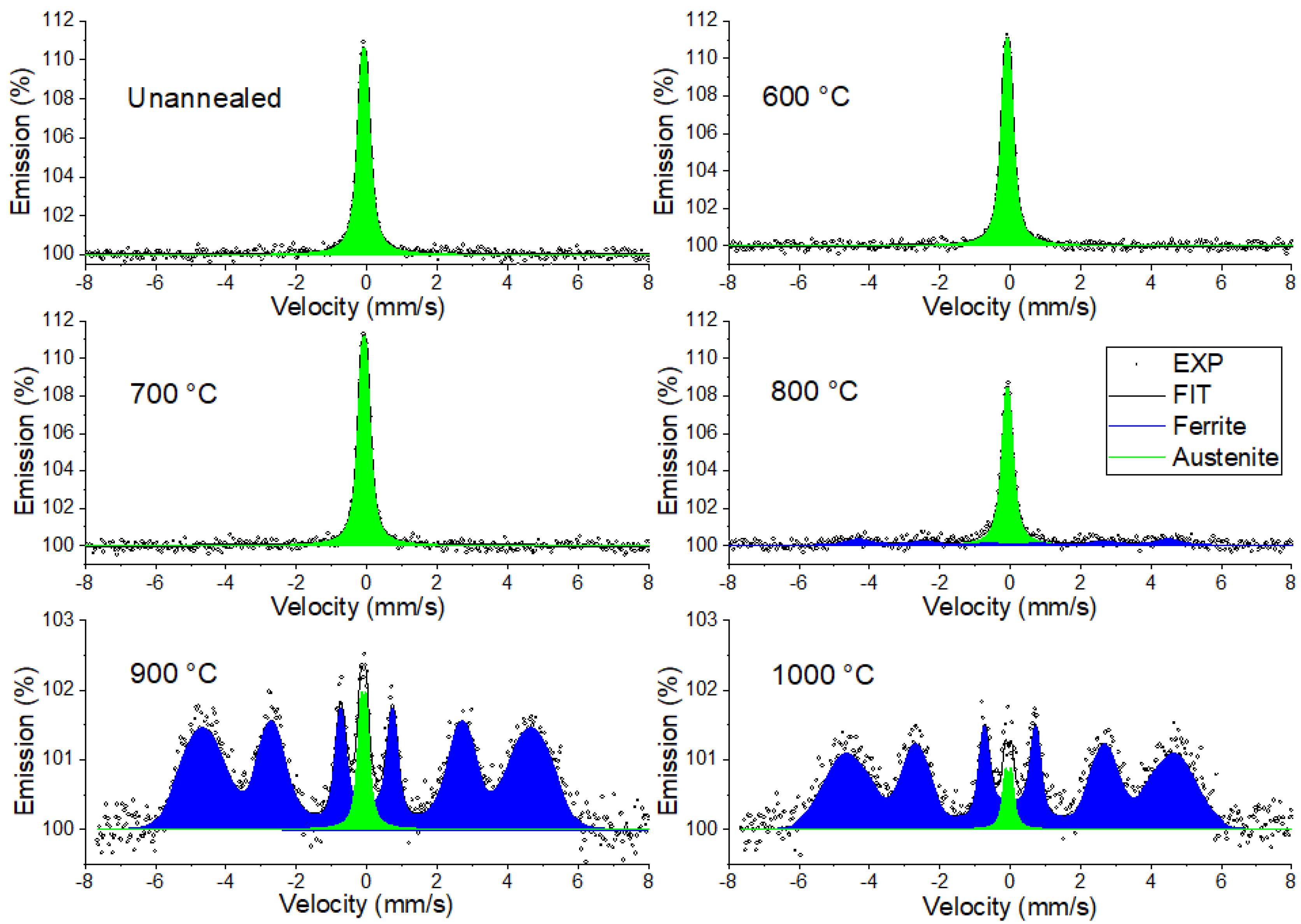
3.3.1. Conversion X-Ray Mössbauer Spectroscopy

Conversion X-ray Mössbauer spectroscopy (CXMS) is applicable to the investigation of bulk parts of a sample due to the detection of X-ray escape depth. By the CXMS method, the surface layer of the original and annealed cast 304 stainless steel samples was examined at a depth of 10 μm. Figure 7 shows all the CXMS spectra of the cast 304 stainless steel annealed at temperatures of 600–1000 °C for 36 h.
The results obtained by fitting the Mössbauer spectra using MOSSWIN 4.0 are shown in Table 2. The model with small quadrupole splitting due to the presence of alloying elements [58] was used for the fitting process of Fe atoms arranged in a face-centered (FCC) lattice (austenitic phase). As can be seen in Figure 7, the CXMS spectrum of the unannealed sample was the same as for the samples after annealing at 600 °C and 700 °C, and only the austenitic phase (γ) was present in the bulk region. The austenitic phase in the Mössbauer spectrum corresponded to a doublet with small quadrupole splitting, and its hyperfine parameters are shown in Table 2. With increasing exposure temperature (800–1000 °C), austenitic–ferritic transformations (γα) began in the bulk region. As can be seen from the spectra obtained, the resulting singlet spectral line amplitude decreased with increasing annealing temperature and increasing austenitic–ferritic transformation. The ferritic phase (α) in Figure 7 is shown as a sextet (blue spectrum) and corresponds to the iron atoms arranged in a body-centered (BCC) crystal lattice. The hyperfine parameters of the ferritic phase are also shown in Table 2. According to the quantitative analysis represented by coefficient A in Table 2, during annealing at 900 °C and 1000 °C, a large proportion of the bulk region belonged to the ferritic phase.

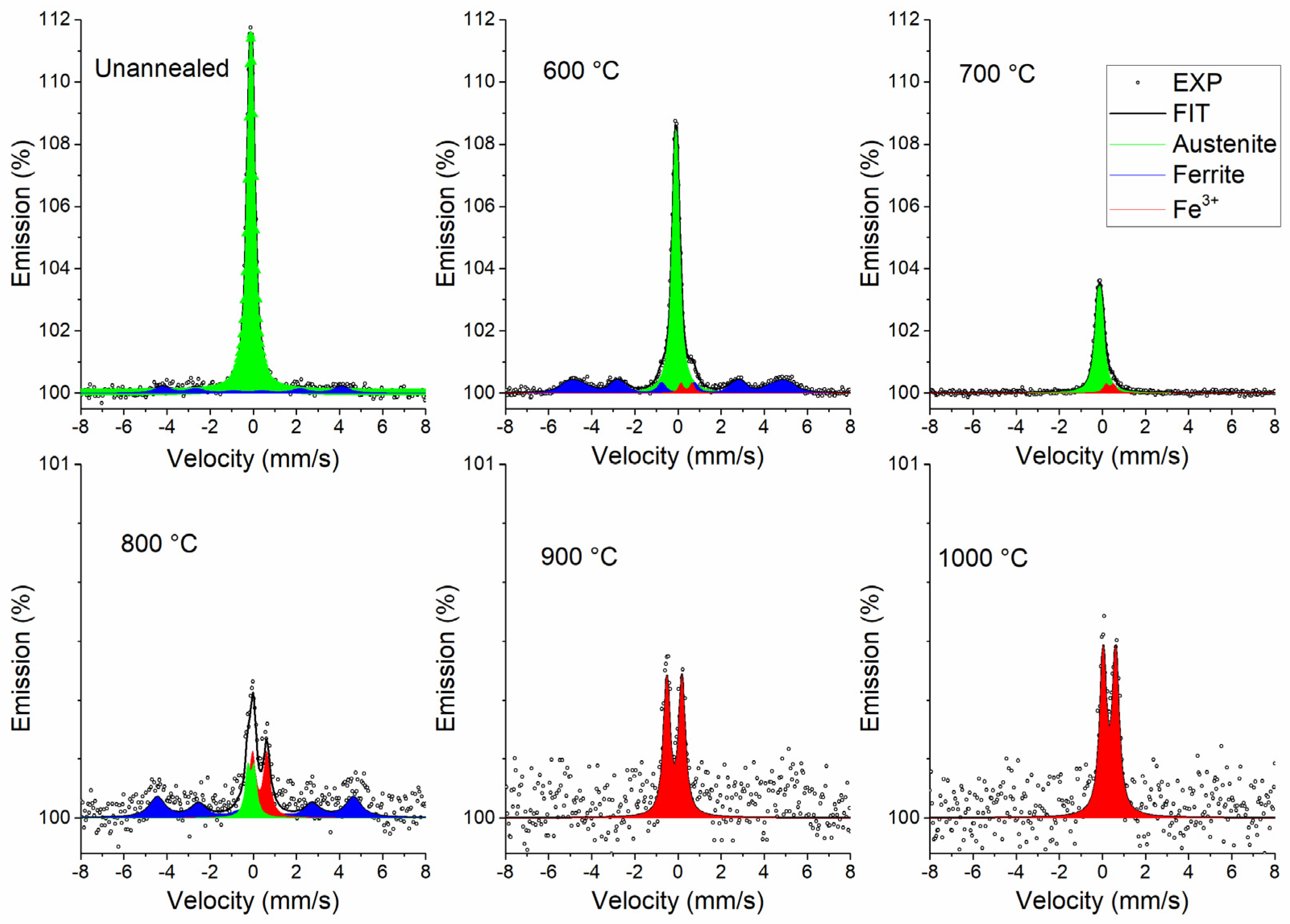
3.3.2. Conversion Electron Mössbauer Spectroscopy

In comparison to CXMS, conversion electron Mössbauer spectroscopy (CEMS) was based on the diffusion depth in the material of conversion electrons emitted from resonance atoms. The penetration depth was approximately 0.3 μm and could be calculated using an equation [59].
The CEMS spectra for all annealed 304 stainless steel samples and the fitting results from the MOSSWINN 4.0 software are shown in Figure 8 and Table 3, respectively. As can be seen, an unannealed cast sample consisted of two phases of iron in a thin surface layer: austenitic (green singlet) and ferritic (blue sextet). This CEMS observation was correlated with the XRD results for an unannealed sample. Austenitic–ferritic (γα) transformation could be observed by CEMS spectra at a temperature impact of 600 °C and 800 °C. The austenite–ferritic transformation was not observed at an annealing temperature of 700 °C. The ferritic phase disappeared in the CEMS spectrum of the sample annealed at this temperature. A doublet (red spectra) corresponded to trivalent iron according to the value of the isomer shift (Table 3) and was present in all spectra of the annealed samples. As the temperature increased, the amplitude of the spectral lines of the iron phases (austenite and ferrite) decreased while the doublet area increased. When fitting Mössbauer spectra, the doublet Fe3+ parameters were fixed for the low content of Fe3+ in the samples. The fixed parameters in Table 3 were taken from the CEMS spectra of the annealed sample containing only doublet Fe3+. The area and intensity of the doublet slightly increased with annealing temperature and, with exposure to temperatures of 900 °C and 1000 °C, the CEMS spectra contained only Fe3+. The decrease in the number of iron-containing phases and the growth of Fe3+ can be explained by the growth of the oxide layer under the influence of temperature. This doublet corresponded to iron oxides, in agreement with the XRD results of mixed oxides with Cr and Mn. A decrease in iron content indicated the formation of other oxides on the surface layer (0.3 μm).

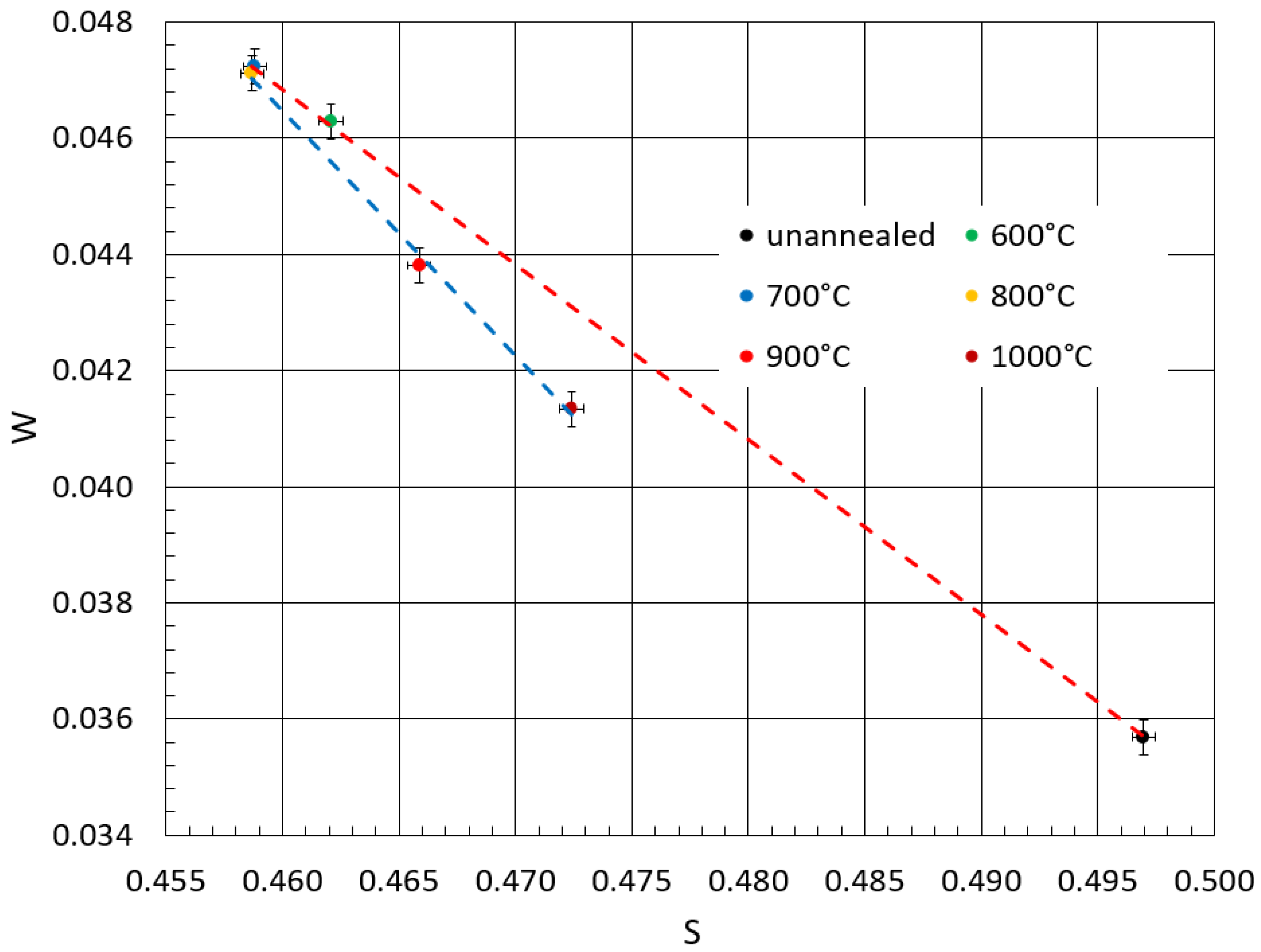
3.4. Positron Annihilation Oxidation Study

The defects associated with the annealing of cast 304 stainless steel were studied by Doppler broadening spectroscopy (DBS). The S-W plot used to evaluate the defect structure of the metallic material is shown in Figure 9. The S-W graph shows the following trends. The unannealed sample (original) had an S value and a W value, indicating a certain density of defects. With an increase in annealing temperature to 700 °C, the value of S decreased and the value of W increased. This behavior of both the S and W parameters indicated a reduction in the vacancy and defect concentration [60]. At the same time, this behavior of the S and W parameters may have been associated with a phase transformation (αγ) [61], which, however, was not confirmed by Mössbauer spectroscopy. From the XRD recordings (Figure 6), we see that the α phase completely disappeared at the annealing temperature of 700 °C, but this phase was preserved at the annealing temperature of 600 °C. We can conclude that the large changes in the values of the S and W parameters between the unannealed and annealed samples were related to the reduction in the defect concentration. Small changes in these parameters were related to the phase transformation (αγ). At an annealing temperature of 800 °C, in accordance with both XRD and MS, the phase transformation (γα) began. At a temperature of 900 °C, when the original austenitic phase had changed from approximately 90% austenitic to ferritic, there was a striking change in the S-W plot. The S value began to increase slightly again and the W value decreased, corresponding to significant structural changes and the formation of a new phase with different properties. At 1000 °C, the values further confirmed the ongoing microstructural changes, likely related to the coarsening of the grain and the growth of the ferritic phase. Perhaps at the same time, the high annealing temperature induced the formation of new other defects in the crystal lattice. In summary, the transformation of austenite to ferrite (γα) at higher temperatures led to characteristic changes in the S and W parameters that reflected both the changes in the electronic structure caused by the phase transformation and the change in specific types of defects and their concentration.

4. Conclusions

The effect of temperature on the surface layer of the 304 stainless steel samples was investigated. Annealing was carried out in air at a temperature range of 600–1000 °C for 36 h. A study of the surface of the steel parts showed that annealing led to the formation of an inhomogeneous surface layer that prevented deep oxidation. According to the results obtained, surface oxidation occurred on the surface of cast 304 stainless steel under the influence of temperature to form corundum-type oxides ((Fe-Cr)2O3 and (Fe-Cr)2MnO4). The formation of these oxides was demonstrated primarily by XRD, and CEMS confirmed the presence of iron-bearing oxides in the 0.3 μm thick surface layer. The formation of mixed oxides was related to the diffusion of alloying Cr and Mn elements to the surface of the samples. EDS showed the diffusion of Cr and Mn towards the surface. It was shown by XRD that in addition to the formation of mixed oxides, a phase transformation of the austenitic phase to the ferritic phase occurred at temperatures of 800–1000 °C. CXMS confirmed an austenite–ferrite phase transformation in the surface layer up to about 30 μm. The reverse transformation of the ferritic phase to austenitic occurred during annealing at 700 °C, which was confirmed by both XRD and CEMS. The corresponding phase transformations were confirmed by DBS, and these transformations were manifested by changes in the S and W parameters.

Supplementary Materials

The following supporting information can be downloaded at: https://www.mdpi.com/article/10.3390/met15070748/s1, References [27,60,62,63,64,65,66] are cited in the supplementary materials.

Author Contributions

Conceptualization, M.M.; Data curation, M.M., M.K. and T.I.; Formal analysis, M.K., T.I. and M.F.; Investigation, T.I., M.K. and M.F.; Methodology, M.K., T.I. and M.M.; Project administration, M.M.; Resources, M.K. and M.M.; Validation, T.I., M.K., M.M. and M.F.; Writing—original draft, T.I. and M.M.; Writing—review and editing, M.M., M.K., T.I. and M.F. All authors have read and agreed to the published version of the manuscript.

Funding

This research was funded by an internal IGA grant of Palacký University (IGA_PrF_2025_007) and the Czech Ministry of Education, Youth and Sports, grants CZ.02.1.01/0.0/0.0/17_049/0008408 and CZ.02.01.01/00/23_021/0008954.

Data Availability Statement

The original contributions presented in this study are included in the article/Supplementary Materials. Further inquiries can be directed to the corresponding authors.

Conflicts of Interest

Author Martin Fryšák was employed by the company SIGMA GROUP a.s. The remaining authors declare that the research was conducted in the absence of any commercial or financial relationships that could be construed as a potential conflict of interest.

References

  1. Podder, A.; Bhanja, A. Application of Stainless Steel in Automobile Industry. Adv. Mater. Res. 2013, 794, 731–740. [Google Scholar] [CrossRef]
  2. Kangas, P.; Chai, G. Use of advanced austenitic and duplex stainless steels for applications in Oil & Gas and Process industry. Adv. Mater. Res. 2013, 794, 645–669. [Google Scholar] [CrossRef]
  3. Cetin, M. Effect of boron added corrosion behavior of cast 304 stainless steel. Prot. Met. Phys. Chem. Surf. 2019, 55, 1217–1225. [Google Scholar] [CrossRef]
  4. Hartwig, A.; Decker, M.; Klein, O.; Karl, H. Stoichiometric titanium dioxide ion implantation in AISI 304 stainless steel for corrosion protection. Nucl. Inst. Met. Phys. Res. Sect. B 2015, 365, 94–99. [Google Scholar] [CrossRef]
  5. Cetin, M.; Ölmez, E. Corrosion, Wear and Mechanical Properties of Boron Added Cast 304 Stainless Steel. Prot. Met. Phys. Chem. Surf. 2020, 56, 619–627. [Google Scholar] [CrossRef]
  6. Zhang, H.; Li, C.; Yao, G.; Shi, Y.; Zhang, Y. Effect of annealing treatment on microstructural evolution and deformation behavior of 304L stainless steel made by laser powder bed fusion. Int. J. Plast. 2022, 155, 103335. [Google Scholar] [CrossRef]
  7. Xu, Q.; Zhu, J.; Zong, Y.; Liu, L.; Zhu, X.; Zhang, F.; Luan, B. Effect of drawing and annealing on the microstructure and mechanical properties of 304 austenitic stainless steel wire. Mater. Res. Express 2021, 8, 126530. [Google Scholar] [CrossRef]
  8. Albritton, O.W. A Study of Sensitization in Types 304 and 304L Stainless Steels Using Mössbauer Spectroscopy. Corrosion 1968, 24, 389–392. [Google Scholar] [CrossRef]
  9. Souza, P.A., Jr.; Macêdo, M.C.S.; Queiroz, R.S.; Klingelhöfer, G. Atmospheric Corrosion Investigation in Industrial, Marine and Rural Environments in South-East Brazil. Hyperfine Interact. 2002, 139, 183–191. [Google Scholar] [CrossRef]
  10. Carbucicchio, M.; Palombarini, G.; Savorelli, G. Wet corrosion of a cw power laser melted AISI 304 stainless steel. Nucl. Inst. Met. Phys. Res. Sect. B 1993, 76, 170–172. [Google Scholar] [CrossRef]
  11. Chabica, M.E.; Williamson, D.L.; Wei, R.; Wilbur, P.J. Microstructure and corrosion of nitrogen implanted AISI 304 stainless steel. Surf. Coat. Technol. 1992, 51, 24–29. [Google Scholar] [CrossRef]
  12. Arshed, M.; Butt, N.M.; Siddique, M.; Anwar-ul-Islam, M. Mössbauer study of corrosion of SS-304 exposed to sea water. J. Radioanal. Nucl. Chem. Lett. 1992, 165, 107–113. [Google Scholar] [CrossRef]
  13. Kasparova, O.V.; Baldokhin, Y.V. New Concept of the Mechanism of Intergranular Corrosion of Stainless Steels. Prot. Met. 2007, 43, 235–240. [Google Scholar] [CrossRef]
  14. León-Silva, U.; Nicho, M.E.; González-Rodríguez, J.G. Effect of thermal annealing of poly(3-octylthiophene) films covered stainless steel on corrosion properties. J. Solid State Electrochem. 2010, 14, 1089–1100. [Google Scholar] [CrossRef]
  15. Etienne, A.; Radiguet, B.; Genevois, C.; Le Breton, J.M.; Valiev, R.; Pareige, P. Thermal stability of ultrafine-grained austenitic stainless steels. Mater. Sci. Eng. A 2010, 527, 5805–5810. [Google Scholar] [CrossRef]
  16. Chen, A.Y.; Zhang, J.B.; Song, H.W.; Lu, J. Thermal-induced inverse γ/α′ phase transformation in surface nanocrystallization layer of 304 stainless steel. Surf. Coat. Technol. 2007, 201, 7462–7466. [Google Scholar] [CrossRef]
  17. Merakeb, N.; Messai, A.; Ayesh, A.I. Investigation of phase transformation for ferrite-austenite structure in stainless steel thin films. Thin Solid Film. 2016, 606, 120–126. [Google Scholar] [CrossRef]
  18. Wu, Y.C.; Zhang, R.; Chen, H.; Li, Y.; Zhang, J.; Zhu, D.M.; Jean, Y.C. Corrosion of iron and stainless steels studied using slow positron beam technique. Rad. Phy. Chem. 2003, 68, 599–603. [Google Scholar] [CrossRef]
  19. Wu, Y.C.; Chen, C.Q.; Chen, Y.Q.; Hu, Y.; Cheng, J.; Wang, S.J.; Jean, Y.C. Hydrogen- and corrosion-related defect characterization of iron and steels using a slow positron beam. Positron Positronium Chem. 2009, 607, 111–113. [Google Scholar] [CrossRef]
  20. Yabuuchi, A.; Maekawa, M.; Kawasuso, A. Vacancy defects in a stress-corrosion-cracked Type 304 stainless steel investigated by positron annihilation spectroscopy. J. Nucl. Mat. 2011, 419, 9–14. [Google Scholar] [CrossRef]
  21. Thaveeprungsriporn, V.; Swatewacharkul, P.; Talerngsuk, K.; Punnachaiya, S.; Thong-Aram, D. Microstructure characterization using compensated Doppler-broadened positron annihilation spectroscopy. Nucl. Inst. Met. Phys. Res. Sect. B 1999, 155, 459–467. [Google Scholar] [CrossRef]
  22. Horodek, P.; Siemek, K.; Kobets, A.G.; Kulik, M.; Meshkov, I.N. Positron beam and RBS studies of thermally grown oxide films on stainless steel grade 304. Appl. Surf. Sci. 2015, 333, 96–103. [Google Scholar] [CrossRef]
  23. Maekawa, M.; Yabuuchi, Y.; Kawasuso, A. Evaluation of stainless steel under tensile stress using positron microbeam. J. Phys. Conf. Ser. 2010, 225, 012033. [Google Scholar] [CrossRef]
  24. Horodek, P.; Dryzek, J.; Kobets, A.; Kulik, M.; Lokhmatov, V.; Meshkov, I.; Orlov, O.; Pavlov, V.; Rudakov, A.; Sidorin, A.; et al. Slow Positron Beam Studies of the Stainless Steel Surface Exposed to Sandblasting. Acta Phys. Pol. A 2014, 125, 714–717. [Google Scholar] [CrossRef]
  25. Dryzek, E.; Sarnek, M.; Wrobel, M. Reverse transformation of deformation-induced martensite in austenitic stainless steel studied by positron annihilation. J. Mater. Sci. 2014, 49, 8449–8458. [Google Scholar] [CrossRef]
  26. Zeman, A.; Novotny, R.; Uca, O.; Krsjak, V.; Macak, J.; Debarberis, L. Behavior of cold-worked AISI-304 steel in stress-corrosion cracking process: Microstructural aspects. Appl. Surf. Sci. 2008, 255, 160–163. [Google Scholar] [CrossRef]
  27. Principi, G. The Mössbauer Effect: A Romantic Scientific Page. Metals 2020, 10, 992. [Google Scholar] [CrossRef]
  28. Hori, F.; Oshima, R. Positron Annihilation Study of Fatigue in Type 304 Stainless Steel. Mat. Sci. Forum 2001, 363–365, 204–206. [Google Scholar] [CrossRef]
  29. Hartley, J.H.; Howell, R.H.; Asoka-Kumar, P.; Sterne, P.A.; Akers, D.; Denison, A. Positron annihilation studies of fatigue in 304 stainless steel. App. Sur. Sci. 1999, 149, 204–206. [Google Scholar] [CrossRef]
  30. Yan, H.; Zhang, Z.; Wang, J.; Okonkwo, B.O.; Han, E.-H. Effect of MeV Fe Ions Irradiation on the Microstructure and Property of Nuclear Grade 304 Stainless Steel: Characterized by Positron Annihilation Spectroscopy, Transmission Electron Microscope and Nanoindentation. Acta Metall. Sin. (Engl. Lett.) 2021, 34, 1695–1703. [Google Scholar] [CrossRef]
  31. Wu, Y.C.; Zhang, X.H.; Jean, Y.C.; Suzuki, R.; Ohdaira, T. Positron Annihilation Study of Hyrogen-Induced Defects in AISI 304 Stainless Steel. Mat. Sci. Forum 2004, 445–446, 213–215. [Google Scholar] [CrossRef]
  32. Horodek, P.; Siemek, K.; Dryzek, J.; Kobets, A.G.; Wróbel, M. Positron Annihilation and Complementary Studies of Stainless Steel Exposed to Sandblasting at Different Angles. Tribol. Lett. 2017, 65, 30. [Google Scholar] [CrossRef]
  33. Kellai, A.; Kahla, S.; Dehimi, S.; Kaba, L.; Boutaghour, Z. Effect of post weld heat treatment on the microstructure and mechanical properties of a gas-tungsten-arc-welded 304 stainless steel. Int. J. Adv. Manuf. Technol. 2022, 121, 8171–8186. [Google Scholar] [CrossRef]
  34. Khodadadi, A.; Shamanian, M.; Karimzadeh, M. Microstructure and Mechanical Properties of Dissimilar Friction Stir Spot Welding Between St37 Steel and 304 Stainless Steel. J. Mater. Eng. Perform. 2017, 26, 2847–2858. [Google Scholar] [CrossRef]
  35. Nam, T.-H.; An, E.; Kim, B.J.; Shin, S.; Ko, W.-S.; Park, N.; Kang, N.; Jeon, J.B. Effect of Post Weld Heat Treatment on the Microstructure and Mechanical Properties of a Submerged-Arc-Welded 304 Stainless Steel. Metals 2018, 8, 26. [Google Scholar] [CrossRef]
  36. Abioye, T.E.; Omotehinse, I.S.; Oladele, I.O.; Olugbade, T.O.; Ogedengbe, T.I. Effects of post-weld heat treatments on the microstructure, mechanical and corrosion properties of gas metal arc welded 304 stainless steel. World J. Eng. 2020, 17, 87–96. [Google Scholar] [CrossRef]
  37. Sedláčková, A.; Ivanova, T.; Mashlan, M.; Doláková, H. Phase Changes in the Surface Layer of Stainless Steel Annealed at a Temperature of 550 °C. Materials 2022, 15, 8871. [Google Scholar] [CrossRef]
  38. Chandra-ambhorn, S.; Saranyachot, P.; Thublaor, T. Higher temperature oxidation behavior of Fe-15.7 wt% Cr-8.5 wt% Mn in oxygen without and with water vapor at 700 °C. Corros. Sci. 2019, 148, 39–47. [Google Scholar] [CrossRef]
  39. Huang, X.; Xiao, K.; Fang, X.; Xiong, Z.; Wei, L.; Zhu, P.; Li, X. Oxidation behavior of 316L austenitic stainless steel in higher temperature air with long-term exposure. Mater. Res. Express 2020, 7, 066517. [Google Scholar] [CrossRef]
  40. Pechousek, J.; Mashlan, M. Mössbauer spectrometer as a virtual instrument in the PXI/Compact PCI modular system. Czechoslov. J. Phys. 2005, 55, 853. [Google Scholar] [CrossRef]
  41. Kholmetskii, A.L.; Misevich, O.V.; Mashlan, M.; Chudakov, V.A.; Anashkevich, A.F.; Gurachevskii, V.L. Air scintillation detector for conversion electrons Mössbauer spectroscopy (CEMS). Nucl. Instrum. Methods Phys. Res. B 1997, 129, 110. [Google Scholar] [CrossRef]
  42. Klencsár, Z.; Kuzmann, E.; Vértes, A. User-Friendly Program for Multifold Evaluation of Mössbauer Spectra. Hyperfine Interact. 1998, 112, 269. [Google Scholar] [CrossRef]
  43. Klencsár, Z.; Kuzmann, E.; Vértes, A. User-Friendly software for Mössbauer spectrum analysis. J. Radioanal. Nucl. Chem. 1996, 210, 105. [Google Scholar] [CrossRef]
  44. Dryzek, J.; Wesseling, C.; Dryzek, E.; Cleff, B. Migration of Vacancies in Stainless Steel Measured by Positron Annihilation. Mater. Lett. 1994, 21, 209–214. [Google Scholar] [CrossRef]
  45. Selim, F.A. Positron annihilation spectroscopy of defects in nuclear and irradiated materials—A review. Mater. Charact. 2021, 174, 110952. [Google Scholar] [CrossRef]
  46. Bonny, G.; Konstantinovic, M.I.; Bakaeva, A.; Chao, Y.; Castin, N.; Mergia, K.; Chatzikos, V.; Dellis, S.; Khvan, T.; Bakaev, A.; et al. Trends in vacancy distribution and hardness of high temperature neutron irradiated single crystal tungsten. Acta Mater. 2020, 198, 1–9. [Google Scholar] [CrossRef]
  47. Han, F.; Lin, G.Y.; Wang, Z.L.; Peng, D.S. Oxidation analysis of 304 Austenitic Stainless Steel Sheet after Intermediate Annealing. Key Eng. Mater. 2011, 474–476, 376–380. [Google Scholar] [CrossRef]
  48. Ivanova, T.; Kořenek, M.; Mashlan, M. Using Mössbauer Spectroscopy to Evaluate the Influence of Heat Treatment on the Surface Characteristics of Additive Manufactured 316L Stainless Steel. Materials 2024, 17, 3494. [Google Scholar] [CrossRef]
  49. Ozturk, B.; Matway, R. Oxidation of Type 304 Stainless Steel under Simulated Annealing Conditions. ISIJ Int. 1997, 37, 169–175. [Google Scholar] [CrossRef]
  50. Grayeli-Kopri, A.-R.; Savaloni, H.; Habibi, M. Corrosion inhibition of stainless steel type AISI 304 by Mn coating and subsequent annealing with flow of nitrogen at different temperatures. Appl. Surf. Sci. 2013, 276, 269–275. [Google Scholar] [CrossRef]
  51. Castaing, R. Applications des Sondes Electronique a une Methode D’analyse Ponctuelle Chimique et Cristallographique. Ph.D. Thesis, University of Paris, Paris, France, 1952. [Google Scholar]
  52. Biehler, J.; Hoche, H.; Oechsner, M. Nitriding behavior and corrosion properties of AISI 304L and 316L austenitic stainless steel with deformation-induced martensite. Surf. Coat. Technol. 2017, 324, 121–128. [Google Scholar] [CrossRef]
  53. Zhang, X.; Misra, A.; Schulze, R.; Wetteland, C.; Wang, H.; Nastasi, M. Critical factors that determine face-centered cubic to body-centered cubic phase transformation in sputter-deposited austenitic stainless steel films. J. Mater. Res. 2004, 19, 1696–1702. [Google Scholar] [CrossRef]
  54. Shuro, I.; Umemoto, M.; Todaka, Y.; Yokoyama, S. Phase Transformation and Annealing Behavior of SUS 304 Austenitic Stainless Steel Deformed by High Pressure Torsion. Mater. Sci. Forum 2010, 654–656, 334–337. [Google Scholar] [CrossRef]
  55. Li, Y.; Zhao, L.; Kan, W.; Pan, H. Phase transformations and micro cracks induced by hydrogen in cold-rolled and annealed AISI 304 stainless steels. Int. J. Hydrogen Energy 2012, 37, 8724–8728. [Google Scholar] [CrossRef]
  56. Riffard, F.; Buscail, H.; Rabaste, F.; Caudron, E.; Cueff, R.; Issartel, C.; Karimi, N.; Perrier, S. Manganese effect on isothermal high temperature oxidation behavior of AISI 304 stainless steel. Mater. Sci. Forum 2008, 595, 1127–1134. [Google Scholar] [CrossRef]
  57. Lothongkum, G.; Tiyawatwitthaya, C.; Prawetpai, N.; Lothongkum, A.W. XRD investigation of the Cu and Mn effects on the oxide scale of hot rolled AISI 304L stainless steel after annealing and shot-blasting. Mater. Test. 2020, 62, 568–572. [Google Scholar] [CrossRef]
  58. Cook, D.C. Strain induced martensite formation in stainless steel. Metall. Mater. Trans. A Phys. Metall. Mater. Sci. 1987, 18, 201–210. [Google Scholar] [CrossRef]
  59. Kanaya, K.; Okayama, S. Penetration and energy-loss theory of electrons in solid targets. J. Phys. D Appl. 1972, 5, 43–58. [Google Scholar] [CrossRef]
  60. Procházka, I. Positron annihilation spectroscopy. Mater. Struct. 2001, 8, 55–60. [Google Scholar]
  61. Dryzek, E.; Sarnek, M. Reverse Transformation of Deformation Induced Martensite in Austenitic Stainless Steel. In Proceedings of the 41st Polish Seminar on Positron Annihilation, Lublin, Poland, 9–13 September 2013; Volume 125, pp. 710–713. [Google Scholar] [CrossRef]
  62. Ovchinnikov, V.V. Mössbauer Analysis of the Atomic and Magnetic Structure of Alloys; Cambridge International Science Publishing: Cambridge, UK, 2006; p. 248. [Google Scholar]
  63. Gütlich, P.; Bill, E.; Trautwein, A.X. Mössbauer Spectroscopy and Transition Metal Chemistry, Fundamentals and Applications; Springer: Berlin/Heidelberg, Germany, 2011; p. 568. [Google Scholar]
  64. Kuzmann, E.; Nagy, D.; Vértes, A. Critical Review of Analytical Applications of Mössbauer Spectroscopy Illustrated by Mineralogical and Geological Examples (IUPAC Technical Report). Pure Appl. Chem. 2003, 75, 801–858. [Google Scholar] [CrossRef]
  65. Puska, M.J.; Nieminen, R.M. Theory of positrons in solids and on solid surface. Rev. Mod. Phys. 1994, 66, 841–896. [Google Scholar] [CrossRef]
  66. Schultz, P.J.; Lynn, K.G. Interactions of positron beams with surface, thin films and interfaces. Rev. Mod. Phys. 1994, 60, 701–779. [Google Scholar] [CrossRef]
Figure 1. Annealed samples of cast 304 austenitic steel (sample diameter 25 mm, height 5 mm).
Metals 15 00748 g001
Figure 2. Definition of parameters S and W extracted from the 511 keV annihilation peak. Values for the windows were from 510.2 to 511.8 keV for region A. For the wings C, they were from 507.7 to 508.9 keV and 512.6 to 514.0 keV.
Metals 15 00748 g002
Figure 3. Surface morphology of cast 304 stainless steel annealed at different temperatures in air for 36 h (SEM images at 1000× magnification).
Metals 15 00748 g003
Figure 4. Surface morphology of cast 304 stainless steel annealed at different temperatures in air for 36 h (SEM images at 10,000× magnification).
Metals 15 00748 g004
Figure 5. Depth dependency of elemental concentration of 304 stainless steel after 36 h of annealing in air. The electron penetration depth was related to the accelerating voltage, given by Castaing’s formula, reprinted from Ref. [51], and ranged from 0.3 μm for HV 11 kV to 2.6 μm for HV 30 kV.
Metals 15 00748 g005
Figure 6. The XRD patterns of cast 304 stainless steel annealed in air for 36 h at temperatures of 600–1000 °C.
Metals 15 00748 g006
Figure 7. CXMS spectra of cast 304 stainless steel annealed in air for 36 h.
Metals 15 00748 g007
Figure 8. CEMS spectra of cast 304 stainless steel annealed in air for 36 h.
Metals 15 00748 g008
Figure 9. S-W plot for positron annihilation measurements. The red line corresponds to γ-Fe and the blue line corresponds to α-Fe.
Metals 15 00748 g009
Table 1. Chemical composition of 304 cast stainless steel.
SteelElement Concentration, wt %
FeCSiMnPSCrNiN
304Bal.0.071.02.00.045≤0.0117.50–19.508.00–10.500.10
Table 2. Hyperfine parameters for CXMS spectra of cast 304 stainless steel annealed in air for 36 h (IS—isomer shift; QS—quadrupole splitting; W—full width at half maximum; A—spectrum area).
Annealing TemperaturePhaseδ [mm/s]Δ [mm/s]B [T]W [mm/s]A [%]
Unannealedaustenite−0.08 ± 0.010.15 ± 0.01-0.28 ± 0.01100
600 °Caustenite−0.08 ± 0.010.14 ± 0.01-0.27 ± 0.01100
700 °Caustenite−0.08 ± 0.010.15 ± 0.01-0.27 ± 0.01100
800 °Caustenite−0.08 ± 0.010.14 ± 0.01-0.28 ± 0.0170 ± 2
ferrite0.09 ± 0.05-27.4 ± 0.50.91 ± 0.1430 ± 2
900 °Caustenite−0.09 ± 0.010.16 ± 0.01-0.22 ± 0.039 ± 2
ferrite0.01 ± 0.01-27.7 *0.25 ± 0.0291 ± 2
1000 °Caustenite−0.05 ± 0.030.17 ± 0.01-0.19 ± 0.035 ± 2
ferrite-0.01 ± 0.01-26.8 *0.22 ± 0.0295 ± 2
* Fixed parameter.
Table 3. Hyperfine parameters for CEMS spectra of cast 304 stainless steel annealed in air for 36 h (IS—isomer shift; QS—quadrupole splitting; W—full width at half maximum; A—spectrum area).
Annealing TemperaturePhaseδ [mm/s]Δ [mm/s]B [T]W [mm/s]A [%]
Unannealedaustenite−0.11 ± 0.010.17 ± 0.01-0.32 ± 0.0186 ± 2
ferrite−0.08 ± 0.06-25.9 ± 0.50.98 ± 0.1514 ± 2
600 °Caustenite−0.10 ± 0.010.15 ± 0.01-0.34 ± 0.0161 ± 2
ferrite0.01 ± 0.02-30.1 ± 0.10.97 ± 0.0435 ± 2
Fe3+ oxide0.35 *0.58 *-0.37 *4 ± 2
700 °Caustenite−0.11 ± 0.010.19 ± 0.01-0.37 ± 0.0192 ± 2
Fe3+ oxide0.35 *0.58 *-0.37 *8 ± 2
800 °Caustenite−0.08 ± 0.020.22 ± 0.02-0.29 ± 0.0519 ± 2
ferrite0.12 ± 0.05-28.2 ± 0.40.86 ± 0.1449 ± 2
Fe3+ oxide0.37 ± 0.020.56 ± 0.03-0.35 ± 0.0432 ± 2
900 °CFe3+ oxide0.36 ± 0.010.63 ± 0.02-0.30 ± 0.03100
1000 °CFe3+ oxide0.35 ± 0.010.58 ± 0.02-0.37 ± 0.02100
* Fixed parameter.
Disclaimer/Publisher’s Note: The statements, opinions and data contained in all publications are solely those of the individual author(s) and contributor(s) and not of MDPI and/or the editor(s). MDPI and/or the editor(s) disclaim responsibility for any injury to people or property resulting from any ideas, methods, instructions or products referred to in the content.

