Abstract
This review summarizes the development of surface treatments applied to dental implants with the aim of improving their clinical performance. It covers the advancement of various techniques, from the conventional to the more advanced ones. Among the recent advancements, surface texturing has enabled atomic and structural modifications of implant surfaces at the micro- and nanoscales, improving tissue–material interactions. Acid etching and atomic layer deposition applied onto implant surfaces results in optimized osseointegration by stimulating the deposition and proliferation of osteoblasts and fibroblasts. The atomic layer deposition of TiO2, ZnO, ZrO2, and CaCO3 has proven effective in improving osseointegration and tackling corrosion. Corrosion is still an important issue, whereby metals released from titanium implants and their associated degradation products cause local and systemic side effects, leaving a wide avenue for future research. The development of hybrid dental implants is envisaged through new materials and technologies, such as additive manufacturing, which may play a critical role in the fabrication of patient-specific implants with tailored nano-topography capable of enhancing such properties as antibacterial activity and osseointegration.
1. Introduction
One of the desired clinical abilities of dental implants is osseointegration, which implies a dynamic biological process by which bone tissue grows directly into the surface of an implant. Many factors affect osseointegration, such as the surface structure of the implants, the specific conditions at the surgical site, the type of bone taken into consideration, and the patient’s general health condition [1,2,3,4]. The surface structure of the implant can be engineered to enhance its osseointegration capabilities by means of various surface treatments, including sandblasting with fine particles, acid etching, and heat treatments, which modify the implant’s surface structure to promote bonding with the bone [5,6,7,8,9,10,11,12].
Beside affecting osseointegration, the surface structure of dental implants also affects the implant–abutment interface, especially those made of titanium (Ti) alloys; these are subjected to wear, fatigue, and corrosion. These tribological phenomena can lead to the release of the metallic ions of the alloy’s component, including Ti, Al and V, potentially causing local complications [13]. At the implant–abutment interface, the quality of the Ti-based implant, manufacturing, and the precision of the connection fit are critical to ensuring the interface’s stability against occlusal forces [13,14,15,16]. Deviations greater than 10 μm in the connection fit can cause biological issues and biomechanical deficiencies [17,18]. An increasing stress on the implant and the surrounding bone can potentially lead to fractures and implant loosening. In addition, the subgingival space formed by an unfit connection can cause micro-infiltration, promote bacterial growth, and cause peri-implantitis [19,20,21].
Apart from the surface treatment of Ti-based implants, attention has recently been focused on zirconium dioxide (ZrO2)-based surfaces, which have shown encouraging results but still lack long-term clinical data to confirm their osseointegration capabilities. Previous studies have identified significant problems, including high failure rates and peri-implant crestal bone loss associated with ZrO2-based surfaces [22]. Although it is considered to be an inert biomaterial, there is evidence that ZrO2 is involved in biological interactions such as the adsorption of blood proteins and cell migration [23,24]. These interactions, coupled with the chemical bonding and degradation properties of ZrO2, make further study worthwhile. More efforts are underway to enhance ZrO2-based surfaces, focusing particularly on their morphological and bioactive properties to optimize cell attachment, proliferation, and differentiation during bone healing [24,25].
The objective of this study is to review the main surface treatment methods that improve the clinical performance of Ti- and ZrO2-based dental implants. It explores in detail the specificities of each method while examining the impact of changes in surface composition and morphology on osseointegration. This review was conducted following a systematic literature search to provide proper coverage of the latest developments in dental implant surface treatment, using PubMed, Google Scholar, and Web of Science, between 2017 and 2025. Boolean operators were utilized to limit the search strategy for the choice of proper studies. The key search terms utilized are shown in Table 1. In order to ensure scientific integrity, certain exclusion and inclusion criteria were utilized. Studies were included if they dealt with surface alterations in titanium- and zirconia-based dental implants, i.e., those that reported osseointegration, mechanical stability, antimicrobial properties, and bioactivity. Only peer-reviewed journals from Web of Science from 2017 to 2025 were included. The exclusion criteria were studies not specifically related to dental implants or biomaterial coatings, non-English-language publications in the absence of translated copies, and studies lacking experimental or clinical data. Finally, the papers included in this review were evaluated on the basis of their scientific contribution to the surface treatment of dental implants prior to their inclusion. Through this systematic process, the very best highly applicable and scientifically rigorous studies were selected, enabling the most detailed examination of recent developments within the field.

Table 1.
Literature review strategy using Web of Science, PubMed, and Google Scholar.
2. Conventional Surface Treatments for Dental Implants
The techniques included in this category are machining, grit blasting, acid etching, blasting–acid combinations, anodizing, and plasma spraying, as summarized in Table 2.
Table 2.
Conventional surface treatments applied to dental implants.
2.1. Machining
Branemark introduced the first machining process used for biomedical and dental implants by using a large lathe with high-speed rotation to shape implants from hard metals [26]. This manufacturing method produces precise dimensions but leaves a surface with macroscopic or microscopic roughness, thus clinically lengthening the bone-healing time to four to six months for rehabilitation [26,36]. Current digitally controlled lathes speed up the shaping process, improve surface quality, and reduce human error [37].
2.2. Grit Blasting
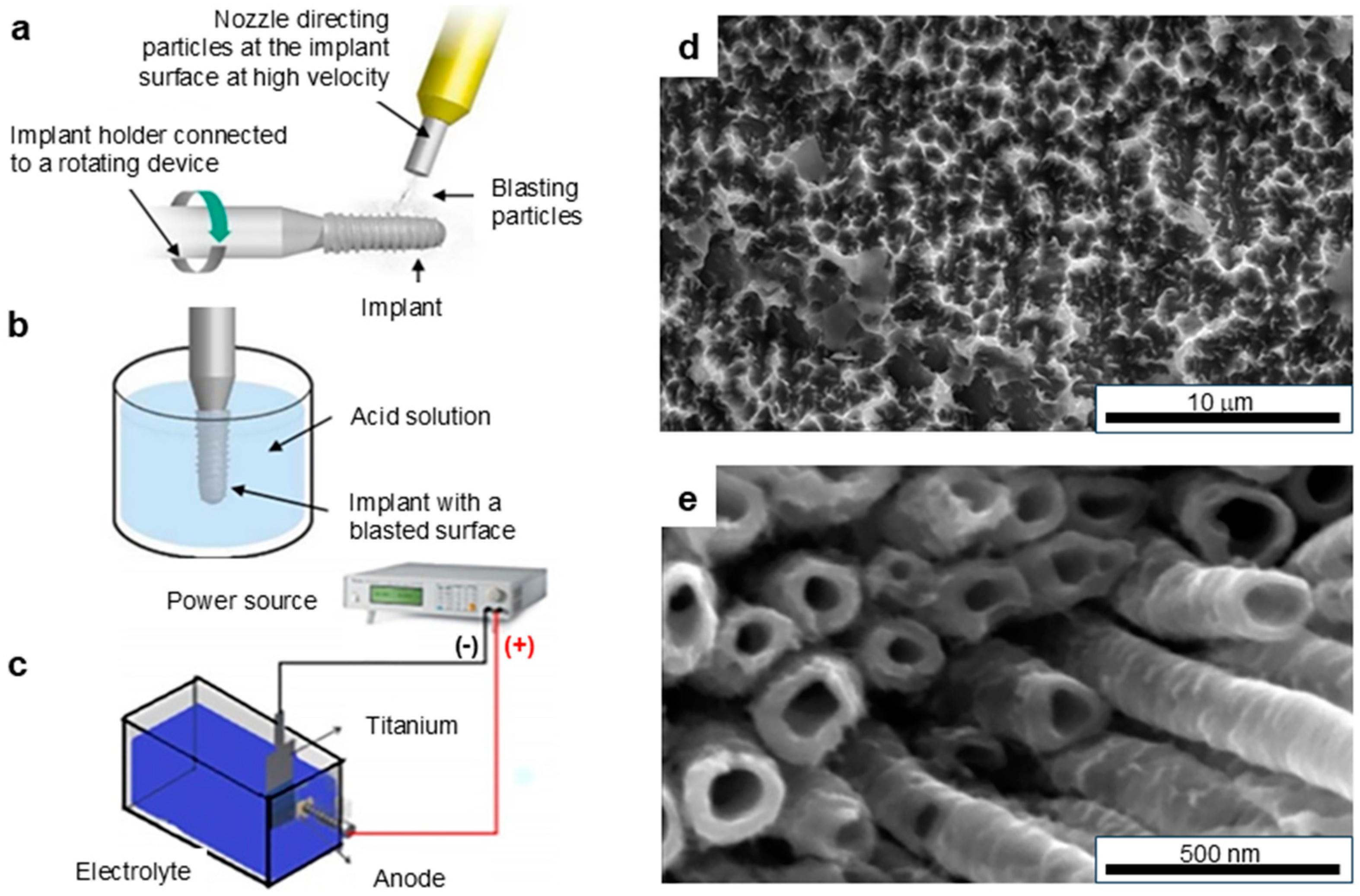
One of the most widely used methods for improving the osseointegration of dental implants in less dense bone areas is surface treatment by sandblasting with micro- or nanoparticles, offering efficiency and low costs (Figure 1a). This process projects particles such as alumina, Al, Ti, or hydroxyapatite onto the implant surface using high-pressure machines, creating numerous indentations. These indentations have varying sizes depending on the size and material nature of the used particles. However, too often, the absence of standardization in measuring the roughness of a surface limits uniformity [5,6]. Sandblasted surfaces show an improved osseointegration time compared to machined or smooth surfaces [5,6,36].
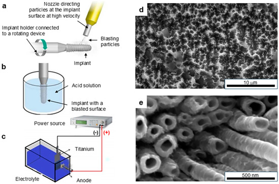
Figure 1.
Conventional surface treatment techniques: (a) sandblasting; (b) acid etching [10]; (c) anodization; (d) SEM image of a surface after double acid etching; (e) SEM image of a surface after anodization [27], 2021 Elsevier.
2.3. Acid Etching
In general, to a certain degree, a rougher implant surface promotes increased cell colonization and accelerates the osseointegration process [38]. Acid etching is another widely used technique for enhancing surface roughness or clean implant surfaces (Figure 1b). The most common acids used in this method are nitric acid (HNO3) [39] and phosphoric acid (H3PO4) [29]. This chemical treatment depends on the type, concentration, temperature, and exposure time of the acid and thus produces different topographies [5,6,9,11]. Acid etching removes manufacturing residues and produces roughness at both the micro- and nanometric levels [27,40]. Its advantages are the acceleration of osseointegration and the reduction of biofilm formation. However, over-etching can result in undesirable surface deformations; rigorous standardization and control of the process are therefore required [41].
2.4. Grit Blasting–Acid Etching
Sandblasted coarse-grained acid etching (SLA) is a combined method incorporating physical and chemical modifications in developing favorable morphology [5,6]. This enhanced approach increases osseointegration and cell adhesion, and the desired effects may be obtained within as little as one to two months of use [5,6]. The acidic agents not only clean the residues from sandblasting but also enhance the surface by making the process self-reinforcing. This is widely recognized as the most effective surface modification technique and provides outstanding success rates in various applications over the long term [11,29,30]. However, proprietary approaches adopted by manufacturers make it challenging to evaluate and optimize specific parameters.
2.5. Anodizing
In recent years, anodization has become another popular method for producing nanotubes and nanopores on the implant surface (Figure 1c). The process is electrochemical, with the utilization of an electrolytic solution and a cathode–anode system, and the application of a controlled temperature and voltage or current [9,27,31,42]. It is usually carried out in combination with acid etching or other methods, and it stimulates the growth of oriented nanotubes that enhance osseointegration to a significant degree within one to two months [6,33]. The current research focuses on functionalizing anodized surfaces for drug delivery applications [9]. The surface topography characterization, conducted via scanning electron microscopy (SEM), revealed the microscale and nanoscale features of the treated Ti-based implant before and after double acid etching with subsequent anodizing (Figure 1d and Figure 1e, respectively).
3. Recent Surface Treatment Methods Applied to Ti-Based Implants
3.1. Anodic Spark Deposition Technique
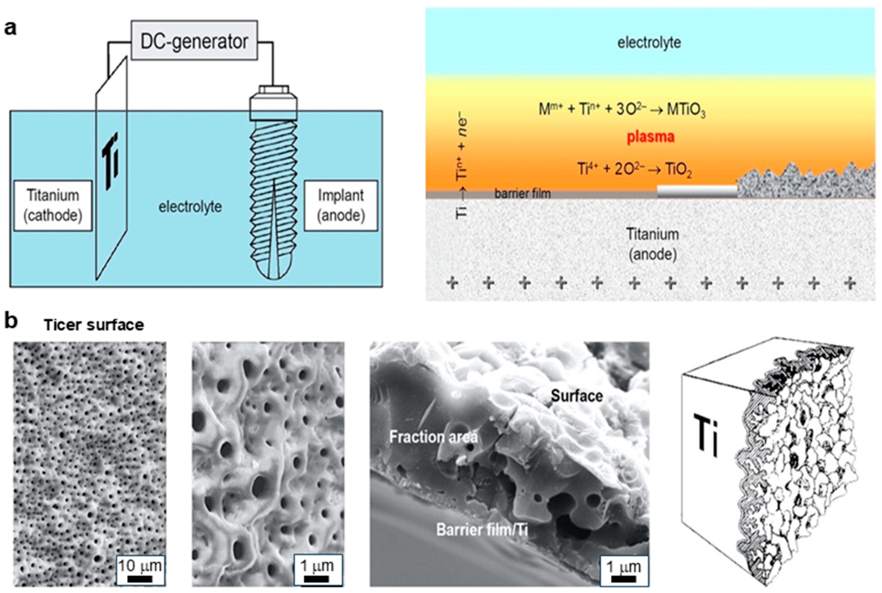
Electrochemical surface modification plays a crucial role in the preparation of biocompatible coatings and microstructures and is a considerable feature of manufacturing methods in this field. Anodic spark deposition technique (ASD) is an advanced plasma chemical and electrochemical technique used to fabricate ceramic surfaces on anodic metal substrates, normally made of Ti and Ti alloys, as shown in Figure 2. This process is known under various designations in the literature, namely “Anodische Oxidation unter Funkenentladung”, dielectric decomposition process, micro-arc oxidation, plasma electrolytic oxidation, and anodic plasma chemical process. ASD has been instrumental in developing surfaces such as Ticer and Ti Unite [43,44,45,46,47], demonstrating long-term clinical effectiveness. The coatings produced using this method incorporate elements from the electrolyte and the anode material. For instance, calcium or phosphorus can be integrated into the surface oxide by optimizing electrolyte composition and coating conditions, enhancing cell growth and bone adhesion on implant surfaces [48]. This process enhances the thickness of the oxide film at a given anode potential, which decreases the resistance at the critical electrical breakdown point, enabling the ASD process to be terminated in a controlled manner based on film thickness. Fast cooling with a cooling rate of approximately 10⁵ K/s of the molten oxides prevents the crystallization of the solid compounds formed and grants the properties desired for the surface.

Figure 2.
Ticer fabrication using anodic spark deposition: (a) electrolytic cell and ASD process on the anode, (b) surface area at processing (SEM and schematic representation) [49].
The voltage and duration of anodizing directly influence the thickness of the oxide layers on pure Ti implants [50]. Different oxide layer thicknesses can be achieved using various electrolytes, including sulfuric, phosphoric, or acetic acid, sodium hydroxide, or calcium hydroxide. Anodizing in acidic electrolytes generally requires higher voltages than in alkaline solutions [51]. Increasing the electrolyte concentration and temperature lowers the anodic formation voltage, formation rate (nm/s), and current efficiency (nm·cm2/°C). Conversely, increasing the current density and anode–cathode surface ratio enhances these parameters. Coatings produced via ASD are typically amorphous to X-rays. The anodic oxidation of pure Ti predominantly yields anatase, while higher voltages during spark discharge produce a thicker oxide layer composed of a rutile–anatase TiO2 mixture [50]. Anatase, which crystallizes in a tetragonal system and is rarely pure, transitions to rutile at around 915 °C. The most important modification of TiO2 is rutile, which is also tetragonal, while another modification, brookite, crystallizes in an orthorhombic system. These ceramic oxide layers provide several advantages: a specific porous structure (Figure 2b), very good adhesion to the implant base up to 26 MPa, the possibility of modifying the chemical composition of the coating, and durability in the long term. This method has been successfully applied to produce Ti-based implants with osseointegration coatings [43,45,46,52,53,54].
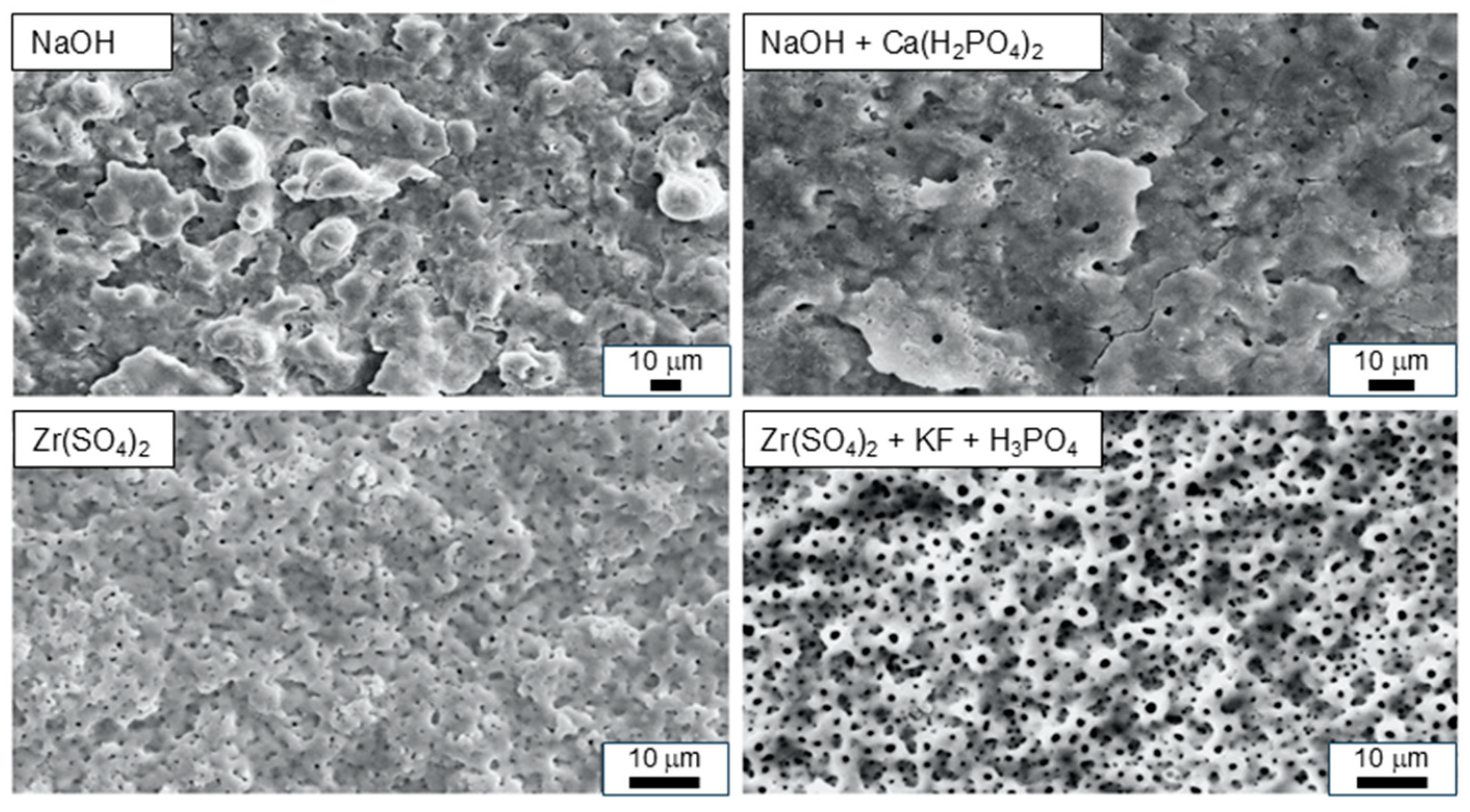
One of the disadvantages of Ti-based materials is their gray color, which can be discolored through thin mucous membranes or after recession; it thus represents an aesthetic problem, especially in anterior teeth. Zirconia, also known as zirconium dioxide (ZrO2), is a synthetic, hard, white crystalline oxide. The Ti-ZrO2 combination has been emphasized for its potential features, taking advantage of ZrO2’s mechanical strength, which is equal to that of stainless steel, along with its superior biocompatibility and natural tooth color [55,56]. The early implants made of ZrO2 bodies and coatings of TiO2 demonstrated problems related to mechanical stability and toxicity [57]. Very recently, Ti-based implants were prepared with ZrO2 coatings [58,59] via anodic plasma-electrochemical oxidation in aqueous electrolyte solutions with different concentrations of Zr(SO4)2, supplemented by KF and/or H3PO4 (Figure 3). These surfaces in vitro exhibited equally superior effects on osteoblastic cells with Ticer by markedly accelerating osteoblast differentiation when compared to smooth-surface topography, represented here by pure Ti and ZrO2. It promoted morphological changes in osteoblasts, generating the cell cluster that could facilitate quicker bone formation. The structure design keeps the integrity of titanium for mechanical performance, with its good biocompatibility from the nature of preserved ZrO2.
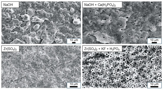
Figure 3.
SEM images of Ti surfaces developed using various electrolyte systems, resulting in two distinct types of surfaces: at the top, two white surfaces (white Ticer); at the bottom, two ZrO2-coated pure Ti surfaces [49].
3.2. Atomic Layer Deposition Technique
Biological inertness reduces the possibility of osseointegration for ZrO2. The atomic layer deposition technique (ALD) of depositing ZrO2 on Ti-based implants appears to be a promising solution to optimizing osseointegration in order to improve osteogenesis and reduce the risk of bacterial infections [60]. To enhance osteogenesis and reduce bacterial infections, by Tatsuhide Hayashi et al. [61] studied the deposition of Zn oxide onto ZrO2. Four samples were prepared and tested in this study: pure ZrO2, ZnO deposited by ALD onto sandblasted ZrO2 and acid-etched ZrO2, and ZnO deposited on sandblasted and acid-etched ZrO2. The in vitro assays showed the effectiveness of ZnO against S. aureus, E. coli, and P. gingivalis and also enhanced MC3T3-E1 cell adhesion and promoted osteogenic differentiation [62]. In another study [63], it was shown that determining the optimal ZnO thickness is critical to preventing toxic metal release during clinical use. In addition, an optimized combination of TiO2 layers and Ag nanoparticles applied to the nanostructured 316L stainless steel demonstrated remarkable antimicrobial efficacy, providing an innovative solution to enhance the biocompatibility and durability of medical implants [64]. Alumina (Al2O3) is a widely used ceramic in dentistry; it is applied in implants, sealants for dental composites, bone cement materials, bases for fixed prostheses, and orthodontic supports [65]. Another researcher fabricated porous alumina membranes coated with thin layers of TiO2 using ALD for two different pore sizes: 20 nm and 100 nm in diameter [66]. In vitro studies using epidermal keratinocytes showed no significant differences in cell viability between the TiO2-coated and uncoated alumina [67]. ALD also showed promise in creating passive films that resist corrosion and bioactivity; it has been successful in protecting metallic and non-metallic surfaces across various fields.
3.3. Plasma Spraying and Plasma Electrolytic Oxidation
Biomimetic implant surfaces were created by applying hydroxyapatite (HA)-calcium-phosphate (CaP) coatings, which were intended to mimic bone’s atomic composition [34,68]. These coatings are applied via plasma systems in a vacuum or low-pressure environment, creating micro- and nanoscale surface layers. Commonly used materials include Ti, Au, Ag, and ceramics. Plasma effects produce the partial melting of surface oxides, together with high-temperature chemical reactions that provide solid phases, including spinel or mixed oxides. Their drawback is that they necessitate careful surgical handling because of their fragility at the interface, as well as increased bacterial contamination risks [68]. Modern plasma electrolytic oxidation (PEO) techniques modify the surfaces of Ti-based dental implants, ensuring predictable and long-lasting results in implant dentistry. PEO-treated implant surfaces show excellent osseointegration properties, regardless of the treatment variants applied. In addition, they have strong potential for application in the context of osteoporosis [69].
4. Recent Surface Treatment for ZrO2-Based Implants
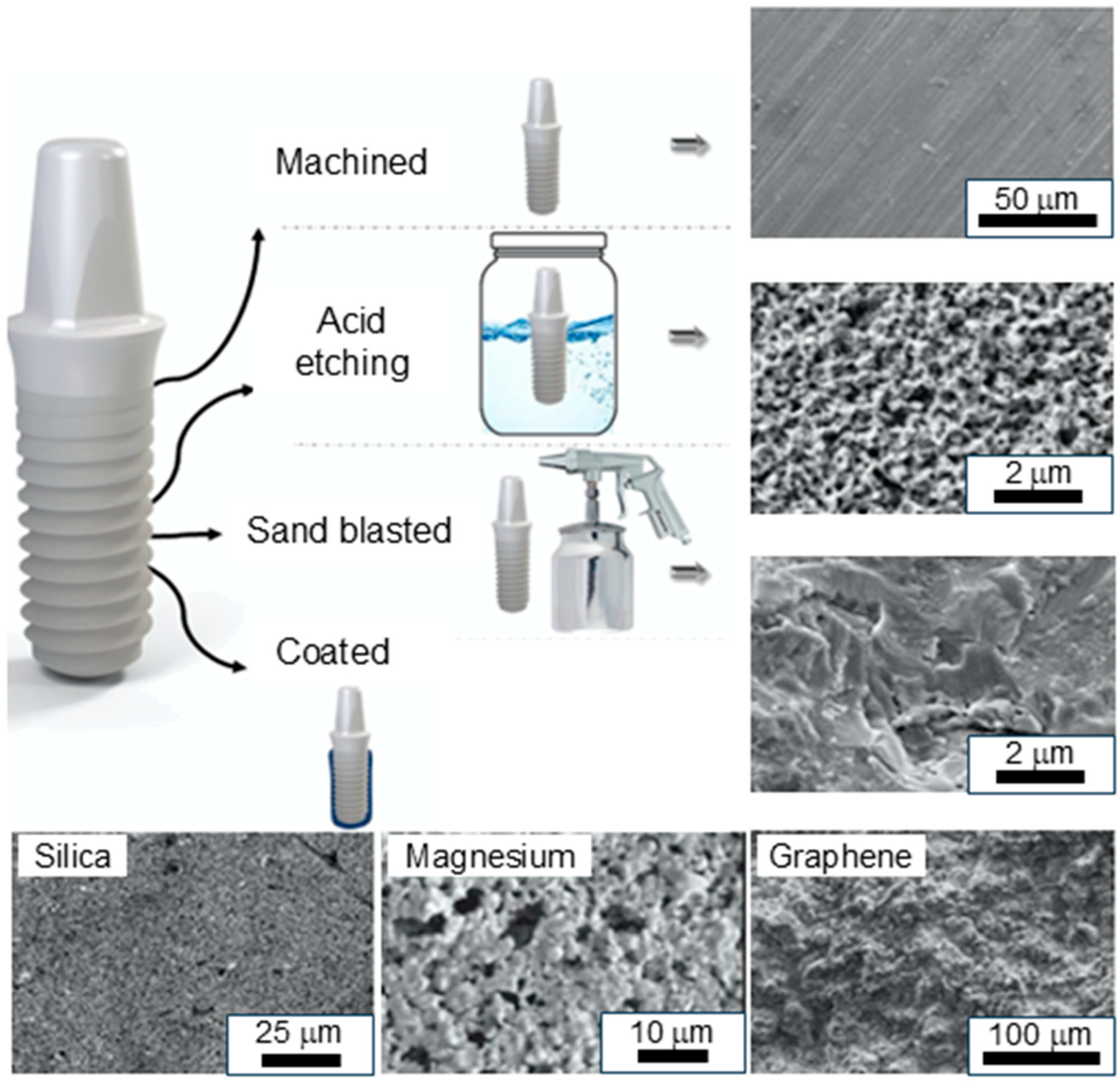
ZrO2-based implants are distinguished by their excellent clinical success rate, high functional resilience, and high resistance to bending and compression while providing outstanding aesthetics [70]. Previous studies have shown that a significant alteration in the ZrO2 surface significantly affects the adhesion, growth, morphology, and differentiation of fibroblasts and osteoblasts, enhancing the osseointegration of implant surfaces [49,71]. Alterations in the surface physicochemical properties influence biocompatibility and osseointegration. Their effect is evaluated using histomorphometry analyses, which allow for the forecasting of clinical outcomes and the long-term stability of peri-implant tissues [72,73]. Therefore, several methods have been developed to improve the physicochemical properties of dental implants, to optimize both the initial bone response, the rapid and efficient integration of the implant into the bone upon insertion, and its long-term stability. The latter aspect relates to the durability of the implant, ensuring that it withstands mechanical stresses, biological interactions, and the environmental conditions of the body over time [3], as shown in Figure 4.
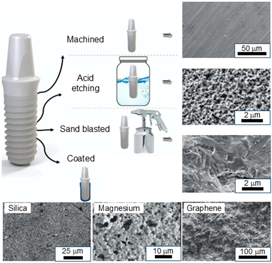
Figure 4.
Schematic diagrams and SEM images of different surface treatments on ZrO2: machined, acid-etched, sandblasted, and coated [74]. Adapted with permission from Elsevier.
4.1. Laser Treatment
Laser treatment is one of the most promising methods for improving the osseointegration of ZrO2-based implants [75,76,77,78,79]. Souza et al. [80] applied laser texturing to enhance the bonding of prosthetic surfaces with lithium-disilicate-reinforced glass ceramics and zirconia resin cements. The use of lasers, which was performed under power densities of 1.0 and 1.5 kW/cm2, resulted in significant changes in material properties on the surface. The changes reduced surface roughness from 0.45 μm to 0.30 μm and improved the surface energy, as well as its wettability. As a result, laser-treated surfaces showed an increase in the bonding strength of resin cements; this is crucial to the long-term success of dental prostheses [80]. Additionally, laser treatment has been used to create micro-grooved surfaces on ZrO2-based implants. These surfaces can influence the collagen fiber organization, peri-implant bone architecture, and human cell metabolism. Various studies show that osteoblast proliferation, adhesion, diffusion, and protein synthesis occur on surfaces with regular micro-geometries [81]. In research conducted by Carvalho et al. in 2020 [82], femtosecond laser microstructuring was used to alter the ATZ surface for dental implant purposes. The topography of the laser-processed surfaces was enhanced, including micro-grooves, which provided improved cell adhesion and proliferation. The results showed that the microstructured ATZ surfaces enhanced bone formation and vascularization compared to untreated zirconia. The micro-grooves on the surface improved the orientation of the collagen fibers, improving the efficiency of the bone remodeling process. The enhanced bone integration is attributed to the increased surface roughness and the delivery of topographical cues that affect cell behavior, improving rapid osseointegration and improving the long-term stability of dental implants. These findings point to the potential of femtosecond laser microstructuring to improve the performance of zirconia-based implants in clinical applications [82]. Though the laser-treated micro-grooved ZrO2-based implants look very promising, further human studies should be carried out to establish their clinical efficacy.
4.2. ZrO2-Based Coatings
One of the main challenges in implant dentistry is the improvement of the biological and mechanical properties of implants by advanced surface coatings. Bioactive coatings on ZrO2 surfaces play a significant role in improving biocompatibility, antibacterial properties, and bioactivity. Such coatings promote the formation of HA in biological environments, which is a key factor for bone growth and osseointegration. These materials, which showed a variety of biological advantages, were among those studied for improving implant performance through reinforcement with silica [25,79,83,84,85,86,87], magnesium, nitrogen, carbon, CaP, HA, dopamine, and graphene. Recent studies have shown that the surface modification of zirconia using bioactive coatings such as hydroxyapatite and calcium phosphate significantly enhances osseointegration and mechanical strength [88].
4.2.1. Magnesium, Nitrogen, and Carbon Coatings
Magnesium coatings on ZrO2 have been shown to enhance osteoblast growth, hence improving its bioactivity in various research studies [89]. The nitrogen and carbon plasma immersion ion implantation (N2-PIII) of zirconia enhances its bioactivity, causing more cell adhesion and osteogenic differentiation compared to untreated zirconia. The coatings exhibit enhanced hydrophilicity and surface roughness, generating an environment that is conducive to bone cell attachment. Soaking in simulated body fluid leads to the formation of hydroxyapatite (HA) with cauliflower-like morphology on nitrogen- and carbon-implanted zirconia [90]. The modification of ZrO2 with the addition of nitrogen and carbon continues to improve its bioactivity and mechanical properties. The improvements include the deposition of a nitrogen-doped hydrogenated amorphous carbon (a-C:H:N) layer, resulting in increased hydrophilicity and bioactivity against bacterial growth. This layer reduces bacterial adhesion on Y-TZP surfaces compared to unmodified ones. Moreover, functionalized carbon nanotubes have been shown to increase the roughness and wettability of ZrO2 surfaces, improving cell adhesion by optimizing their osseointegration potential [91,92].
4.2.2. Hydroxyapatite and Calcium Phosphate Coatings
HA has a mineral composition similar to that of bone; it therefore favors tissue reaction and enhances osseointegration. Coating ZrO2 with HA changes it from a stable implant surface to a bioactive implant surface, thus enhancing osseointegration. HA-coated porous ZrO2 scaffolds have been used as drug carriers to enhance bone response and osseointegration [93,94,95]. HA-enriched CAD/CAM porous ZrO2 scaffolds significantly increased new bone formation compared to those without HA, highlighting the positive impact of HA on osteogenesis [96]. Combining various ratios of Yttria-stabilized Tetragonal ZrO2 Polycrystal ceramics (Y-TZP) and HA and a higher content of CaP resulted in the reduced chemical and mechanical stability and bioactivity of the material. The material was also found to precipitate an apatite layer spontaneously under certain conditions when submerged in simulated body fluids (SBF) of various concentrations, a biomimetic process with a composition close to blood plasma [97]. This phenomenon improves the bioactivity and osseointegration of the implants. The coatings obtained demonstrated excellent interfacial adhesion and good mechanical strength, especially those containing a higher proportion of tetragonal ZrO2 [83]. CaP, which has the same composition as bone, is bioactive itself and is thus considered one of the stimuli for bone repair. The coatings, however, exhibit limited stability and weak substrate adhesion. It is for these reasons that tri-CaP-reinforced HA coatings on ZrO2 have been studied. Indeed, coatings with open pores higher than 100 μm and bond strength as high as 24 MPa guarantee excellent interfacial adhesion in addition to absorbable and osteoconductive properties [98,99]. Recent studies highlight the importance of adding HA nanoparticles to implant surfaces modified by sandblasting and acid etching, along with examining nanolayered metal phosphate coatings as biocompatible drug reservoirs of antimicrobial medicines, such as silver nanoparticles, for more potent antibacterial protection and bioactivity assurance [100]. This approach improves peri-implant bone healing, thus promoting the improved osseointegration of the implants.
4.2.3. Graded ZrO2
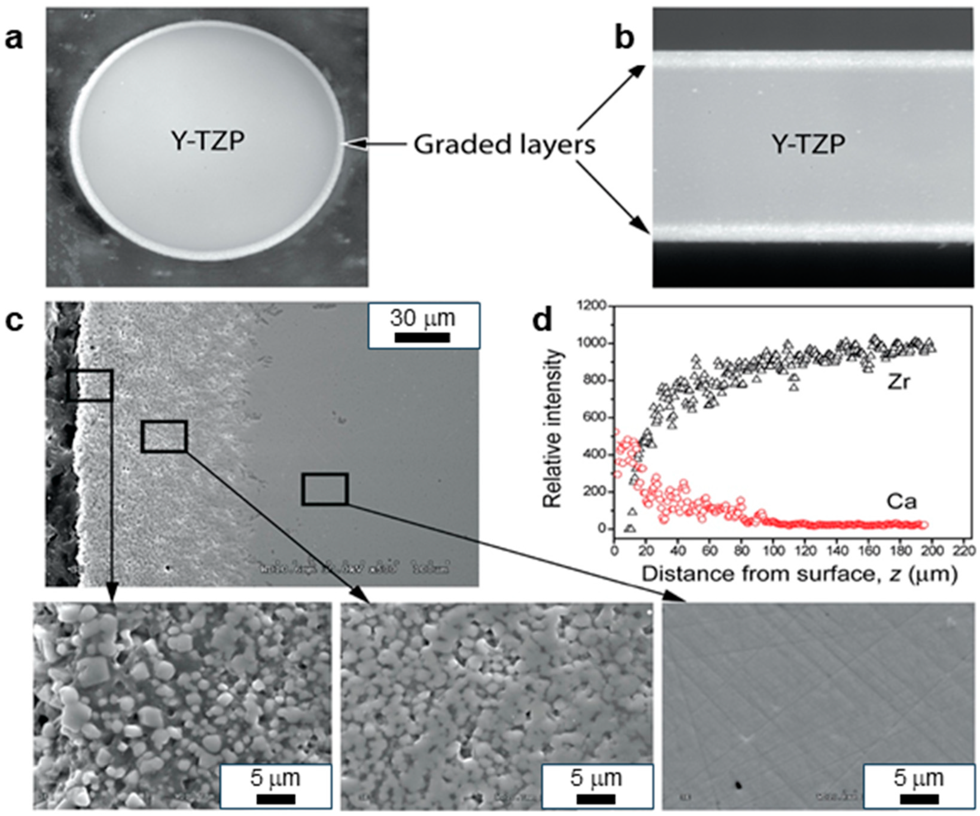
CaP glass (CPG) osteoconductive coatings can accelerate osseointegration by preventing micro-movements between the implant and the surrounding tissue [100,101]. The residual outer layer of CPG acts as a protection barrier to the Y-TZP interior against hydrothermal degradation, as shown in Figure 5. In addition, CPG transforms into a layer of carbonated HA (CHA) and induces the direct adhesion of newly formed bone directly to the CHA layer when embedded in a mineral precipitation solution or simulated body fluid. Despite these advantages, there are relatively few investigations into the properties of the CPG/Y-TZP system, compared with extensive studies into glass-infiltrated ZrO2 structures [102,103,104,105,106,107,108,109]. Based on a recent comparison of the surface modifications of ZrO2-based implants available on the market, several techniques are used, including sandblasting, acid etching, lasers, and their combinations. Laser-treated surfaces have the best roughness and bone-implant contact (BIC). In addition, they promote cell adhesion and early bone formation through increased hydrophilicity. Finally, combination treatments, such as sandblasting followed by laser treatment or acid etching, also show favorable biological results [110].
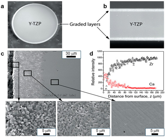
Figure 5.
Cross-sectioned Y-TZP coating: (a) circular section (2 mm of radius), (b) rectangular section (1.2 mm thick), (c) SEM image of CPG-infiltrated Y-TZP, (d) EDS mapping of the gradual transition of Ca and Zr contents. The inset shows a gradual transition from the surface GC remnant layer (left) to the graded CPG/Y-TZP structure (center) and dense Y-TZP interior (right) [74]. Adapted with permission from Elsevier.
5. Challenges and Limitations in Current Technologies
One of the principal concerns with dental implants is the need for them to be mechanically compatible with the surrounding bone. The mismatch in the elastic modulus between the implant materials (such as titanium and zirconia) and native bone can lead to stress shielding [111], wherein the over-rigidity of the implant reduces the mechanical loading on the bone, which can lead to bone resorption over time. New methods such as gradient materials, surface treatment, and composite implants have been proposed to minimize this issue, but there remains an issue related to balancing mechanical strength with flexibility [112]. Materials such as polyetheretherketone (PEEK) and its composite have been studied to modify the mechanical properties of the implant to closely match natural bone to reduce stress shielding and provide implant stability. It is difficult to optimize the maximum mechanical strength to a good level of flexibility. Additionally, occlusal loading and fatigue resistance should be taken into consideration to prevent implant fracture and ensure long-term clinical success [113]. Occlusal loading and fatigue strength also need to be addressed to prevent implant fracture and early failure during clinical procedures [114].
Peri-implant infection and biofilm formation remain the primary concerns regarding implant survival. Biofilm formation by Porphyromonas gingivalis and Streptococcus mutans on the surface of an implant has the potential to lead to peri-implantitis, which can result in the instability of the implant [115]. New trends in antimicrobial coatings, such as silver nanoparticles, zinc oxide, titanium dioxide, and bioactive peptides, have proven to be highly promising in preventing bacterial adhesion [116]. However, cytotoxicity issues, the release control of antimicrobial molecules, and the durability of these films over the long term still need to be addressed. Smart films that release antimicrobial species in response to bacterial activity but do not affect normal tissue need to be the subject of future investigations [117].
The success of implantation depends on osseointegration (direct bone–implant contact) and osteoconduction (the development of bone along the implant surface). Surface treatments such as acid etching, anodization, and plasma spraying enhance bone integration, but achieving quick and stable osseointegration remains problematic, particularly in osteoporotic bone or in the presence of systemic disease (e.g., diabetes, smoking-related disease) [118,119]. Ongoing research focuses on topics including biochemical surface alteration through bioactive molecules such as bone morphogenetic proteins (BMPs), calcium phosphate deposition, and collagen interfaces for the enhancement of cell adhesion and differentiation [120]. The stability of such systems in the long term, as well as their biological acceptance, must be proven clinically.
6. Prospective Approaches for Dental Implant Surface Engineering
Hybrid dental implants will soon follow as surface technology and material science technologies evolve further. Additive manufacturing, or 3D printing, is fast emerging as a revolutionary method for creating patient-specific implants with customized nano-topographies. Such modifications improve antibacterial functionality and osseointegration, but the mass production of homogeneous nanostructures is an enormous challenge [120]. Even though the technology has the potential to customize implants, the fabrication time and cost must be minimized for more widespread clinical applications [121]. Future dental implants can be integrated with advanced drug-delivery systems with the externally controlled release of therapeutic molecules in response to temperature, pH, or electromagnetic fields [122]. This would optimize the therapeutic effect due to controlled drug release, avoid burst effects, and enhance the healing process. Smart implants equipped with biosensors can monitor cell attachment and tissue growth and control drug release accordingly, beyond their structural function [123].
Tailorable commercially available 3D-printed implants have attracted a great deal of attention due to their ability to be designed in accordance with patient-specific anatomical structures. Various companies have developed FDA-approved and CE-marked 3D-printed dental implants [124] using selective laser melting (SLM) and electron beam melting (EBM) technologies. The implants provide enhanced osseointegration with porous architectures that mimic the natural bone architecture [125]. In addition, developments in composite and bioresorbable materials for 3D printing [126] are setting the stage for next-generation hybrid implants that offer both mechanical toughness and optimized biocompatibility.
The second important trend in implant surface engineering is the immobilization of bioactive molecules for augmenting bone healing. This includes functionalization with growth factors, bioactive proteins, and non-viral gene delivery to enhance osseointegration and regeneration [127]. Covalent or non-covalent immobilization methods for biomolecules have to be tailored for increased therapeutic efficacy. For this purpose, bioactive ceramic coatings, in combination with mechanical surface treatments, confer synergistic benefits for biocompatibility, corrosion resistance, and fatigue strength [128]. One of the more intriguing paths is the development of degradable implants, which possess certain advantages over traditional permanent devices. Nevertheless, providing coating uniformity and efficacy is a challenge in complex geometries such as porous 3D-printed scaffolds [129]. Studies of degradation kinetics are required to prevent the failure of the implant due tot he uncontrolled dissolution of the material before complete tissue regeneration. These ongoing advancements in implant surface engineering will shape the future of dental implantology by bringing about improved clinical results and outcomes, as well as improved patient care [130].
7. Conclusions
This review presents the evolution of surface modification and treatment techniques for dental implants, which have significantly improved their biocompatibility, osseointegration, and durability. For Ti-based implants, methods such as machining, sandblasting, and acid etching remain common approaches to optimizing surface roughness and promoting cell adhesion. In parallel, more advanced technologies, including plasma spraying, plasma electrolytic oxidation, anodic spark deposition, and atomic layer deposition, enhance corrosion resistance and optimize biological interactions with bone. Bioactive ceramic surface coatings and mechanical surface treatments enhance corrosion resistance, fatigue life, and biocompatibility. Resorbable dental implants are another promising avenue, but uniform coating and controlled degradation kinetics need to be optimized. Controlled therapeutic release via sophisticated drug-delivery systems and smart implants with biosensors can radically transform treatment. For ZrO2-based implants, several surface treatments have been developed to compensate for their initially low biological reactivity. Coatings based on hydroxyapatite and calcium phosphate aim to improve their bioactivity and bone integration. The application of laser techniques, along with the addition of magnesium, nitrogen, and carbon coatings, also provides significant improvements in terms of cell adhesion and mechanical resistance. Overall, these technological advancements help optimize the performance of dental implants by enhancing their interaction with biological tissues. The selection of the treatment method depends on several factors, including the implant material, specific clinical requirements, and desired durability. The continuous optimization of these techniques remains a crucial area of research for improving the success of implant treatments and enhancing patients’ quality of life.
Author Contributions
Conceptualization, M.A. and N.M.; methodology, M.A. and Q.T.; formal analysis, M.A. and Q.T.; investigation, M.A. and Q.T.; resources, A.E.-R., H.H. and N.M.; writing—original draft, M.A. and Q.T.; writing—review and editing, Q.T., A.E.-R., H.H. and N.M.; supervision, A.E.-R., H.H. and N.M. All authors have read and agreed to the published version of the manuscript.
Funding
This work was supported by the Natural Sciences and Engineering Research Council of Canada through the Discovery Grant (RGPIN-2023-03884) and the Alliance International Catalyst Grant (ALLRP 571273-2021) (H.H.).
Data Availability Statement
No new data were created or analyzed in this study.
Acknowledgments
Mohamed Aissi gratefully acknowledges the Canadian Government for providing the Study in Canada scholarship, allowing a six-month research internship at Laval University.
Conflicts of Interest
The authors declare no conflicts of interest.
References
- Safaee, S.; Nesabi, M.; Nesabi, M.; Mamizad Moghtader, F. Osseointegration Dynamics: Insights into the Dental Bone-Implant Interface. J. Appl. Tissue Eng. 2023, 9, 1–9. [Google Scholar] [CrossRef]
- Ruggiero, A.; De Stefano, M. On the Biotribological Surfaces of Dental Implants: Investigation in the Framework of Osseointegration. Biotribology 2023, 35–36, 100254. [Google Scholar] [CrossRef]
- Łosiewicz, B.; Osak, P.; Nowińska, D.; Maszybrocka, J. Developments in Dental Implant Surface Modification. Coatings 2025, 15, 109. [Google Scholar] [CrossRef]
- Albrektsson, T.; Tengvall, P.; Amengual, L.; Coli, P.; Kotsakis, G.A.; Cochran, D. Osteoimmune Regulation Underlies Oral Implant Osseointegration and Its Perturbation. Front. Immunol. 2023, 13, 1056914. [Google Scholar] [CrossRef] [PubMed]
- Pandey, C.; Rokaya, D.; Bhattarai, B.P. Contemporary Concepts in Osseointegration of Dental Implants: A Review. Biomed. Res. Int. 2022, 2022, 6170452. [Google Scholar] [CrossRef]
- Liu, Z.; Liu, X.; Ramakrishna, S. Surface Engineering of Biomaterials in Orthopedic and Dental Implants: Strategies to Improve Osteointegration, Bacteriostatic and Bactericidal Activities. Biotechnol. J. 2021, 16, e2000116. [Google Scholar] [CrossRef] [PubMed]
- Han, W.; Fang, S.; Zhong, Q.; Qi, S. Influence of Dental Implant Surface Modifications on Osseointegration and Biofilm Attachment. Coatings 2022, 12, 1654. [Google Scholar] [CrossRef]
- Lin, D.-J.; Fuh, L.-J.; Chen, W.-C. Nano-Morphology, Crystallinity and Surface Potential of Anatase on Micro-Arc Oxidized Titanium Affect Its Protein Adsorption, Cell Proliferation and Cell Differentiation. Mater. Sci. Eng. C 2020, 107, 110204. [Google Scholar] [CrossRef]
- Kunrath, M.F.; Vargas, A.L.M.; Sesterheim, P.; Teixeira, E.R.; Hubler, R. Extension of Hydrophilicity Stability by Reactive Plasma Treatment and Wet Storage on TiO2 Nanotube Surfaces for Biomedical Implant Applications. J. R. Soc. Interface 2020, 17, 20200650. [Google Scholar] [CrossRef]
- Thakur, A.; Kumar, A.; Kaya, S.; Marzouki, R.; Zhang, F.; Guo, L. Recent Advancements in Surface Modification, Characterization and Functionalization for Enhancing the Biocompatibility and Corrosion Resistance of Biomedical Implants. Coatings 2022, 12, 1459. [Google Scholar] [CrossRef]
- Jambhulkar, N.; Jaju, S.; Raut, A. Surface Modification Techniques for Different Materials Used in Dental Implants: A Review. Mater. Today Proc. 2022, 60, 2266–2269. [Google Scholar] [CrossRef]
- Sharma, P.; Mishra, V.; Murab, S. Unlocking Osseointegration: Surface Engineering Strategies for Enhanced Dental Implant Integration. ACS Biomater. Sci. Eng. 2025, 11, 67–94. [Google Scholar] [CrossRef]
- Fangaia, S.I.G. Tribological Study of Metal Alloys Subject to Dental Wear. Ph.D. Thesis, University of Coimbra, Coimbra, Portugal, 2022. Available online: https://estudogeral.uc.pt/handle/10316/101749 (accessed on 10 March 2025).
- Li, J.; Zhou, P.; Attarilar, S.; Shi, H. Innovative Surface Modification Procedures to Achieve Micro/Nano-Graded Ti-Based Biomedical Alloys and Implants. Coatings 2021, 11, 647. [Google Scholar] [CrossRef]
- Körtvélyessy, G. The Impact of Different Cone-Angle Implant-Abutment Relationships on the Long-Term Success of Implant Restorations. Ph.D. Thesis, University of Szeged, Szeged, Hungary, 2023. Available online: https://doktori.bibl.u-szeged.hu/id/eprint/11803/ (accessed on 10 March 2025).
- Tramontana, D. Custom Abutments for Dental Implants, Their Evolution and Uses: A Narrative Review. Ph.D. Thesis, Universidade Fernando Pessoa, Porto, Portugal, 2022. Available online: https://bdigital.ufp.pt/entities/publication/9d7f967d-48b5-49e1-8ef0-d83a4e7ad52c (accessed on 10 March 2025).
- Gasik, M.; Lambert, F.; Bacevic, M. Biomechanical Properties of Bone and Mucosa for Design and Application of Dental Implants. Materials 2021, 14, 2845. [Google Scholar] [CrossRef] [PubMed]
- Pan, Y.; Tsoi, J.K.H.; Lam, W.Y.H.; Pow, E.H.N. Implant Framework Misfit: A Systematic Review on Assessment Methods and Clinical Complications. Clin. Implant Dent. Relat. Res. 2021, 23, 244–258. [Google Scholar] [CrossRef] [PubMed]
- Kowalski, J.; Lapinska, B.; Nissan, J.; Lukomska-Szymanska, M. Factors Influencing Marginal Bone Loss Around Dental Implants: A Narrative Review. Coatings 2021, 11, 865. [Google Scholar] [CrossRef]
- Kim, J.C.; Lee, M.; Yeo, I.-S.L. Three Interfaces of the Dental Implant System and Their Clinical Effects on Hard and Soft Tissues. Mater. Horiz. 2022, 9, 1387–1411. [Google Scholar] [CrossRef]
- Malmqvist, S.; Erdenborg, J.; Johannsen, G.; Johannsen, A. Patient’s Experiences of Dental Implants, Peri-Implantitis, and Its Treatment—A Qualitative Interview Study. Int. J. Dent. Hyg. 2024, 22, 530–539. [Google Scholar] [CrossRef]
- Cesar, P.F.; Miranda, R.B.P.; Santos, K.F.; Scherrer, S.S.; Zhang, Y. Recent Advances in Dental Zirconia: 15 Years of Material and Processing Evolution. Dent. Mater. 2024, 40, 824–836. [Google Scholar] [CrossRef]
- Kunrath, M.F.; Gupta, S.; Lorusso, F.; Scarano, A.; Noumbissi, S. Oral Tissue Interactions and Cellular Response to Zirconia Implant-Prosthetic Components: A Critical Review. Materials 2021, 14, 2825. [Google Scholar] [CrossRef]
- Sun, J.; Ding, Q.; Chen, Y.; Li, J.; Wang, Z.; Wei, Z.; Ge, X.; Zhang, L. Effects and Underlying Mechanism of Micro-Nano-Structured Zirconia Surfaces on Biological Behaviors of Human Gingival Fibroblasts Under Inflammatory Conditions. Acta Biomater. 2024, 183, 356–370. [Google Scholar] [CrossRef] [PubMed]
- Avila, J.d.J. Calcium Phosphate and Zirconia Toughened Alumina Reinforced Titanium and CoCrMo Matrix Structures for Articulating Surfaces of Load-Bearing Implants. Ph.D. Thesis, Washington State University, Pullman, WA, USA, 2021. [Google Scholar] [CrossRef]
- Jain, V.K.; Patel, D.S.; Ramkumar, J.; Bhattacharyya, B.; Doloi, B.; Sarkar, B.R.; Ranjan, P.; Sankar, E.S.S.; Jayal, A.D. Micro-Machining: An Overview (Part II). J. Micromanuf. 2022, 5, 46–73. [Google Scholar] [CrossRef]
- Kunrath, M.F.; Muradás, T.C.; Penha, N.; Campos, M.M. Innovative Surfaces and Alloys for Dental Implants: What About Biointerface-Safety Concerns? Dent. Mater. 2021, 37, 1447–1462. [Google Scholar] [CrossRef] [PubMed]
- Wu, Y.; Wan, K.; Lu, J.; Yuan, C.; Cui, Y.; Duan, R.; Yu, J. Research Progress on Surface Modification of Titanium Implants. Coatings 2025, 15, 229. [Google Scholar] [CrossRef]
- García, I.; Trobajo, C.; Amghouz, Z.; Alonso-Guervos, M.; Díaz, R.; Mendoza, R.; Mauvezín-Quevedo, M.; Adawy, A. Ag- and Sr-Enriched Nanofibrous Titanium Phosphate Phases as Potential Antimicrobial Cement and Coating for a Biomedical Alloy. Mater. Sci. Eng. C 2021, 126, 112168. [Google Scholar] [CrossRef] [PubMed]
- Minguela Díaz, J. Surface Characterization and Cell Instructive Properties of Superficially Modified Dental Zirconia. Ph.D. Thesis, Universitat Politècnica de Catalunya, Barcelona, Spain, 2021. [Google Scholar] [CrossRef]
- Pereira, R.M.; Ribas, R.G.; Montanheiro, T.L.D.A.; Schatkoski, V.M.; Rodrigues, K.F.; Kito, L.T.; Kobo, L.K.; Campos, T.M.B.; Bonfante, E.A.; Gierthmuehlen, P.C.; et al. An Engineering Perspective of Ceramics Applied in Dental Reconstructions. J. Appl. Oral Sci. 2023, 31, e20220421. [Google Scholar] [CrossRef]
- Poznyak, A.; Pligovka, A.; Salerno, M. Anodizing of Hydrogenated Titanium and Zirconium Films. Materials 2021, 14, 7490. [Google Scholar] [CrossRef]
- Shahmohammadi, M.; Sun, Y.; Yuan, J.C.-C.; Mathew, M.T.; Sukotjo, C.; Takoudis, C.G. In Vitro Corrosion Behavior of Coated Ti6Al4V with TiO2, ZrO2, and TiO2/ZrO2 Mixed Nanofilms Using Atomic Layer Deposition for Dental Implants. Surf. Coat. Technol. 2022, 444, 128686. [Google Scholar] [CrossRef]
- Hadzik, J.; Kubasiewicz-Ross, P.; Gębarowski, T.; Waloszczyk, N.; Maciej, A.; Stolarczyk, A.; Gedrange, T.; Dominiak, M.; Szajna, E.; Simka, W. An Experimental Anodized Titanium Surface for Transgingival Dental Implant Elements—Preliminary Report. J. Funct. Biomater. 2023, 14, 34. [Google Scholar] [CrossRef]
- Hussain, S.; Sabiruddin, K. Surface Modification Methods of Titanium (Ti)-Based Implant Materials for Biomedical Applications. In Bioimplants Manufacturing, 1st ed.; CRC Press: Boca Raton, FL, USA, 2024; p. 22. ISBN 9781003509943. [Google Scholar]
- Saha, S.; Roy, S. Metallic Dental Implants Wear Mechanisms, Materials, and Manufacturing Processes: A Literature Review. Materials 2023, 16, 161. [Google Scholar] [CrossRef]
- Huang, S.; Wei, H.; Li, D. Additive Manufacturing Technologies in the Oral Implant Clinic: A Review of Current Applications and Progress. Front. Bioeng. Biotechnol. 2023, 11, 1100155. [Google Scholar] [CrossRef] [PubMed]
- Cruz, M.B.; Silva, N.; Marques, J.F.; Mata, A.; Silva, F.S.; Caramês, J. Biomimetic Implant Surfaces and Their Role in Biological Integration—A Concise Review. Biomimetics 2022, 7, 74. [Google Scholar] [CrossRef]
- Tahan, F.I.; Abdelhamid, A.M.; Osman, E.; Harouny, R.; Hachem, R.; Aly, Y.M. The Effect of Nitric Acid–Hydrofluoric Acid Etching Solution on the Shear Bond Strength and Mode of Failure of Resin Cement to Zirconia (In Vitro Study). Ann. Stomatol. 2024, 15, 85–93. [Google Scholar] [CrossRef]
- Abdelraouf, R. Introduction to Materials–Tissue Interface. Biomater. Sci. 2022, 1, 35–52. [Google Scholar]
- Assaf, A.; Azer, S.S.; Sfeir, A.; Al-Haj Husain, N.; Özcan, M. Risk Factors with Porcelain Laminate Veneers Experienced During Cementation: A Review. Materials 2023, 16, 4932. [Google Scholar] [CrossRef] [PubMed]
- Osak, P.; Skwarek, S.; Łukowiec, D.; Przeliorz, G.; Łosiewicz, B. Preparation and Characterization of Oxide Nanotubes on Titanium Surface for Use in Controlled Drug Release Systems. Materials 2024, 17, 3753. [Google Scholar] [CrossRef] [PubMed]
- Benčina, M.; Resnik, M.; Starič, P.; Junkar, I. Use of Plasma Technologies for Antibacterial Surface Properties of Metals. Molecules 2021, 26, 1418. [Google Scholar] [CrossRef]
- Aljafery, A.M.A.; Fatalla, A.A.; Haider, J. Cytotoxicity, Corrosion Resistance, and Wettability of Titanium and Ti-TiB2 Composite Fabricated by Powder Metallurgy for Dental Implants. Metals 2024, 14, 538. [Google Scholar] [CrossRef]
- Sapiro, I. Comparative Roughness Analysis of Dental Implants and Comparison of Methods Using Scanning Electron Microscopy and White Light Interferometry. Ph.D. Thesis, Charité—Universitätsmedizin Berlin, Berlin, Germany, 2024. Available online: https://refubium.fu-berlin.de/handle/fub188/41881 (accessed on 8 March 2025).
- Ming, X.; Wu, Y.; Zhang, Z.; Li, Y. Micro-Arc Oxidation in Titanium and Its Alloys: Development and Potential of Implants. Coatings 2023, 13, 2064. [Google Scholar] [CrossRef]
- Pesode, P.; Barve, S. Biocompatibility of Plasma Electrolytic Oxidation Coated Titanium Alloy for Biomedical Applications. BioNanoScience 2025, 15, 232. [Google Scholar] [CrossRef]
- Kyrylenko, S.; Warchoł, F.; Oleshko, O.; Husak, Y.; Kazek-Kęsik, A.; Korniienko, V.; Deineka, V.; Sowa, M.; Maciej, A.; Michalska, J.; et al. Effects of the Sources of Calcium and Phosphorus on the Structural and Functional Properties of Ceramic Coatings on Titanium Dental Implants Produced by Plasma Electrolytic Oxidation. Mater. Sci. Eng. C 2021, 119, 111607. [Google Scholar] [CrossRef]
- Mishchenko, O.; Volchykhina, K.; Maksymov, D.; Manukhina, O.; Pogorielov, M.; Pavlenko, M.; Iatsunskyi, I. Advanced Strategies for Enhancing the Biocompatibility and Antibacterial Properties of Implantable Structures. Materials 2025, 18, 822. [Google Scholar] [CrossRef] [PubMed]
- Golalipour, S.; Jalalian, E.; Koosha, S.; Khorshidi, S.; Torshabi, M.; Sayyari, M. In Vitro Effect of Anodization on Surface Roughness and Bacterial Adhesion to Titanium Abutments. J. Prosthet. Dent. 2025, 133, 291.e1–291.e8. [Google Scholar] [CrossRef] [PubMed]
- Vattanasup, C.; Kuntiyaratana, T.; Rungsiyakull, P.; Chaijareenont, P. Color Formation on Titanium Surface Treated by Anodization and the Surface Characteristics: A Review. Chiang Mai Dent. J. 2023, 44, 13–21. Available online: https://www.dent.cmu.ac.th/cmdj/frontend/web/?r=site/viewarticle&id=199 (accessed on 8 March 2025). [CrossRef]
- Meng, F.; Yin, Z.; Ren, X.; Geng, Z.; Su, J. Construction of Local Drug Delivery System on Titanium-Based Implants to Improve Osseointegration. Pharmaceutics 2022, 14, 1069. [Google Scholar] [CrossRef] [PubMed]
- Sotova, C.; Yanushevich, O.; Kriheli, N.; Grigoriev, S.; Evdokimov, V.; Kramar, O.; Nozdrina, M.; Peretyagin, N.; Undritsova, N.; Popelyshkin, E.; et al. Dental Implants: Modern Materials and Methods of Their Surface Modification. Materials 2023, 16, 7383. [Google Scholar] [CrossRef]
- Kandavalli, S.R.; Wang, Q.; Ebrahimi, M.; Gode, C.; Djavanroodi, F.; Attarilar, S.; Liu, S. A Brief Review on the Evolution of Metallic Dental Implants: History, Design, and Application. Front. Mater. 2021, 8, 646383. [Google Scholar] [CrossRef]
- Al-Amin, M.D. Effects of Multiple Metal Oxides on Properties of ZTA-5TiO2-5MgO Composites. Ph.D. Thesis, University of Dhaka, Dhaka, Bangladesh, 2023. Available online: http://reposit.library.du.ac.bd:8080/xmlui/handle/123456789/2656 (accessed on 8 March 2025).
- Hichem, N.; Hadjer, Z.; Fateh, S.; Feriel, L.; Wang, Z. The Potential Exposure and Hazards of Zirconia Nanoparticles: A Review. Ecotoxicol. Environ. Contam. 2022, 17, 1–21. [Google Scholar] [CrossRef]
- Gautam, S.; Bhatnagar, D.; Bansal, D.; Batra, H.; Goyal, N. Recent Advancements in Nanomaterials for Biomedical Implants. Biomed. Eng. Adv. 2022, 3, 100029. [Google Scholar] [CrossRef]
- Losic, D. Advancing Titanium Medical Implants by Surface Engineering: Recent Progress and Challenges. Expert Opin. Drug Deliv. 2021, 18, 1355–1378. [Google Scholar] [CrossRef]
- Steblevskaya, N.I.; Belobeletskaya, M.V.; Yarovaya, T.P.; Nedozorov, P.M. Preparation and Luminescence Properties of the Composite of Mixed Europium and Tantalum Oxides/ZrO2 + TiO2/Ti. Russ. J. Inorg. Chem. 2022, 67, 415–423. [Google Scholar] [CrossRef]
- Baishya, K.; Bacova, J.; Al Chimali, B.; Capek, J.; Michalicka, J.; Gautier, G.; Le Borgne, B.; Rousar, T.; Macak, J.M. Ultrathin ALD Coatings of Zr and V Oxides on Anodic TiO2 Nanotube Layers: Comparison of the Osteoblast Cell Growth. ACS Appl. Mater. Interfaces 2025, 17, 739–749. [Google Scholar] [CrossRef]
- Hayashi, T.; Asakura, M.; Koie, S.; Hasegawa, S.; Mieki, A.; Aimu, K.; Kawai, T. In Vitro Study of Zirconia Surface Modification for Dental Implants by Atomic Layer Deposition. Int. J. Mol. Sci. 2023, 24, 10101. [Google Scholar] [CrossRef] [PubMed]
- Ghosh, S.; Webster, T.J. Metallic Nanoscaffolds as Osteogenic Promoters: Advances, Challenges and Scope. Metals 2021, 11, 1356. [Google Scholar] [CrossRef]
- Puspasari, V.; Ridhova, A.; Hermawan, A.; Amal, M.I.; Khan, M.M. ZnO-Based Antimicrobial Coatings for Biomedical Applications. Bioprocess Biosyst. Eng. 2022, 45, 1421–1445. [Google Scholar] [CrossRef] [PubMed]
- Jadon, M.S.; Kumar, S. Fabrication of TiO2 Nanoparticle Coating on Stainless Steel 316L and Its Assessment for Orthopaedic Applications. Int. J. Online Biomed. Eng. 2024, 20, 47–63. [Google Scholar] [CrossRef]
- Gürsoy, E.; Yılmaz, H. Nanoparticles and Their Application in Prosthetic Dentistry. Clin. Exp. Health Sci. 2023, 13, 685–695. [Google Scholar] [CrossRef]
- Itoh, H.; Yanagishita, T. Effects of Pore Arrangement of TiO2-Coated Porous Alumina Membranes on Photocatalytic Properties. J. Phys. Chem. C 2024, 128, 6478–6486. [Google Scholar] [CrossRef]
- Hashemi Astaneh, S. Surface Functionalization and Enhancement of Properties of Biomaterials Through Atomic Layer Deposition. Ph.D. Thesis, University of Illinois Chicago, Chicago, IL, USA, 2021. [Google Scholar] [CrossRef]
- Rudolf, R.; Majerič, P.; Lazić, V.; Raić, K.T. Advanced Dental Metallic Materials; Springer: Berlin/Heidelberg, Germany, 2024; ISBN 9783031473500. [Google Scholar]
- Momesso, G.A.C.; de Souza Santos, A.M.; Fonseca e Santos, J.M.; da Cruz, N.C.; Okamoto, R.; Barão, V.A.R.; Siroma, R.S.; Shibli, J.A.; Perez Faverani, L. Comparison Between Plasma Electrolytic Oxidation Coating and Sandblasted Acid-Etched Surface Treatment: Histometric, Tomographic, and Expression Levels of Osteoclastogenic Factors in Osteoporotic Rats. Materials 2020, 13, 1604. [Google Scholar] [CrossRef]
- Sen, A.; Banerjee, N.; Biswas, A.R.; Ghosh, T.K.; Samanta, A.; Kumar, M.; Das, S.; Srivastava, U.; Sengupta, D.; Maity, S.R. Standards of Mechanical, Physical, Chemical, and Biological Properties of Bioceramics. In Advanced Bioceramics; CRC Press: Boca Raton, FL, USA, 2023; pp. 1–17. ISBN 9781003258353. [Google Scholar]
- Paul, S.; Hanisch, O.; Nesic, D. Human Gingival Fibroblast Proliferation on Materials Used for Dental Implant Abutments: A Systematic Review. Int. J. Prosthodont. 2021, 34, 811–828. [Google Scholar] [CrossRef]
- van Oirschot, B.A.J.A.; Zhang, Y.; Alghamdi, H.S.; Cordeiro, J.M.; Nagay, B.E.; Barao, V.A.R.; de Avila, E.D.; van den Beucken, J.J.J.P. Surface Engineering for Dental Implantology: Favoring Tissue Responses Along the Implant. Tissue Eng. Part A 2022, 28, 555–572. [Google Scholar] [CrossRef]
- Abdulla, M.A.; Hasan, R.H.; Al-Hyani, O.H. Radiographic and Histologic Assessment of Osseointegration for Surface-Treated Titanium Dental Implants: An Experimental Study in Dogs. J. Dent. Res. Dent. Clin. Dent. Prospects 2024, 18, 44–54. [Google Scholar] [CrossRef] [PubMed]
- Schünemann, F.H.; Galárraga-Vinueza, M.E.; Magini, R.; Fredel, M.; Silva, F.; Souza, J.C.M.; Zhang, Y.; Henriques, B. Zirconia Surface Modifications for Implant Dentistry. Mater. Sci. Eng. C 2019, 12, 1294–1305. [Google Scholar] [CrossRef]
- Kunrath, M.F. Customized Dental Implants: Manufacturing Processes, Topography, Osseointegration and Future Perspectives of 3D Fabricated Implants. Bioprinting 2020, 20, e00107. [Google Scholar] [CrossRef]
- Ispas, A.; Iosif, L.; Murariu-Măgureanu, C.; Craciun, A.; Constantiniuc, M. Zirconia in Dental Medicine: A Brief Overview of Its Properties and Processing Techniques. Hum. Vet. Med. 2021, 13, 33–39. [Google Scholar]
- Agut López, R. Microstructural Design of a New Zirconia-Based Material for Dental Implants. Bachelor’s Thesis, Universitat Politècnica de Catalunya, Barcelona, Spain, 2021. [Google Scholar]
- Eftekhar Ashtiani, R.; Alam, M.; Tavakolizadeh, S.; Abbasi, K. The Role of Biomaterials and Biocompatible Materials in Implant-Supported Dental Prosthesis. Evid. Based Complement. Alternat. Med. 2021, 2021, 3349433. [Google Scholar] [CrossRef] [PubMed]
- Weng, W.; Wu, W.; Hou, M.; Liu, T.; Wang, T.; Yang, H. Review of Zirconia-Based Biomimetic Scaffolds for Bone Tissue Engineering. J. Mater. Sci. 2021, 56, 8309–8333. [Google Scholar] [CrossRef]
- Souza, J.C.M.; Raffaele-Esposito, A.; Carvalho, O.; Silva, F.; Özcan, M.; Henriques, B. Surface Modification of Zirconia or Lithium Disilicate-Reinforced Glass Ceramic by Laser Texturing to Increase the Adhesion of Prosthetic Surfaces to Resin Cements: An Integrative Review. Clin. Oral Investig. 2023, 27, 3331–3345. [Google Scholar] [CrossRef]
- Rodríguez Montaño, Ó. Influence of the Micro-Geometry of a Scaffold for Bone Regeneration on the Stresses of the Newly Formed Tissue. Ph.D. Thesis, National University of Colombia, Bogotá, Colombia, 2021. [Google Scholar]
- Carvalho, A.; Grenho, L.; Fernandes, M.H.; Daskalova, A.; Trifonov, A.; Buchvarov, I.; Monteiro, F.J. Femtosecond Laser Microstructuring of Alumina Toughened Zirconia for Surface Functionalization of Dental Implants. Ceram. Int. 2020, 46, 1383–1389. [Google Scholar] [CrossRef]
- Migliorini, F.; Schenker, H.; Betsch, M.; Maffulli, N.; Tingart, M.; Hildebrand, F.; Lecouturier, S.; Rath, B.; Eschweiler, J. Silica Coated High-Performance Oxide Ceramics Promote Greater Ossification Than Titanium Implants: An In Vivo Study. J. Orthop. Surg. Res. 2023, 18, 31. [Google Scholar] [CrossRef]
- Drevet, R.; Fauré, J.; Benhayoune, H. Electrophoretic Deposition of Bioactive Glass Coatings for Bone Implant Applications: A Review. Coatings 2024, 14, 1084. [Google Scholar] [CrossRef]
- Avelino, S.O.M.; Alvares Sobral-Silva, L.; Thim, G.P.; de Almeida-Silva, L.A.; Dos Santos Lupp, J.; Campos, T.M.B.; de Vasconcellos, L.M.R. Development, Characterization, and Biological Study of Bioglass Coatings 45S5 and BioK on Zirconia Implant Surfaces. J. Biomed. Mater. Res. B Appl. Biomater. 2024, 112, e35380. [Google Scholar] [CrossRef] [PubMed]
- Upadhyay, A.; Pradhan, L.; Yenurkar, D.; Kumar, K.; Mukherjee, S. Advancement in Ceramic Biomaterials for Dental Implants. Int. J. Appl. Ceram. Technol. 2024, 21, 2796–2817. [Google Scholar] [CrossRef]
- Liang, J.; Lu, X.; Zheng, X.; Li, Y.R.; Geng, X.Y.; Sun, K.X.; Cai, H.X.; Jia, Q.; Jiang, H.B.; Liu, K. Modification of Titanium Orthopedic Implants with Bioactive Glass: A Systematic Review of In Vivo and In Vitro Studies. Front. Bioeng. Biotechnol. 2023, 11, 1269223. [Google Scholar] [CrossRef] [PubMed]
- Amghouz, Z.; García, J.R.; Adawy, A. A Review on the Synthesis and Current and Prospective Applications of Zirconium and Titanium Phosphates. Eng 2022, 3, 161–174. [Google Scholar] [CrossRef]
- Chen, H.; Huang, Z.-Y.; Chen, W. The Fabrication and In Vitro Bioactivity of HA/ZrO2/MgO Gradient Composite Coatings. Ceram. Int. 2024, 50, 1814–1825. [Google Scholar] [CrossRef]
- Guo, S.; Liu, N.; Liu, K.; Li, Y.; Zhang, W.; Zhu, B.; Gu, B.; Wen, N. Effects of Carbon and Nitrogen Plasma Immersion Ion Implantation on Bioactivity of Zirconia. RSC Adv. 2020, 10, 35917–35929. [Google Scholar] [CrossRef]
- Fróis, A.; Francisco, R.; Morais, P.V.; Santos, L.F.; Peres, M.; Lorenz, K.; Santos, A.C.; Louro, C.S. The Influence of Low Nitrogen Doping on Bacterial Adhesion of Sputtered a-C:H Coatings. Diam. Relat. Mater. 2024, 147, 111309. [Google Scholar] [CrossRef]
- Rogala-Wielgus, D.; Zieliński, A. Preparation and Properties of Composite Coatings, Based on Carbon Nanotubes, for Medical Applications. Carbon Lett. 2024, 34, 565–601. [Google Scholar] [CrossRef]
- Kim, H.; Lee, Y.-H.; Kim, N.-K.; Kang, I.-K. Bioactive Surface of Zirconia Implant Prepared by Nano-Hydroxyapatite and Type I Collagen. Coatings 2022, 12, 1335. [Google Scholar] [CrossRef]
- Rathee, G.; Bartwal, G.; Rathee, J.; Mishra, Y.K.; Kaushik, A.; Solanki, P.R. Emerging Multimodal Zirconia Nanosystems for High-Performance Biomedical Applications. Adv. NanoBiomed Res. 2021, 1, 2100039. [Google Scholar] [CrossRef]
- Kim, J.; Kang, I.G.; Cheon, K.H.; Lee, S.; Park, S.; Kim, H.E.; Han, C.M. Stable Sol-Gel Hydroxyapatite Coating on Zirconia Dental Implant for Improved Osseointegration. J. Mater. Sci. Mater. Med. 2021, 32, 81. [Google Scholar] [CrossRef]
- Aboushelib, M.N.; Shawky, R. Osteogenesis Ability of CAD/CAM Porous Zirconia Scaffolds Enriched with Nano-Hydroxyapatite Particles. Int. J. Implant Dent. 2017, 3, 21. [Google Scholar] [CrossRef]
- Ding, S.J.; Chu, Y.H.; Chen, P.T. Mechanical Biocompatibility, Osteogenic Activity, and Antibacterial Efficacy of Calcium Silicate-Zirconia Biocomposites. ACS Omega 2021, 6, 7106–7118. [Google Scholar] [CrossRef]
- Hou, X.; Zhang, L.; Zhou, Z.; Luo, X.; Wang, T.; Zhao, X.; Lu, B.; Chen, F.; Zheng, L. Calcium Phosphate-Based Biomaterials for Bone Repair. J. Funct. Biomater. 2022, 13, 187. [Google Scholar] [CrossRef]
- Arcos, D.; Vallet-Regí, M. Substituted Hydroxyapatite Coatings of Bone Implants. J. Mater. Chem. B 2020, 8, 1781–1800. [Google Scholar] [CrossRef]
- García, I.; Trobajo, C.; Amghouz, Z.; Adawy, A. Nanolayered Metal Phosphates as Biocompatible Reservoirs for Antimicrobial Silver Nanoparticles. Materials 2021, 14, 1481. [Google Scholar] [CrossRef]
- Lalzawmliana, V.; Mukherjee, P.; Roy, S.; Roy, M.; Nandi, S.K. Ceramic Biomaterials in Advanced Biomedical Applications. In Functional Biomaterials; Jana, S., Jana, S., Eds.; Springer: Singapore, 2022; pp. 239–254. [Google Scholar] [CrossRef]
- Sarasati, A.; Wihadmadyatami, H.; Ana, I.D. Carbonate Apatite Nanoparticles: A Novel Nano-Adjuvant for Oral Mucosal Vaccines and Immunomodulator. OpenNano 2023, 12, 100149. [Google Scholar] [CrossRef]
- Schünemann, F.H. Development of Bioactive-Zirconia Surfaces for Biomedical Applications. Ph.D. Thesis, Universidade Federal de Santa Catarina, Florianópolis, Brazil, 2023. Available online: https://repositorio.ufsc.br/handle/123456789/249886 (accessed on 8 March 2025).
- He, Q.; Zhang, W.; Zhan, X.; Qin, Y.; Ye, J. Enhanced Bioactivity and Hydrothermal Aging Resistance of Y-TZP Ceramics for Dental Implant. J. Biomed. Mater. Res. B Appl. Biomater. 2023, 111, 1824–1839. [Google Scholar] [CrossRef]
- Parekh, A.; Moore, M.; Janorkar, A.V.; Roach, M.D. Mg-Doped Carbonated Hydroxyapatite and Tricalcium Phosphate Anodized Coatings on Titanium Implant Alloys. Appl. Sci. 2024, 14, 11831. [Google Scholar] [CrossRef]
- Sari, M.; Kristianto, N.A.; Chotimah, A.I.D.; Yusuf, Y. Carbonated Hydroxyapatite-Based Honeycomb Scaffold Coatings on a Titanium Alloy for Bone Implant Application—Physicochemical and Mechanical Properties Analysis. Coatings 2021, 11, 941. [Google Scholar] [CrossRef]
- Kengtanyakich, S.; Peampring, C. An Experimental Study on Hydrothermal Degradation of Cubic-Containing Translucent Zirconia. J. Adv. Prosthodont. 2020, 12, 265–272. [Google Scholar] [CrossRef] [PubMed]
- Yu, M.K.; Oh, E.J.; Lim, M.J.; Lee, K.W. Change of Phase Transformation and Bond Strength of Y-TZP with Various Hydrofluoric Acid Etching. Restor. Dent. Endod. 2021, 46, e54. [Google Scholar] [CrossRef]
- Li, K.; Rao, J.; Ning, C. Optimized Sintering and Mechanical Properties of Y-TZP Ceramics for Dental Restorations by Adding Lithium Disilicate Glass Ceramics. J. Adv. Ceram. 2021, 10, 1326–1337. [Google Scholar] [CrossRef]
- Stich, T.; Alagboso, F.; Křenek, T.; Kovářík, T.; Alt, V.; Docheva, D. Implant-Bone-Interface: Reviewing the Impact of Titanium Surface Modifications on Osteogenic Processes In Vitro and In Vivo. Bioeng. Transl. Med. 2021, 1, 10239. [Google Scholar] [CrossRef]
- Gaur, P.; Muthukumar, C.; Parthasarathy, V. Friction and Wear Properties of Biocomposites for Dental, Orthopedic, and Biomedical Applications. In Tribological Properties, Performance and Applications of Biocomposites; Muthukumar, C., Krishnasamy, S., Thiagamani, S.M., Chinnachamy, G., Eds.; Wiley-VCH: New York, NY, USA, 2023; Chapter 12. [Google Scholar] [CrossRef]
- Kligman, S.; Ren, Z.; Chung, C.H.; Perillo, M.A.; Chang, Y.C.; Koo, H.; Zheng, Z.; Li, C. The Impact of Dental Implant Surface Modifications on Osseointegration and Biofilm Formation. J. Clin. Med. 2021, 10, 1641. [Google Scholar] [CrossRef]
- Verma, V.; Hazari, P.; Verma, P. Do Implants Made of Polyetheretherketone and Its Composites Have Reduced Stress Shielding Effects Compared to Other Dental Implant Materials? A Systematic Review. Evid. Based Dent. 2023, 24, 193–194. [Google Scholar] [CrossRef]
- Manfredini, M.; Poli, P.P.; Giboli, L.; Beretta, M.; Maiorana, C.; Pellegrini, M. Clinical Factors on Dental Implant Fractures: A Systematic Review. Dent. J. 2024, 12, 200. [Google Scholar] [CrossRef]
- Esteves, G.M.; Esteves, J.; Resende, M.; Mendes, L.; Azevedo, A.S. Antimicrobial and Antibiofilm Coating of Dental Implants—Past and New Perspectives. Antibiotics 2022, 11, 235. [Google Scholar] [CrossRef]
- Cui, Z.; Wang, P.; Gao, W. Microbial Dysbiosis in Periodontitis and Peri-implantitis: Pathogenesis, Immune Responses, and Therapeutics. Front. Cell. Infect. Microbiol. 2025, 15, 1517154. [Google Scholar] [CrossRef]
- Gao, X.; Li, Y.; Li, J.; Xiang, X.; Wu, J.; Zeng, S. Stimuli-Responsive Materials in Oral Diseases: A Review. Clin. Oral Investig. 2024, 28, 497. [Google Scholar] [CrossRef] [PubMed]
- Abdulghafor, M.A.; Mahmood, M.K.; Tassery, H.; Tardivo, D.; Falguiere, A.; Lan, R. Biomimetic Coatings in Implant Dentistry: A Quick Update. J. Funct. Biomater. 2024, 15, 15. [Google Scholar] [CrossRef] [PubMed]
- Kocak-Oztug, N.A.; Ravali, E.I. Titanium Dental Implants in Compromised Conditions: Need for Enhanced Bioactivity and Therapy. In Surface Modification of Titanium Dental Implants; Gulati, K., Ed.; Springer: Cham, Switzerland, 2023. [Google Scholar] [CrossRef]
- Khaohoen, A.; Sornsuwan, T.; Chaijareenont, P.; Poovarodom, P.; Rungsiyakull, C.; Rungsiyakull, P. Biomaterials and Clinical Application of Dental Implants in Relation to Bone Density—A Narrative Review. J. Clin. Med. 2023, 12, 6924. [Google Scholar] [CrossRef]
- Karmakar, R.; Dixit, M.; Rengan, A.K.; Pati, F. Nanostructures Using 3D Printing. In Advances in Nanostructures; Gautam, S., Singh, J.P., Rai, D.P., Kumar, A., Mishra, A., Eds.; Elsevier: Amsterdam, The Netherlands, 2025; pp. 195–229. [Google Scholar] [CrossRef]
- Hakim, L.K.; Yari, A.; Nikparto, N.; Mehraban, S.H.; Cheperli, S.; Asadi, A.; Darehdor, A.A.; Nezaminia, S.; Dortaj, D.; Nazari, Y.; et al. The Current Applications of Nano and Biomaterials in Drug Delivery of Dental Implants. BMC Oral Health 2024, 24, 126. [Google Scholar] [CrossRef]
- Cicha, I.; Priefer, R.; Severino, P.; Souto, E.B.; Jain, S. Biosensor-Integrated Drug Delivery Systems as New Materials for Biomedical Applications. Biomolecules 2022, 12, 1198. [Google Scholar] [CrossRef]
- Meng, M.; Wang, J.; Huang, H.; Liu, X.; Zhang, J.; Li, Z. 3D Printing Metal Implants in Orthopedic Surgery: Methods, Applications and Future Prospects. J. Orthop. Transl. 2023, 42, 94–112. [Google Scholar] [CrossRef]
- Raheem, A.A.; Hameed, P.; Whenish, R.; Elsen, R.S.; G, A.; Jaiswal, A.K.; Prashanth, K.G.; Manivasagam, G. A Review on Development of Bio-Inspired Implants Using 3D Printing. Biomimetics 2021, 6, 65. [Google Scholar] [CrossRef] [PubMed]
- Maintz, M.; Tourbier, C.; de Wild, M.; Cattin, P.C.; Beyer, M.; Seiler, D.; Honigmann, P.; Sharma, N.; Thieringer, F.M. Patient-Specific Implants Made of 3D Printed Bioresorbable Polymers at the Point-of-Care: Material, Technology, and Scope of Surgical Application. 3D Print. Med. 2024, 10, 13. [Google Scholar] [CrossRef]
- Accioni, F.; Vázquez, J.; Merinero, M.; Begines, B.; Alcudia, A. Latest Trends in Surface Modification for Dental Implantology: Innovative Developments and Analytical Applications. Pharmaceutics 2022, 14, 455. [Google Scholar] [CrossRef]
- Kumar, A.; Singh, G. Surface Modification of Ti6Al4V Alloy via Advanced Coatings: Mechanical, Tribological, Corrosion, Wetting, and Biocompatibility Studies. J. Alloys Compd. 2024, 989, 174418. [Google Scholar] [CrossRef]
- Varma, M.V.; Kandasubramanian, B.; Ibrahim, S.M. 3D Printed Scaffolds for Biomedical Applications. Mater. Chem. Phys. 2020, 255, 123642. [Google Scholar] [CrossRef]
- Dixit, S.; Gupta, S.; Sharma, A. Surgical Devices for Biomedical Implants. In Additive Manufacturing for Biomedical Applications; Dixit, A., Kumar, A., Pathak, D.K., Eds.; Biomedical Materials for Multi-Functional Applications; Springer: Singapore, 2024. [Google Scholar] [CrossRef]
Disclaimer/Publisher’s Note: The statements, opinions and data contained in all publications are solely those of the individual author(s) and contributor(s) and not of MDPI and/or the editor(s). MDPI and/or the editor(s) disclaim responsibility for any injury to people or property resulting from any ideas, methods, instructions or products referred to in the content. |
© 2025 by the authors. Licensee MDPI, Basel, Switzerland. This article is an open access article distributed under the terms and conditions of the Creative Commons Attribution (CC BY) license (https://creativecommons.org/licenses/by/4.0/).