Article

Effect of ω-Phase Precipitation on Magnetic Susceptibility and Corrosion Resistance of Meta-Stable β-Phase Zr-Nb-Ti-Cr Alloy

1
Research & Development Group, Hitachi, Ltd., 1-1, Omika-cho 7-chome, Hitachi-shi 319-1292, Ibaraki-ken, Japan
2
Nuclear Energy Production Division, Hitachi-GE Nuclear Energy, Ltd., 1-1, Saiwai-cho 3-chome, Hitachi-shi 317-8511, Ibaraki-ken, Japan
*
Author to whom correspondence should be addressed.
Metals 2025, 15(11), 1208; https://doi.org/10.3390/met15111208
Submission received: 24 June 2025 / Revised: 17 September 2025 / Accepted: 20 September 2025 / Published: 30 October 2025

Abstract

As well as having corrosion resistance and mechanical properties, medical metallic biomaterials used in metal implants must allow imaging by MRI for prognostic diagnosis. Alloys based on Ti, Fe, Co, etc., have the disadvantage that those constituent elements have higher magnetic susceptibility than the tissue surrounding the metallic implant, and this condition results in defects and distortions (“artifacts”) in MR images during MRI imaging. In consideration of this issue, MRI-compatible low-magnetic-susceptibility materials are currently being researched and developed. In this study, microstructural control of Zr-based alloys by alloy design and heat treatment was investigated. The problem with pure Zr is its low corrosion resistance due to the α-phase of its hexagonal-close-packed (HCP) structure. However, alloys that were alloyed and solution heat-treated to a β-phase (body-centered cubic (BCC) structure) showed high corrosion resistance. In particular, when Zr-15Nb-5Ti-3Cr, which has relatively high corrosion resistance, was subjected to aging heat treatment at 673 K for 1.8 ks, precipitation of fine ω-phase in the β-phase was confirmed. The metallographic structure in which the ω-phase precipitated in the β-phase provided high corrosion resistance [≧1000 mV (vs. SHE)] derived from the β-phase, as well as low magnetic susceptibility (approximately 1.2 × 10⁻⁶ cm3/g), due to the effect of the ω-phase. This study provides guidelines for microstructural control to achieve both low magnetic susceptibility and high corrosion resistance in Zr-based metallic biomaterials for medical use.

1. Introduction

Traditionally, metallic biomaterials for medical use, such as stainless steel, cobalt–chromium (Co-Cr) alloys, and titanium (Ti) alloys, have been used for devices such as artificial joints and dental implants. Stainless steels and Co-Cr alloys, however, contain elements such as chromium (Cr) and nickel (Ni), which, because they are cytotoxic, may cause inflammatory reactions in the body [1]. Moreover, it is known that because the Young’s modulus of a metal implant is higher than that of the surrounding bone tissue, if the implant is left in the body for a long period of time, the load transmission around the implant during the daily life of the implantee is hindered in a manner that causes the surrounding bone to atrophy. On the contrary, having almost no cytotoxicity, titanium (Ti) has a high affinity with bone and can integrate with bone after long-term implantation in the body; accordingly, it has been mainly used for implanted devices that contact bone. Moreover, the application of Ti alloys as metallic biomaterials for medical use has been researched; for example, a low-elasticity β-type Ti-29Nb-13Ta-4.6Zr alloy, which has a Young’s modulus similar to that of bone, has been developed [2,3,4]. As for devices such as bone-fixation screws that are removed from the body after healing, however, the high affinity between Ti and bone can be an issue, and cases of re-fracture during removal surgery have been reported [5,6].
In addition, if a metal implant is implanted in the body, the status of the implant must be visually diagnosed by magnetic resonance imaging (MRI). MRI is a useful diagnostic tool that can obtain cross-sectional views of the human body and diagnostic results non-invasively. However, metal implants containing elements with high magnetic susceptibility (such as Fe and Ti) become magnetized by the magnetic field applied during MRI imaging, and that magnetization causes defects and distortions (“artifacts”) in the acquired MR images. Such artifacts can distort MRI images of organs and tissues surrounding metal implants and hinder accurate diagnosis [7,8,9]. To alleviate artifacts, it is necessary to reduce the magnetic susceptibility of the medical metallic biomaterials used in metal implants to the same level as that of the tissue surrounding the implant; consequently, it is increasingly necessary to develop MRI-compatible low-magnetic-susceptibility materials. From the perspective of low magnetic susceptibility, gold–niobium (Au-Nb) alloys [10] and zirconium–niobium (Zr-Nb) alloys [11], which are mainly composed of Au and Zr, respectively, and have lower magnetic susceptibility than Ti, are being developed. Of these elements, Zr is expected to have the effect of suppressing bone fusion, so it is regarded as promising as a new metallic biomaterial for medical use [12].
Studies on Zr alloys for biomedical applications are, however, fewer than those on Ti alloys. The reason for this situation is explained as follows. Zirconium has a hexagonal-close-packed (HCP) structure (α-phase Zr) at room temperature, in which the amount of solid solution of added elements is much smaller compared to that in Ti. As a result, when Zr is alloyed, intermetallic compounds (which act as the initiation points of pitting corrosion) precipitate, and that precipitation decreases pitting potential [13].
In contrast, the high-temperature phase of Zr, namely, the β-phase, has a body-centered cubic (BCC) structure with a higher solid solubility limit than that of the α-phase; it is therefore expected that the pitting potential will be increased by reducing the amount of intermetallic compounds. In addition to having the α-phase and β-phase, Zr also has a thermodynamically non-equilibrium phase, called the ω-phase, which is produced during the transformation from the β-phase to the α-phase. There are two types of ω-phases: athermal ω-phase and isothermal ω-phase. In particular, the latter isothermal ω-phase is produced by aging heat treatment (“aging” hereafter) of the β-phase at 373 to 723 K, and it has been reported that the relationship between each phase in terms of magnetic susceptibility χ is expressed as χβ > χα > χω; that is, the ω-phase has lower magnetic susceptibility than the β-phase and α-phase [11,14]. In other words, it is possible to reduce the magnetic susceptibility of Zr alloys by controlling the phase as well as the composition of the alloy.
From the above, we thought that Zr alloys could achieve both high corrosion resistance and low magnetic susceptibility by appropriate phase control through additive elements and heat treatment. This will contribute significantly to the application of Zr in MRI-compatible metallic biomaterials for medical use. In this study, aiming to develop Zr-based metallic biomaterials with artifact-resistant properties for medical applications, we investigated alloy design and microstructure control by heat treatment to achieve both low magnetic susceptibility and high corrosion resistance.

2. Materials and Methods

2.1. Alloy Design

We designed four metastable β-type Zr alloys—for achieving both low magnetic susceptibility and high corrosion resistance—by adding (to Zr) all-β-phase solid-solution Nb and Ti and β-eutectoid Cr. The chemical compositions of the four composed Zr-Nb-Ti-Cr alloys are listed in Table 1.
The purpose of the added elements in each designed alloy is explained as follows.
Nb: The phase diagram of Zr-Nb shows an all-β-phase solid solution, and the addition of Nb expands the region in which the β-phase is stable toward the low-temperature side. By rapid cooling from a temperature above the β-phase transformation point, the β-phase (which is in a non-equilibrium state at room temperature) is fixed in a manner that produces a metastable Zr alloy. It has been previously reported that β-Zr-based alloys can be obtained by adding 20 to 40 wt.% of Nb to the base metal (Zr) [11]. And the addition of about 30 wt.% Nb increases pitting potential [13]. However, since Nb has a higher magnetic susceptibility than Zr, the Nb addition in this study was limited to 15 wt.% or less.
Ti and Cr: The phase diagram of Zr-Ti shows an all-β-phase solid solution similar to that of Zr-Nb, while that of Zr-Cr shows a β-phase eutectoid; therefore, the addition of Ti and Cr stabilizes the β-phase. It has also been reported that the addition of about 2.5 wt.% Ti increases the pitting potential [13]. Furthermore, the combined addition of Ti and Cr is expected to stabilize the β-phase further by suppressing martensitic transformation due to lattice contraction caused by the negative lattice distortion of Cr and the strong bond formed by Ti with Cr [15,16]. In this study, the amount of added Cr was set at an upper limit of approximately 3 wt.%, which is the solid solution limit according to the phase diagram. Moreover, so that Ti can interact with Cr, Ti must exist at the nearest lattice point of Cr in the BCC structure. When Cr is placed at an arbitrary lattice point, there are eight nearest lattice points of Cr, so the minimum amount of added Ti is 12 at.% or more. In this study, in consideration of the effect of Ti in increasing pitting corrosion potential, the amount of added Ti was set to 10 wt.% (20 at.%) or less [13].
Sn (tin): It can be assumed that the magnetic susceptibility of an alloy depends on the magnetic susceptibility and content of the added elements. The addition of β-phase stabilizing elements, including Nb, Ti, and Cr, which have higher magnetic susceptibility than pure Zr, may increase the magnetic susceptibility of the alloy. Therefore, we aimed to reduce the alloy’s magnetic susceptibility by adding elements with lower magnetic susceptibility than that of Zr. It has been reported that the addition of antimagnetic elements such as copper (Cu) and Sn to zirconium reduces the alloy’s magnetic susceptibility [17,18]. In this study, we studied the addition of Sn, which also contributes to the stabilization of the β-phase [18]. On the other hand, excessive addition of Sn may decrease the pitting potential due to the precipitation of intermetallic compounds; therefore, the Sn addition was set to 5wt.%.

2.2. Alloy Preparation and Heat Treatment Conditions

To obtain the chemical compositions of each of the four alloys listed in Table 1, 40 g of high-purity element powder or billet (Kojundo Chemical Laboratory Co., Ltd., Sakado-shi, Saitama-ken, Japan) was weighed out and melted into button-shaped ingots in a high-purity argon gas atmosphere (purity 5N) by using a non-consumable electrode automatic arc-melting furnace (DAIVAC, Ltd., Yachiyo-shi, Chiba-ken, Japan). The heat treatment diagram of the alloys is shown in Figure 1. The melted ingots were subjected to solution treatment at 1273 K, which is 130 K higher than the β-transus temperature of Zr (approximately 1143 K), for 1.8 ks, and then water-quenched. Moreover, some of the solution-treated ingots were subjected to aging at 673 K for 0.6 to 10.8 ks. Since all heat treatment steps were performed in air, we expected the formation of the oxide film on the sample surface. Therefore, the oxide film (with a thickness of about 1 mm) was removed from the surface of the ingots by mechanical polishing before their various properties were evaluated.

2.3. Characterization of the Designed and Aged Alloys

The crystal structure of the samples was evaluated by X-ray diffraction using Cu-Kα radiation in the 2θ range of 30° to 70° at a scanning speed of 0.6°/min. The microstructure was evaluated by bright-field TEM and electron-beam diffraction using a transmission electron microscope (TEM; Hitachi High-Technologies Corporation, Minato-ku, Tokyo, Japan, HF-2100 Cold FE-TEM).
The magnetization of the samples was evaluated using a vibrating-sample magnetometer (VSM, Riken Denshi Co., Ltd., Meguro-ku, Tokyo, Japan) with a maximum magnetic field of 10,000 Oe and a magnetic field sweep rate of 5 min/loop. The mass magnetic susceptibility of the samples was calculated by determining the magnetic susceptibility from the slope of the magnetization curve and dividing it by the sample weight. The test specimens for magnetization measurement were prepared by taking a rectangular prism (with dimensions of approximately 9 × 9 × 6 mm3) from each ingot and polishing all six faces with waterproof abrasive paper with roughness/grade up to #220.
The corrosion resistance of the samples was evaluated by measuring the pitting potential in physiological saline (0.9 mass% NaCl) to simulate corrosion conditions in the human body. A corrosion-monitoring system (manufactured by Toho Technical Research Co., Ltd., Yokohama-shi, Kanagawa-ken, Japan) was used for the measurements. The corrosion test conditions (excluding surface area) were in accordance with the JIST 0302 method for evaluating the corrosion resistance of metallic biomaterials by anodic polarization testing [19] at a liquid temperature of 37 °C and a potential sweep rate of 40 mV/min under a nitrogen atmosphere. The physiological saline was prepared from 99.5% NaCl (Wako Pure Chemical Industries, Ltd., Osaka-shi, Osaka-fu, Japan) and ultrapure water. A stainless-steel wire was soldered to each test piece, and a cold-mounting resin (manufactured by Marumoto Struers Co., Ltd., Shinagawa-ku, Tokyo, Japan) was used to make a rectangular test piece measuring approximately 10 × 15 × 20 mm3. The surface roughness of the specimens was adjusted by polishing with grit size #1200 with water-resistant abrasive paper, and single-component RTV rubber (manufactured by Shin-Etsu Silicone, Chiyoda-ku, Tokyo, Japan) was applied to the interface between the specimen and the resin to prevent crevice corrosion. The pitting potential of each sample was measured once, and the highest value of the potential corresponding to a current value of 100 μA/cm2 in the anodic polarization curve was taken as the pitting potential.

3. Results

3.1. Phase Structure of Designed Alloys

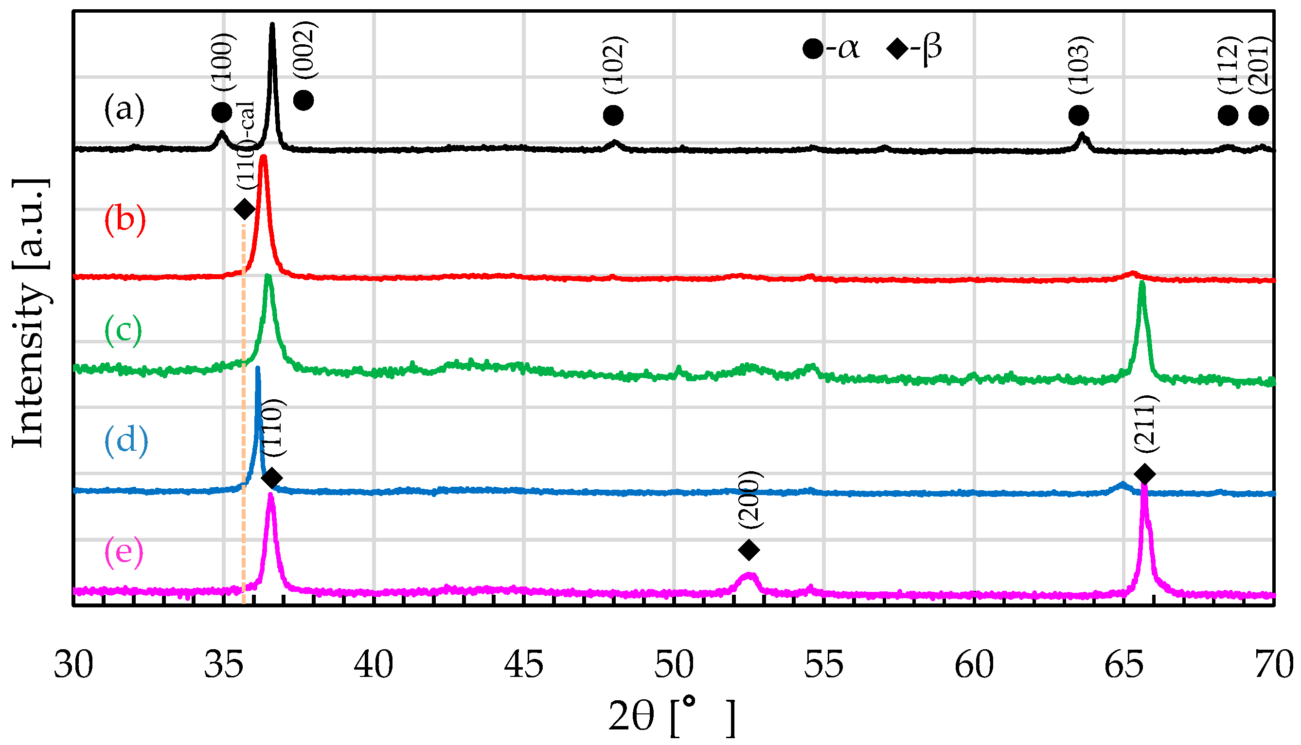
The measured X-ray diffraction patterns of the designed alloys after solution treatment (and pure Zr for comparison) are shown in Figure 2. For pure Zr, only the α-Zr diffraction peak was observed [Figure 2a]. For Zr-15Nb-10Ti, the α-Zr diffraction peak disappeared, and only the β-Zr diffraction peak was observed [Figure 2b]. And similar to the Zr-15Nb-10Ti alloy, the alloys containing Cr and Sn, in addition to Nb and Ti, also showed a single β-phase [Figure 2c–e]. It can be inferred from these XRD patterns that the addition of Nb and Ti and the solution heat treatment created alloys with the β-phase only. Moreover, if the (110) peak at approximately 36° is focused on, the observed β-Zr diffraction peak is shifted to the higher-angle side compared to the calculated diffraction peak position (vertical dotted yellow line). This peak shift can be interpreted as the result of negative lattice distortion (i.e., contraction of the unit lattice) caused by the addition of a substitutional element with a smaller atomic radius than that of Zr. For all the designed alloys, no diffraction peaks from phases other than the β-phase were observed [Figure 2b–e].

3.2. Measurements of Magnetization

The magnetic susceptibility of each designed alloy is listed in Table 2. The magnetic susceptibility of all the designed alloys is approximately 2.0 × 10⁻⁶ cm3/g, which is about 1.0 × 10⁻⁶ cm3/g lower than that (approximately 3.0 × 10⁻⁶ cm3/g) of the Ti-6Al-4V alloy, namely, a typical alloy used as a medical metallic biomaterial [11]; however, all the designed alloys had higher magnetic susceptibility than that of pure Zr (1.29 × 10⁻⁶ cm3/g). It is presumed that this higher magnetic susceptibility than that of pure Zr is due to the addition of Cr, Nb, and Ti, which are elements with higher magnetic susceptibility than that of Zr. Moreover, for Zr-15Nb-10Ti-5Sn, to which a diamagnetic element (Sn) was added, the magnetic susceptibility was reduced by about 0.2 × 10⁻⁶ cm3/g compared to that of Zr-15Nb-10Ti. This result confirms that the addition of diamagnetic elements reduces magnetic susceptibility.

3.3. Measurement of Pitting Potential by the Anodic Polarization Test

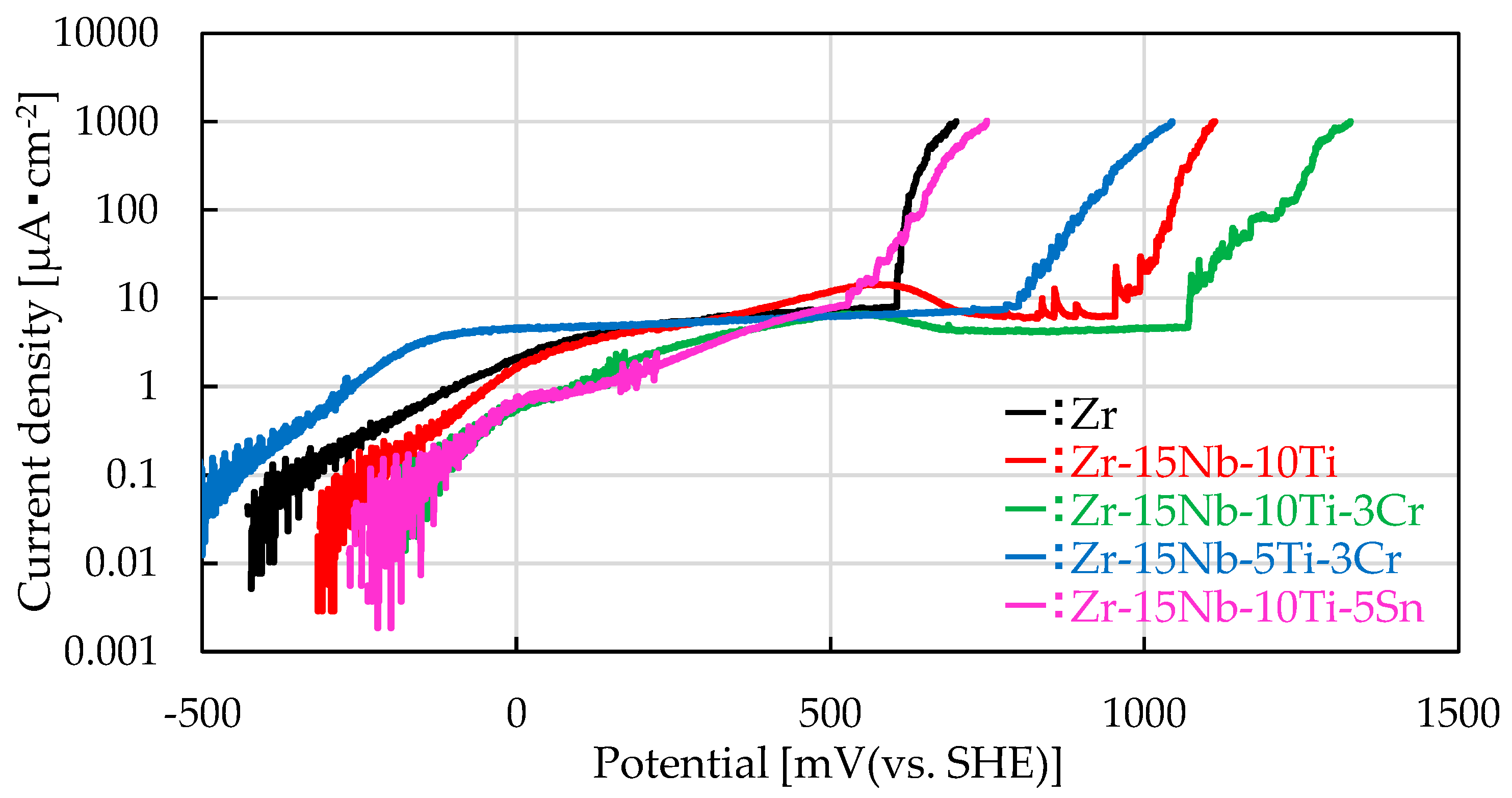
To verify that the pitting potential is raised by adding β-phase and additional elements, we measured the pitting potential of the four designed alloys in physiological saline. The pitting potential of Zr-15Nb-10Ti, to which Nb and Ti were added, was shifted to the more-noble (higher potential) direction by about 400 mV compared with that of pure Zr. Furthermore, the Cr-added alloys, Zr-15Nb-10Ti-3Cr and Zr-15Nb-5Ti-3Cr, like Zr-15Nb-10Ti, showed a higher pitting potential than pure Zr. While the pitting corrosion mechanism of Zr in chloride environments is still unknown, it has been reported that this is caused not by a single factor but by the interaction of various factors, including the type and concentration of additive elements, phase composition, heat treatment conditions, precipitates, localized segregation of impurity elements, and surface condition [13,20,21]. At present, we speculate that the pitting corrosion mechanism in the designed alloys is caused by galvanic corrosion due to localized segregation of precipitates and additive elements [22]. In other words, these results indicate that galvanic corrosion is suppressed by forming an all β-phase (phase composition), which is effective in suppressing precipitates, and adding Nb and Ti (alloy elements), which exhibit superior resistance to localized corrosion caused by chlorides. On the contrary, the pitting potential of the Sn-added alloy (Zr-15Nb-10Ti-5Sn) was shifted to the less-noble direction (lower potential) compared to those of the other designed alloys, even though the alloy was transformed into the β-phase, and the value was equivalent to that of pure Zr. This negative potential shift is thought to be due to the precipitation of Zr-Sn intermetallic compounds that could not fully form a solid solution in the β-phase, and these high Sn concentration precipitates caused the occurrence of pitting corrosion [21] (Table 3, Figure 3).

4. Discussion

4.1. Change in Magnetic Susceptibility Due to Aging Heat Treatment

Among the designed alloys, Zr-15Nb-5Ti-3Cr, which had both a higher pitting potential than that of pure Zr and a lower magnetic susceptibility than that of the other designed alloys, was subjected to aging at 673 K for 0.6 to 10.8 ks to evaluate the effect of aging on magnetic susceptibility. As shown in Figure 4a, it was confirmed that the magnetic susceptibility of the Zr-15Nb-5Ti-3Cr alloy was decreased by aging for 0.6 to 1.8 ks. In particular, after aging for 1.8 ks, its magnetic susceptibility decreased to approximately 1.2 × 10⁻⁶ cm3/g. This value is approximately one-third that of the Ti-6Al-4V alloy (i.e., a common medical metallic biomaterial). In standard MRI imaging, typical artifacts are reported to occur with Ti-based alloys [23]. Therefore, the 1.8 ks aged Zr-15Nb-5Ti-3Cr alloy is expected to reduce artifacts more than Ti-based alloys. However, after aging for over 1.8 ks or more, the magnetic susceptibility was higher than that after aging for 1.8 ks. Since the magnetic susceptibility χ of an alloy with a certain composition depends on the crystal structure, such that χβ > χα > χω [11,14], we speculate that (i) a transformation from β-phase to ω-phase occurs during aging for less than 1.8 ks and (ii) a transformation from β/ω-phase to α-phase occurs during aging for more than 1.8 ks. To evaluate the effect of aging on pitting potential, pitting potential was measured, and the measurement results are plotted in Figure 4b. The pitting potential of the as-solution-treated Zr-15Nb-5Ti-3Cr alloy is about 900 mV, whereas that of the aged alloy is over 1400 mV, and the pitting potential was not noticeably decreased by the aging.

4.2. Effect of Changes in Phase Composition on Magnetic Susceptibility

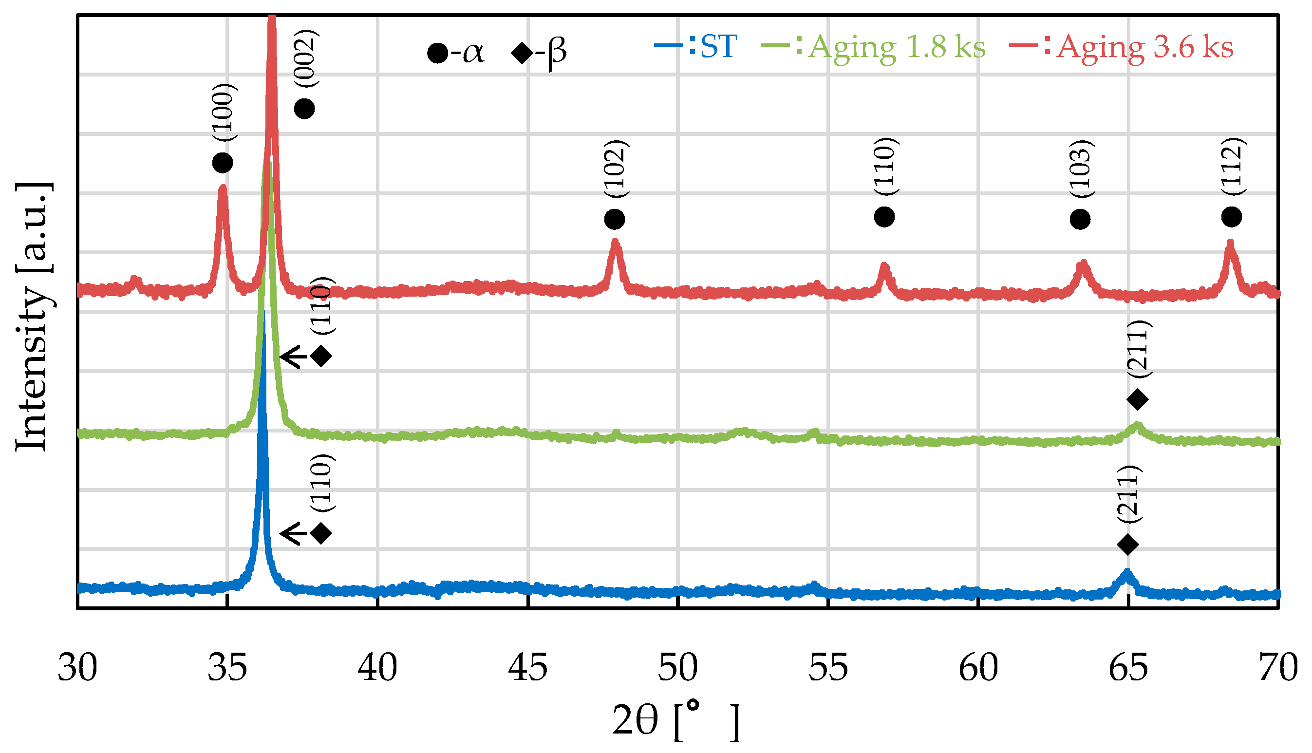
The phase structures of the as-solution-treated Zr-15Nb-5Ti-3Cr alloy, whose magnetic susceptibility changed significantly due to aging, and the same alloy aged for 1.8 and 3.6 ks were evaluated by XRD. The obtained XRD patterns are shown in Figure 5. Similar to the as-solution-treated alloy, the 1.8 ks aged Zr-15Nb-5Ti-3Cr alloy showed only the diffraction peaks of β-Zr. However, it is supposed that since no other phases were identified, the reason for the decrease in magnetic susceptibility due to 1.8 ks aging could not be identified by XRD. On the contrary, when the alloy was aged for 3.6 ks, only the diffraction peaks of α-Zr were observed. That result confirms that when an alloy that has already transformed to β-phase is subjected to long-term aging, a phase transformation from β-phase to α-phase occurs through diffusion transformation. Moreover, the increase in magnetic susceptibility by aging for 1.8 to 3.6 ks is thought to be due to a change in the crystal structure caused by the α-phase transformation [11,14].
XRD was able to identify the factor that increased magnetic susceptibility by aging for 1.8 to 3.6 ks; however, it was unable to identify the cause of the decrease in magnetic susceptibility by aging up to 1.8 ks. Accordingly, the microstructure of the 1.8 ks aged Zr-15Nb-5Ti-3Cr alloy was observed by TEM, which enables higher-resolution observation than XRD. The observed bright-field TEM image and [110]-incidence electron-diffraction pattern in the same field are shown in Figure 6a and 6b, respectively. As shown in Figure 6a, precipitates with a long-axis diameter of approximately 50–100 nm are homogeneously dispersed in the matrix. As shown in Figure 6b, the observed diffraction peaks were assigned to β-Zr (cubic)-phase [with a = 3.551 Å] and ω-Zr (hexagonal)-phase [with a = 5.021 Å and c = 3.075 Å]. These results confirm that the parent phase was β-Zr and that the precipitates within the β-Zr-phase grains were ω-phase [24,25,26]. The ω-phase observed consisted of two types, namely, a normal state (green, ω) and a mirror state (yellow, ω*), with (110)β as the rotation axis. The formation pattern of the ω-phase within the β-phase grains had mirror symmetry. Moreover, according to compositional analysis of the ω-phase by energy-dispersive X-ray spectroscopy (EDX), the Cr and Nb contents in the ω-phase were lower than those in the matrix β-phase. This result suggests that an isothermal ω-phase formed with concentration changes of Zr, Cr, and Nb in the Zr-15Nb-5Ti-3Cr alloy aged for 1.8 ks (Table 4).
The change in magnetic susceptibility with aging time shown in Figure 4 can be explained in terms of the relationship between the change in phase composition due to aging and the magnetic susceptibility of each phase (expressed as χβ > χα > χω) as follows. Under aging for up to 1.8 ks, the β-phase transformed to the ω-phase as the concentration of Cr and Nb in the β matrix decreased, and the phase structure changed from a single β-phase to a β + ω-phase. Accordingly, the magnetic susceptibility of an alloy is thought to be decreased by a transformation from χβ to χβ + χω. When the aging time was between 1.8 and 3.6 ks, the ω-phase to α-phase or β-phase to α-phase transformations progressed due to thermal diffusion, and the phase structure changed from β + ω-phase to α-phase. As the ω-phase disappears and the α-phase appears, magnetic susceptibility increases because it approaches χα. It is presumed that under aging for 3.6 and 10.8 ks thereafter, the β(ω)-phase continues to transform to the α-phase, the phase structure approaches a single α-phase, and magnetic susceptibility is thereby slightly decreased.

5. Conclusions

As a first step in the development of zirconium-based artifact-resistant alloys for medical use, alloy design that can achieve both low magnetic susceptibility and high corrosion resistance, as well as microstructural control by heat treatment, was investigated, and the key results of the investigation are summarized as follows.
(1) The pitting potential of the alloys transformed to β-phase by combined addition of Nb, Ti, and Cr and solution heat treatment was shifted in the noble direction (higher potential) compared to that of pure Zr; on the contrary, that of the alloy with added Sn shifted to the less-noble direction (lower potential) despite the transformation to β-phase.
(2) All of the designed alloys had lower magnetic susceptibility than that of Ti-6Al-4V. However, in the case of all four designed alloys, it was approximately twice that of pure Zr, indicating that the addition of high-magnetic-susceptibility elements such as Cr, Nb, and Ti increases the magnetic susceptibility of pure Zr. On the contrary, the addition of Sn, a low-magnetic-susceptibility element, slightly reduced the magnetic susceptibility.
(3) When the Zr-15Nb-5Ti-3Cr alloy was subjected to aging at 673 K for up to 1.8 ks, its magnetic susceptibility decreased, but when the aging was applied from 1.8 to 3.6 ks, it increased. Evaluation of the phase structure by XRD and TEM showed that the up to 1.8 ks aged alloy underwent a transformation from β-phase to ω-phase, and the 1.8 to 3.6 ks aged alloy underwent a transformation from β(ω)-phase to α-phase, which presumably caused the increase in magnetic susceptibility. Moreover, no effect on the pitting potential due to the precipitation of ω-phase into β-phase was confirmed.
In particular, the magnetic susceptibility of the 1.8 ks aged Zr-15Nb-5Ti-3Cr designed alloy was approximately one-third that of the Ti-6Al-4V alloy (i.e., a common medical metallic biomaterial), and its pitting potential exceeded 1000 mV. In light of these properties, it can be considered useful as a metallic biomaterial for medical devices used under MRI. However, since ω-phase precipitation may degrade mechanical properties, the authors plan to evaluate the effect of ω-phase precipitation on mechanical properties (in addition to magnetic susceptibility and corrosion resistance) as a future study.

Author Contributions

Conceptualization, T.K. and Y.A.; methodology, Y.A.; validation, S.T. and T.K.; formal analysis, Y.A.; investigation, Y.A.; resources, S.T., T.K. and Y.A.; data curation, S.T.; writing—original draft preparation, S.T.; writing—review and editing, S.T.; visualization S.T.; supervision, S.T., T.K. and Y.A.; project administration, T.K.; funding acquisition, T.K. All authors have read and agreed to the published version of the manuscript.

Funding

This research received no external funding.

Data Availability Statement

The original contributions presented in this study are included in the article. Further inquiries can be directed to the corresponding author.

Conflicts of Interest

Authors Shinya Tamura and Yasuhisa Aono were employed by the company Research & Development Group, Hitachi, Ltd. Author Tomonori Kimura was employed by the company Nuclear Energy Production Division, Hitachi-GE Nuclear Energy, Ltd. The remaining authors declare that the research was conducted in the absence of any commercial or financial relationships that could be construed as a potential conflict of interest.

References

  1. Niinomi, M.; Akahori, T. Improvement of the fatigue life of titanium alloys for biomedical devices through microstructural control. Expert Rev. Med. Devices 2010, 7, 481–488. [Google Scholar] [CrossRef] [PubMed]
  2. Niinomi, M. Development of high biocompatible titanium alloys. Funct. Mater. 2000, 20, 36–44. [Google Scholar] [CrossRef]
  3. Niinomi, M. Recent Titanium R&D for Biomedical Applications in Japan. JOM 1999, 51, 32–34. [Google Scholar] [CrossRef]
  4. Niinomi, M. Development of Beta Titanium Alloys for Biomedical Applications. Mater. Jpn. 1998, 37, 843–846. [Google Scholar] [CrossRef]
  5. Sanderson, P.L.; Ryan, W.; Turner, P.G. Complications of metalwork removal. Injury 1992, 23, 29–30. [Google Scholar] [CrossRef]
  6. Seipel, R.C.; Pintar, F.A.; Yoganandan, N.; Boynton, M.D. Biomechanics of Calcaneal Fractures A Model for the Motor Vehicle. Clin. Orthop. Relat. Res. 2001, 388, 218–224. [Google Scholar] [CrossRef] [PubMed]
  7. Matsuura, H.; Inoue, T.; Konno, H.; Sasaki, M. Quantification of susceptibility artifacts produced on high-field magnetic resonance images by various biomaterials used for neurosurgical implants. J. Neurosurg. 2002, 97, 1472–1475. [Google Scholar] [CrossRef]
  8. Matsuura, H.; Inoue, T.; Ogasawara, K.; Sasaki, M.; Konno, H.; Kuzu, Y.; Nishimoto, H.; Ogawa, A. Quantitative Analysis of Magnetic Resonance Imaging Susceptibility Artifacts Caused by Neurosurgical Biomaterials: Comparison of 0.5, 1.5, and 3.0 Tesla Magnetic Fields. Neurol. Med.-Chir. 2005, 45, 395–399. [Google Scholar] [CrossRef]
  9. Ernstberger, T.; Heidrich, G.; Buchhorn, G. Postimplantation MRI with cylindric and cubic intervertebral test implants: Evaluation of implant shape, material, and volume in MRI artifacting—An in vitro study. Spine J. 2007, 7, 353–359. [Google Scholar] [CrossRef]
  10. Inui, S.; Uyama, E.; Hamada, K. Volume magnetic susceptibility design and hardness of Au–Ta alloys and Au–Nb alloys for MRI-compatible biomedical applications. Biomed. Phys. Eng. Express 2017, 3, 015025. [Google Scholar] [CrossRef]
  11. Nomura, N.; Tanaka, Y.; Suyalatu; Kondo, R.; Doi, H.; Tsutsumi, Y.; Hanawa, T. Effects of Phase Constitution of Zr-Nb Alloys on Their Magnetic Susceptibilities. Mater. Trans. 2009, 50, 2466–2472. [Google Scholar] [CrossRef]
  12. Kobayashi, E.; Ando, M.; Tsustumi, Y.; Doi, H.; Yoneyama, T.; Kobayashi, M.; Hanawa, T. Inhibition Effect of Zirconium Coating on Calcium Phosphate Precipitation of Titanium to Avoid Assimilation with Bone. Mater. Trans. 2007, 48, 301–306. [Google Scholar] [CrossRef]
  13. Tsutsumi, Y. Corrosion Resistance of Zr as Metallic Biomaterials. Zair. Kankyo 2014, 63, 360–364. [Google Scholar] [CrossRef]
  14. Collings, E.W. Magnetic studies of phase equilibria in Ti-Ai (30 to 57 At. Pct) alloys. Metall. Trans. A 1979, 10, 463–474. [Google Scholar] [CrossRef]
  15. Chui, P. Near β-type Zr-Nb-Ti biomedical alloys with high strength and low modulus. Vacuum 2017, 143, 54–58. [Google Scholar] [CrossRef]
  16. Morinaga, M. Local lattice distortion and martensitic transformation near alloying elements in titanium and iron. Toyota Res. Rep. 2017, 70, 35–44. [Google Scholar]
  17. Hong, S.-P.; Ko, Y.-M.; Kim, C.-S. Magnetic Susceptibility of Zr-Cu Binary Alloys. Mater. Trans. 2014, 55, 1634–1636. [Google Scholar] [CrossRef]
  18. Xue, R.-H.; Wang, D.; Yue-yan, T.; Zi-xuan, D.; Li-bin, L.; Li-gang, Z. Effect of Sn on elastic modulus and magnetic susceptibility of Zr-16Nb-xTi (x = 4 wt%, 6 wt%) alloys. J. Cent. South Univ. 2023, 30, 412–418. [Google Scholar] [CrossRef]
  19. JIS T 0302; Method for Evaluating Corrosion Resistance of Metallic Biomaterials by Anodic-Polarization Test. JSA GROUP Webdesk: Tokyo, Japan, 2000.
  20. Motooka, T.; Komatsu, A.; Tsukada, T.; Yamamoto, M. Effect of gamma radiolysis on pit initiation of zircaloy-2 in water containing sea salt. J. Nucl. Sci. Technol. 2014, 51, 987–995. [Google Scholar] [CrossRef]
  21. Tsutsumi, Y.; Muto, I.; Nakano, S.; Tsukada, J.; Manaka, T.; Chen, P.; Ashida, M.; Sugawara, Y.; Shimojo, M.; Hara, N.; et al. Effect of Impurity Elements on Localized Corrosion of Zirconium in Chloride Containing Environment. J. Electrochem. Soc. 2020, 167, 141507. [Google Scholar] [CrossRef]
  22. Palit, G.C. Pit Simulation Study of Zirconium Under Simple Immersion Conditions in Oxidizing Chloride Solutions. Corrosion 2000, 56, 523–532. [Google Scholar] [CrossRef]
  23. Georg, C.; Feuerriegel, R. Sutter. Managing hardware-related metal artifacts in MRI: Current and evolving techniques. Skelet. Radiol. 2024, 53, 1737–1750. [Google Scholar] [CrossRef]
  24. Zhanal, P.; Harcuba, P.; Hajek, M.; Smola, B.; Strasky, J.; Smilauerova, J.; Vesely, J.; Janecek, M. Evolution of ω phase during heating of metastable β titanium alloy Ti–15Mo. J. Mater. Sci. 2018, 53, 837–845. [Google Scholar] [CrossRef]
  25. Prima, F.; Vermaut, P.; Ansel, D.; Debuigne, J. ω Precipitation in a Beta Metastable Titanium Alloy, Resistometric Study. Mater. Trans. 2000, 41, 1092–1097. [Google Scholar] [CrossRef]
  26. Settefrati, A.; Aeby-Gaytier, E.; Dehmas, M.; Geandier, G.; Appolaire, B.; Audion, S.; Delfosse, J. Precipitation in a near β titanium alloy on ageing: Influence of heating rate and chemical composition of the β-metastable phase. Solid State Phenom. 2011, 172–174, 760–765. [Google Scholar] [CrossRef]
Figure 1. Heat-treatment diagram.
Metals 15 01208 g001
Figure 2. X-ray-diffraction patterns of the designed alloys after solution treatment: (a) pure Zr, (b) Zr-15Nb-10Ti, (c) Zr-15Nb-10Ti-3Cr, (d) Zr-15Nb-5Ti-3Cr, and (e) Zr-15Nb-10Ti-5Sn.
Metals 15 01208 g002
Figure 3. Anodic polarization curves of each designed alloy.
Metals 15 01208 g003
Figure 4. (a) Change in magnetic susceptibility of the Zr-15Nb-5Ti-3Cr alloy due to aging and (b) anodic polarization curve of the Zr-15Nb-5Ti-3Cr alloy after 1.8 ks aging heat treatment.
Metals 15 01208 g004
Figure 5. XRD patterns of the Zr-15Nb-5Ti-3Cr alloy specimen for two aging times.
Metals 15 01208 g005
Figure 6. TEM images of the Zr-15Nb-5Ti-3Cr alloy specimen after 1.8 ks aging: (a) bright-field image and (b) [110]-incidence electron diffraction image.
Metals 15 01208 g006
Table 1. Composition of the designed alloys.
AlloysNbTiCrSnZr
Zr-15Nb-10Ti15100075
13.517.50069.0
Zr-15Nb-10Ti-3Cr15103072
13.317.24.7064.8
Zr-15Nb-5Ti-3Cr1553077
13.89.04.9072.3
Zr-15Nb-10Ti-5Sn15100570
13.717.703.665.0
Top and bottom amounts show wt.% and at.%, respectively.
Table 2. Magnetic susceptibility of each designed alloy.
AlloysZrZr-15Nb-10TiZr-15Nb-10Ti-3CrZr-15Nb-5Ti-3CrZr-15Nb-10Ti-5Sn
Mass magnetic
susceptibility [×10−6 cm3/g]
1.292.132.171.791.91
Table 3. Pitting potential of each designed alloy.
AlloysZrZr-15Nb-10TiZr-15Nb-10Ti-3CrZr-15Nb-5Ti-3CrZr-15Nb-10Ti-5Sn
Pitting potential
[mV (vs. SHE)]
62210451219905647
Table 4. Composition ratios of elements in β-phase and ω-phase for the Zr-15Nb-5Ti-3Cr alloy.
Zr [at.%]Cr [at.%]Nb [at.%]
β-phase76.02.8011.24
ω-phase85.50.866.19
Disclaimer/Publisher’s Note: The statements, opinions and data contained in all publications are solely those of the individual author(s) and contributor(s) and not of MDPI and/or the editor(s). MDPI and/or the editor(s) disclaim responsibility for any injury to people or property resulting from any ideas, methods, instructions or products referred to in the content.