Article

Biosorption and Bioaccumulation Capacity of Arthrospira platensis toward Yttrium Ions

1
Department of Nuclear Physics, Joint Institute for Nuclear Research, 141980 Dubna, Russia
2
Doctoral School of Biological, Geonomic, Chemical and Technological Science, State University of Moldova, MD-2009 Chisinau, Moldova
3
Department of Nuclear Physics, Horia Hulubei National Institute for R&D in Physics and Nuclear Engineering, MG-6, Magurele, 077125 Ilfov, Romania
4
Laboratory of Physical and Quantum Chemistry, Institute of Chemistry, MD-2028 Chisinau, Moldova
5
Laboratory of Phycobiotechnology, Institute of Microbiology and Biotechnology, MD-2028 Chisinau, Moldova
*
Author to whom correspondence should be addressed.
Metals 2022, 12(9), 1465; https://doi.org/10.3390/met12091465
Submission received: 15 August 2022 / Revised: 26 August 2022 / Accepted: 29 August 2022 / Published: 31 August 2022

Abstract

:
Yttrium is an element of critical importance for industry and technology. Cyanobacteria Arthrospira platensis was employed for Y(III) recovery from contaminated wastewater through biosorption and bioaccumulation processes. The effect of pH of a solution, contact time, temperature, and initial Y(III) concentration on the adsorption behaviour of Arthrospira platensis were studied. The maximum adsorption capacity of 719.8 mg/g was attained at a pH of 3, temperature of 20 °C, and adsorption time of 3 min. The Langmuir and Freundlich isotherm models were suitable to describe the equilibrium of the biosorption, while kinetic of the process followed the pseudo-first-order model. Thermodynamic parameters showed that the biosorption process was spontaneous and exothermic in nature. In bioaccumulation experiments, Arthrospira platensis was able to remove up to 70% of Y(III) from the solution. Beside biomass uptake capacity, the toxic effect of Y(III) on the biomass productivity and biochemical composition was assessed. Thus, Y(III) in concentration of 10–30 mg/L led to a decrease in the content of proteins, carbohydrates, and phycobiliproteins in the biomass and had no significant negative impact on productivity and photosynthetic pigments content. Experiments performed using Arthrospira platensis showed that biological objects have a great potential to be applied for the recovery of rare earth elements from wastewater.

1. Introduction

Rare earth elements, because of their excellent physical and chemical properties, are widely used in various fields of industry, specifically in global clean energy technology development and consumer products [1,2]. Today, rare earth elements are defined as the “industrial vitamin” [2]. Yttrium, along with the other four elements Dy, Eu, Nd, and Tb amount up to ~ 85% of the total weight of rare earth elements in final products [2,3]. Yttrium has been widely used as a catalyst in a wide array of industries, as a deoxidizer for non-ferrous metals, in fiber optics, and in making fireproof bricks [2,4].
Today, Y(III) released from mining activity, processing, and the disposal of wastes contaminate objects of the environment [5]. For, example, it was reported that concentrations of rare earth elements downstream of the river and wastewater of mining and refining factories can range from 1 to 200 mg/L [3]. The discharge of waste containing rare earth elements, including Y(III), in the environment not only causes the loss of rare earth resources but also negatively affect humans and other living organisms [3].
Information regarding Y(III) toxicity is very controversial. Thus, Ding and co-authors [6] demonstrated that YCl3 treatment greatly promoted neuron cells’ death in rats. Among the three freshwater organisms Daphnia magna, Chironomus riparius, and Oncorhynchus mykiss, only Chironomus riparius showed toxicity at Y(III) exposure concentrations close to environmental ones [5]. In the study of Nakamura et al. [7], Y(III) in rats was mainly accumulated in the liver, spleen, and bones, but it did not provoke any visible toxic effects.
Recovery of the rare earth elements from wastewater, in which their concentrations range from 1 to 100 mg/L is a key concern for the economy and environmental protection. Various techniques have been designed to ensure the efficient recovery of rare earth elements, including chemical precipitation, filtration, solvent extraction, membrane technology, and adsorption [2]. However, conventional methods applied for industrial wastewater treatment are considered inefficient for diluted effluents, are relatively expensive, and require a high energy and chemical consumption. Some of these methods generate large volumes of waste and consequently can lead to secondary environmental pollution, while others have limited applications [8,9].
The cyanobacterium Arthrospira platensis (spirulina) is a well-known model object used in bioremediation studies to elaborate effective procedures of pollutant removal from aquatic environments [10,11,12,13,14,15,16]. Spirulina application in bioremediation studies is explained by the fact that cyanobacteria are polyextremophile organisms able to cope with high alkalinity, temperature, salt concentration, and the presence of different pollutants in culture media. Moreover, spirulina can accumulate a large amount of biomass under these conditions. Similarly, spirulina is a technologically convenient object, being easy to handle under both laboratory and industrial conditions, which significantly facilitates the implementation of the developed bioremediation technology [17].
Microbial bioremediation of metal-polluted industrial effluents is considered one of the most effective eco-friendly tools to combat pollution [8]. Biological pathways of metal removal from wastewater are low-cost, ecologically viable, and safe for the environment [9]. It is suggested that biological objects possess high uptake capacities and tolerances toward rare earth elements.
In the present study, the biosorption and bioaccumulation capacity of Arthrospira platensis (A. platensis) toward Y(III) was tested. Biomass biosorption capacity was investigated regarding the influence of pH, contact time, initial Y(III) ions concentration, and temperature. The sorption process was described using well-known kinetics, equilibrium, and thermodynamics models. The effect of the Y(III) in different concentrations on the spirulina accumulation capacity, productivity, and biochemical parameters (proteins, carbohydrates, lipids, pigments) was assessed.

2. Materials and Methods

2.1. Chemicals

All the chemical reagents of analytical grade used in the study were purchased from Sigma-Aldrich (Darmstadt, Germany).

2.2. Object of Study

The biosorption and bioaccumulation experiments were carried out with the cyanobacterium Arthrospira platensis (spirulina) CNMN-CB-02 from the collection of non-pathogenic microorganisms (Institute Microbiology and Biotechnology, Chisinau, Moldova). The composition of the cultivation medium and conditions of biomass growth are presented in [10]. Spirulina biomass was used as biosorbent after the full cultivation cycle was separated from the medium by filtration, dried at 105 °C, and homogenized. The conditions of biomass growth in bioaccumulation experiments are presented in Section 2.4.

2.3. Biosorption

Biosorption experiments were performed in a 50-milileter Erlenmeyer flask containing 20 mL of yttrium solution and 100 mg of biomass. Single-factor experiments were performed in order to reveal the effect of pH (2.0–6.0), yttrium initial concentration (10–100 mg/L), contact time (2–120 min), and temperature (20–50 °C). Studying the effect of parameters, except time, experiments lasted for 30 min, then the solution was filtrated and yttrium concentration was determined using ICP-OES PlasmaQuant PQ 9000 Elite spectrometer (Analytik Jena, Jena, Germany). Results are presented as average values obtained from the triplicate analysis. The removal efficiency and adsorption capacity of the spirulina were calculated according to Equations (1) and (2):
q = V ( C i C f ) m
E = C i C f C i × 100
where V is the volume of the solution, ml; Ci and Cf are the initial and final metal concentrations in mg/L, and m is the mass of sorbent in g.

2.4. Bioaccumulation

In order to assess the bioaccumulation capacity of spirulina biomass Y(III) in concentrations of 10–30 mg/L, metal was added to the cultivation medium on the first day of biomass growth. Then, biomass was cultivated for six days at a temperature of 25–28 °C, illumination ~37 µM photons/m2/s, and pH 8–9. At the end of the cultivation cycle, the spirulina biomass was separated from the culture liquid by filtration. The amount of biomass, expressed in g/L, was determined spectrophotometrically based on the calibration curve. The samples were standardized to the concentration of 10 mg/mL of distilled water. For the biochemical tests, the biomass was subjected to the repeated freezing–thawing procedure. The concentration of Y(III) in experimental solutions was determined by ICP-OES.
Served as a control in both types of experiments, spirulina biomass was cultivated in the medium without the addition of Y(III).

2.5. Biochemical Tests

Protein content was determined based on the Lowry method in samples of 10 mg/mL biomass. Biomass was processed with 0.1N NaOH solution for 30 min. Then, reagent (49 parts of 2% Na2CO3 solution in 0.1N NaOH and 1 part of 0.5% CuSO4 solution in 1.0% Na3C6H5O7) and 0.2 mL of Folin–Ciocalteu reagent (Sigma Aldrich, Darmstadt, Germany) were added to 0.2 mL of basic extract. The absorbance at 750 nm was recorded through 30 min. The amount of protein was calculated based on the calibration curve plotted for bovine serum albumin [18].
Carbohydrates content was determined using Anthracene-9(10H)-one. The reaction mixture consisted of 2.5 mg of biomass and 2.5 mL of 0.5% Antron reagent (Sigma Aldrich, Darmstadt, Germany) solution in 66% H2SO4. The obtained samples were boiled on a water bath for 30 min. After cooling and incubation in the dark for 30 min, the absorbance of the samples was recorded at 620 nm. The quantitative calculation was performed based on the calibration curve plotted for glucose [12].
Quantitative determination of lipids was performed using the phosphovanillin reagent (Sigma Aldrich, Darmstadt, Germany). The lipid extract from the spirulina biomass was obtained by adding 1 mL of a mixture of chloroform and ethanol in a ratio of 9:1 (v:v) to 10 mg of biomass. The extraction was carried out by continuous stirring for 2 h at room temperature. The lipid extract underwent hydrolysis with sulfuric acid. The reaction mixture consisted of 0.1 mL of hydrolyzate and 2.9 mL of phosphovanillin reagent. After 30 min, the absorbance at 560 nm was recorded. The quantitative calculation was performed based on the calibration curve constructed for oleic acid [19].
The content of phycobiliproteins in the biomass was calculated using formulas based on molar coefficients for pigments. The hydrous extract of phycobiliproteins was obtained as a result of applying the procedure of repeated freezing–thawing of standardized biomass with distilled water. The absorbance of the extract at 620 nm was recorded for phycocyanin and at 650 nm for allophycocyanin [20].
The content of chlorophyll α and β-carotene in the biomass was determined using ethanolic extracts. The mixture of 10 mg of biomass with 1.0 mL of 96% ethyl alcohol was prepared. The pigments extraction was performed by continuous stirring at room temperature for 12 h. Chlorophyll content was determined at an absorbance of 665 nm, and β-carotene at an absorbance of 450 nm. The quantitative calculation was performed based on the calibration curves.
The determination of the content of malonic dialdehyde (MDA) in the biomass was carried out based on the reactive substances of thiobarbituric acid (TBA). Three milliliters of 0.76% TBA solution in 20% C2HCl3O2 were added to 10 mg of biomass. The obtained mixture was boiled on a water bath at 95 °C for 20 min, then the mixture was cooled and the absorbance was recorded at wavelengths of 532 nm and 600 nm. The content of MDA in samples was calculated using the extinction coefficient of the complex product of the MDA-TBA [21].

2.6. Antioxidant Activity

The antioxidant activity of the ethanolic and water extracts was determined using the radical cation ABTS (2,2 azinobis 3-ethylbenzothiazoline-6-sulfonic acid) [22]. The ethanolic extract from the biomass was obtained in a similar way to the extract for the determination of pigments. The water extract was obtained identically to the extracts for the determination of phycobiliproteins. ABTS radical cation was obtained by oxidation with potassium persulfate. ABTS stock solution was prepared by mixing 7 mM ABTS in deionized water with 2.45 mM K2S2O8 in a 1:1 ratio. The ABTS oxidation process took place in the dark at room temperature for 12–16 h. The working radical ABTS solution had an absorbance value of 0.700 ± 0.020 at 734 nm. The samples were obtained by mixing 0.3 mL of biomass extract and 2.7 mL of ABTS solution. The absorbance of the samples was measured after 6 min. The % of inhibition was calculated relative to the absorbance of the ABTS reagent.

2.7. Statistical Analysis

The values in the manuscript are presented as an average of three experiments ± standard deviation. One-way analysis of variance (ANOVA) was performed using Student’s t-test.

3. Results and Discussion

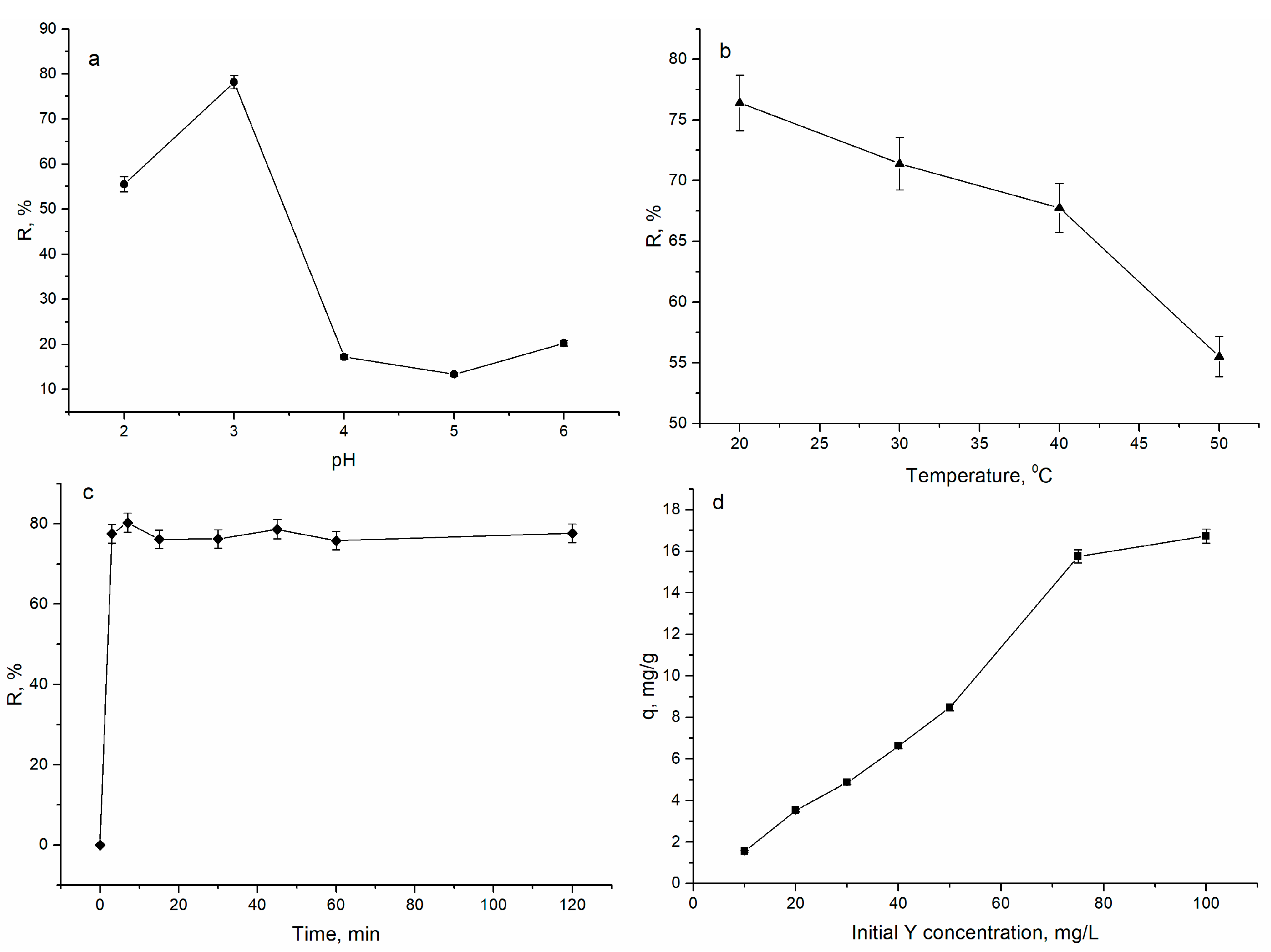
3.1. Yttrium Removal by Biosorption

The effect of pH, Y(III) concentration, contact time, and temperature on the biosorption capacity of A. platensis is shown in Figure 1.
The pH of the solution plays an important role in adsorption experiments affecting the charge of the adsorbent surface and the chemical state of the metal. The spirulina biosorption capacity increased from 55 to 78% with a rise in the pH from 2.0 to 3.0, and at a further increase in pH, a decline in the sorption was noticed (Figure 1a). At a pH of 2.0, relatively low biomass removal efficiency (55%) can be associated with the predominance of H+ in the solution and their competition with Y(III) for binding sites. At a pH of 4.0–6.0, the removal efficiency of spirulina significantly decreased (up to 20%), which can be explained by the increase in the concentration of OH groups in the solution and Y(III) precipitation in the form of Y(OH)3 [2]. According to [23], Y(III) at a pH range of 2.0–5.5 is present in the solution in the form of Y3+, and at higher pH values, Y(OH)3 is formed. Thus, further experiments were performed at a pH of 3.0. For a comparison maximum, Y(III) biosorption on Serratia marcescens was achieved at a pH of 5.5 [2]. The process of Y(III) sorption on bacteria Microbacterium sp., Curtobacterium sp., Bacillus subtilis, Pseudomonas putida, and Bacillus pumilis better proceeded at a pH of 4 [24].
An increase in temperature from 20 to 50 °C resulted in a decrease in the spirulina adsorption capacity from 76 to 55 % (Figure 1b). The decrease in biomass sorption capacity occurred with the temperature point at the exothermic character of Y(III) biosorption. The result is opposite to that reported by [2], which showed that Y(III) sorption on Serratia marcescens was an endothermic process. At the same time, the results are in line with [25], which reported that a temperature increase produced a decrease in Y(III) sorption by microbial biomass.
The process of Y(III) biosorption was very rapid; the maximum metal removal was achieved in 3 min of interaction when 76% of ions were removed from the solution. An increase in the time of biosorption up to 120 min has no noticeable impact on the A. platensis adsorption efficiency. Rapid Y(III) adsorption in the first minutes of sorbent interaction with the sorbate is explained by a large number of unoccupied biosorption sites. Reduction in the rate of Y(III) sorption and equilibrium achievement is associated with the occupation of binding sites. The highest sorption rate for Y(III) was registered during the first 10 min for Microbacterium sp., Bacillus pumilis, and Pseudomonas putida strains, while the highest sorption rate for Curtobacterium sp. and Bacillus subtilis strains was detected after 35 min of the experiment [24].
Y(III) initial concentration in the solution was directly proportional to its sorption on the spirulina; the adsorption capacity of 1.6 mg/g recorded at a Y(III) concentration of 10 mg/L increased to 16.7 mg/g at a metal concentration of 100 mg/L. Spirulina biosorption capacity rapidly increased at a Y(III) concentration range of 10–75 mg/L, which is in agreement with [2], and then the rise was less pronounced.
Experimentally obtained data were used to describe equilibrium, kinetics, and thermodynamics of Y(III) biosorption on A. platensis biomass. Pseudo-first- and pseudo-second-order, as well as Elovich models, have been employed to assess the adsorption reaction mechanism (see Equations (3)–(5)):
q =   q e   ( 1 e k 1 t )
q = q e 2 k 2 t 1 + q e k 2 t
q t   = 1 β ln ( 1 + α β t )
where qe and qt are the content of metal (mg/g) adsorbed at equilibrium and at t time, k2 (g/mg·min) and k1 (1/min) are rates constant of pseudo-first-order and pseudo-second-order, and α (g/mg∙min) and β (g/mg) are the Elovich equation constants.
In addition to the coefficient of determination, the applicability of applied models was assessed using the sum of error squares (SSE, %) and nonlinear chi-square test (χ2)-te, given by Equations (6) and (7):
S S E = i = 1 n ( q e ,   c a l q e ,   e x p ) 2
χ 2 = i = 1 n ( q e ,   c a l q e ,   e x p ) 2 q e ,   e x p
where n is the number of data points.
The graphical presentation of the data is illustrated in Figure 2a and the parameters of the applied models are listed in Table 1. According to coefficient of determination values, all three models can be considered applicable for the description of experimental data. Values of SSE and χ2 , 0.006 and 0.003, respectively (the same values for both models were obtained), showed that the pseudo-first model and pseudo-second-order model were suitable to satisfactorily describe Y(III) biosorption. However, extremely high values of α in the Elovich model and a negative value of k2 in the pseudo-second model exclude the possibility of their use for the description of experimental findings. Additionally, the Akaike Information Criterion (AIC) test was applied to emphasize which model best describes the experimentally obtained values. According to the AIC test, the pseudo-first-order model better describes Y(III) biosorption on spirulina biomass. The applicability of the pseudo-first-order model suggests that the process of Y(III) sorption is physical, with the formation of a monolayer on a heterogeneous surface [26,27,28].
Equilibrium sorption data were modelled using Langmuir, Freundlich, and Temkin sorption isotherms (Figure 2b).
q e = q m   bC e 1 + bC e
where Ce is Y(III) concentration at equilibrium in mg/L, qe and qm are amount of metal adsorbed at equilibrium and maximum adsorption capacity in mg/g, and b is Langmuir constant in L/mg.
q e = K F C 1 n
where KF and n are Freundlich constants.
The Temkin isotherm model takes into account the effects of indirect adsorbate/adsorbate interactions on the adsorption process [13]:
q e = RT b T ln ( a T C e )
1/bT indicates the sorption potential of the sorbent, aT is the Temkin constant, R is the universal gas constant (8.314 J K−1 mol−1), and T is the temperature (K).
The calculated isotherm constants are given in Table 1. Langmuir and Freundlich models well-represented the data for the Y(III) biosorption on A. platensis since the determination coefficients for both models were 0.97. The coefficient of determination for the Temkin model was significantly lower. The maximum adsorption capacity calculated by the Langmuir model was 719.8 mg/g, and it was considerably higher than the values obtained for the other sorbents (Table 2). Application of the Langmuir models supports the sorption on homogenous sites, facilitating chemisorption [29], while the Freundlich model is suitable for heterogeneous surfaces with a non-uniform distribution of active adsorption sites. Since both models fit well with the experimental data, it can be suggested that Y(III) biosorption by A. platensis follows an intermediate behaviour between mono- and multilayer adsorption mechanisms [30]. According to the AIC test, the Langmuir model was more applicable for the description of experimental data. The Freundlich isotherm model better described the adsorption of Y(III) on magnetic nitrogen functionalized mesoporous expanded perlite [31].
The three thermodynamic parameters, the Gibbs free energy (∆G°), enthalpy (∆H°), and entropy (∆S°) were obtained by using Equations (11) to (13) [26,27].
lnK d = Δ S ° R Δ H ° RT
Δ G ° = Δ H ° T Δ S °
K d = ( C 0 C e ) V mC e
where Kd is the distribution coefficient, R is the gas constant, and T is the absolute temperature.
The thermodynamic parameters evaluated by plotting lnKd versus 1/T are presented in Figure A1, and the thermodynamic parameters are listed in Table 3.
The values of the ∆G° ranging from −13.2 to −12.1 kJ/mol indicate the feasibility and spontaneity of the biosorption process. The negative value of ∆H° indicates the exothermic nature, and the positive value of ∆S° points to the randomness at the solid and solution interface. Since ∆H° was less than °25 kJ/mol, the sorption can be considered physical. Since the value of ∆S° was higher than −10 J/mol·K, it can be suggested that the adsorption reaction complies with a dissociative mechanism [33].
Biological sorbents, such as other types of sorbents, are characterized by the possibility of their regeneration and multiple use. Thus, using 0.2 M HCl/0.5 M CaCl2, the elution of rare earth elements from new phosphorylated hydrogel (algal biomass/PEI) was complete and remarkably stable for the five cycles [34]. A number of sulphate and chloride salts were successfully used for rare earth elements desorption and the desorption efficiency of the sulphates was higher compared with chloride [35].

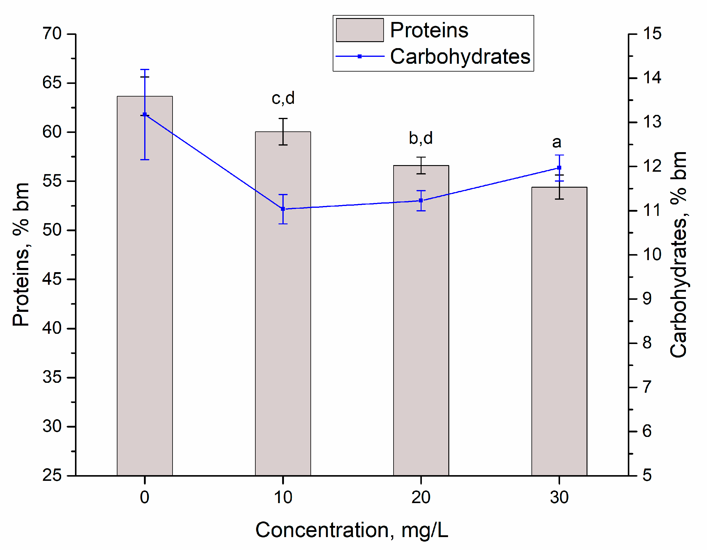
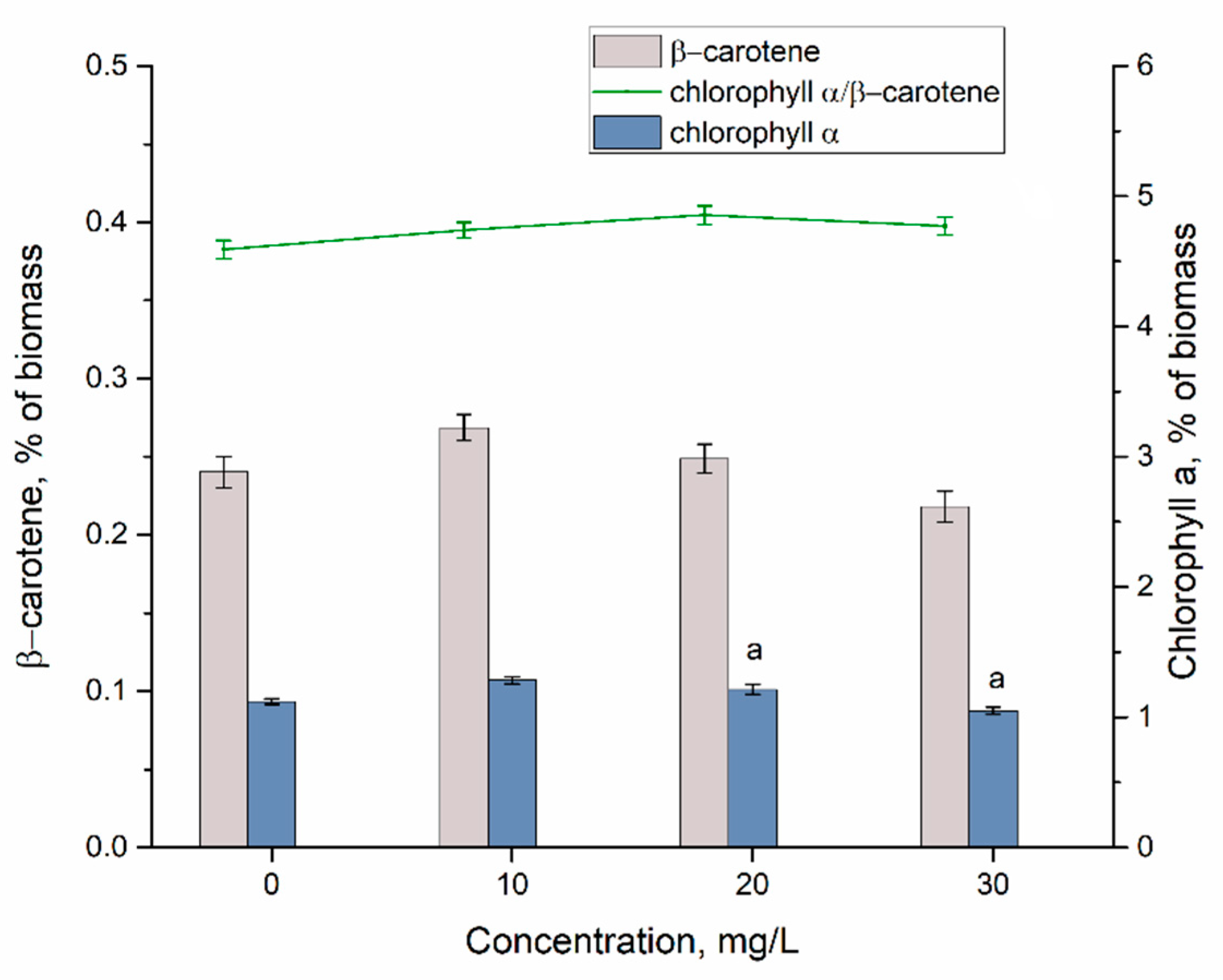
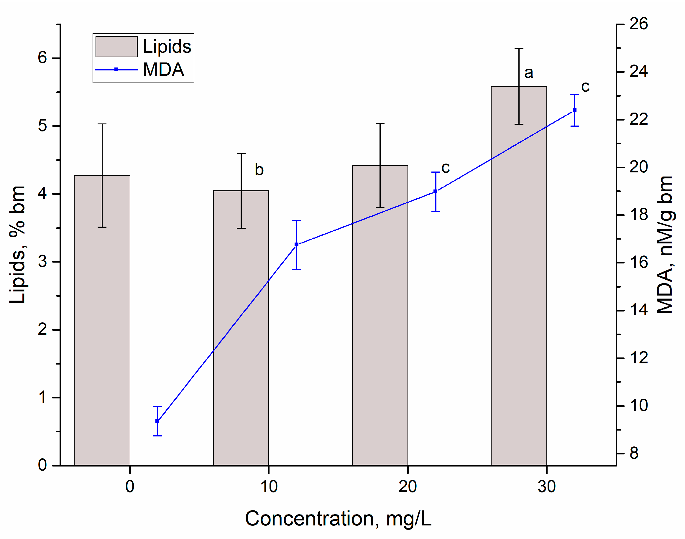
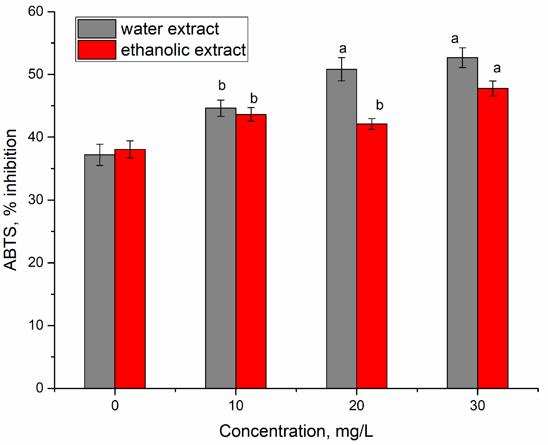
3.2. Yttrium Removal by Bioaccumulation

In bioaccumulation experiments, when spirulina was exposed to 10–30 mg/L of Y(III), the lowest rate of Y(III) removal of 29% was observed at the initial Y(III) concentration of 10 mg/L. At higher concentrations, the efficiency of Y(III) removal was approximately two times higher (on the level of 60–70%). Microalgae Ulva lactuca was able to remove 70–80% of Y(III) from the solutions containing 10–500 μg/L of metal, Gracilaria sp. contributed to the removal of 50–65% of Y(III), while removal efficiency of Ulva intestinalis, Fucus spiralis, Fucus vesiculosus, and Osmundea pinnatifida was not higher than 20–30% [36]. Fungus Penicillium sp. ZD28 accumulated up to 99% of Y(III) at all applied metal concentrations [1].
One of the main indicators of the toxicity of different chemical compounds for microorganisms is the ability to grow and multiply cells, which is expressed as the amount of biomass accumulated in a unit or per a period of time. The accumulation of cyanobacterium A. platensis biomass in a cycle of cultivation in a closed system was monitored both under standard conditions (control) and under the application of Y(III). According to results presented in Figure 3, at a Y(III) concentration of 10 mg/L, no difference between the control and the experimental values of biomass productivity was observed. The concentration of 20 mg/L caused a veridical increase in the amount of biomass by 9.3% compared to control (p = 0.0011) and that of 30 mg/L—a decrease by 4.6% (p = 0.0076).
At the same time, it should be mentioned that from a biotechnological point of view, the amount of biomass in both control and experimental samples was within the physiological norm characteristic for spirulina. Thus, the amount of biomass at the end of the cultivation cycle was 0.927–1.071 g/L.
The stimulating effect of rare earth elements on spirulina productivity was reported in other studies as well. For example, dysprosium in concentrations of 10–30 mg/L stimulated biomass accumulation by 19.3% compared to the control and lanthanum—by 18.1% [37]. A pattern revealed for Y(III) was also observed in the case of Yb, when a metal concentration of 20 mg/L led to a slight increase in the amount of biomass and a concentration of 30 mg/L resulted in its decrease by 26.8% [38]. Such response of spirulina biomass is probably an expression of the phenomenon of hormesis, when the dose-response effect is biphasic, with stimulation at low doses and inhibition at higher doses. The stimulation of certain processes at low doses of xenobiotics is generated by an overcompensation reaction of the culture.
At the same time, spirulina appears to be much more resistant to Y(III) compared to other cyanobacteria. Thus, the growth of the Microcystis aeruginosa culture was inhibited by 0.3 mg/L of Y(III) and the amplitude of the effect depended on the amount of phosphorus in the medium [38]. In the case of stress caused by lead, the concentration of Y(III) of up to 0.5 mg/L stimulated the growth of the Microcystis aeruginosa culture, while concentrations of 5 and 10 mg/L enhanced the negative effect provoked by lead [39]. Other rare earth elements applied in concentrations similar to those used in the present study caused inhibitory effects in other cyanobacteria. For example, lanthanum at a concentration of 72 µmol/L (10 mg/L) had a strong inhibitory effect on the cyanobacterium Microcystis aeruginosa [40], and cerium at a concentration of 5 mg/L on the cyanobacterium Anabaena flosaquae [41]. Concentrations of Nd 5 and 10 mg/L significantly inhibited the growth of Microcystis aeruginosa by reducing the amount of accumulated biomass two times. The stationary phase occurred more rapidly, was shorter, and was followed by a very pronounced biomass decline [42]. Thus, according to the obtained results, spirulina is a cyanobacterium with a high tolerance to rare earth elements, in particular to Y(III), with mild toxic effects on biomass accumulation being observed only at metal concentration of 30 mg/L.
Although applied Y(III) concentrations led to different responses of the spirulina culture in terms of the amount of biomass, changes in the protein content in the biomass indicate a similar effect at all concentrations (Figure 4). The content of proteins decreased in all experimental variants, which denoted an impairment of physiological processes in the presence of Y(III). The content of proteins in the control biomass was 63.65% of the dry biomass and in the experimental variants 54.4–60.05%, corresponding to a decrease of 5.714.5% compared to control. At a Y(III) concentration of 10 mg/L, a slight but statistically veridical decrease (p = 0.0269) in protein content was observed. At concentrations of 20 and 30 mg/L, the amount of protein decreased by 11.1 and 14.5%, respectively, compared to the control (p = 0.0013 and p = 0.0002).
A slight decrease in the content of protein was observed for other rare earth elements applied in the same range of concentrations. Thus, the concentration of Nd, Tb, and Sm of 30 mg/L caused a statistically significant decrease in the content of protein in the spirulina biomass. The most pronounced inhibitory effect was provoked by Sm at a concentration of 30 mg/L, when the content of proteins in the biomass decreased by 16% compared to the control [37]. Eu(III) also had a negative effect on protein content, reducing it by 10.2% at an Eu(III) concentration of 20 mg/L and by 17.7% at a concentration of 30 mg/L [10].
The decrease in the amount of protein in spirulina biomass under conditions of stress is already a proven fact and it can be caused by both physical and chemical factors [11,12,13]. At the addition of Y(III) in concentrations of 10–30 mg/L in a cultivation medium, the content of protein has not decreased below 54% of dry biomass, which ensures an adequate adaptation of the culture to the created conditions of growth.
The content of carbohydrates in the spirulina biomass in the present study varied between 13.18 and 11.03%. The highest level of this parameter was determined in the control sample and in experimental variants its content decreased by 17–16.25%. At Y(III) concentrations of 10 and 20 mg/L, the decrease in the content of carbohydrates is statistically significant (p = 0.013 and p = 0.016, respectively), while at the concentration of 30 mg/L, it only can be said regarding a decreasing trend.
It was previously shown that rare earth elements can differently affect the content of carbohydrates in spirulina biomass. Thus, in the presence of Sm, Tb, La, and Dy in the cultivation medium, the content of carbohydrates increased by up to 48% compared to the control, the increase being dependent on the element and its concentration. On the other hand, Nd and Yb significantly reduced the content of carbohydrates in spirulina—up to 21.9% compared to the control [37]. In the case of Eu, only a concentration of 30 mg/L led to a decrease in carbohydrates content by 27.4% with respect to the control [10].
Phycobiliproteins in cyanobacterial cells have a dual function. Firstly, there are secondary photosynthetic pigments, which absorb solar energy with a wavelength of 495–650 nm and transfer it to chlorophyll in the reactive centres of the photosynthetic apparatus, thus increasing the efficiency of the photosynthesis process. Secondly, these molecules have the function of antioxidant protection. As effective antioxidants, phycobiliproteins manifest themselves in the process of neutralizing free radicals. Thus, it was established that these compounds eliminate oxyl, hydroxyl, and peroxyl radicals. Their protective effect is expressed by the protection of physiologically active membranes from the peroxidation process [43]. Spirulina cells contain two phycobilin pigments—phycocyanin and allophycocyanin. The change in their content in Y-loaded spirulina biomass is presented in Figure 5.
The content of phycocyanin in the control biomass was 9.64 ± 0.72% of the dry biomass, and of allophycocyanin 0.76 ± 0.41%. The sum of phycobiliproteins in the control biomass was 17.41 ± 1.14%, very close to values of 17.93 ± 0.65% obtained at the addition of 10 mg/L of Y(III) in the cultivation medium. In this case, the difference between control and experimental samples was statistically insignificant. Instead, the other two applied Y(III) concentrations reduced the phycobilin content by 18.17% at a concentration of 20 mg/L (p = 0.0059) and by 27.44% at a concentration of 30 mg/L (p = 0.0015). It is known that phycobiliproteins react very quickly to changes in the state of the spirulina culture, and intense or moderate stress can result in a very pronounced decrease in their content.
In the case of other rare earth elements, the same pattern was observed. Thus, concentrations of 30 mg/L of Nd and Yb caused a reduction in the content of phycobiliproteins by 10.7–19.0% compared to the control, and the same concentration of La, Dy, Sm, and Tb reduced the content of phycobiliproteins twice [37]. In the present study, the reduction in the content of phycobiliproteins in the biomass at Y(III) concentrations of 20 and 30 mg/L could be associated with the decrease in the efficiency of photosynthesis (which is most likely valid only for the concentration of 30 mg/L at which the amount of biomass was reduced) and with a decrease in the antioxidant capacity of the spirulina biomass.
The change in the content of basic photosynthetic pigments under the action of Y(III) is illustrated in Figure 6.
In the control biomass, the amount of α-chlorophyll was 1.07 ± 0.021% and of β-carotene 0.24 ± 0.01%. The ratio of chlorophyll α/β–carotene, a parameter that can be considered as an indicator of photosynthetic activity, was also taken into account. The content of β-carotene in the experimental samples was very close to control values and felt within the limits of 0.22–0.27% of the biomass, which are characteristic physiological values for spirulina.
Similar results were obtained for other rare earth elements—Sm, La, Dy, Nd, and Yb—in concentrations similar to those applied in the present study ensured in most cases the maintenance of the amount of β-carotene at the control level or a moderately high one [37]. Only Eu in a concentration of 30 mg/L led to a slight decrease in the content of β-carotene [10].
The content of α-chlorophyll in the experimental samples varied between 1.05 and 1.28% of the dry spirulina biomass. Y(III) concentrations of 10 and 20 mg/L caused an increase in the content of this pigment by 19.92 and 13.34, respectively. Cultivation of spirulina in the presence of other rare earth elements did not influence significantly the content of α-chlorophyll, except for Sm (concentrations 10–30 mg/L) and Eu (concentration 30 mg/L), when a significant decrease in the content of α-chlorophyll in the biomass was observed [10,37].
The chlorophyll α/β-carotene ratio indicates not only photosynthetic activity but also a possible state of stress. A decrease in the ratio is usually associated with stress. According to values in Figure 7, the ratio did not change significantly in the presence of Y(III) in the medium, which also correlates with the results obtained for the amount of biomass accumulated during the spirulina growth.
Thus, the results obtained for primary and secondary photosynthetic pigments in the spirulina biomass show that they remained at a level characteristic of the good physiological state of the culture, which allowed maintaining the productivity of the cyanobacterium at a level acceptable for spirulina.
The content of lipids in the biomass and the level of malonic dialdehyde—a product of the oxidative degradation of lipids—are two parameters that can indicate a state of stress caused by different types of factors on the spirulina culture. The results obtained for these parameters are shown in Figure 7.
The content of lipids in the control sample was 4.27 ± 0.20% of the dry biomass and in experimental variants, at Y(III) concentrations of 10 and 20 mg/L, it was almost on the same level (4.04 ± 0.43 and 4.42 ± 0.16%, respectively). The concentration of 30 mg/L of Y(III) led to an increase in the content of lipids in the biomass of up to 5.58 ± 0.29% of the dry biomass, which is 30.69% higher compared to the control.
The data obtained for other rare earth elements showed that inhibition of lipid accumulation occurred more often compared to the stimulation effect. Thus, Tb, Dy, Yb, and Eu, in concentrations similar to those applied in the present study, led to a decrease in lipid content in spirulina biomass by up to 35% compared to the control. Some of the rare earth elements, for example, Sm and Nd, did not affect the content of lipids in the biomass. A lipid-enhancing effect was observed at La concentrations of 20 and 30 mg/L, when lipids content in the dry spirulina biomass was 14.7 and 29.1% higher compared to the control [10,37,38,39,40,41,42].
The amount of MDA in the control biomass was 9.35 ± 1.48 nM per gram of dry biomass, and in the experimental variants the amount of this marker of oxidative stress was 1.78–2.38 times higher. The amount of MDA increased proportionally to the increase in the Y(III) concentration in the medium. In this case, a clear dose-effect type dependence was obtained, which proved the toxic action of Y(III) toward spirulina.
An increase in the level of MDA in spirulina biomass grown on a medium with the addition of rare earth elements is a common phenomenon. Thus, La, Dy, Sm, Nd, Yb, and Eu in concentrations of 10–30 mg/L provoked a moderate to a very pronounced increase in the MDA level [10,37]. In the case of other cyanobacteria, the doubling of the MDA content under the influence of rare earth elements occurred at much lower concentrations. For example, in the case of Microcystis aeruginosa, the concentration of 1 mg/L of Y(III) increased the MDA content in biomass two times [38].
The activity of the ethanolic and water extracts from spirulina biomass grown on a medium with the addition of Y(III) can be seen in Figure 8. In the experimental variants, the water and ethanolic extracts had a higher activity compared to those obtained for control samples. Thus, the water extract obtained from the biomass grown on the medium with 10 mg/L of Y(III) was 19.97% more active compared to the control, for a concentration of 20 mg/L—36.62% more active—and at the concentration of 30 mg /L—41.61% more active.
The ethanolic extracts in the experimental variants were 10.63–25.53% more active compared to the control.
In previously performed studies, it was also observed a pronounced change in the level of activity of the water and ethanolic extracts obtained from spirulina biomass grown on media with the addition of rare earth elements in concentrations of 10–30 mg/L. The antioxidant activity of the extracts from Eu-loaded spirulina increased by 10–30% compared to the control [10]. Similarly, an increase in the antioxidant activity of the water and ethanolic extracts was observed when La, Dy, Sm, and Tb were added to the cultivation medium in the range of concentrations applied in the present study. Nd and Yb, on the contrary, caused a considerable decrease in the inhibition activity of the ABTS radical [37].

4. Conclusions

The potential of A. platensis to recover Y(III) by applying biosorption and bioaccumulation was shown. The process of Y(III) biosorption was dependent on the pH of the solution, adsorption time, Y(III) concentration, and temperature. A maximum theoretical sorption capacity of 719.8 mg/g can be achieved at a pH of 3, temperature of 20 °C, and adsorption time of 3 min. The equilibrium of the biosorption was described using the Langmuir and Freundlich isotherm models, while the kinetic of the biosorption was better presented by the pseudo-first-order model. From the thermodynamic point of view, Y(III) biosorption was a spontaneous and exothermic process. The interaction of Y(III) with living biomass in bioaccumulation experiments resulted in the removal of up to 70% of Y(III) from the media. At applied concentrations, Y(III) caused serious changes in the composition of the spirulina biomass. Thus, in all experimental variants, a decrease in the content of proteins, phycobiliproteins, and carbohydrates, and an increase in MDA content and antioxidant activity were observed. An adequate level of spirulina productivity in Y-loaded biomass was ensured by maintaining the content of photosynthetic pigments and the chlorophyll α/β-carotene ratio on the level characteristic for the control biomass. According to high values of spirulina adsorption capacity, biosorption seems to be a more efficient process for the Y(III) removal from wastewater. The sorption of Y(III) on dead spirulina biomass showed to be more efficient in acidic environments, while Y(III) is prone to hydrothermal mobilization in alkaline solutions. Thus, depending on the experimental conditions, both processes are promising to be realized. The results obtained using the cyanobacteria Arthrospira platensis can serve as a basis for further research using other cyanobacteria with higher productivity and lower biotechnological values, thus making it possible to reduce considerably the costs of bioremediation technologies.
A study of the Y(III) effect on the biomass composition allows for the consideration of the present work also as a study of the toxic effects produced by yttrium ions in the aquatic environment.

Author Contributions

Conceptualization, N.Y., I.Z. and L.C.; methodology, N.Y., T.C. and L.R.; software, D.G.; formal analysis, D.G.; investigation, N.Y., I.Z., L.C., T.C. and L.R.; data curation, I.Z. and L.C.; writing—original draft preparation, I.Z. and L.C.; writing—review and editing, all authors; visualization, I.Z. and N.Y. All authors have read and agreed to the published version of the manuscript.

Funding

The study was carried out with the financial support of the Association of Young Scientists and Specialist of the JINR Grant No. 22-402-11.

Institutional Review Board Statement

Not applicable.

Informed Consent Statement

Not applicable.

Data Availability Statement

Not applicable.

Conflicts of Interest

The authors declare no conflict of interest.

Appendix A

Figure A1. Graph of lnKd versus 1/T.
Metals 12 01465 g0a1

References

  1. Wang, W.; Xu, C.; Jin, Y.; Zhang, Z.; Yan, R.; Zhu, D. The accumulation of rare-earth yttrium ions by Penicillium sp. ZD28. AMB Express 2020, 10, 25. [Google Scholar] [CrossRef] [PubMed]
  2. Liang, C.L.; Shen, J.L. Removal of yttrium from rare-earth wastewater by Serratia marcescens: Biosorption optimization and mechanisms studies. Sci. Rep. 2022, 12, 4861. [Google Scholar] [CrossRef] [PubMed]
  3. Du, X.; Graedel, T.E. Global in-use stocks of the rare earth elements: A first estimate. Environ. Sci. Technol. 2011, 45, 4096–4101. [Google Scholar] [CrossRef] [PubMed]
  4. Dubey, S.S.; Grandhi, S. Sorption studies of yttrium(III) ions on surfaces of nano-thorium(IV) oxide and nano-zirconium(IV) oxide. Int. J. Environ. Sci. Technol. 2019, 16, 59–70. [Google Scholar] [CrossRef]
  5. Cardon, P.Y.; Triffault-Bouchet, G.; Caron, A.; Rosabal, M.; Fortin, C.; Amyot, M. Toxicity and Subcellular Fractionation of Yttrium in Three Freshwater Organisms: Daphnia magna, Chironomus riparius, and Oncorhynchus mykiss. ACS Omega 2019, 4, 13747–13755. [Google Scholar] [CrossRef] [PubMed]
  6. Ding, Y.; Tian, Y.; Zeng, Z.; Shuai, P.; Lan, H.; Zhu, X.; Zhong, Y.; Wu, L.; Fan, X. YCl3 promotes neuronal cell death by inducing apoptotic pathways in rats. Biomed Res. Int. 2017, 2017, 2183658. [Google Scholar] [CrossRef]
  7. Nakamura, Y.; Tsumura, Y.; Tonogai, Y.; Shibata, T.; Ito, Y. Differences in Behavior among the Chlorides of Seven Rare Earth Elements Administered Intravenously to Rats. Toxicol. Sci. 1997, 37, 106–116. [Google Scholar] [CrossRef]
  8. Hamdan, A.M.; Abd-El-Mageed, H.; Ghanem, N. Biological treatment of hazardous heavy metals by Streptomyces rochei ANH for sustainable water management in agriculture. Sci. Rep. 2021, 11, 9314. [Google Scholar] [CrossRef]
  9. Razzak, S.A.; Faruque, M.O.; Alsheikh, Z.; Alsheikhmohamad, L.; Alkuroud, D.; Alfayez, A.; Hossain, S.M.Z.; Hossain, M.M. A comprehensive review on conventional and biological-driven heavy metals removal from industrial wastewater. Environ. Adv. 2022, 7, 100168. [Google Scholar] [CrossRef]
  10. Yushin, N.; Zinicovscaia, I.; Cepoi, L.; Chiriac, T.; Rudi, L.; Grozdov, D. Biosorption and Bioaccumulation Capacity of Arthospiraplatensis toward Europium Ions. Water 2022, 14, 2128. [Google Scholar] [CrossRef]
  11. Cepoi, L. Environmental and Technological Stresses and Their Management in Cyanobacteria. In Cyanobacteria: From Basic Science to Applications; Elsevier: Amsterdam, The Netherlands, 2018; pp. 217–244. ISBN 9780128146682. [Google Scholar]
  12. Cepoi, L.; Zinicovscaia, I.; Rudi, L.; Chiriac, T.; Miscu, V.; Djur, S.; Strelkova, L.; Grozdov, D. Spirulina platensis as renewable accumulator for heavy metals accumulation from multi-element synthetic effluents. Environ. Sci. Pollut. Res. 2020, 27, 31793–31811. [Google Scholar] [CrossRef] [PubMed]
  13. Cepoi, L.; Zinicovscaia, I.; Rudi, L.; Chiriac, T.; Miscu, V.; Djur, S.; Strelkova, L.; Vergel, K.; Nekhoroshkov, P. Growth and heavy metals accumulation by Spirulina platensis biomass from multicomponent copper containing synthetic effluents during repeated cultivation cycles. Ecol. Eng. 2020, 142, 105637. [Google Scholar] [CrossRef]
  14. Gunasundari, E.; Kumar, P.S. Higher adsorption capacity of Spirulina platensis alga for Cr(VI) ions removal: Parameter optimisation, equilibrium, kinetic and thermodynamic predictions. IET Nanobiotechnol. 2017, 11, 317–328. [Google Scholar] [CrossRef]
  15. Zinicovscaia, I.; Safonov, A.; Ostalkevich, S.; Gundorina, S.; Nekhoroshkov, P.; Grozdov, D. Metal ions removal from different type of industrial effluents using Spirulina platensis biomass. Int. J. Phytoremediation 2019, 21, 1442–1448. [Google Scholar] [CrossRef] [PubMed]
  16. Almomani, F.; Bohsale, R.R. Bio-sorption of toxic metals from industrial wastewater by algae strains Spirulina platensis and Chlorella vulgaris: Application of isotherm, kinetic models and process optimization. Sci. Total Environ. 2021, 755, 142654. [Google Scholar] [CrossRef]
  17. Cepoi, L.; Zinicovscaia, I. Spirulina platensis as a model object for the environment bioremediation studies. In Handbook of Algal Science, Technology and Medicine; Academic Press: Cambridge, MA, USA, 2020; pp. 629–640. [Google Scholar]
  18. Lowry, O.H.; Rosebrough, N.J.; Farr, A.L.; Randall, R.J. Protein measurement with the Folin phenol reagent. J. Biol. Chem. 1951, 193, 265–275. [Google Scholar] [CrossRef]
  19. Park, J.; Jeong, H.J.; Yoon, E.Y.; Moon, S.J. Easy and rapid quantification of lipid contents of marine dinoflagellates using the sulpho-phospho-vanillin method. Algae 2016, 31, 391–401. [Google Scholar] [CrossRef]
  20. Carra, P.O. Algal biliproteins. Biochem. J. 1970, 119, 2P–3P. [Google Scholar] [CrossRef]
  21. Hodges, D.M.; DeLong, J.M.; Forney, C.F.; Prange, R.K. Improving the thiobarbituric acid-reactive-substances assay for estimating lipid peroxidation in plant tissues containing anthocyanin and other interfering compounds. Planta 1999, 207, 604–611. [Google Scholar] [CrossRef]
  22. Re, R.; Pellegrini, N.; Proteggente, A.; Pannala, A.; Yang, M.; Rice-Evans, C. Antioxidant activity applying an improved ABTS radical cation decolorization assay. Free Radic. Biol. Med. 1999, 26, 1231–1237. [Google Scholar] [CrossRef]
  23. Fialkovsky, I.S.; Lutskiy, D.S.; Litvinova, T.E.; Oleijnik, O.I. Effect of anion composition on the extraction of cerium (III) and yttrium (III) by oleic acid. ARPN J. Eng. Appl. Sci. 2018, 13, 3152–3161. [Google Scholar]
  24. Kazak, E.S.; Kalitina, E.G.; Kharitonova, N.A.; Chelnokov, G.A.; Elovskii, E.V.; Bragin, I.V. Biosorption of Rare-Earth Elements and Yttrium by Heterotrophic Bacteria in an Aqueous Environment. Moscow Univ. Geol. Bull. 2018, 73, 287–294. [Google Scholar] [CrossRef]
  25. Karavaiko, G.I.; Kareva, A.S.; Avakian, Z.A.; Zakharova, V.I.; Korenevsky, A.A. Biosorption of scandium and yttrium from solutions. Biotechnol. Lett. 1996, 18, 1291–1296. [Google Scholar] [CrossRef]
  26. Umar, A.; Khan, M.S.; Alam, S.; Zekker, I.; Burlakovs, J.; dC Rubin, S.S.; Bhowmick, G.D.; Kallistova, A.; Pimenov, N.; Zahoor, M. Article synthesis and characterization of Pd-Ni bimetallic nanoparticles as efficient adsorbent for the removal of acid orange 8 present in wastewater. Water 2021, 13, 1095. [Google Scholar] [CrossRef]
  27. Rahman, N.U.; Ullah, I.; Alam, S.; Khan, M.S.; Shah, L.A.; Zekker, I.; Burlakovs, J.; Kallistova, A.; Pimenov, N.; Vincevica-Gaile, Z.; et al. Activated ailanthus altissima sawdust as adsorbent for removal of acid yellow 29 from wastewater: Kinetics approach. Water 2021, 13, 2136. [Google Scholar] [CrossRef]
  28. Treto-Suárez, M.A.; Prieto-García, J.O.; Mollineda-Trujillo, Á.; Lamazares, E.; Hidalgo-Rosa, Y.; Mena-Ulecia, K. Kinetic study of removal heavy metal from aqueous solution using the synthetic aluminum silicate. Sci. Rep. 2020, 10, 10836. [Google Scholar] [CrossRef]
  29. Nithya, R.; Gomathi, T.; Sudha, P.N.; Venkatesan, J.; Anil, S.; Kim, S.K. Removal of Cr(VI) from aqueous solution using chitosan-g-poly(butyl acrylate)/silica gel nanocomposite. Int. J. Biol. Macromol. 2016, 87, 545–554. [Google Scholar] [CrossRef]
  30. Zinicovscaia, I.; Yushin, N.; Abdusamadzoda, D.; Grozdov, D.; Shvetsova, M. Efficient Removal of Metals from Synthetic and Real Galvanic Zinc–Containing Effluents by Brewer’s Yeast Saccharomyces cerevisiae. Materials 2020, 13, 3624. [Google Scholar] [CrossRef]
  31. Liu, J.; Zeng, L.; Liao, S.; Liao, X.; Liu, J.; Mao, J.; Chen, Y.; Qiu, T.; Ren, S. Highly efficient enrichment and adsorption of rare earth ions (yttrium(III)) by recyclable magnetic nitrogen functionalized mesoporous expanded perlite. Chin. Chem. Lett. 2020, 31, 2849–2853. [Google Scholar] [CrossRef]
  32. Sakr, A.K.; Cheira, M.F.; Hassanin, M.A.; Mira, H.I.; Mohamed, S.A.; Khandaker, M.U.; Osman, H.; Eed, E.M.; Sayyed, M.I.; Hanfi, M.Y. Adsorption of yttrium ions on 3-amino-5-hydroxypyrazole impregnated bleaching clay, a novel sorbent material. Appl. Sci. 2021, 11, 10320. [Google Scholar] [CrossRef]
  33. Li, Y.; Xia, B.; Zhao, Q.; Liu, F.; Zhang, P.; Du, Q.; Wang, D.; Li, D.; Wang, Z.; Xia, Y. Removal of copper ions from aqueous solution by calcium alginate immobilized kaolin. J. Environ. Sci. 2011, 23, 404–411. [Google Scholar] [CrossRef]
  34. He, C.; Salih, K.A.M.; Wei, Y.; Mira, H.; Abdel-Rahman, A.A.H.; Elwakeel, K.Z.; Hamza, M.F.; Guibal, E. Efficient recovery of rare earth elements (Pr(iii) and tm(iii)) from mining residues using a new phosphorylated hydrogel (algal biomass/pei). Metals 2021, 11, 294. [Google Scholar] [CrossRef]
  35. Moldoveanu, G.A.; Papangelakis, V.G. Recovery of rare earth elements adsorbed on clay minerals: I. Desorption mechanism. Hydrometallurgy 2012, 117–118, 71–78. [Google Scholar] [CrossRef]
  36. Pinto, J.; Henriques, B.; Soares, J.; Costa, M.; Dias, M.; Fabre, E.; Lopes, C.B.; Vale, C.; Pinheiro-Torres, J.; Pereira, E. A green method based on living macroalgae for the removal of rare-earth elements from contaminated waters. J. Environ. Manag. 2020, 263, 110376. [Google Scholar] [CrossRef]
  37. Zinicovscaia, I.; Cepoi, L.; Rudi, L.; Chiriac, T.; Grozdov, D.; Pavlov, S.; Djur, S. Accumulation of dysprosium, samarium, terbium, lanthanum, neodymium and ytterbium by Arthrospira platensis and their effects on biomass biochemical composition. J. Rare Earths 2021, 39, 1133–1143. [Google Scholar] [CrossRef]
  38. Wang, Y.; Li, Y.; Luo, X.; Ren, Y.; Gao, E.; Gao, H. Effects of yttrium and phosphorus on growth and physiological characteristics of Microcystis aeruginosa. J. Rare Earths 2018, 36, 781–788. [Google Scholar] [CrossRef]
  39. Wu, Y.; Wang, Y.; Du, J.; Wang, Z.; Wu, Q. Effects of yttrium under lead stress on growth and physiological characteristics of Microcystis aeruginosa. J. Rare Earths 2016, 34, 747–756. [Google Scholar] [CrossRef]
  40. Jin, X.; Chu, Z.; Yan, F.; Zeng, Q. Effects of lanthanum(III) and EDTA on the growth and competition of Microcystis aeruginosa and Scenedesmus quadricauda. Limnologica 2009, 39, 86–93. [Google Scholar] [CrossRef]
  41. Wang, Y.; Li, J.; Lü, Y.; Jin, H.; Deng, S.; Zeng, Y. Effects of cerium on growth and physiological characteristics of Anabaena flosaquae. J. Rare Earths 2012, 30, 1287–1292. [Google Scholar] [CrossRef]
  42. Wang, Y.; Jin, H.; Deng, S.; Chen, Y.; Yu, Y. Effects of neodymium on growth and physiological characteristics of Microcystis aeruginosa. J. Rare Earths 2011, 29, 388–395. [Google Scholar] [CrossRef]
  43. Sekar, S.; Chandramohan, M. Phycobiliproteins as a commodity: Trends in applied research, patents and commercialization. J. Appl. Phycol. 2008, 20, 113–136. [Google Scholar] [CrossRef]
Figure 1. Influence of adsorption parameters (a) pH; (b) temperature, (c) time, and (d) Y(III) concentration on the biosorption capacity of spirulina biomass.
Metals 12 01465 g001
Figure 2. (a) Kinetics and (b) isotherms of Y(III) biosorption on A. platensis biomass.
Metals 12 01465 g002
Figure 3. The influence of Y(III) applied in concentrations range of 10–30 mg/L on the amount of biomass of Arthrospira platensis (a—p < 0.005, b—p < 0.01 for the difference between experimental samples and control).
Metals 12 01465 g003
Figure 4. The content of proteins and carbohydrates in Arthrospira platensis exposed to Y(III) in concentrations of 10–30 mg/mL (a—p < 0.0005, b—p < 0.005, c—p < 0.05 for the difference between the experimental sample and control—proteins; d—p < 0.05 for the difference between the experimental sample and control—carbohydrates).
Metals 12 01465 g004
Figure 5. The content of phycobiliproteins in the biomass of Arthrospira platensis exposed to Y(III) in a concentration of 10–30 mg/L (a—p < 0.005, b—p < 0.01, for the difference between the sample and the control—the content of phycobiliproteins).
Metals 12 01465 g005
Figure 6. The content of pigments in the biomass of Arthrospira platensis exposed to Y(III) at a concentration of 10–30 mg/L (a—p < 0.01 for the difference between sample and control—chlorophyll).
Metals 12 01465 g006
Figure 7. The content of lipids and malonic dialdehyde in the biomass of Arthrospira platensis exposed to Y(III) in a concentration of 10–30 mg/L (a—p < 0.005 for the difference between the sample and the control—lipids; b—p < 0.005 and c—p < 0.0005 for the difference between sample and control—MDA).
Metals 12 01465 g007
Figure 8. The antioxidant activity of extracts from A. platensis biomass exposed to Y(III) at concentrations 10–30 mg/L (a—p < 0.0005, b—p < 0.005 for the difference between sample and control).
Metals 12 01465 g008
Table 1. Parameters of the kinetics and isotherm of Y(III) sorption on A. platensis.
KineticsModelParametersY(III)IsothermsModelParametersY(III)
Pseudo-First-Orderqe1.53Langmuirqm 719.8
k11.79b 0.0002
R20.99
SSE0.006R20.97
χ2 0.003
Pseudo-Second-Orderqe1.5FreundlichKF0.17
k2–2.54
R20.99n0.99
SSE0.006
χ2 0.003R20.97
Elovichα1.15·1043TemkinbT0.53
β69.4
R20.99aT0.2
SSE32286
χ2 21054R20.75
Table 2. Comparison of maximum sorption capacity for Y(III) achieved in the present study with the literature data.
SorbentpHqmax, mg/gReference
A. platensis3719.8Present study
Serratia marcescens5.5123.65[2]
Nano-thorium (IV) Oxide6.910.5[4]
Nano-zirconium (IV) Oxide6.918[4]
3-Amino-5-Hydroxypyrazole
Impregnated Bleaching Clay
6.0172.41[32]
Magnetic Nitrogen Functionalized Mesoporous Expanded Perlite5.5383.2[31]
Table 3. Thermodynamics parameters for Y(III) sorption on A. platensis.
Temperature, K∆G°, kJ/mol∆H°, kJ/mol∆S°, J/mol·K
293−12.1−2.134.1
303−12.5
313−12.8
323−13.2
Publisher’s Note: MDPI stays neutral with regard to jurisdictional claims in published maps and institutional affiliations.

