Article

Study on the Effects of Multiple Laser Shock Peening Treatments on the Electrochemical Corrosion Performance of Welded 316L Stainless Steel Joints

Science and Technology on Plasma Dynamics Laboratory, Air Force Engineering University, Xi’an 710038, China
*
Author to whom correspondence should be addressed.
Metals 2022, 12(7), 1215; https://doi.org/10.3390/met12071215
Submission received: 8 June 2022 / Revised: 8 July 2022 / Accepted: 9 July 2022 / Published: 19 July 2022

Abstract

:
To study the influence of laser shock peening on the electrochemical corrosion resistance of welded 316L stainless steel joints, welded 316L stainless steel joints are treated with different laser shock peening treatments (i.e., one, two, and three times). Our analysis employs electron backscattering diffraction (EBSD), scanning electron microscopy (SEM), X-ray diffraction (XRD), an X-ray stress meter, and electrochemical corrosion tests to observe and analyze the microstructure, structural composition, residual stress, and corrosion resistance in different areas of the surface of 316L before and after the laser shock peening. The results show that the residual stress distribution of the welded joints is optimized after laser shock peening, with a maximum residual compressive stress near the matrix of 171 MPa. When the number of laser shock peening treatments is two, the corrosion current reaches a minimum of 9.684×10⁻⁷ A/cm2, and optimal pitting resistance is obtained. However, when the number of laser shock peening treatments is further increased to three, the corrosion current increase and the pitting resistance decreases. In summary, the electrochemical corrosion resistance of the welded joints effectively improves after laser shock peening, but its performance begins to decline after three repeated shocks, which is related to the combined effects of stress change and microstructure phase transformation.

1. Introduction

316L stainless steel is an austenitic stainless steel with good oxidation resistance, formability, and corrosion resistance, which is widely used in the manufacturing of nuclear reactor main pipes [1]. However, the welded part is often the first place where corrosion occurs after long-term work; therefore, research on the corrosion resistance of stainless-steel welds has attracted the attention of many scholars in recent years. Lv Hongwei [2] used tungsten argon arc welding to weld 316L stainless steel, and studied the corrosion resistance of welded parts and their corrosion resistance in chlorine-containing solutions. The results showed that the corrosion resistance of the welded samples was significantly lower than that of the substrate samples. With the increase in chloride ion concentration, the corrosion resistance of the welded samples decreased. Sánchez-Tovar et al. used potentiodynamic measurements and laser scanning confocal microscopy to measure the corrosion of 316L welds in situ, and concluded that the weld zone acts as the cathode of the corrosion galvanic cell when corrosion occurs [3]. Study have shown that the majority of welded structural failures are related to performance deterioration in the heat-affected zones of the welds [4]. In practical industrial applications, heat treatment, hammering treatment, and other processes are usually used to eliminate the residual tensile stress formed by welding [5,6,7]. In particular, the hammering treatment method has requirements regarding the size and shape of the material, and heat treatment methods have certain restrictions on the temperature of the welded material.
Laser shock peening is a surface strengthening process developed in the 1960s, which irradiates the target with a high-power laser, induces plasma shock waves, and acts on the surface of the target [8,9]. During this process, plastic deformation occurs on the surface of the material, residual compressive stress is generated, and the microstructure changes, all of which improve the electrochemical corrosion resistance of the target component’s performance [10,11,12]. Among the parameters of laser shock peening, the number of shocks has a significant effect on the surface peening effect of the material. Karthik [13] carried out research on the effects of multiple laser shocks on the microstructure, crystal texture, and pitting behavior of 2060-T8 Al-Li alloy, finding that different crystal texture transitions were induced after single and multiple shocks. After a single shock, the corrosion behavior improved due to the formation of a dense and uniform α-Al2O3-type oxide film, whereas three and five repetitions of shock led to the formation of porous γ-Al2O3 and θ-Al2O3 oxides, and copper-rich regions, which activated deeper and wider pitting. Geng et al. [14] conducted high-temperature, multiple laser shock peening, hot-corrosion experiments on IN718 alloy. They found that multiple laser shocks promoted the outward diffusion of Cr elements and formed a Cr-rich oxide layer, thereby slowing high-temperature hot corrosion. Zhou et al. [15] carried out multiple laser shocks on Ti-5Al-4Mo-4Cr-2Sn-2Zr titanium alloy, and studied the effects of different shock times on the microstructure and mechanical properties of the material. Their results showed that multiple laser shocks had a great influence on the plastic deformation zone and residual stress generated on the surface of the material. The depth and amplitude of the peening could be increased by increasing the number of shocks; however, this increase declined as the number of shocks increased. Wang [16] used multiple laser shocks to study the mechanical properties and microstructure evolution of 2A14 aluminum alloy, and their results showed that the strength and microhardness of the material improved with an increase in the instances of peening. Compared to samples that did not undergo any peening, the tensile strength and surface microhardness of the material increased by 20.69% and 72.37%, respectively, after being peened three times. Meanwhile, high-density dislocations were observed on the surface of the sample.
At present, research of multiple laser shocks mainly focuses on the influence of the microstructure and mechanical properties of materials, such as aluminum and magnesium alloys. There are few reports on stainless steel weldments and their electrochemical corrosion properties. Therefore, this paper takes 316L stainless steel as a research material, to which welding and different laser shock peening treatments are applied. Using electrochemical, electron backscattering diffraction (EBSD), scanning electron microscopy (SEM), X-ray diffraction (XRD), and residual stress analyses, we characterize the microstructure and properties of the welded joints, exploring the influence of laser shock on the electrochemical corrosion performance of the welded joints, so as to provide scientific guidance for improving the electrochemical corrosion resistance of welded joints.
The remainder of this paper is organized as follows. In Section 2, it introduces the materials, instruments, and how to test the parameters of the laser shock processing scheme. In Section 3, it describes the organization of the material before and after laser shock morphology, phase changes in the corrosion resistance, and the residual stress. Section 4, analyzes the results, and our conclusions are given in Section 5.

2. Materials and Methods

2.1. Experiment Material

The base metal was 316L austenitic stainless-steel plates, the size of which was 15 mm wide, 40 mm long, and 3 mm thick, and its chemical composition (i.e., mass fractions) is shown in Table 1; the data come from Zhongbaotai Metal Co., Ltd. (Xi’an, China). The filler metal was also 316L stainless steel. NSA-300 argon tungsten arc welding equipment was used to melt the base plate and the filler metal, which was placed in the middle of the base plate, so as to form a 5 mm welded joint at the center of the weldment in Figure 1. During the welding process, 99.999% high-purity argon was used for protection. In order to strictly control the heat input in the welding process, small line energy was used for welding; it is low current, low voltage, and rapid welding. In the welding experiment, the current was 85 A, voltage was 10 V, and argon flow rate was 10L/min. To avoid welding defects, the test piece was ground and decontaminated, then cleaned and dried before welding.

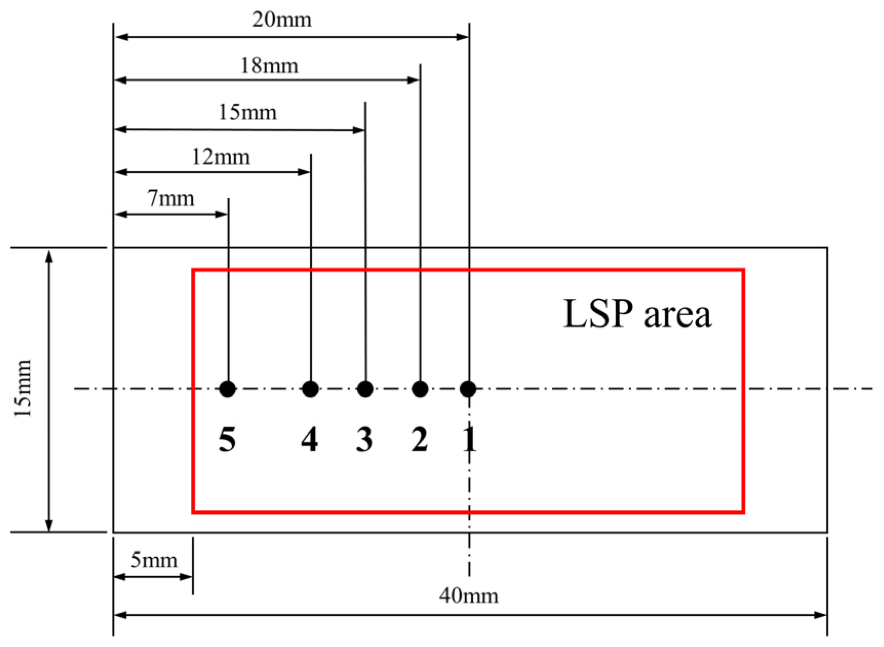
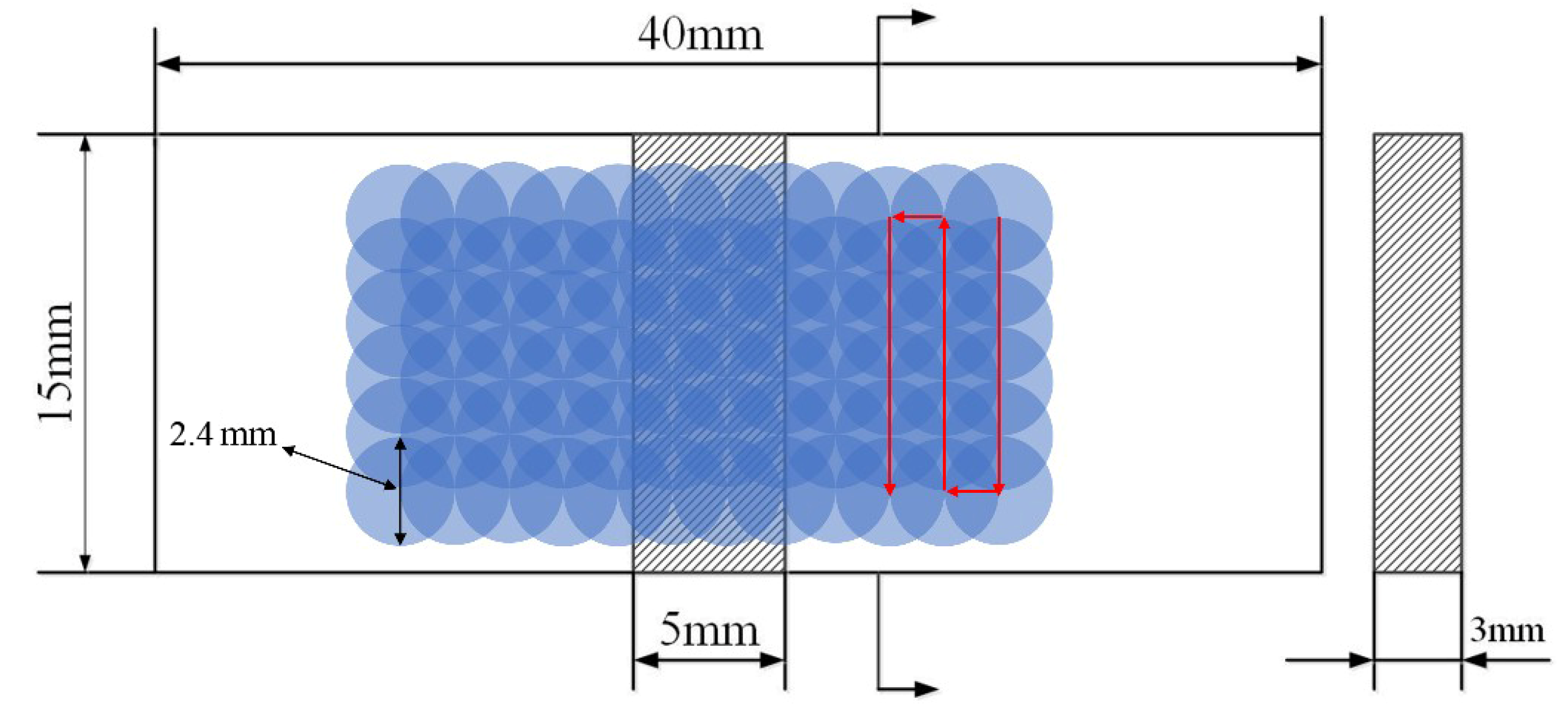
2.2. Laser Shock Peening Scheme

The surface of each welded specimen was grinded with silicon carbide paper using different roughness grades (from 400 to 2000), then cleaned in deionized water. An Nd YAG solid-state laser was then used for laser shock peening. The experimental parameters were designed as follows: wavelength of 1064 nm, pulse width of 20 ns, pulse energy of 5 J, power density of 5.52 GW/cm2, spot diameter of 2.4 mm, repetition frequency of 1 Hz, lap rate of 50%, and laser shock peening numbers of 1, 2, and 3. In the experiments, the constraining layer was water, and the absorbing layer was 0.1 mm thick aluminum foil, which was replaced after each peening. A schematic diagram of the laser shock area is shown in Figure 1. The shaded area in Figure 1 represents the weld, which is about 5 mm in width.

2.3. Performance Testing

The residual stress of the surface layer was measured by an X-350A residual stress tester produced by Aster. The experimental parameters were designed as follows: with the roll method, using roll angles of 0°, 24.2°, 35.3°, and 45°; the diffraction crystal plane was (220); Cr target Kα characteristic radiation was used; the scanning range was 121–138°; the stress constant was -601 MPa; the acceleration voltage of the X-ray tube was 26 kV; and the current of the X-ray tube was 6mA. In the experiments, the measurement error was ±20 MPa.
Next, we employed a Quanta200 field emission scanning electron microscope equipped with EBSD to observe the microstructure, grain size, and other microstructural properties of the weld’s structural section. We used an MFS-7000 X-ray diffractometer to analyze the welded area of the welded samples before and after laser shock peening. The radiation was Cu-Kα, the tube voltage was 40 kV, the tube current was 40 mA, the scanning angle ranged from 30° to 90°, the step size was 0.02°, and the counting time was 2 s. A TS-750C salt spray corrosion test chamber manufactured by Jiangsu Antewen was used to conduct the corrosion tests on the welded area after laser shock peening with the continuous spray method. The corrosive solution was a 22.2 NaCl prepared solution, and the temperature was (40 ± 1) °C. We used a PARSTAT-2273 electrochemical workstation produced by Ametek Company in the United States to test the electrochemical corrosion. A three-electrode system was used, in which the sample was the working electrode. The sample was exposed to the area of 1 cm2, and other parts were sealed with paraffin. During the potentiodynamic polarization curve tests, the corrosive medium was a 5% NaCl neutral aqueous solution, the temperature was (30 ± 1) °C, and the scanning rate was 1 mV∕s. It is noted that the potential scan rate has an important role in order to minimize the effects of distortion in Tafel slopes and corrosion current density analyses, as previously reported [17,18,19,20]. However, based on these reports, the adopted 1 mV/s has no deleterious effects on the Tafel extrapolation method in order to determine the corrosion current densities of the examined samples.

3. Results

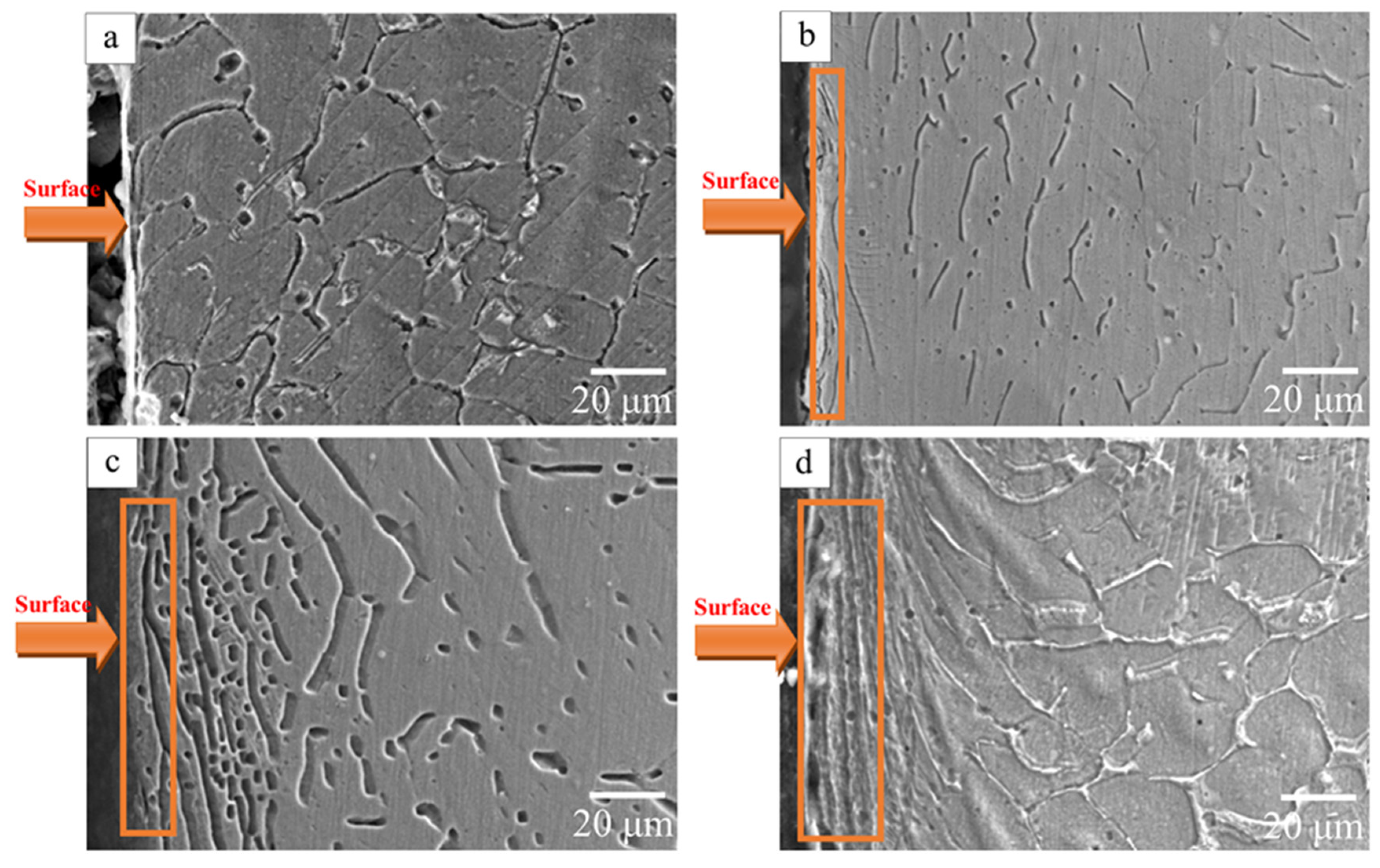
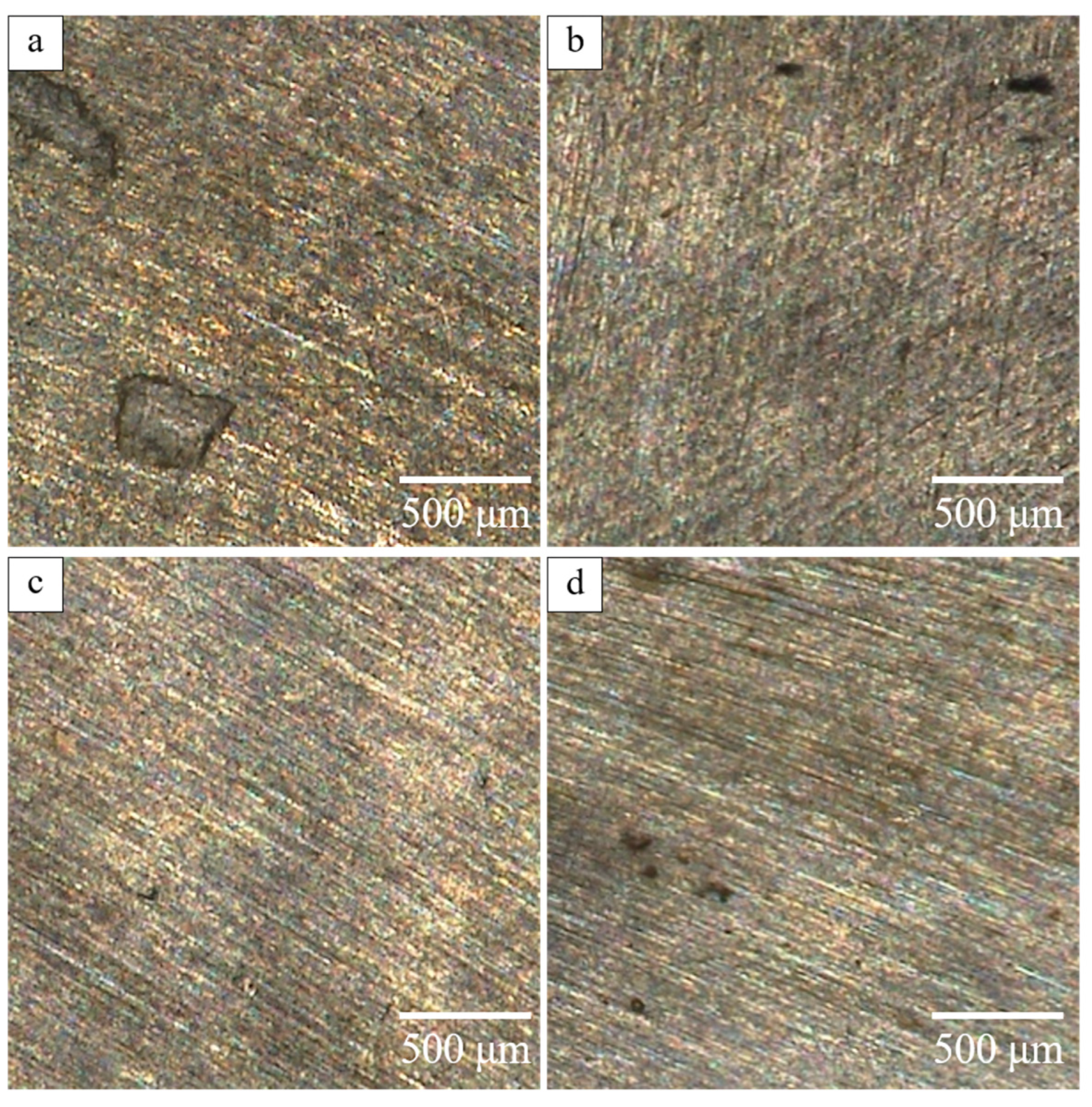
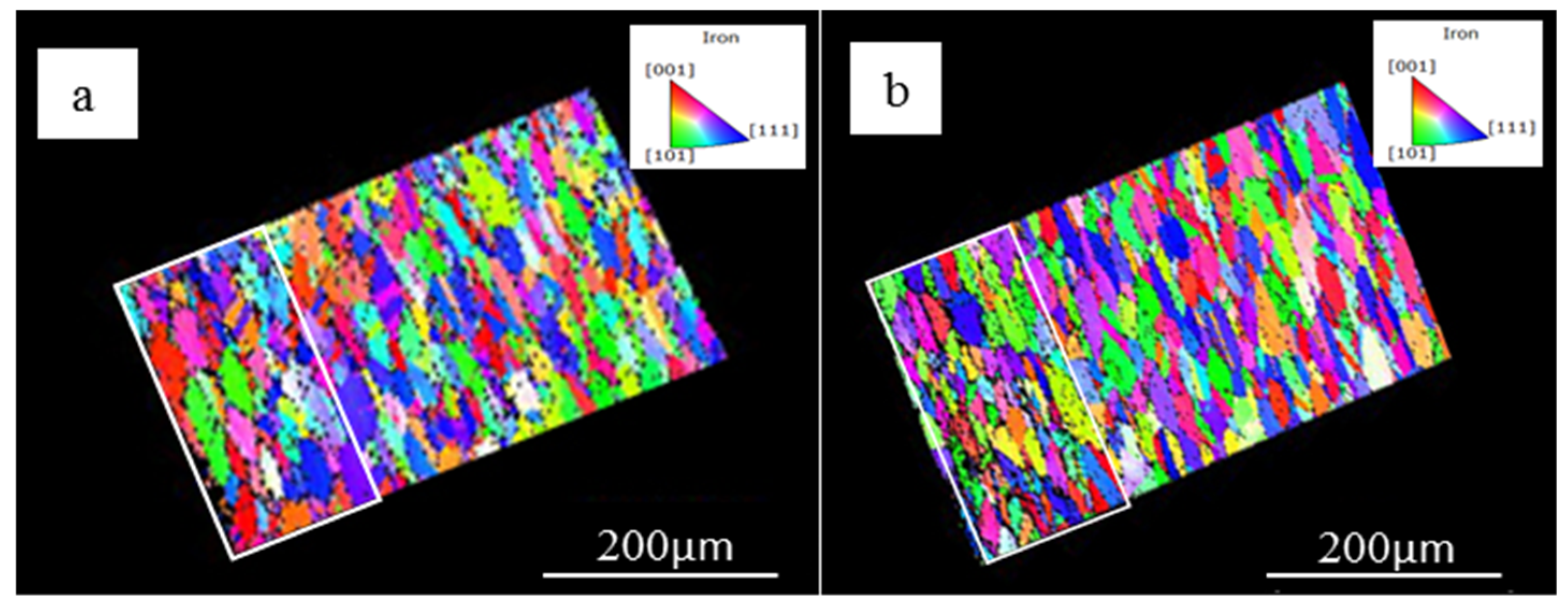
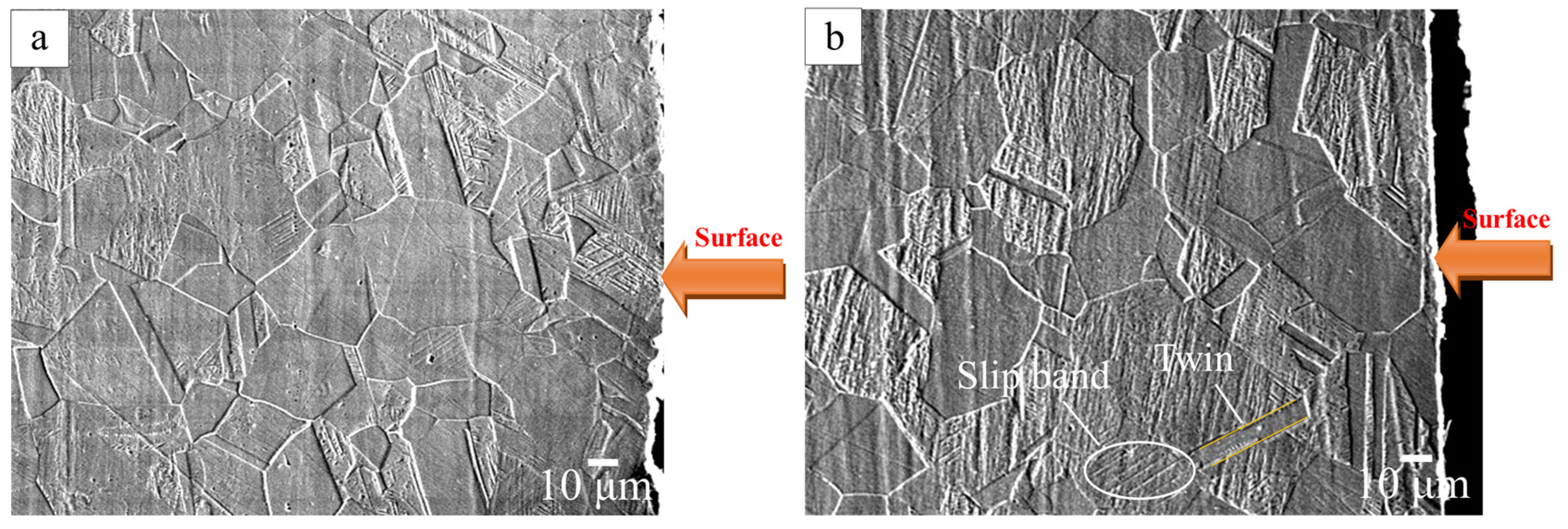
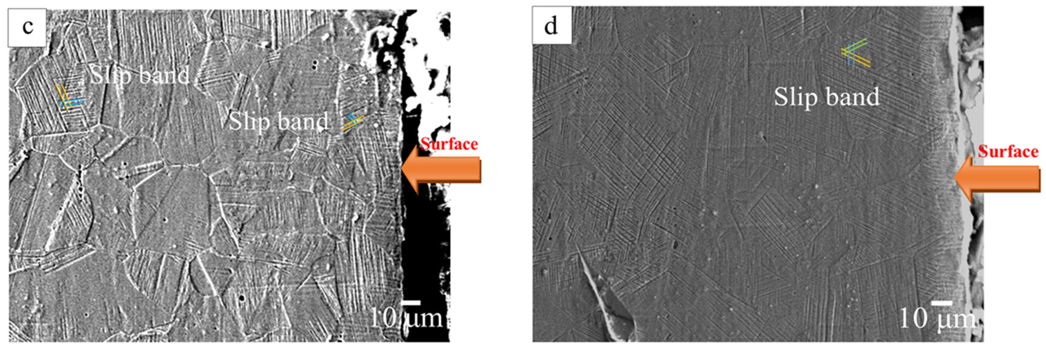
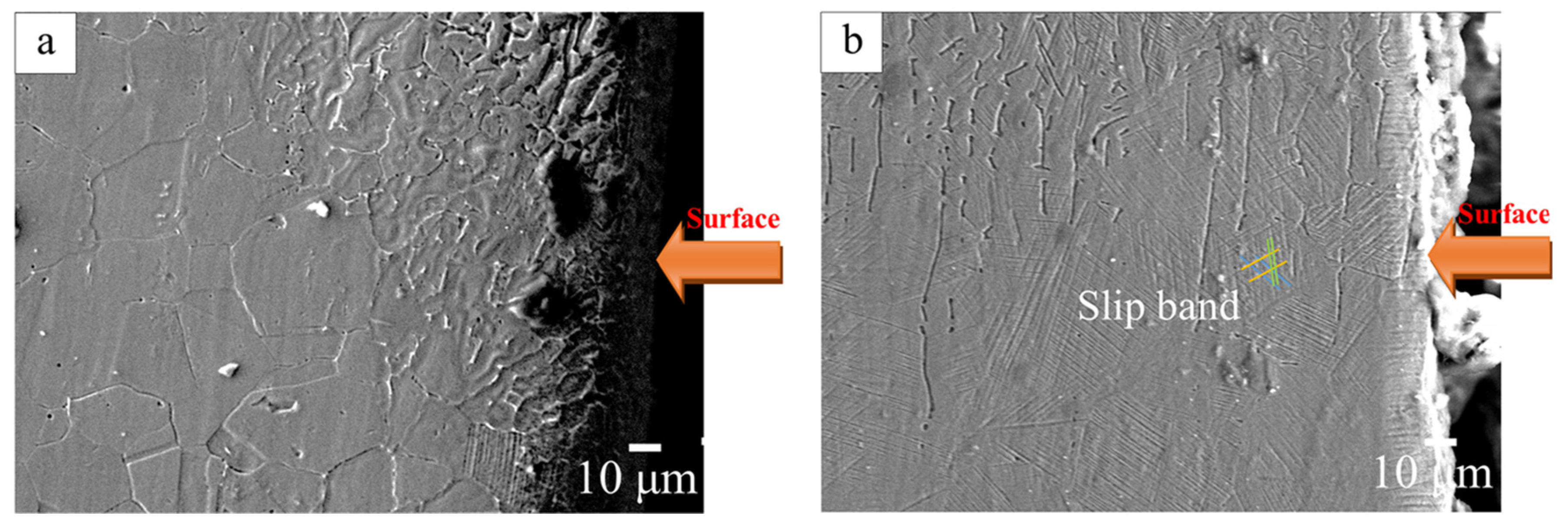
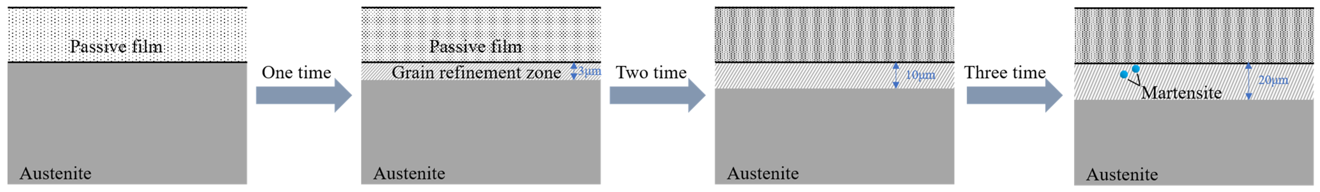
3.1. Effects of Different Laser Shock Treatments on the Microstructure of 316L

An EBSD diagram of the cross section of a 316L stainless steel sample before and after laser shock is shown in Figure 2. In Figure 2a, the matrix of the 316L stainless steel shows a typical austenite structure with clear grain boundaries. After one laser shock peening treatment (Figure 2b), the 316L stainless steel sample had a certain depth of grain refinement area along the depth direction.
A reverse pole diagram and grain boundary orientation angle diagram of a 316L stainless steel sample before and after laser shock are shown in Figure 3. The grains of the 316L sample’s steel section without laser shock peening are randomly distributed and relatively uniform. After one laser shock peening treatment, the orientation of the grains changed to be preferential in the [100] directions. The angle of the grain boundary also changed from a large angle to a wide distribution, and there a large number of small angle grain boundaries could be seen.
The microstructure of a section of the surface layer of the 316L stainless steel samples was observed by SEM, and the results are shown in Figure 4. The matrix of the 316L stainless steel sample was single-phase austenite (Figure 4a). When the number of laser shock treatments was one (Figure 4b), deformation twins and a slip band could be seen in one direction. When the peening number was two (Figure 4c), the number of slip bands in the grains increased, and the grains that had not formed slip bands started to form slip bands, but the local grain plastic deformation along the depth direction was small. When the peening number was three (Figure 4d), from the surface to the inside, and after three deformations coordinated in the same grain, plastic deformation of the slip bands in three directions appeared, while grains without any slip bands deflected to a soft orientation; meanwhile, slip and twinning formed in the grains, and the degree of plastic deformation further increased, all of which are conducive to the formation of residual compressive stress [21,22]. The plastic deformation of slip bands in three directions appeared in the same grain. When the peening number was lower, grains without slip bands deflected to a soft orientation, and slipping and twinning formed in the grain. The degree of plastic deformation further increased, which was conducive to the formation of residual compressive stress.
SEM images of a section of the surface microstructures of base metal zone of 316L stainless steel samples after different laser shock peening are shown in Figure 5. The welds of the 316L stainless steel samples show many larger grains (Figure 5a). When the number of laser shock peening treatments was one (Figure 5b), the grain along the depth direction compressed, the presence of grain boundaries was not obvious, and a severe plastic deformation zone of fibrous structures formed. With an increase in the instances of peening, the severe plastic deformation zone increased in size; when the number of peening treatments was three (Figure 5d), the severe plastic deformation zone reached about 20 μm. SEM images of the microstructure of cross sections of the heat-affected zone of the 316L stainless steel welded samples by different laser shock peening are shown in Figure 6. Compared to the un-peened sample (Figure 6a), when the number of laser shock peening treatments increased to three (Figure 6b), the grains deformed and coordinated, rotated, and reached a position where they could slip. The number of slip bands increased, and three directions of slip bands appeared in the same grain.
After laser shock peening, the plastic deformation of the material and the dislocation density conformed to the following relationship [23]; ε = Kρχb where ε is the amount of plastic deformation, K is the dislocation coefficient, ρ is the dislocation density, χ is the distance between dislocations, and b is the Parker vector.
It can be seen from the above formula that the plastic deformation of the material on the macroscopic level corresponds to the movement of dislocations inside the crystal on the microscopic level. As the dislocation density increases and the distance between dislocations decreases, the amount of plastic deformation of the material increases. High pressure shock waves induced by the laser cause severe plastic deformation of the materials, resulting in a large number of multiplied dislocation slips that are hindered by grain boundaries in the process of movement. [24] Therefore, the dislocation density of the surface layer greatly increases, and at the same time, the aggregation of high-density dislocations produces dislocation tangles. In the process of increasing the number of shocks, part of the energy is absorbed by the black tape, and the other part serves as the driving force for the annihilation and resetting of high-density dislocations inside the material. As a result, the dislocation entanglement transforms to form new grain boundaries, and the original coarse grains are divided by the new grain boundaries to form fine grains, which is called grain refinement [25,26,27,28].

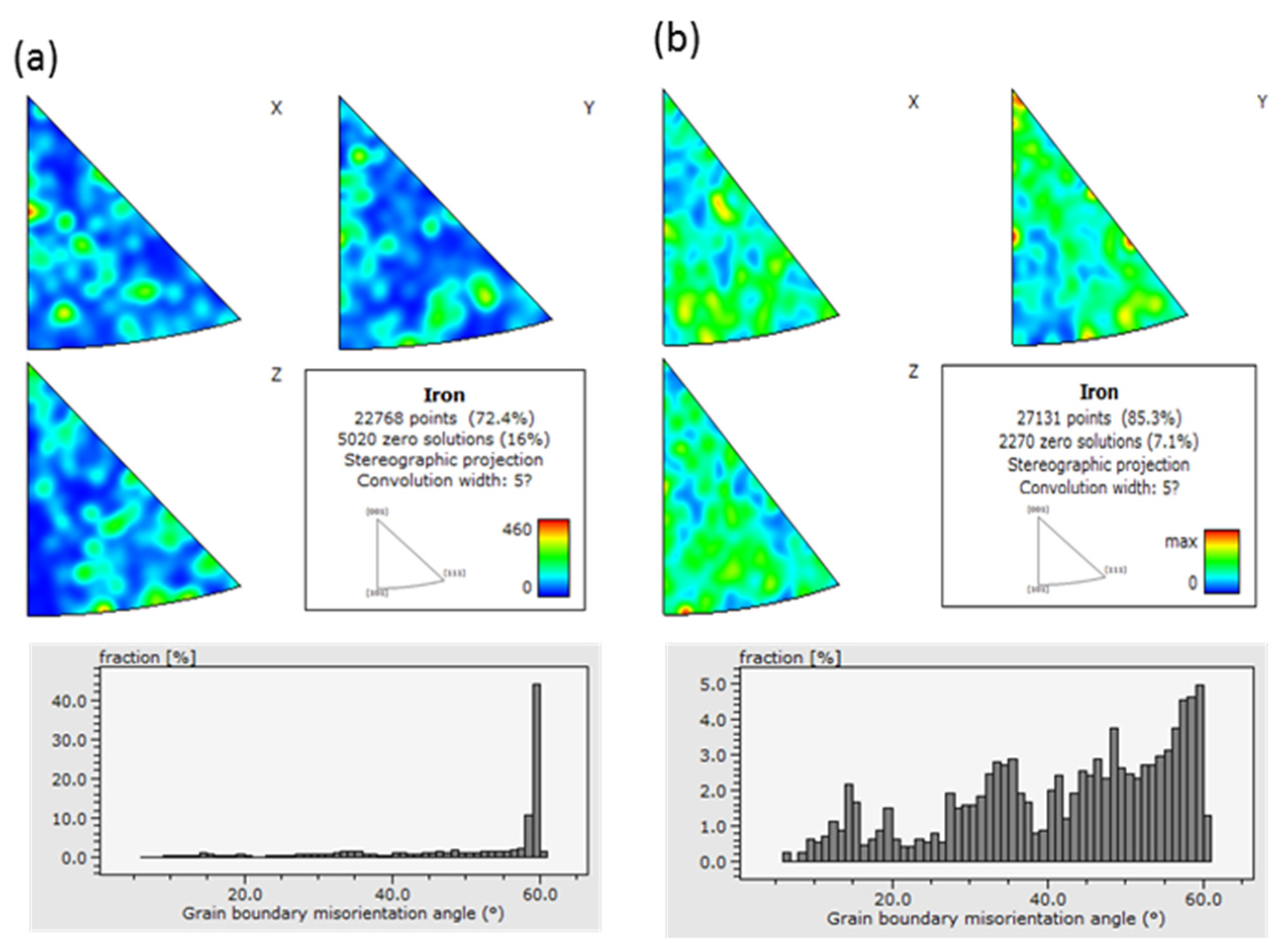
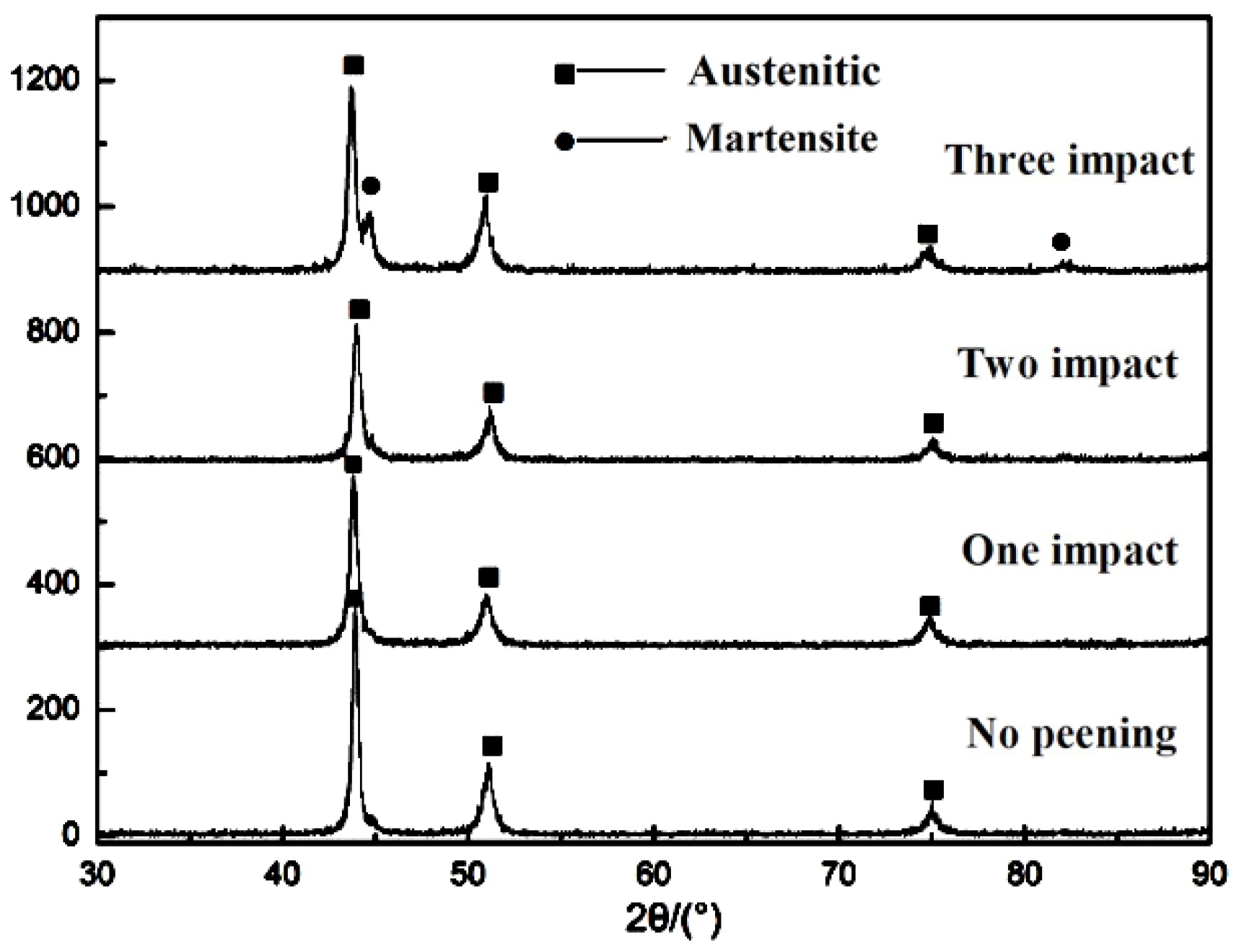
3.2. Phase Analysis of the Welded Area

XRD patterns of different samples of 316L stainless steel are shown in Figure 7. It can be seen that the 316L stainless steel was composed of single-phase austenite. Compared to the untreated sample, when the number of laser shocks was one and two, the corresponding spectral peak positions in the figure remained unchanged and the phase structure of the samples did not change. At the same time, the diffraction peaks corresponding to 316L stainless steel began to decrease and broaden, indicating that the surface grains of the welded area were refined; meanwhile, the peaks shifted to lower angles, indicating that the atomic spacing had changed, where the difference in atomic spacing produced different micro-stresses, which have been verified in subsequent residual stress measurements [29,30]. When the number of shocks was three, a new diffraction peak appeared, and transformation from austenite to martensite occurred.

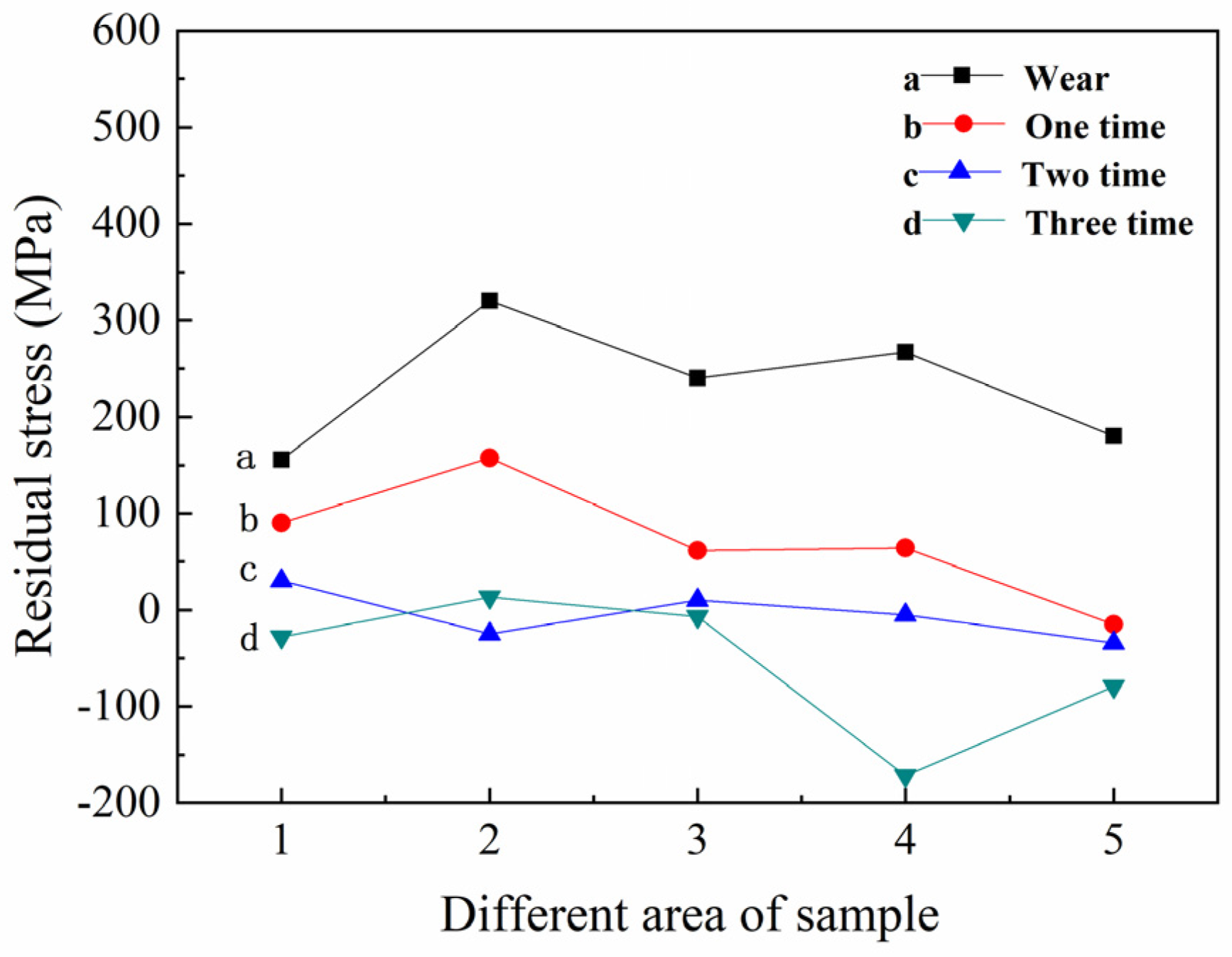
3.3. Effects of Laser Shock Peening on Welded 316L Stainless Steel Joints

Residual stress tests were carried out on different areas on the surface of the welded 316L stainless steel specimens treated with different laser parameters; the distribution of the residual stress test points is shown in Figure 8.
The residual stress distribution of the 316L stainless steel samples along the vertical weld seam under different peening times is shown in Figure 9. The residual tensile stress on the surface of the welded specimen was high when it was not treated, especially in the area at the junction of the weld zone and the heat-affected zone, and the maximum residual tensile stress was 330 MPa. After one laser shock treatment, the residual tensile stress in the weld zone and heat-affected zone decreased. When two laser shock treatments were applied, the residual stress value of the weld’s heat-affected zone, and that of the base metal, was close to 0 MPa; the distribution was uniform, which reduced the stress concentration and the local maximum stress gradient, and significantly improved the residual stress state of the welded joint. With an increase to three shock treatments, a residual compressive stress of tens of megapascals was formed in the weld’s heat-affected zone and matrix area, and the residual compressive stress near the matrix area was the highest, reaching 171 MPa.

3.4. Effects of Laser Shock on the Salt Spray Corrosion of Welded 316L Stainless Steel Joints

The morphologies of the welding zone of 316L stainless steel samples after salt spray corrosion with different laser shock peening are shown in Figure 10. After salt spray corrosion, the untreated sample (Figure 10a) had a large corrosion area where the corrosion pits were dense. After one laser shock treatment (Figure 10b), local pitting corrosion appeared on the surface of the sample, but the sizes of the corrosion pits were significantly smaller than those of the untreated sample. After two laser shock treatments (Figure 10c), the pitting phenomenon on the surface of the sample was not obvious, and there were few corrosion pits. When three laser shock treatments were applied (Figure 10d), the number of local corrosion pits increased compared to the number for one shock, but their sizes were much smaller than those of the corrosion area without peening. It was seen that, after laser shock peening, the number and size of the corrosion pits on the surface was reduced, and their depths were shallower. The anti-salt spray corrosion effect was best when two peening treatments were applied, indicating that laser shock peening was able to improve the pitting resistance of the material.

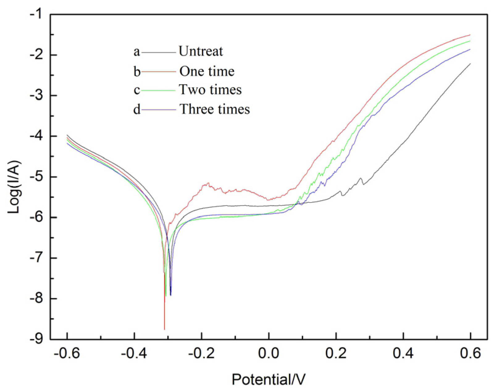
3.5. Effects of Laser Shock on the Electrochemical Corrosion of Welded 316L Stainless Steel Joints

Electrochemical spectra of the surfaces of the 316L stainless steel samples treated with different laser parameters are shown in Figure 11. The corrosion current of the untreated sample was 1.49 × 10⁻⁶ A/cm2, with a measurement error within 0.4%. After one peening, the corrosion current decreased to 1.089 × 10⁻⁶ A/cm2, which was about 28.8% lower than that of the untreated sample. When the corrosion conditions remained unchanged, as the number of laser shock treatments increased, the corrosion current showed a trend of first decreasing and then increasing, but it was lower than that of the untreated sample. When the number of laser shocks was two, the corrosion current reached a minimum value of 9.684 × 10⁻⁷ A/cm2. Since the corrosion current represents the dissolution rate of the metal, the polarization curves also showed that the laser-shock-peening samples had lower corrosion rates and better pitting resistance than the untreated sample. When the number of laser shocks was three, the corrosion current increased compared to when the number of shocks was two, and the pitting resistance decreased.

4. Discussion

To further explain the effects of laser shock peening on the electrochemical properties of the welded area of the stainless-steel samples, Figure 12 presents a schematic diagram of the changes in the welded area after different laser shock treatments.
As mentioned previously, the welded 316L stainless steel matrix was austenite, and the surface contained a double-layer passivation film with an inner layer rich in Cr and an outer layer rich in Fe [31,32,33,34]. After one laser shock treatment, the residual tensile stress on the surface of the sample was reduced, a layer consisting of a grain refinement area appeared near the surface, and some defects and dislocations appeared, which improved the diffusion of Cr elements and surface aggregation [20]. Therefore, the surface passivation film became uniform and compact, and the pitting resistance improved. After two laser shock treatments, the residual tensile stress was almost eliminated, the stress distribution was more uniform, and the depth of the grain refinement zone near the surface increased. At the same time, the dislocation density and the number of defects increased, which further enhanced the diffusion and surface aggregation of Cr elements; meanwhile, the surface passivation film became more compact, and its resistance to pitting corrosion improved compared to after a single-peening. When the number of shocks reached three, residual compressive stress was generated on the surface of the sample. Compared to two-peening treatments, the stress distribution gradient increased, and the depth of the grain refinement zone near the surface further increased. At this time, some of the sample underwent austenite to martensite transformation, which was induced by the action of laser shock peening. Since there is a certain potential difference between austenite and martensite, martensite is prone to pitting corrosion as an anode. However, due to the introduction of residual compressive stress and the relatively thick depth of the grain refinement zone near the surface, it played a certain defensive role against corrosive ions such as Cl-; therefore, the pitting corrosion resistance was lower than that of the samples that underwent two shock treatments, but it was higher that of the untreated sample.

5. Conclusions

In this paper, 316L stainless steel welded joints were treated with multiple laser shock peening treatments. By analyzing the microstructure, phase change, residual stress, and corrosion resistance of the materials before and after the shocks, the influencing mechanisms and effects of multiple laser shocks on the electrochemical corrosion performance of 316L stainless steel welded joints were finally revealed. The results of this study are summarized in the following:
(1) Laser shock peening induced a severe plastic deformation zone on the surfaces of the welded samples. As the number of laser shock treatments increased, the severe plastic deformation zone enlarged, which assisted the formation of residual compressive stress.
(2) Compared to the untreated samples, the pitting corrosion resistance of the samples after laser shock peening improved. When the number of laser shock treatments was two, the corrosion current reached a minimum value of 9.684 × 10⁻⁷ A/cm2, about 37% less than the untreated sample, and the pitting resistance was the best. However, when the number of peening treatments was further increased to three, the corrosion current increased and the pitting resistance decreased.
In summary, after the two laser shock treatments, the effect of the residual stress on electrochemical corrosion was the dominant factor. However, when the number of laser shocks treatments was three, due to the formation of new phases, the microstructure played a dominant role in electrochemical corrosion; thus, pitting resistance decreased.

Author Contributions

Conceptualization, Y.L. and J.F.; methodology, Y.L.; validation, J.F. and J.W.; formal analysis, Y.L. and J.F.; investigation, Y.L. and J.F.; resources, Y.L., X.N. and L.Z.; data curation, J.F.; writing—original draft preparation, Y.L. and J.F.; writing—review and editing, X.N. and L.Z.; visualization, J.F. and J.W.; supervision, Y.L.; project administration, Y.L. All authors have read and agreed to the published version of the manuscript.

Funding

This research was funded by “Basic Research Program, grant number JCKY2019802B001” and “Youth Science Fund Project, grant number 51305456”.

Institutional Review Board Statement

Not applicable.

Informed Consent Statement

Not applicable.

Data Availability Statement

The data used to support the findings of this study are available from the corresponding author upon request.

Acknowledgments

The authors gratefully acknowledge financial support from the Fundamental Research Project of Aero-Engine and Gas Turbine, No. J2019-IV-0014-0082.

Conflicts of Interest

The authors declare no conflict of interest.

References

  1. Liu, J.-Z. Nuclear Structural Material; Chemical Industry Press: Beijing, China, 2007; pp. 202–203. [Google Scholar]
  2. Sánchez-Tovar, R.; Montaes, M.T.; García-Antón, J. Effect of the micro-plasma arc welding technique on the microstructure and pitting corrosion of AISI 316L stainless steels in heavy LiBr brines. Corros. Sci. 2011, 53, 2598–2610. [Google Scholar] [CrossRef]
  3. Lv, H.-W.; Dong, S.-G.; Wang, J.-J.; Li, N.; Lin, C.-J. Electrochemical Behaviors of the 316L Stainless Steel Welding Seam Corrosion. Sci. Technol. Rev. 2013, 31, 25–28. [Google Scholar]
  4. Lian, M.-Y. Study on Microstructure and Properties of 40CrNiMo Quenched and Tempered Steel CMT Surfacing HAZ for Gear; Zhengzhou University: Zhengzhou, China, 2020. [Google Scholar]
  5. Gao, H.-F. Seismic Performance of Fabricated Steel Lattice Columns with Lateral Force Resistance; Beijing University Of Civil Engineering and Architecture: Beijing, China, 2021. [Google Scholar]
  6. Wang, J.-H.; Lu, H. Some discussions on principle of causing and relieving welding residual stress. J. Weld. 2002, 23, 75–79. [Google Scholar]
  7. Zhao, R. Study of Welding Residual Stress’s Numerical Simulation and Reliving; Dalian University of Technology: Dalian, China, 2006. [Google Scholar]
  8. Luo, G.-L.; Zhang, L.-F.; Xiong, Y.; Zhang, B.-F.; Chen, X.-P. Effect of Laser Shock Peening on Microstructure and Properties of Ti-6Al-3Nb-2Zr-1Mo Titanium Alloy. Chin. J. Lasers 2022, 49, 215–226. [Google Scholar]
  9. Xu, S.; Su, B.-Y.; Hua, G.-R.; Wang, H.; Cao, Y.-P. Effect of Laser Shock Peening on the Interfacial Bonding Properties of TiN Coatings on TC4 Titanium Alloy. Surf. Technol. 2022, 51, 315–325. [Google Scholar]
  10. He, Z.-R.; Shen, Y.-Z.; Zhou, J.; Liu, W.-L.; Sun, R.-J. Microstructure evolution and performance enhancement of laser shock peening. Aeronaut. Manuf. Technol. 2021, 64, 48–58. [Google Scholar]
  11. Nie, X.-F.; Li, Y.-H.; He, W.-F.; Luo, S.-H.; Zhou, L.-C. Research Progress and Prospect of Laser Shock Peening Technology in Aero-engine Components. J. Mech. Eng. 2021, 57, 293–305. [Google Scholar]
  12. Jiao, Q.-Y.; Han, P.-P.; Lu, Y.; Zhao, D.; Qiao, H.-C.; Zhao, J.-B. Effect of laser shock peening on residual stress and mechanical properties of TA15 titanium alloy. J. Plast. Eng. 2021, 28, 146–152. [Google Scholar]
  13. Karthik, D.; Jiang, J.-C.; Hu, Y.-X.; Yao, Z. Effect of multiple laser shock peening on microstructure, crystallographic texture and pitting corrosion of Aluminum-Lithium alloy 2060-T8. Surf. Coat. Technol. 2021, 421, 127354. [Google Scholar] [CrossRef]
  14. Geng, Y.-X.; Xia, D.; Wang, K.-D.; Yan, X.; Duan, W.; Fan, Z.; Wang, W.; Mei, X. Effect of microstructure evolution and phase precipitations on hot corrosion behavior of IN718 alloy subjected to multiple laser shock peening. Surf. Coat. Technol. 2019, 370, 244–254. [Google Scholar] [CrossRef]
  15. Zhou, L.-C.; Li, Y.-H.; He, W.-F.; Chen, D. Effect of multiple laser shock processing on microstructure and mechanical properties of Ti-5Al-4Mo-4Cr- 2Sn-2Zr titanium alloy (English). Rare Metal. Mat. Eng. 2014, 43, 1067–1072. [Google Scholar]
  16. Wang, J.; Lu, Y.-L.; Zhou, D.-S.; Sun, L.; Xie, L.; Wang, J. Mechanical properties and microstructural response of 2A14 aluminum alloy subjected to multiple laser shock peening impacts. Vacuum 2019, 165, 193–198. [Google Scholar] [CrossRef]
  17. Duarte, T.; Meyer YAOsório, W.R. The Holes of Zn Phosphate and Hot Dip Galvanizing on Electrochemical Behaviors of Multicoatings on Steel Substrates. Metals 2022, 12, 863. [Google Scholar] [CrossRef]
  18. Osório, W.R.; Freitas, E.S.; Garcia, A. EIS and potentiodynamic polarization studies on immiscible monotectic Al–In alloys. Electrochim. Acta 2013, 102, 436–445. [Google Scholar] [CrossRef]
  19. Zhang, X.L.; Jiang, Z.H.; Yao, Z.P.; Song, Y.; Wu, Z.D. Effects of scan rate on the potentiodynamic polarization curve obtained to determine the Tafel slopes and corrosion current density. Corros. Sci. 2009, 51, 581–587. [Google Scholar] [CrossRef]
  20. McCafferty, E. Validation of corrosion rates measured by Tafel extrapolation method. Corros. Sci. 2005, 47, 3202–3215. [Google Scholar] [CrossRef]
  21. Li, Y.; Guan, L.; Wang, G.; Zhang, B.; Ke, W. Influence of mechanical stresses on pitting corrosion of stainless steel. J. Chin. Soc. Corros. Prot. 2019, 39, 215–226. [Google Scholar]
  22. Wang, Y.-J.; Wang, X.; Sha, A.-X.; Li, X. Simulation of residual stress and fatigue test in hole extrusion process for TiAlNb alloy. J. Aeronaut. Mater. 2021, 41, 66–74. [Google Scholar]
  23. Zhang, M.-Y.; Zhu, Y.; Guo, W.; Huang, S.; Hou, G. Effects of laser shock processing on fatigue properties of TC17 titanium alloy. Laser Technol. 2017, 41, 231–234. [Google Scholar]
  24. Lu, J.Z.; Luo, K.Y.; Zhang, Y.K.; Cui, C.Y.; Sun, G.F.; Zhou, J.Z.; Zhang, L.; You, J.; Chen, K.M.; Zhong, J.W. Grain refinement of LY2 aluminum alloy induced by ultra-high plastic strain during multiple laser shock processing impacts. Acta Mater. 2010, 58, 3984–3994. [Google Scholar] [CrossRef]
  25. Zhang, W.; Lu, J.; Luo, K. Residual Stress Distribution and Microstructure at a Laser Spot of AISI 304 Stainless Steel Subjected to Different Laser Shock Peening Impacts. Metals 2016, 6, 6. [Google Scholar] [CrossRef] [Green Version]
  26. Lan, L.; Xin, R.; Jin, X.; Gao, S.; He, B.; Rong, Y.; Min, N. Effects of Laser Shock Peening on Microstructure and Properties of Ti–6Al–4V Titanium Alloy Fabricated via Selective Laser Melting. Materials 2020, 13, 3261. [Google Scholar] [CrossRef]
  27. Lu, Y.; Yang, Y.; Zhao, J.; Yang, Y.; Qiao, H.; Hu, X.; Wu, J.; Sun, B. Impact on Mechanical Properties and Microstructural Response of Nickel-Based Superalloy GH4169 Subjected to Warm Laser Shock Peening. Materials 2020, 13, 5172. [Google Scholar] [CrossRef]
  28. Sun, R.; He, G.; Bai, H.; Yan, J.; Guo, W. Laser Shock Peening of Ti6Al4V Alloy with Combined Nanosecond and Femtosecond Laser Pulses. Metals 2022, 12, 26. [Google Scholar] [CrossRef]
  29. Li, Y.-Q.; Meng, C.-J.; Wang, X.-D.; Luo, S.; Xu, W. Corrosion resistance property of 316L stainless steel welding joints treated by laser shock peening. Laser Optoelectron. Prog. 2017, 54, 165–169. [Google Scholar]
  30. Jiao, Y.; He, W.-F.; Luo, S.-H.; Zhou, L.-C.; Li, Y.-H. Study of micro-scale laser shock processing without coating improving the high cycle fatigue performance of K24 simulated blades. Chin. J. Lasers 2015, 42, 73–79. [Google Scholar]
  31. Liu, B.-J. Color Image Grading Evaluation of 316L Stainless Steel Passivation Film Damage and Its Application; Dalian University of Technology: Dalian, China, 2021. [Google Scholar]
  32. Guo, J.-G.; Chen, H.; Li, J.-R.; Tang, Z.-L.; Chen, Q.-M. Study on Corrosion Resistance Characteristics of 316L Stainless Steel Bipolar Plate Element Enrichment. Mater. Rep. 2021, 35, 391–394. [Google Scholar]
  33. Zhang, Y.; Wu, C.-H.; Fu, M.-J.; Li, Q.; Qin, Z.-W. Research Progress of Electrodeposited Corrosion Resistant Coatings on Surface of Stainless Steel. Mater. Prot. 2022, 55, 126–135. [Google Scholar]
  34. Wang, Z.; Feng, Z.; Zhang, L.; Lu, M.-X. Current application and development trend in electrochemical measurement methods for the corrosion study of stainless steels. Chin. J. Eng. 2020, 42, 549–556. [Google Scholar]
Figure 1. Schematic map of laser shock peening.
Metals 12 01215 g001
Figure 2. EBSD diagram of the cross section of a 316L stainless steel sample before and after laser shock treatment: (a) untreated and (b) after one laser shock peening treatment.
Metals 12 01215 g002
Figure 3. Reverse pole diagram and grain boundary orientation angle diagram of a 316L stainless steel sample before and after laser shock treatment: (a) untreated and (b) after one laser shock treatment.
Metals 12 01215 g003
Figure 4. SEM of a section of the surface microstructures of the base metal zone of the 316L stainless steel samples after different laser shock peening: (a) substrate, (b) peening once, (c) peening twice, and (d) peening three times.
Metals 12 01215 g004aMetals 12 01215 g004b
Figure 5. SEM images of the microstructure of cross sections of the welding zone of the 316L stainless steel samples after different laser shock peening: (a) matrix, (b) peening once, (c) peening twice, and (d) peening three times.
Metals 12 01215 g005
Figure 6. SEM images of the microstructure of cross sections of the heat-affected zone of the 316L stainless steel welded samples after different laser shock peening: (a) matrix and (b) peening three times.
Metals 12 01215 g006
Figure 7. XRD patterns of different samples of 316L stainless steel.
Metals 12 01215 g007
Figure 8. Distribution of residual stress test points.
Metals 12 01215 g008
Figure 9. Residual stress distribution of the 316L stainless steel samples along the vertical weld seam after different numbers-of peening.
Metals 12 01215 g009
Figure 10. The morphologies of the welding zone of 316L stainless steel samples after salt spray corrosion with different numbers of laser shock peening: (a) substrate, (b) peening once, (c) peening twice, and (d) peening three times.
Metals 12 01215 g010
Figure 11. Polarization curves of the welded 316L stainless steel samples treated with different numbers of laser shock treatment.
Metals 12 01215 g011
Figure 12. Schematic diagram of laser shock treatment applied to the welded area of the 316L stainless steel samples.
Metals 12 01215 g012
Table 1. Chemical composition of the 316L stainless steel used in this study.
ElementCCrNiMoMnSiPSFe
Content0.02017.00010.3002.4101.5100.5100.0300.003bal
Publisher’s Note: MDPI stays neutral with regard to jurisdictional claims in published maps and institutional affiliations.

