Article

Bio-Oxidation of a Double Refractory Gold Ore and Investigation of Preg-Robbing of Gold from Thiourea Solution

School of Minerals Processing and Bioengineering, Central South University, Changsha 410083, China
*
Author to whom correspondence should be addressed.
Metals 2020, 10(9), 1216; https://doi.org/10.3390/met10091216
Submission received: 13 August 2020 / Revised: 1 September 2020 / Accepted: 4 September 2020 / Published: 10 September 2020
(This article belongs to the Section Extractive Metallurgy)

Abstract

:
Carbonaceous sulfidic gold ores are commonly double refractory and thus require pretreatment before gold extraction. In this paper, the capacity of pre-bio-oxidation can simultaneously decompose sulfides or deactivate carbonaceous matters (CM) from a double refractory gold ore (DRGO) using pure cultures of A. ferrooxidans or L. ferrooxidans, and a mixed culture containing A. ferrooxidans and L. ferrooxidans was investigated. The results showed that direct thiourea leaching of the as-received DRGO yielded only 28.7% gold extraction, which was due to the encapsulation of sulfides on gold and the gold adsorption of CM. After bio-oxidation, thiourea leaching of the DRGO resulted in gold extraction of over 75–80%. Moreover, bio-oxidation can effectively reduce the adsorption of carbon to gold. XRD, SEM-EDS and FTIR analysis showed that many oxygen-containing groups were introduced on the surface of DRGO during bio-oxidation, while the C=C bond was cleaved and the O–C–O and C–N bonds were degraded, causing a decrease in active sites for gold adsorption. Moreover, passivation materials such as jarosite were formed on the surface of DRGO, which might reduce the affinity of CM for gold in solutions. In addition, the cleavage of the S–S band indicated that sulfides were oxidized by bacteria. This work allows us to explain the applicability of pre-bio-oxidation for degrading both sulfides and CM and increasing gold recovery from DRGO in the thiourea system.

1. Introduction

Environmentally friendly gold extraction from refractory ores has gradually dominated gold production, with a decrease in the quality of gold ores [1]. Currently, refractory gold ores account for around one-third of the total gold production of natural ores, and their refractory behavior is mainly due to the fact that the fine gold particles are often encased in sulfide matrices such as pyrite and arsenopyrite, or the leached gold are preg-robbed by carbonaceous matters (CM) [2,3,4]. In some cases, the gold ore contains both sulfide minerals and CM, and it can be classified as a double refractory gold ore (DRGO), which is usually abandoned for economic reasons [5]. In such ores, the gold is encased in sulfides, which prevents direct contact between the leaching reagent and gold particles, thus interfering with the dissolution of the gold [6]. Moreover, CM has a good affinity for dissolved gold complex (preg-robbing), leading to gold losses [7,8]. Commercially, pretreatment of the DRGO is required in order to degrade sulfides and deactivate CM prior to leaching to improve gold extraction [9].
Bio-oxidation has been proven to be a viable pretreatment process for DRGO, which can reduce the consumption of chemical solvents for gold leaching in subsequent operations. It presents several attractive features such as environmental friendliness and low cost [10,11,12]. The commonly used large-scale leaching bacteria now mainly include Acidithiobacillus ferrooxidans (A. ferrooxidans), Leptospirillum ferrooxidans (L. ferrooxidans) and Acidithiobacillus thiooxidans [13]. These autotrophic bacteria are reliant on oxidizing ferrous (Fe2+) to ferric (Fe3+) ion or oxidizing sulfur compounds or elemental S to H2SO4 as their energy source [2,14]. This enables bacteria to oxidize and decompose the sulfide matrices, releasing the entrapped gold. In addition, there are two main types of views on the interaction between microorganisms and CM, reflecting the passivation and degradation of CM by microorganisms and their metabolites. It is reported that Phanerochaete chrysosporium (P. chrysosporium) can secrete manganese peroxidase, lignin peroxidase and oxidative enzymes during metabolism, which are capable of degrading aromatic CM [15,16,17]. Ofori-Sarpong et al. [18,19] used P. chrysosporium to deactivate different grades of CM and found that biotransformation of CM reduced the graphitization of carbon and increased the oxygen content and average pore diameter, which limited the adsorption of gold by CM. Amankwah et al. [20] also pointed out in a two-stage microbial process that Streptomyces setonii has the ability to degrade CM, and gold extraction increased by 13.6% after degradation of CM. This may be due to the production of alkaline metabolites which was responsible for CM degradation. Though there have been numerous studies on biological pretreatment of DRGO, the results are rather controversial, and there is no general agreement about whether A. ferrooxidans and L. ferrooxidans can simultaneously decompose sulfides and deactivate CM. Moreover, according to previous studies, the mixed bacteria could result in synergistic effects that can improve the bio-oxidation efficiency [21,22,23]. Up to now, there has not been a clear correlation between the mixture of these two dominant strains and the efficiency of the bio-oxidation process. Thus, it is interesting to compare the behavior of A. ferrooxidans, L. ferrooxidans and a mixed culture composed of A. ferrooxidans and L. ferrooxidans in a bio-oxidation of DRGO system.
Cyanidation is generally used for leaching gold ores because of its high stability, low cost and simple process [24,25]. However, its poor selectivity, slow leaching kinetics and harmful environmental effects have called for an alternative lixiviant. Thiourea has been considered to be a promising alternative to cyanide due to its high leaching rates and good selectivity [26], and it can be used to leach refractory gold ores containing sulfides (such as pyrite and arsenopyrite) or carbonaceous sulfides [27].
The initial leaching rate of gold at high concentration thiourea is comparable to that of cyanide leaching [28]. Thiourea is a water-soluble organic compound, which is relatively stable in acidic solutions but decomposes rapidly in basic solutions. Therefore, thiourea leaching of gold is normally carried out in a pH range of 1–2 [29]. In acidic solutions, thiourea dissolves gold and forms a cationic complex; this reaction is rapid and gold extractions can reach 99% [30]. Previous research has revealed that gold particles only dissolve in thiourea solutions in the presence of oxidizing agents, and ferric sulfate has been shown to be the most effective [31]. The dissolution of gold in thiourea solutions can be expressed as the following reaction.
Au + 2 CS ( N H 2 ) 2 + Fe 3 + = Au ( CS ( NH 2 ) 2 ) 2 + + Fe 2 +
Unlike the cyanide system, no adequate explanation for gold adsorption by CM in the thiourea system has been made in previous studies. The available literature indicates that the ability of CM to adsorb gold in cyanide, thiourea and thiosulfate systems is in the order of Au(CN)2 > Au(CS(NH2)2)2+ > Au(S2O3)23 [32]. This conclusion has been proven by Ofori-Sarpong and Osseo-Asare [19]. It has also been reported that CM can adsorb not only gold but also thiourea, and the adsorbed gold is difficult to elute [33]. Considering the potential of thiourea to be used as an alternative to cyanide, it is necessary to evaluate the behavior of preg-robbing of CM in the thiourea system.
The objective of this study was to investigate the ability of decomposing sulfides and decreasing gold adsorption on CM by using pure cultures of A. ferrooxidans or L. ferrooxidans and a mixed culture composed of A. ferrooxidans and L. ferrooxidans in a thiourea system, so as to assess the potential of A. ferrooxidans and L. ferrooxidans for the pretreatment of DRGO. This work can help to expand the current understanding of the biological pretreatment of DRGO by A. ferrooxidans and L. ferrooxidans, especially the impact of the pretreatment on the preg-robbing of CM for gold in the thiourea system.

2. Experimental

2.1. Materials

The DRGO was obtained from a gold mine in Yunnan Province, China. The ore was finely ground to a particle size of 90.0% < 0.074 mm. The chemical component analysis showed that the gold content in this ore was 18.05 g/t, and the ore had high contents of C (6.95%) and S (20.76%) (Table 1). The elemental and organic carbon that has a preg-robbing effect on gold accounted for 75.40% of the total carbon [8]. Mineralogical studies of Au showed that only 1.72% of gold was exposed and 87.31% was encapsulated in arsenopyrite and pyrite.

2.2. Microorganisms

A. ferrooxidans and L. ferrooxidans were obtained from the Key Lab of Biohydrometallurgy of Ministry of Education, Central South University, Changsha, China. Bacterial cells were cultivated in 100 mL of 9 K medium in 250 mL Erlenmeyer flasks were located in a shaking incubator at 30 °C and 160 rpm. The initial solution pH was adjusted to 1.8 using 3 M sulfuric acid. The 9 K medium contains 3 g (NH4)2SO4, 0.1 g KCl, 0.5 g MgSO4·7H2O, 0.5 g K2HPO4, 0.01 g Ca(NO3)2 in 1000 mL deionized water. In addition, 44.7 g/L of FeSO4·7H2O was supplemented as the energy source for strains. For domestication, the microorganisms were serially cultured in 9 K medium in the presence of 10 g/L DRGO particles for three months prior to bio-oxidation experiments.
All chemical reagents used in this study were analytically graded, and all solutions were prepared with ultrapure water.

2.3. Bio-Oxidation Experiments

The bio-oxidation experiments were conducted in 250 mL flasks containing 100 mL 9 K medium, and the pulp density was 10 g/L of DRGO. Inoculations were separately achieved with 20 mL inoculum of A. ferrooxidans and L. ferrooxidans which had adapted to the DRGO (Section 2.2), and the cell concentration in the two inoculated solutions was around 108 cells/mL. The flasks were located in a shaking incubator at 160 rpm and 30° for 8 days, with an initial pH of 1.8. In this study, A. ferrooxidans and L. ferrooxidans were mixed in 1:1 (v/v) culture to obtain a mixed culture. The experimental conditions of bio-oxidation using the mixed culture were the same as those using pure cultures. Parallel experiments without bacterial inoculation, but with the same medium and DRGO, were performed as sterile control.
During the bio-oxidation process, pH and redox potential (Eh) were measured daily, and aliquot samples were periodically taken from the supernatant of each flask for determining total concentrations of Fe and As, as well as total organic carbon (TOC). The pulp from the bio-oxidation experiment was filtered with a neutral filter paper at different phases, rinsed with copious ultrapure water and dried in a vacuum dryer at 35 °C overnight to obtain the residue for the subsequent preg-robbing studies, thiourea leaching process and other analyses.

2.4. Preg-Robbing Experiments

The thiourea leaching solution with known gold concentration (2.612 mg/L) was used for the preg-robbing experiments. Triplicate samples of as-received and bio-oxidized DRGO (each weighing 0.5 g) were contacted with 100 mL of 2.612 mg/L gold solution and agitated with a magnetic stirrer at a rotating speed of 400 rpm at 30 °C for 3 h. The initial pH of the solution was adjusted to around 1.5. After the preg-robbing experiments, the residual solutions were subsequently separated by filtration for the analysis of gold concentration. The difference in gold concentration between as-received and bio-oxidized DRGO gave an indication of the effect of A. ferrooxidans and L. ferrooxidans on preg-robbing for gold by the DRGO.

2.5. Thiourea Leaching Experiments

Leaching experiments were carried out in a 200 mL beaker using an agitator. Each beaker contained 10 g samples of as-received and bio-oxidized DRGO, respectively, and the liquid–solid ratio was set at 5:1. Then, each beaker was supplemented with 14 g/L of ferric sulfate, and the initial pH was adjusted to 1.5. Finally, thiourea was added to each beaker in a quantity of 6 g/L. The formed pulp was agitated at 400 rpm, 30 °C and the experiments lasted for 4 h. Triplicate leaching experiments were performed under identical conditions. After the experiment, the pulp was filtered and the concentration of gold was measured.

2.6. Thermodynamic Calculation

The bio-oxidation process was based on the redox reactions occurring in an aqueous electrochemical system. To illustrate the thermodynamic probability of the formation of intermediate products during the bio-oxidation process of arsenopyrite and pyrite, the Eh−pH diagrams of Fe-As-S-H2O and Fe-S-H2O systems were implemented using the thermodynamic software HSC Chemistry 6.0. In addition, some special species, such as H3OFe3(SO4)2(OH)6, H2S and SO42−, are so thermodynamically stable that they would be preferentially shown in the E–pH diagram. Therefore, to reflect the dissolution of arsenopyrite and pyrite and the possible intermediate products in the process of thiourea leaching, the thermodynamic calculation was conducted by considering or neglecting some of the special species. The thermodynamic data were available from the software itself. The temperature and pressure were respectively set at 25 °C and 1 atm in the process of plotting the diagrams, within the ranges of Eh (−1–1 V) and pH value (0–6). Moreover, the concentration of Fe2+ used to plot the diagrams was 0.16 mol/L, and the concentration of FeAsS was 7.4 × 10⁻⁴ mmol/L and FeS2 was 3 × 10⁻⁴ mmol/L (i.e., around 10 g/L DRGO).

2.7. Analysis Methods

The pH and Eh were measured using a pH meter (PHSJ-4A, Shanghai Leici, Shanghai, China) equipped with a Pt electrode and a standard Ag/AgCl (saturated KCl) as a reference electrode. The total concentrations of Fe, As and Au in the solutions were detected by inductively coupled plasma–atomic emission spectrometer (ICP-AES) (PS-6, Baird, TX, USA). TOC concentration in the sample was analyzed using a total organic carbon analyzer (LCPH, Shanghai, China). The contents of C, Fe, As and S in the DRGO were determined by X-ray fluorescence (XRF, Panakot, Netherlands). Mineralogical compositions of as-received and bio-oxidized DRGO were analyzed by X-ray diffractometer (XRD) (D/Max 2500, Rigaku, Tokyo, Japan). The morphology of as-received and bio-oxidized DRGO was examined using a scanning electron microscope (SEM) (Helios NanoLab G3 UC, FEI, Hillsboro, OR, USA) equipped with X-ray energy dispersive analysis (EDS). The chemical bonds and functional groups of as-received and bio-oxidized DRGO were analyzed by Fourier transform infrared spectroscopy (FTIR) (IRAffinity-1, Shimadzu, Kyoto, Japan), and the KBr disk method was employed in the wavelength range of 4000 to 400 cm−1 with a resolution of 4 cm−1.

3. Results and Discussion

3.1. Bio-Oxidation Behavior of DRGO

Variations in the parameters of the solution over time during the bio-oxidation process of DRGO using different bacterial cultures are shown in Figure 1. As can be seen from Figure 1a, the pH increased from 1.8 to 2.26–2.29 within 1 day for all samples except for the sterile control, in which it did not change significantly throughout the entire process. This was due to the fact that A. ferrooxidans and L. ferrooxidans were able to oxidize Fe2+ to Fe3+ to obtain energy, with a mass of H+ being consumed at this time, and it could be summarized by Equation (3). Furthermore, the pH increase might be caused by the decomposition of a small amount of alkaline mineral presented in the DRGO [34]. After this period, pH started to decrease due to the fact that the oxidation and decomposition of sulfide produced acid (Equations (4)–(7)). The Eh generally indicated bacterial activity and bio-oxidation efficiency during the bio-oxidation process (Figure 1b). The Eh within the range of 330–390 mV was detected in the sterile control, while a remarkable initial increase in Eh values was detected in the bacterial system, reaching 595–627 mV after 8 days, which was conducive to decomposing the sulfide minerals [35]. This was mainly because the Eh in the bio-oxidation solution was related to the Fe3+/Fe2+ ratio using the Nernst equation (Equation (2)), and it was documented that the oxidation rate of Fe2+ in the bio-oxidation process was 105 to 106 times faster than that in the non-bio-oxidation process at low pH (<3) [36]. Figure 1c clearly shows that the total Fe concentration decreased for all the samples as the bio-oxidation reaction proceeded, except for the sterile control. This was because Fe3+ was extremely unstable in almost all environmental conditions except for high Eh and low pH, and it could rapidly form hydroxides or iron oxides such as jarosite (Equation (8)).
E = E θ + R T Z F ln Fe 3 + Fe 2 +
Figure 1d generally shows that both A. ferrooxidans and L. ferrooxidans were able to decompose arsenopyrite (that encapsulated 66.87% of gold) in DRGO, and the As removal efficiency of mixed cultures achieved the highest value (98.8%). As will be shown in the subsequent thiourea leaching experiments, this result is consistent with the gold recovery rate (>80%). In terms of removal efficiency, the effect of the different cultures on As removal could be ranked as follows: mixed cultures > L. ferrooxidans > A. ferrooxidans > sterile control. The oxidation of As could be explained as stated in Equation (5). It is well known that this oxidation occurs through a polysulfide pathway previously described by Shippers and Sand [37] and clearly confirmed by Rohwerder et al. [38]. The changes in TOC concentration in the solutions are shown in Figure 1e, which presented a trend of increasing first and then decreasing during the oxidation process of CM, indicating that some organic carbon fractions in the ore were solubilized when the sulfur and arsenic were oxidized. After 4 days, the TOC values tended to decrease until the end of the experiment, which might be caused by the lack of energy, restraining the bacterial activity, and the TOC started to adhere to the surface of the ore. In addition, the change range of TOC concentration in the sterile system was weaker than the bacterial systems, which was possibly caused by its much slower chemical oxidation.
4 Fe 2 +   +   O 2   +   4 H + b a c t e r i a 4 Fe 3 +   +   2 H 2 O  
2 S 0   +   3 O 2   +   2 H 2 O   b a c t e r i a   4 H +   +   2 SO 4 2  
FeAsS   +   5 Fe 3 +   +   3 H 2 O   6 Fe 2 +   +   H 3 AsO 3   +   S 0   +   3 H +  
  2 Fe 3 +   +   2 H 3 AsO 4     2 FeAsO 4   +   6 H +
FeS 2   +   14 Fe 3 +   +   8 H 2 O   15 Fe 2 +   +   16 H +   +   2 SO 4 2  
3 Fe 3 + +   K + +   6 H 2 O   +   2 SO 4 2 KFe 3 ( SO 4 ) 2 ( OH ) 6   +   6 H +
FeS 2 + 2 Fe 3 + 3 Fe 2 + + 2 S 0

3.2. Characterization of DRGO

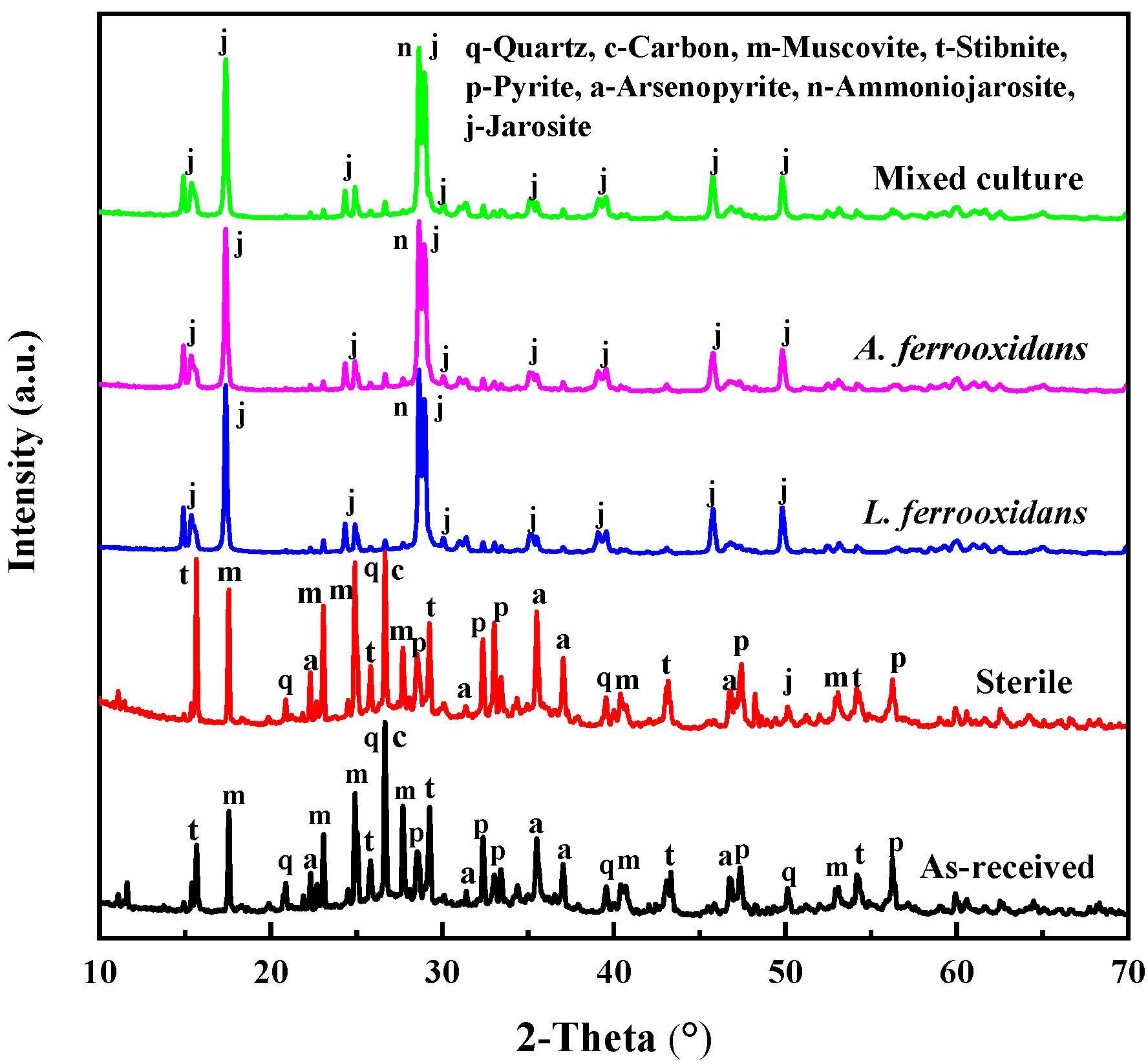
3.2.1. Mineralogical Composition Analysis by XRD

The XRD patterns of as-received, bio-oxidized and sterile control DRGO are shown in Figure 2. It can be seen that the DRGO consisted of muscovite, graphite, quartz, arsenopyrite and pyrite, etc. Evidently, photocatalysts with the bio-oxidized DRGO exhibited similar XRD patterns, with peaks at 17.37° (012), 28.78° (021), 45.55° (303) and 50.08° (220), which corresponded to the jarosite or ammoniojarosite phase. This was consistent with the results in Figure 1d, meaning that the oxidative dissolution of Fe-bearing sulfides was caused by A. ferrooxidans and L. ferrooxidans but not observed obviously in the sterile control. This finding was consistent with the work of Nazari et al. [39] in which A. ferrooxidans was used to investigate jarosite formation during the bioleaching of a pyrite. Previous studies suggested that the formation of jarosite in bioleaching systems reduces the dissolution kinetics of metallic sulfide [40,41]. However, it was found in this study that the precipitation of jarosite had little effect on the bioleaching process. As previously reported [42], jarosite presented as fluffy or woolly spherical particles, loosely packed in a porous structure on the surface, and a similar conclusion was reached by previous works [43,44]. It should be noted that, considering that the sterile control had no significant effect on the oxidation of DRGO, no analysis was conducted in the subsequent process.

3.2.2. Morphology Analysis by SEM-EDS

High-magnification SEM images and EDS data of DRGO using different cultures are shown in Figure 3. In Figure 3a, the SEM image showed that the DRGO surface was smooth before treatment, and the atomic percentage of C, As and S in the as-received DRGO was 16.87%, 0.91% and 22.07%, respectively (spot a-1). After 1 day of bio-oxidation, there were many small crystalline flocs visible on the mineral surface, as seen in Figure 3b,e,h. According to the EDS analysis, the atomic percentages of elemental C, O and S on the mineral surface were significantly increased, indicating that DRGO was oxidized and CM was released. In addition, it was accompanied by the formation of a large amount of elemental sulfur and precipitates with high oxygen content (Equations (5), (8) and (9)). However, the element content varied greatly in different ore sites—this was because the bacterial attachment to the metal sulfide did not occur randomly. In other words, these bacteria, such as A. ferrooxidans, were preferentially attached to sites with visible defects on the surface [38]. Within 3 days of bio-oxidation, holes and cracks were clearly shown in Figure 3c,f,i. This suggested that the crystal lattice structure of the DRGO surface was partly disrupted following bio-oxidation. Meanwhile, the content of O on the DRGO surface was increased, but the C, As and S contents were decreased, suggesting that the DRGO was further oxidized and elemental sulfur was oxidized by bacteria (Equation (4)). After 6 days of bio-oxidation, the mineral surface became granular and larger cracks or pits also appeared on the surface, and the SEM images (Figure 3d,g,j) show that it was covered with many solid particles. According to the XRD analysis (Figure 2), it could be deduced that jarosite and ammoniojarosite were formed. In particular, the corrosion on mineral surfaces seemed more severe in the mixed culture (Figure 3j) than pure cultures.

3.2.3. Chemical Bonds and Functional Groups Analysis by FTIR

FTIR analysis was further carried out to verify the conversion of DRGO in the bio-oxidation process. By using differential spectra, where the “zero-moment” spectra (the spectra collected before the treatment) were subtracted from a series of spectrograms collected at different times, the difference spectra obtained would contribute to the understanding of the evolution of chemical structural changes [45,46]. The negative peaks in difference spectra mean the loss of chemical structure, while the positive peaks are a sign of the formation of new chemical structures [46]. As expected, the FTIR spectral peaks’ shape and trend of samples were basically the same as shown in Figure 4a–c, indicating that these treatments had a parallel influence on the structure and composition distribution of functional groups in bio-oxidized DRGO. The differences in the bio-oxidized samples were mainly concentrated in the peaks and the intensity of some functional groups. Compared to the FTIR spectra of as-received DRGO, the main peaks of bio-oxidized samples included a strong broad peak of 3419 cm−1, which was attributed to O-H stretching vibration of water or Fe oxyhydroxides [47]. The new peak at 1427 cm−1 could be assigned to the v3 stretching vibration of the C-O bond [48], suggesting that a mass of oxygen-containing groups were introduced on the surface of DRGO after bio-oxidation. Peaks at 1238 cm−1 and 1079 cm−1 could be assigned to the stretching vibration of SO42− [49], which was due to the bio-oxidation of pyrite and arsenopyrite, producing SO42− ions. The peak at around 971 cm−1 corresponded to O–H bending vibration, which was ascribed to the adsorption of water onto the mineral surface [50].
In addition, negative peaks also occurred at 1617 cm−1, 1376 cm−1, 1141 cm−1, 1051 cm−1 and 603 cm−1. The C=C band around 1617 cm−1 was considered to be characteristic of the graphite hexagonal ring structure [51]. However, there was no significant interaction between highly ordered structures (such as graphite) and infrared radiation, and this absorption band might be due to the presence of polar functional groups or the irregularities of the aromatic structure, such as carboxylic acid groups or phenolic groups attached to aromatic rings [51]. Peaks at 1376 cm−1 and 1141 cm−1 were designated to the stretching vibration of the O–C–O and C–N bands [52], respectively, suggesting that A. ferrooxidans and L. ferrooxidans could remove the aromatic compounds contained in the DRGO in the process of bio-oxidation. The peak at 1051 cm−1 referred to the bending vibration of the O–H band [52], which was decreased, and this might be due to the fact that some substances of DRGO were coated with the sediment or converted from hydrophobic to hydrophilic nature brought about by bio-oxidation. In addition, the peak at 603 cm−1 was referenced as the stretching vibration of the S–S band [53], which indicated that sulfides were oxidized by A. ferrooxidans and L. ferrooxidans.
These results implied that DRGO could be effectively oxidized by A. ferrooxidans and L. ferrooxidans with time. However, the intensity of some functional groups tended to increase faster in mixed culture than in pure cultures, which was consistent with the results obtained from the changes in various indexes in solutions and the analyses of XRD, SEM and EDS in previous sections. In addition, bio-oxidation by A. ferrooxidans and L. ferrooxidans could significantly decrease the content of aromatic structures and introduce large quantities of oxygen-containing groups on the surface of DRGO. This phenomenon might result in reduced preg-robbing for gold in the subsequent thiourea leaching process.

3.3. The Process of Sulfides Bio-Oxidation

Gold occurred mainly in arsenopyrite and pyrite in the DRGO used in this study, and the diagrams of arsenopyrite and pyrite oxidation in Eh–pH coordinates are presented in Figure 5. Since the bio-oxidation process was carried out under acidic conditions, the relevant range of pH values was 0–3, where the phase changes were considered. As indicated, arsenopyrite was relatively unstable and it could be oxidized when Eh > −0.4 V under bio-oxidation conditions (Figure 5a–c). When the solution exhibited a low Eh (−0.4 V–0.4 V), iron existed mostly as Fe2+, while arsenic and sulfur presented mainly as As2S2, As2S3, H3AsO3 and S0. In the solutions of high Eh (>0.2 V (pH 3)–0.4 V (pH 0)), the elements of iron, arsenic and sulfur in sulfide lattices were oxidized into FeO·OH, HAsO3/H3AsO4/H2AsO4 and HS2O3/S2O32−/HS2O4, respectively. Furthermore, jarosite was easily formed between H3OFe3(SO4)2(OH)6 and K+ in 9 K medium [54], and the precipitate could coat the surface of arsenopyrite in the presence of jarosite, FeO·OH, As2S2, As2S3 and S0, which would impede the subsequent oxidation and leaching process. However, As2S2, As2S3 and S0 could be oxidized by increasing Eh (>0.4 V).
The data presented in the phase diagram for pyrite in the course of bio-oxidation (Figure 5d,e) show that the oxidation potential of pyrite was −0.26 V to 0.33 V within the pH range of 0–3. The oxidation potential of pyrite was significantly higher than arsenopyrite, indicating that the pyrite was more difficult to oxidize than arsenopyrite [55]. In the subsequent deeper oxidation process, the iron and sulfur species became Fe3+, S0 and S2Ox2− at a higher Eh (>0.33 V), while Fe2+, elemental iron and HS occurred at a lower Eh (<−0.26 V). It can be seen from the data that the S0 was gradually oxidized and the thiosulphate stabilizing effect could dominate with the increase in oxidation potential, which could accelerate the overall gold dissolution [56].
The above thermodynamic analysis demonstrated that the gold encapsulated in both arsenopyrite and pyrite could be liberated by bio-oxidation. In addition, the possible equilibrium equations involved in the bio-oxidation of arsenopyrite and pyrite are listed in Appendix A.

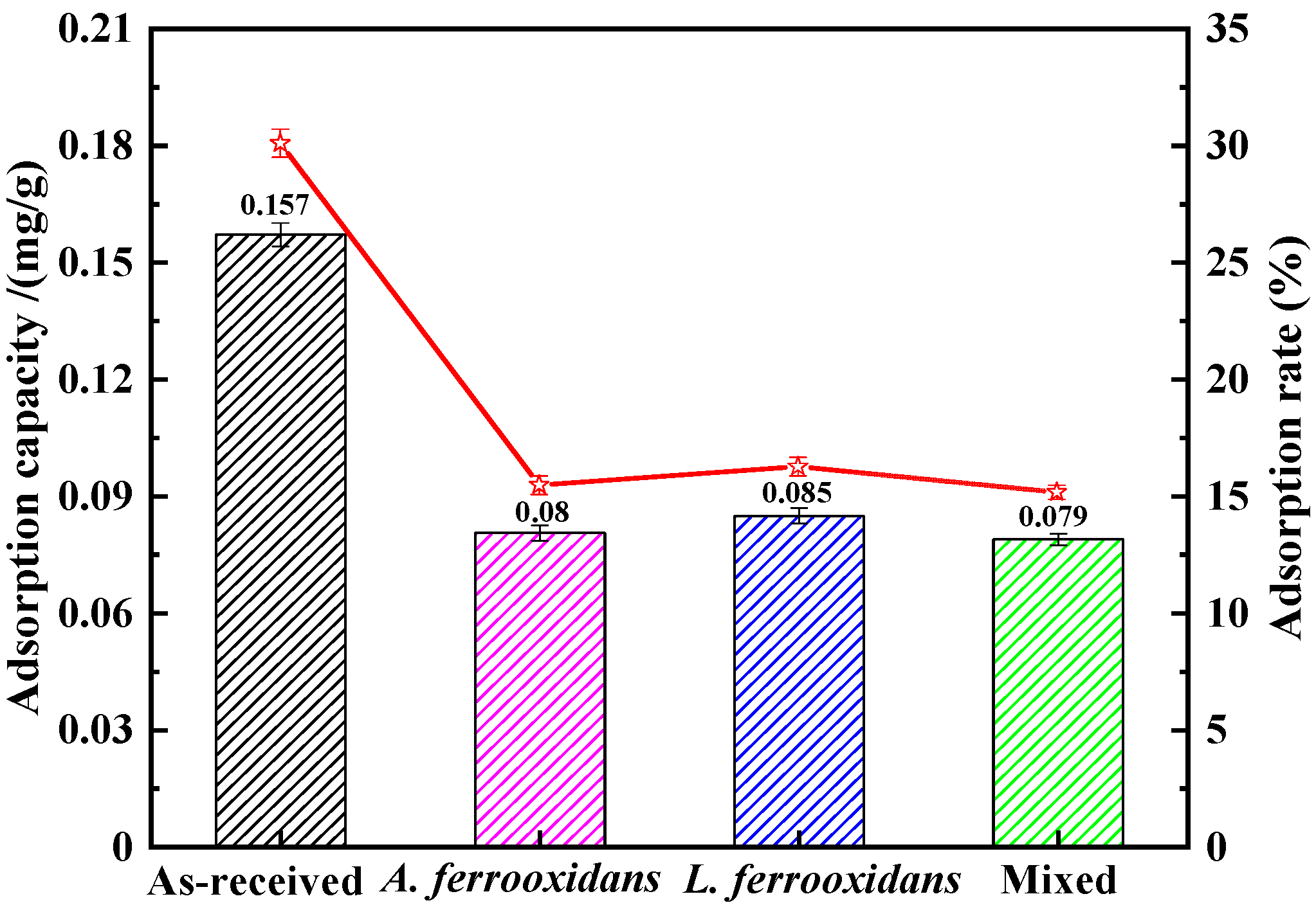
3.4. Preg-Robbing Studies of DRGO in Thiourea System

The effect of different bio-oxidation treatments on gold adsorption in the thiourea system is presented in Figure 6. It can be observed that the adsorption capacity of as-received DRGO to gold was 0.157 mg/g within 3 h. After bio-oxidation, there was a decrease in the preg-robbing capacity of all samples, and the overall decrease in preg-robbing capacity was around 14%. It was thus very clear that A. ferrooxidans and L. ferrooxidans possessed the ability to inactivate CM in DRGO and reduce its gold adsorption activity. It has been reported that the graphitic structure of CM was important for gold adsorption from the thiourea system [19]. However, there was an introduction of oxygen groups on the CM, which had the potential to reduce the degree of graphitization and thus decrease gold adsorption activity. In addition, the cleavage of the aromatic C=C band and the degradation of aromatic O-C-O and C-N bonds reduced the affinity of CM for gold in solution [57]. Another possibility was the reduction in the specific surface area via blocking the pores of CM by biochemicals produced from the microbial metabolism, hence reducing gold adsorption [58]. In addition, the coating of passivation materials such as jarosite reduced the effective contact between CM and gold. This conclusion was also reported in previous research from our team [59], where graphite was used as a substitute for CM. This observation might contribute to the increase in gold extraction from the bio-oxidized samples in the process of thiourea leaching.

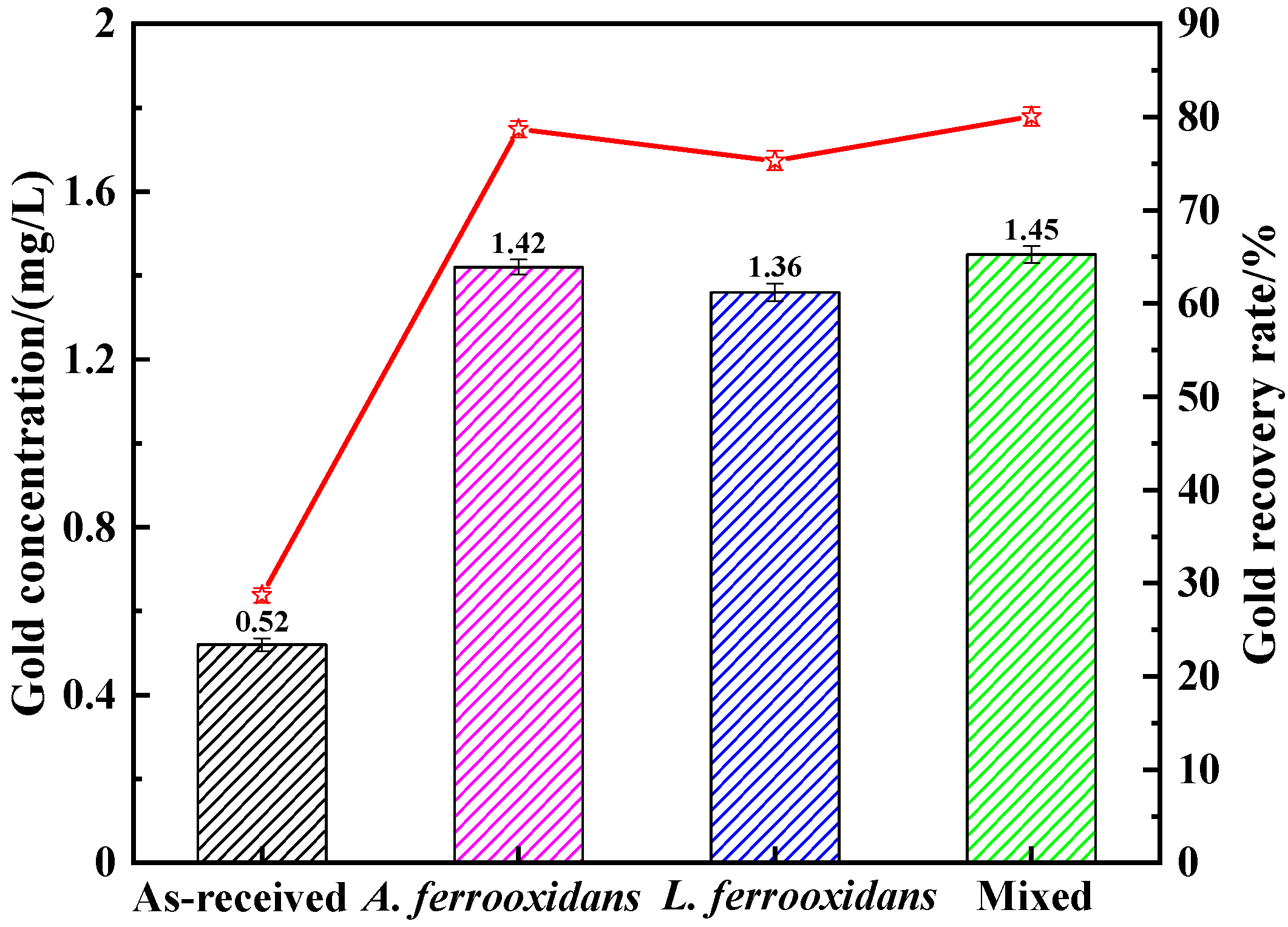
3.5. Thiourea Leaching

Leaching of gold was carried out in acidic thiourea solutions (Figure 7). It can be seen that the as-received DRGO yielded only around 28.7% of gold extraction by direct thiourea leaching, indicating that the present sample was very refractory. The recovery loss was mainly caused by the inaccessibility of thiourea reagent to gold particles and the preg-robbing of CM for gold in solutions. After biological pre-treatment, the gold recovery rate reached 78.69% (A. ferrooxidans) and 75.31% (L. ferrooxidans), and the DRGO oxidized by mixed culture resulted in higher gold extraction of 80.07%. It can be seen that the mixed culture did not show any obvious advantage over the pure A. ferrooxidans and L. ferrooxidans in this study. This is inconsistent with previous reports [22,60]. However, this is possibly because the capacity of the strain varies depending on its source, pre-adaptation to minerals, the pH value of the solution and bacterial growth conditions [61]. In addition, compared with A. ferrooxidans, L. ferrooxidans has a stronger affinity for ferrous iron and is less sensitive to the inhibition of ferric iron [60]. Therefore, the use of mixed culture did not show obviously better bio-oxidation results than pure cultures, which may be related to the inferior iron oxidation ability of A. ferrooxidans at high potential. This finding is similar to the results reported earlier by Yang et al. [61] and Falco et al. [62]; both found that the synergistic effect of microorganisms with different metabolic pathways did not play an important role in their study.

4. Conclusions

This paper demonstrated that the bio-oxidation of a typical DRGO using A. ferrooxidans and L. ferrooxidans could decompose sulfides and simultaneously reduce the gold adsorption capability of CM in a thiourea system. Investigations showed that A. ferrooxidans and L. ferrooxidans were able to oxidize Fe2+ to Fe3+ and thus increase Eh in the solutions, which was conducive to decomposing the sulfides. This result was also proven by thermodynamic analysis. The preg-robbing capacity for gold was reduced by more than 14% after bio-oxidation. XRD, SEM-EDS and FTIR analyses showed that A. ferrooxidans and L. ferrooxidans were able to destroy the graphitization structure by the introduction of oxygen-containing groups, thus decreasing the active sites necessary for gold adsorption. Moreover, biochemicals were produced by microbial metabolism and passivation materials such as jarosite were formed on the surface of the DRGO, which prevented effective contact between CM and gold. In addition, the S-S band was cleaved, indicating that sulfides were oxidized by bacteria. Thiourea leaching results have shown that gold extractions of over 75% were achieved for both pure and mixed cultures. However, the use of mixed culture did not show obviously better bio-oxidation results than pure cultures. Preliminary results were promising, though there was still partial gold adsorption. Therefore, with optimized parameters in the process of bio-oxidation, higher levels of gold extraction and lower levels of preg-robbing are feasible.

Author Contributions

R.X. and Q.L.: Data curation, Writing, Original draft preparation, Conceptualization, Methodology, Funding acquisition. F.M.: Visualization, Investigation, Writing—Reviewing and Editing, Funding acquisition. Y.Y.: Software. B.X. and H.Y.: Writing—Reviewing and Editing. T.J.: Supervision, Validation. All authors have read and agreed to the published version of the manuscript.

Funding

This research was funded by the National Key Research and Development Program of China (grant No. 2018YFE0110200), the National Natural Science Foundation of China (grant No. 51574284) and Science and Technology Planning Project of Yunnan Province (grant No. 2018ZE001)

Conflicts of Interest

The authors declare no conflict of interest.

Appendix A

Table A1. Possible equilibrium equations involved in the bio-oxidation of arsenopyrite and pyrite.
Bio-Oxidation of Arsenopyrite and Pyrite∆G0298/(kcal/mol)No.
FeAsS + 5Fe3+ + 3H2O = 6Fe2+ + H3AsO3 + S0 + 3H+−81.661
2FeAsS + 16Fe3+ + 9H2O = 18Fe2+ + 2HAsO3 + HS2O3 + 15H+−211.1992
6FeAsS + 12Fe3+ = 3As2S2 + 18Fe2+−322.2693
FeAsS + 3H2O + 6Fe3+ = 7Fe2+ + S0 + HAsO3 + 5H+−91.4894
4FeAsS + 4H2O + 12Fe3+ = As2S3 + S2− + 16Fe2+ + 2AsO2 + 8H+−194.9895
2FeAsS + 6H2O + 10Fe3+ = S0 + 2HAsO3 + S2− + 12Fe2+ + 10H+−126.8946
2As2S2 + 15H2O + 16Fe3+ = 4HAsO3 + 2S2− + HS2O3 + 16Fe2+ + 25H+−67.1637
As2S3 + 9H2O + 10Fe3+ = 2HAsO3 + S2− + HS2O3 + 10Fe2+ + 15H+−42.2128
FeS2 + 14Fe3+ + 8H2O = 15Fe2+ + 16H+ + 2SO42−−134.779
FeS2 + 2Fe3+ = 3Fe2+ + 2S0−19.16410
FeS2 + 3H2O + 6Fe3+ = 7Fe2+ + HS2O3 + 5H+−47.38511
S0 + 4H2O + 6Fe3+ = SO42− + 8H+ + 6Fe2+−57.80312
Fe3+ + 3H2O = Fe(OH)3 + 3H+5.50313
2Fe3+ + 4H2O = 2FeO·OH + 6H+1.0714

References

  1. Agorhom, E.A.; Skinner, W.; Zanin, M. Upgrading of low-grade gold ore samples for improved particle characterisation using Micro-CT and SEM/EDX. Adv. Powder Technol. 2012, 23, 498–508. [Google Scholar] [CrossRef]
  2. Kaksonen, A.H.; Perrot, F.; Morris, C.; Rea, S.; Benvie, B.; Austin, P.; Hackl, R. Evaluation of submerged bio-oxidation concept for refractory gold ores. Hydrometallurgy 2013, 141, 117–125. [Google Scholar] [CrossRef]
  3. Wang, G.H.; Xie, S.B.; Liu, X.X.; Wu, Y.H.; Liu, Y.J.; Zeng, T.T. Bio-oxidation of a high-sulfur and high-arsenic refractory gold concentrate using a two-stage process. Miner. Eng. 2018, 120, 94–101. [Google Scholar] [CrossRef]
  4. Yang, H.Y.; Liu, Q.; Song, X.L.; Dong, J.K. Research status of carbonaceous matter in carbonaceous gold ores and bio-oxidation pretreatment. Trans. Nonferrous Met. Soc. China 2013, 23, 3405–3411. [Google Scholar] [CrossRef]
  5. Nanthakumar, B.; Pickles, C.A.; Kelebek, S. Microwave pretreatment of a double refractory gold ore. Miner. Eng. 2007, 20, 1109–1119. [Google Scholar] [CrossRef]
  6. Luo, W.J.; Yang, H.Y.; Jin, Z.A. Study on the Gold Recovery of Double Refractory Gold Ore Concentrate by Biological Oxidation Pretreatment. Adv. Mat. Res. 2015, 1130, 379–382. [Google Scholar] [CrossRef]
  7. Pyke, B.L.; Johnston, R.F.; Brooks, P. Characterisation and behaviour of carbonaceous material in a refractory gold bearing ore. Miner. Eng. 1999, 12, 851–862. [Google Scholar] [CrossRef]
  8. Xu, B.; Yang, Y.B.; Li, Q.; Jiang, T.; Liu, S.Q.; Li, G.H. The development of an environmentally friendly leaching process of a high C, As and Sb bearing sulfide gold concentrate. Miner. Eng. 2016, 89, 138–147. [Google Scholar] [CrossRef]
  9. Rawlings, D.E.; Dew, D.; du Plessis, C. Biomineralization of metal-containing ores and concentrates. Trends Biotechnol. 2003, 21, 38–44. [Google Scholar] [CrossRef]
  10. Brierley, C.L. Biohydrometallurgical prospects. Hydrometallurgy 2010, 104, 324–328. [Google Scholar] [CrossRef]
  11. Ma, S.J.; Luo, W.J.; Mo, W.; Su, X.J.; Liu, P.; Yang, J.L. Removal of arsenic and sulfur from a refractory gold concentrate by microwave heating. Miner. Eng. 2010, 23, 61–63. [Google Scholar] [CrossRef]
  12. Rawlings, D.E.; Johnson, D.B. The microbiology of biomining: Development and optimization of mineral-oxidizing microbial consortia. Microbiology 2007, 153, 315–324. [Google Scholar] [CrossRef] [PubMed] [Green Version]
  13. Gonzalez, R.; Gentina, J.C.; Acevedo, F. Biooxidation of a gold concentrate in a continuous stirred tank reactor: Mathematical model and optimal configuration. Biochem. Eng. J. 2004, 19, 33–42. [Google Scholar] [CrossRef]
  14. Sand, W.; Gerke, T.; Hallmann, R.; Schippers, A. Sulfur chemistry, biofilm, and the (in)direct attack mechanism ? a critical evaluation of bacterial leaching. Appl. Microbiol. Biotechnol. 1995, 43, 961–966. [Google Scholar] [CrossRef]
  15. Glenn, J.K.; Morgan, M.A.; Mayfield, M.B.; Kuwahara, M.; Gold, M.H. An extracellular H2O2-requiring enzyme preparation involved in lignin biodegradation by the white rot basidiomycete Phanerochaete chrysosporium. Biochem. Biophys. Res. Commun. 1983, 114, 1077–1083. [Google Scholar] [CrossRef]
  16. Tien, M.; Kirk, T.K. Lignin-Degrading Enzyme from the Hymenomycete Phanerochaete chrysosporium Burds. Science 1983, 221, 661–663. [Google Scholar] [CrossRef] [Green Version]
  17. Tien, M.; Kirk, T.K. Lignin Peroxidase of Phanerochaete Chrysosporium. In Methods in Enzymology; Academic Press: San Diego, CA, USA, 1988; Volume 161, pp. 238–249. [Google Scholar]
  18. Ofori-Sarpong, G.; Tien, M.; Osseo-Asare, K. Myco-hydrometallurgy: Coal model for potential reduction of preg-robbing capacity of carbonaceous gold ores using the fungus, Phanerochaete chrysosporium. Hydrometallurgy 2010, 102, 66–72. [Google Scholar] [CrossRef]
  19. Ofori-Sarpong, G.; Osseo-Asare, K. Preg-robbing of gold from cyanide and non-cyanide complexes: Effect of fungi pretreatment of carbonaceous matter. Int. J. Miner. Process. 2013, 119, 27–33. [Google Scholar] [CrossRef]
  20. Amankwah, R.K.; Yen, W.T.; Ramsay, J.A. A two-stage bacterial pretreatment process for double refractory gold ores. Miner. Eng. 2005, 18, 103–108. [Google Scholar] [CrossRef]
  21. Zhang, J.; Zhang, X.; Ni, Y.; Yang, X.; Li, H. Bioleaching of arsenic from medicinal realgar by pure and mixed cultures. Process Biochem. 2007, 42, 1265–1271. [Google Scholar] [CrossRef]
  22. Akcil, A.; Ciftci, H.; Deveci, H. Role and contribution of pure and mixed cultures of mesophiles in bioleaching of a pyritic chalcopyrite concentrate. Miner. Eng. 2007, 20, 310–318. [Google Scholar] [CrossRef]
  23. Nguyen, V.K.; Lee, M.H.; Park, H.J.; Lee, J.U. Bioleaching of arsenic and heavy metals from mine tailings by pure and mixed cultures of Acidithiobacillus spp. J. Ind. Eng. Chem. 2015, 21, 451–458. [Google Scholar] [CrossRef]
  24. Anderson, C.G. Alkaline sulfide gold leaching kinetics. Miner. Eng. 2016, 92, 248–256. [Google Scholar] [CrossRef]
  25. Sun, C.B.; Zhang, X.L.; Kou, J.; Xing, Y. A review of gold extraction using noncyanide lixiviants: Fundamentals, advancements, and challenges toward alkaline sulfur-containing leaching agents. Int. J. Miner. Metall. Mater. 2020, 27, 417–431. [Google Scholar] [CrossRef]
  26. Zhou, H.; Song, Y.S.; Li, W.J.; Song, K. Electrochemical behavior of gold and its associated minerals in alkaline thiourea solutions. Int. J. Miner. Metall. Mater. 2018, 25, 737–743. [Google Scholar] [CrossRef]
  27. Syna, N.; Valix, M. Assessing the potential of activated bagasse as gold adsorbent for gold–thiourea. Miner. Eng. 2003, 16, 511–518. [Google Scholar] [CrossRef]
  28. Chen, C.K.; Lung, T.N.; Wan, C.C. A study of the leaching of gold and silver by acidothioureation. Hydrometallurgy 1980, 5, 207–212. [Google Scholar] [CrossRef]
  29. Hilson, G.; Monhemius, A.J. Alternatives to cyanide in the gold mining industry: What prospects for the future? J. Clean. Prod. 2006, 14, 1158–1167. [Google Scholar] [CrossRef]
  30. Yannopoulos, J.C. The Extractive Metallurgy of Gold; Springer: New York, NY, USA, 1991. [Google Scholar]
  31. Li, J.S.; Miller, J.D. Reaction kinetics of gold dissolution in acid thiourea solution using ferric sulfate as oxidant. Hydrometallurgy 2007, 89, 279–288. [Google Scholar] [CrossRef]
  32. Gallagher, N.P.; Hendrix, J.L.; Milosavljevic, E.B.; Nelson, J.H.; Solujic, L. Affinity of activated carbon towards some gold(I) complexes. Hydrometallurgy 1990, 25, 305–316. [Google Scholar] [CrossRef]
  33. Nakbanpote, W.; Thiravetyan, P.; Kalambaheti, C. Preconcentration of gold by rice husk ash. Miner. Eng. 2000, 13, 391–400. [Google Scholar] [CrossRef]
  34. Konadu, K.T.; Huddy, R.J.; Harrison, S.T.L.; Osseo-Asare, K.; Sasaki, K. Sequential pretreatment of double refractory gold ore (DRGO) with a thermophilic iron oxidizing archeaon and fungal crude enzymes. Miner. Eng. 2019, 138, 86–94. [Google Scholar] [CrossRef]
  35. Guo, Y.J.; Guo, X.; Wu, H.Y.; Li, S.P.; Wang, G.H.; Liu, X.X.; Qiu, G.Z.; Wang, D.Z. A novel bio-oxidation and two-step thiourea leaching method applied to a refractory gold concentrate. Hydrometallurgy 2017, 171, 213–221. [Google Scholar] [CrossRef]
  36. Kirby, C.S.; Thomas, H.M.; Southam, G.; Donald, R. Relative contributions of abiotic and biological factors in Fe(II) oxidation in mine drainage. Appl. Geochem. 1999, 14, 511–530. [Google Scholar] [CrossRef]
  37. Schippers, A.; Sand, W. Bacterial Leaching of Metal Sulfides Proceeds by Two Indirect Mechanisms via Thiosulfate or via Polysulfides and Sulfur. Appl. Environ. Microbiol. 1999, 65, 319–321. [Google Scholar] [CrossRef] [Green Version]
  38. Rohwerder, T.; Gehrke, T.; Kinzler, K.; Sand, W. Bioleaching review part A: Progress in bioleaching: Fundamentals and mechanisms of bacterial metal sulfide oxidation. Appl. Microbiol. Biotechnol. 2003, 63, 239–248. [Google Scholar] [CrossRef] [PubMed]
  39. Nazari, B.; Jorjani, E.; Hani, H.; Manafi, Z.; Riahi, A. Formation of jarosite and its effect on important ions for Acidithiobacillus ferrooxidans bacteria. Trans. Nonferrous Met. Soc. China 2014, 24, 1152–1160. [Google Scholar] [CrossRef]
  40. Fantauzzi, M.; Licheri, C.; Atzei, D.; Loi, G.; Elsener, B.; Rossi, G.; Rossi, A. Arsenopyrite and pyrite bioleaching: Evidence from XPS, XRD and ICP techniques. Anal. Bioanal. Chem. 2011, 401, 2237–2248. [Google Scholar] [CrossRef]
  41. Corkhill, C.L.; Wincott, P.L.; Lloyd, J.R.; Vaughan, D.J. The oxidative dissolution of arsenopyrite (FeAsS) and enargite (Cu3AsS4) by Leptospirillum ferrooxidans. Geochim. Cosmochim. Acta 2008, 72, 5616–5633. [Google Scholar] [CrossRef]
  42. Wang, H.M.; Bigham, J.M.; Jones, F.S.; Tuovinen, O.H. Synthesis and properties of ammoniojarosites prepared with iron-oxidizing acidophilic microorganisms at 22–65 degrees C. Geochim. Cosmochim. Acta 2007, 71, 155–164. [Google Scholar] [CrossRef]
  43. Marchevsky, N.; Barroso Quiroga, M.M.; Giaveno, A.; Donati, E. Microbial oxidation of refractory gold sulfide concentrate by a native consortium. Trans. Nonferrous Met. Soc. China 2017, 27, 1143–1149. [Google Scholar] [CrossRef]
  44. Zhu, P.; Liu, X.D.; Chen, A.J.; Liu, H.W.; Yin, H.Q.; Qiu, G.Z.; Hao, X.D.; Liang, Y.L. Comparative study on chalcopyrite bioleaching with assistance of different carbon materials by mixed moderate thermophiles. Trans. Nonferrous Met. Soc. China 2019, 29, 1294–1303. [Google Scholar] [CrossRef]
  45. Nguyen, T.; Martin, J.; Byrd, E.; Embree, N. Relating laboratory and outdoor exposure of coatings: II. J. Coat. Technol. 2002, 74, 65–80. [Google Scholar] [CrossRef]
  46. Martin, J.W.; Nguyen, T.; Byrd, E.; Dickens, B.; Embree, N. Relating laboratory and outdoor exposures of acrylic melamine coatings: I. Cumulative damage model and laboratory exposure apparatus. Polym. Degrad. Stab. 2002, 75, 193–210. [Google Scholar] [CrossRef]
  47. Mazzetti, L.; Thistlethwaite, P.J. Raman spectra and thermal transformations of ferrihydrite and schwertmannite. J. Raman Spectrosc. 2002, 33, 104–111. [Google Scholar] [CrossRef]
  48. Gunasekaran, S.; Anbalagan, G.; Pandi, S. Raman and Infrared Spectra of Carbonates of Calcite Structure. J. Raman Spectrosc. 2006, 37, 892–899. [Google Scholar] [CrossRef]
  49. Lazaroff, N.; Melanson, L.; Lewis, E.; NicholasSantoro; CurtPueschel. Scanning electron microscopy and infrared spectroscopy of iron sediments formed by Thiobacillus ferrooxidans. Geomicrobiol. J. 1985, 4, 231–268. [Google Scholar] [CrossRef]
  50. Fredd, C.N.; Fogler, H.S. The Kinetics of Calcite Dissolution in Acetic Acid Solutions. Chem. Eng. Sci. 1998, 53, 3863–3874. [Google Scholar] [CrossRef]
  51. Painter, P.C.; Starsinic, M.; Squires, E.; Davis, A.A. Concerning the 1600 cm−1 region in the i.r. spectrum of coal. Fuel 1983, 62, 742–744. [Google Scholar] [CrossRef]
  52. Sakthivel, R.; Das, B.; Satpati, B.; Mishra, B.K. Gold supported iron oxide-hydroxide derived from iron ore tailings for CO oxidation. Appl. Surf. Sci. 2009, 255, 6577–6581. [Google Scholar] [CrossRef]
  53. Ofori-Sarpong, G.; Amankwah, R.K.; Osseo-Asare, K. Reduction of preg-robbing by biomodified carbonaceous matter—A proposed mechanism. Miner. Eng. 2013, 42, 29–35. [Google Scholar] [CrossRef]
  54. Liu, J.Y.; Tao, X.X.; Cai, P. Study of Formation of Jarosite Mediated by Thiobacillus Ferrooxidans in 9K Medium. In Proceedings of the International Conference on Mining Science & Technology, Amsterdam, The Netherlands, 12 October 2009; Ge, S., Liu, J., Guo, C., Eds.; Elsevier Science Bv: Amsterdam, The Netherlands, 2009; Volume 1, pp. 706–712. [Google Scholar]
  55. Koslides, T. Pressure oxidation of arsenopyrite and pyrite in alkaline solutions. Hydrometallurgy 1992, 30, 87–106. [Google Scholar] [CrossRef]
  56. Feng, D.; van Deventer, J.S.J. The effect of sulphur species on thiosulphate leaching of gold. Miner. Eng. 2007, 20, 273–281. [Google Scholar] [CrossRef]
  57. Twum Konadu, K.; Sasaki, K.; Ofori-Sarpong, G.; Osseo-Asare, K.; Kaneta, T. Bio-Modification of Carbonaceous Matters in Gold Ore: Model Experiments Using Powdered Activated Charcoal and Cell-Free Extracts of Phanerochaete chrysosporium. Hydrometallurgy 2017, 168, 76–83. [Google Scholar] [CrossRef]
  58. Moreira, M.T.; Feijoo, G.; Lema, J.M. Fungal Bioreactors: Applications to White-Rot Fungi. Rev. Environ. Sci. Biotechnol. 2003, 2, 247–259. [Google Scholar] [CrossRef]
  59. Li, Q.; Shen, H.; Xu, R.; Zhang, Y.; Yang, Y.B.; Xu, B.; Jiang, T.; Yin, H.Q. Effect of Acidithiobacillus ferrooxidans and Leptospirillum ferrooxidans on preg-robbing of gold by graphite from thiourea leaching solution. J. Clean. Prod. 2020, 261, 9. [Google Scholar] [CrossRef]
  60. Rawlings, D.E.; Tributsch, H.; Hansford, G.S. Reasons why ‘Leptospirillum’-like species rather than Thiobacillus ferrooxidans are the dominant iron-oxidizing bacteria in many commercial processes for the biooxidation of pyrite and related ores. Microbiol. Read. 1999, 145, 5–13. [Google Scholar] [CrossRef] [Green Version]
  61. Yang, Y.; Harmer, S.; Chen, M. Synchrotron X-ray photoelectron spectroscopic study of the chalcopyrite leached by moderate thermophiles and mesophiles. Miner. Eng. 2014, 69, 185–195. [Google Scholar] [CrossRef]
  62. Falco, L.; Pogliani, C.; Curutchet, G.; Donati, E. A comparison of bioleaching of covellite using pure cultures of Acidithiobacillus ferrooxidans and Acidithiobacillus thiooxidans or a mixed culture of Leptospirillum ferrooxidans and Acidithiobacillus thiooxidans. Hydrometallurgy 2003, 71, 31–36. [Google Scholar] [CrossRef]
Figure 1. Variations in the (a) pH; (b) Eh; (c) total Fe concentration; (d) As leaching rate; (e) TOC concentration over time during the oxidation process of DRGO.
Metals 10 01216 g001aMetals 10 01216 g001b
Figure 2. XRD diffraction patterns for the as-received, bio-oxidized and sterile control DRGO.
Metals 10 01216 g002
Figure 3. SEM images with EDS data (at.%) for the as-received sample (DRGO) and the DRGO after oxidizing 1 d, 3 d and 6 d in 9 K culture medium. (a) As-received; (bd) A. ferrooxidans; (eg) L. ferrooxidans; (hj) Mixed culture.
Metals 10 01216 g003
Figure 4. FTIR spectra (difference spectrum) of DRGO after oxidizing 1, 3 and 6 in 9 K culture medium. (a) A. ferrooxidans; (b) L. ferrooxidans; (c) Mixed culture.
Metals 10 01216 g004
Figure 5. Eh–pH diagrams of Fe-As-S-H2O systems: (a) Fe species; (b) As species; (c) S species and Fe-S-H2O systems: (d) Fe species; (e) S species. Conditions: [Fe2+]total 0.16 M, [FeAsS] 7.4 × 10⁻⁴ mM, [FeS2] 3 × 10⁻⁴ mM; 25 °C and 1 atm.
Metals 10 01216 g005
Figure 6. Effect of different treatments on gold adsorption by DRGO.
Metals 10 01216 g006
Figure 7. Effect of different treatments on gold recovery rates of DRGO.
Metals 10 01216 g007
Table 1. Chemical composition and the phase constitution analysis of C and Au of the double refractory gold ore.
ConstituentAu *CAsFeSiO2CaOMgOS
Content/wt%18.056.951.6219.627.684.262.0020.76
Phase of CElemental carbonOrganic carbonCarbonateTotal
Content/wt%4.740.501.716.95
Distribution/%68.207.2024.60100.00
Phase of AuExposed goldEncapsulated in arsenopyriteEncapsulated in pyriteEncapsulated in oxidesEncapsulated in silicatesTotal
Content *0.3112.073.691.870.1118.05
Distribution/%1.7266.8720.4410.360.61100.00
* Unit g/t.

