Article

A Novel, Sport-Specific EMG-Based Method to Evaluate Movement Efficiency in Karate Punching

1
Department of Combat Sports, Hungarian University of Sports Science, 1123 Budapest, Hungary
2
Department of Kinesiology, Hungarian University of Sports Science, 1123 Budapest, Hungary
3
Research Center for Sports Physiology, Hungarian University of Sports Science, 1123 Budapest, Hungary
*
Author to whom correspondence should be addressed.
Sports 2025, 13(7), 218; https://doi.org/10.3390/sports13070218
Submission received: 27 May 2025 / Revised: 30 June 2025 / Accepted: 3 July 2025 / Published: 7 July 2025

Abstract

Background: This study aimed to develop a method to analyze the kinetic and kinematic characteristics of the traditional karate Gyaku Tsuki (reverse punch), focusing on the activation sequence of lower and upper extremities and trunk muscles during execution. Methods: An elite male (N = 1) karate athlete (in kata) performed 20 Gyaku Tsuki punches while equipped with 16 wireless surface EMG sensors integrated with 3-axis accelerometers. The five punches with the highest forearm acceleration were selected for analysis. EMG, accelerometer, and synchronized video data were recorded and processed. Results: A novel visualization technique was developed to represent muscle activation over time, distinguishing a spectrum of 0–25–50–75–100% activation levels. Muscle activation times for arm, leg, and trunk muscles ranged from −0.31 to −0.11 s relative to punch execution, indicating rapid, coordinated muscle engagement. Conclusions: This method enables detailed analysis of muscle activation patterns in karate punches. It offers valuable insights for biomechanics researchers and practical applications for coaches aiming to enhance performance and prevent injuries through better understanding of movement dynamics.

Graphical Abstract

1. Introduction

Biomechanical analysis of sports movements such as gait, running, and cycling is well-established. Three-dimensional motion capture systems, force plates, and EMG are some of the most frequently used devices, with which universally accepted measurement protocols are performed [1,2,3,4]. However, in martial arts research, data collection [5] and processing are not as straightforward as in gait or running analysis. Martial arts movements are not as restricted, like, for example, the circular cycling motion, therefore, the individual execution pattern of a movement varies significantly between individuals [6]. Determination and classification of a martial arts movement through biomechanical variables is difficult even with objective measurements [7]. The comparison of executions and determination of good performance is highly subjective without universally accepted measurement methodology. The most significant problem is that as the movement pattern is not pre-determined, therefore, the collection of different variables of biomechanical movement data and the consequent determination of average values to be used as a baseline for comparison is very difficult [8].
Traditional karate is special compared to other martial art forms, because the optimal movement executions are pre-determined through the kata system [9]. Therefore, it would be expected that a usable biomechanical analysis methodology would have already been developed for karate. But, according to our knowledge, no such methodology has been introduced until now. Consequently, the aim of this paper is to present a novel method enabling objective analysis and classification of karate techniques.
To present the system step-by-step we have chosen the Gyaku Tsuki punch, which is one of the most frequented commonly used karate techniques in traditional karate and free style sparring [10]. The Gyaku Tsuki is a punch delivered with the opposite hand of the front leg [11]. A strong, stable stance [12] and a straight trunk perpendicular to the floor is necessary for the execution of this punch [13]. The stance makes stable connection with the ground; the perpendicular trunk posture helps to absorb the reaction force of the target and keeps the balance of the individual in the moment of impact [14]. The execution of a good Gyaku Tsuki needs the rotation of the pelvis, because pelvic rotation introduces additional momentum to the trunk, while the hips remain on the same height [15]. The punch may lose effectiveness if the center of gravity is not pushed forward parallel to the direction of the attack. Punch execution phases are depicted in Figure 1.
The punch starts with the rotation of the pelvis, which transmits power to the trunk, shoulder, arm, and fist and culminates in a strong impact force on the target. This impact can be achieved with the maximum tension of all muscles involved in the motion. At the end of the execution, the body reaches the target as a one-solid mass [16].
The delivered impact is further augmented by the reverse motion of opposite arm (hikite). At the end of the movement the punching arm, the backward pulled opposite arm, and the pelvic rotation is stopped simultaneously [14,15,16].

2. Materials and Methods

The test subject is an elite male karate contestant (male, age: 21 years, height: 168 cm, weight: 66 kg), who holds a third dan black belt and is ranked among the top eight in the All-Japan Championship. The subject has been practicing karate for 18 years, trains regularly and was in good physical condition with no injuries to report at the time of measurement.
The test subject was instrumented with 16 (8-8 on each side) wireless surface EMG (sEMG) sensor units (MiniWave, Cometa Italy, Milan, Italy), which also comprised 3-axis integrated accelerometers. Following appropriate skin preparation, two AgCl2 electrodes were placed on the muscle bellies according to the SENIAM guidelines on both the right and left side of the body [17,18,19,20]. Table 1 lists the muscles we have included in our study.
The simultaneous EMG signals, accelerometer data, and time-synchronized video signals [21,22] were recorded and stored using the EMG and Motion Tools software (version 8.11, Cometa Italy). The sampling rates for the sEMG, accelerometer data, and the video sequences were 2000, 142 Hz, and 60 frames per second, respectively.
For subsequent off-line analysis, EMG and accelerometer data were exported to txt format to be processed by an in-house written Python script (Python v. 3.12.2). Codes are available from the corresponding author, upon reasonable request.

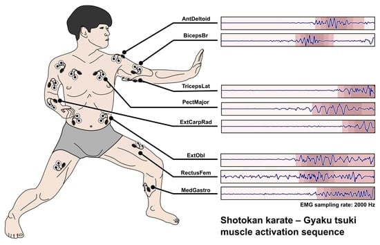
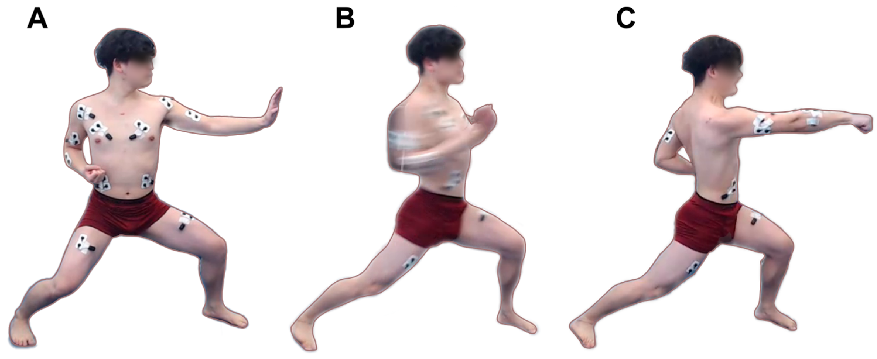
2.1. Movement Execution Protocol

First, the test subject was instrumented with the sensor units and familiarized with the devices. Then the subject was instructed to perform 20 reverse punches with the dominant side (right) with approximately 5 s intervals separating each punch. The punches were delivered to a 35 × 35 cm (13.8 × 13.8 inch) semi-rigid X-ray film hung on a holder frame to maintain constant distance for the attack and to provide a target for the subject to focus on. All punches were executed correctly according to the expert (L.Cs.) supervising the execution. Prior to data collection, a warmup sequence was performed.

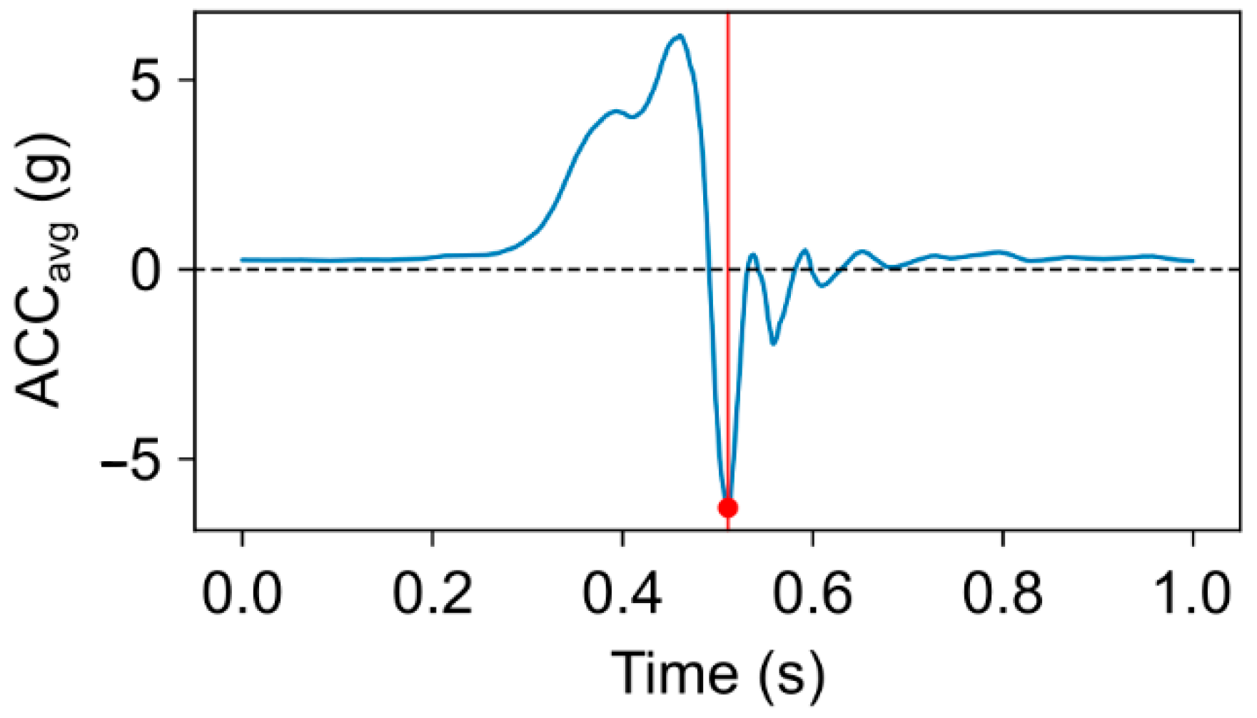
2.2. Data Processing and Evaluation
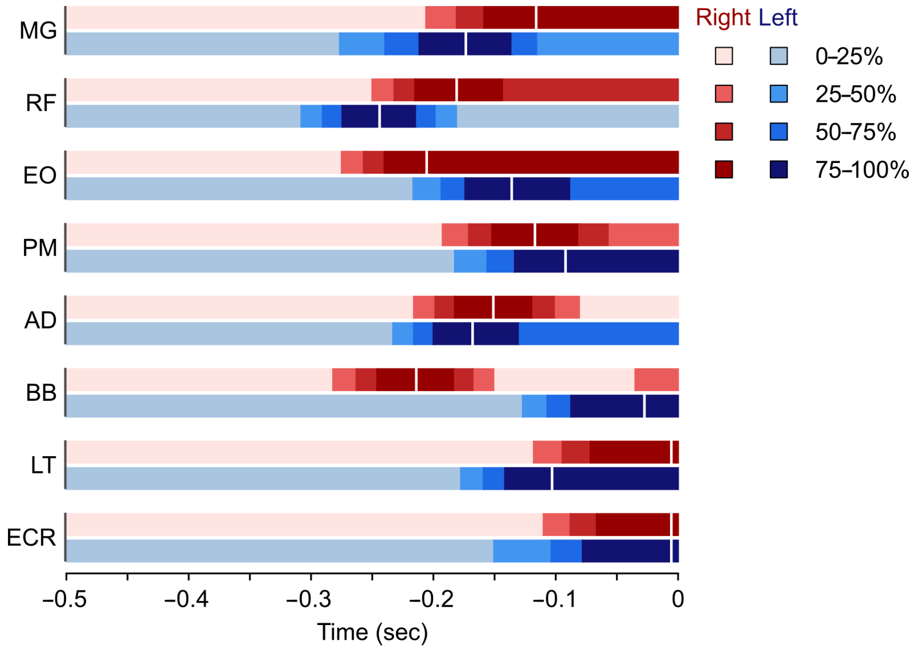
For muscle activation sequence determination, the best 5 out of 20 punches were selected. The best punches were chosen based on the maximum acceleration magnitude (in multiples of g) of the punching forearm, resulting in the greatest velocity at impact with the target. Forearm acceleration magnitudes were calculated as:
accMag = acc x 2 + acc y 2 + acc z 2 ,
where accx, accy, and accz, refer to acceleration vectors in x, y, and z directions.
For each muscle a representative EMG signal was created by signal “averaging” 0.5 s-long EMG segments taken from the 5 best punches. This procedure was carried out by aligning the ends of the EMG segments with the end-of-the-punch time index. The time index of the end-of-punch was determined as the timestamp corresponding to the local minimum of the averaged (accx, accy, accz) forearm acceleration signal see Figure 2. Finally, the representative EMG is calculated by taking the median of the 5 EMG segments. All subsequent calculations were carried out on the “averaged” signals.
The median signal was filtered with a digital 6th order Butterworth bandpass filter between 30 and 300 Hz [23]. Since our wireless EMG system ran on USB power, we did not apply any narrow-band power-line signal filtering.
The filtered and windowed EMG was transformed with a Teager–Kaiser energy operator (TKEO) [24] to improve signal-to-noise ratio. The TKEO transformation is a method of sEMG signal processing recommended for onset detection problems dealing with rapid, explosive movements [25]. The transformation is performed according to:
Ψ x n = x n 2 - x n + 1 · x n - 1 ,
where xn is the nth sample of the filtered EMG signal. To acquire a smoother envelope, the transformed signal was filtered with a 4th order Butterworth low pass filter, with a 7 Hz cutoff frequency.
To visualize the muscle activation sequence pattern, we have implemented a unique technique. Instead of only characterizing muscle activation as active/non-active from the EMG data we have developed an interval representation, where using the processed EMG signal 0–25%, 25–50%, 50–75%, and 75–100%, activation levels were determined and separated as the function of time. Consequently, a more precise visualization and a better understanding of the muscle behavior and activation pattern is achievable.
The smoothed envelope was normalized to a range 0–1, then discrete values of 0, 1, 2, and 3 were assigned to the continuous amplitude values to gain activation levels.
Ψ D = 0 ,   i f   Ψ s m o o t h < 0.25 1 ,   i f   Ψ s m o o t h   [ 0.25 ,   0.5 ) 2 ,   i f   Ψ s m o o t h   [ 0.5 ,   0.75 ) 3 ,   i f   Ψ s m o o t h   0.75 ,
where ΨD is the discretized envelope (activation level) and Ψsmooth is the low pass filtered smoothed envelope, as seen in Figure 3.
Muscle activation onset timestamp is defined as when ΨD value passed from 0 to 1 (when the normalized amplitude first surpassed the value of 0.25). These onset timestamps for each of the examined muscles are listed in Table 2.
Evaluation of the results. The performance was rated based on the recorded and processed data by an expert (8th dan), who is a practicing coach in karate. By analyzing the data, the expert determined the correct and incorrect movement execution components in the discussion section.

3. Results

Forearm accelerations of the best five punches in ascending order were: 25.14 g, 25.20 g, 25.48 g, 25.52 g, and 25.79 g (mean: 25.43 g, SD: 0.26 g).
Activation levels for all examined muscles were determined bilaterally. Results are displayed in Figure 4 as a visual summary of Table 2, which lists the sequence of muscle activation and activation times related to the end-of-punch.

4. Discussion

This study was designed to develop a method to describe the kinetic and kinematic characteristics of the traditional karate Gyaku Tsuki strike. Our aim was to determine the activation sequence of the lower and upper extremities along with the muscles of the trunk throughout the execution of the technique. Although each of the applied methods are well-documented in the literature, the novelty of this paper lies within the unique combination of the above-mentioned techniques.
The study revealed that the athlete first used the rectus femoris muscle of the front leg to stabilize the stance. From the coach’s point of view, this is the correct execution, since leg strength is strongly associated with peak punch force within elite boxers [26] and effective punches require the knee of the front leg to not move backwards, as this causes a significant reduction in efficiently transferring ground reaction forces to the punch.
Next, the right biceps brachii muscle, followed by the left (front) medial gastrocnemius almost simultaneously (0.002 s difference) with the right extensor oblique abdominal muscle, become active. Minimal stretching of the biceps brachii muscle is necessary during the initiation of the punch because the weight of the punching arm starting from the side of the torso must be maintained. Activation and contraction of the left medial gastrocnemius facilitates forward translation of the knee joint (in the direction of the attack), thus preventing backward movement of the joint. The right Abdominal External Oblique muscle plays a significant role in the rotation of the pelvis and trunk, which is supported by the next tensing muscle—the rectus femoris—on the dominant side (rear leg). The rectus femoris contraction assists in the conversion of the vertical ground reaction force component into a horizontally directed force.
The left anterior deltoid and left external oblique abdominal muscles began to be activated almost simultaneously (Δt = 0.001 s) to initiate withdrawal of the extended left arm and to help accelerate the punch performed with the right.
Rotation of the right upper arm in the sagittal plane (initiated earlier by the biceps brachii muscle) is further accelerated by the right anterior deltoid muscle, which was involved in the next movement sequence.
Next, the medial gastrocnemius muscle of the back leg was activated. By analyzing the data, the expert concluded that the right medial gastrocnemius muscle should have engaged much earlier, at about the same time as the rectus femoris of the front leg (left side). Based on this, it allows us to conclude that the subject could not efficiently harness the ground reaction force but tried to fix the back foot by pressing the heel to the ground instead of plantarflexion. This is also clearly visible on the time-synchronized video footage, where the inner sole of the rear foot is pressed against the ground, see Figure 1C. Based on these observations, our recommendation for coaches is that through strengthening lower limb muscles involved in the movement, they can improve stance stability, which results in higher punch impact force.
The right and the left major pectoral muscles are activated next, respectively, with a negligible 0.01 s difference between the sides. While the right pectoral muscle directed the punching arm towards the target positioned at the midline of the body, the left muscle brought the retracting opposite arm closer to the trunk.
Closing to the end of the movement, muscles of the left arm (lateral triceps, extensor carpi radialis and biceps brachii muscles, in order) activated before their counterparts on the right side. Finally, the focus of punch is ensured by fully extending the right elbow joint by the right lateral triceps [27] and stabilizing the right wrist with the assistance of the right extensor carpi radialis muscle.
Overall, it can be said that the pelvis/trunk rotation is the most decisive accelerating element in the entire process. Considering that the whole execution time takes ~0.3 s, it can be said that the karate Gyaku Tsuki movement pattern is the result of very fast, almost simultaneous contraction of the 16 examined muscles.

Future Development Possibilities

To generate a universally useful database, the recording of a large data pool is necessary, and, by using AI machine learning tools, the determination of optimal execution would be possible. Consequently, by pairing the subjective rating of the execution by the experts and the recorded data automated evaluation of the punch can be achieved. To develop and train supervised machine learning models, an expert consensus-based feature set on a pre-defined ordinary scale should be developed, which clearly distinguishes correct/incorrect punch executions.
It is worth mentioning that this method can be applied to superficial muscle components of other kinetic chains in the body and can be generalized to analyze other movement patterns in other sports.

5. Conclusions

By creating unique and simple data recordings, data evaluation, and representation methods for muscle activation, the analysis of the Gyaku Tsuki karate punch is possible. The method provides data that is not only useful for the biomechanics expert but also for the coaches working with the athletes. By implementing the method introduced in the paper determination of faulty execution and identification of correct and incorrect movement patterns is possible.
A novel method for muscle activation analysis gives objective additional information for “coaches eye”, which helps to develop karate techniques through evaluation of the execution, meaning “good form” according the World Karate Federation (WKF) competition rules: “Good form is the properly executed technique which have the characteristics conferring probable effectiveness within the framework of traditional karate concept.” [28] Using the method of our study, either for the coaches or for the athletes, it is possible to motor control the execution by knowing the muscle sequences, in order to reach the highest velocity with minimum effort from the muscles. In addition, it helps to prevent injuries during training.

6. Limitations

Despite the fact that the aim of this study was to introduce our proposed method, the obvious limitation is that only one subject participated in data recording.

Supplementary Materials

The following supporting information can be downloaded at: https://www.mdpi.com/article/10.3390/sports13070218/s1, Data supplement S1: Raw EMG and Accelerometer data file.

Author Contributions

Conceptualization, L.C., T.H. and B.K.; methodology, T.H. and L.C.; software, T.H.; validation, B.K. and T.H.; formal analysis, B.K.; investigation, L.C., T.H. and B.K.; resources, L.C.; data curation, T.H.; writing—original draft preparation, L.C.; writing—review and editing, T.H. and B.K.; visualization, T.H.; supervision, B.K. and T.H.; funding acquisition, L.C. All authors have read and agreed to the published version of the manuscript.

Funding

This research was funded by Ministry of Defense of Hungary and the Hungarian University of Sport Science, Department of Combat Sports, Budo Project, (4872-5/2024.) and T.H. was financed under the Thematic Excellence Program 2021 TKP2021-EGA-37 funding scheme.

Institutional Review Board Statement

The study was conducted in accordance with the Declaration of Helsinki and approved by the Institutional Review Board of the Hungarian University of Sports Science issued for the Combat Sports Department (TE-OKE-KEB/07/2023), approval date: 22 November 2023.

Informed Consent Statement

Informed consent was obtained from the subject involved in the study.

Data Availability Statement

Raw EMG and accelerometer data is available to download as the Supplementary S1.

Conflicts of Interest

The authors declare no conflicts of interest.

Abbreviations

The following abbreviations are used in this manuscript:
ACCAcceleration
ADDeltoid muscle, anterior head
BBBiceps Brachii muscle
ECRExtensor Carpi Radialis muscle
EOAbdominal External Oblique muscle
LTTriceps Brachii, lateral head
MGGastrocnemius, medial head
PMMajor Pectoral muscle
RFRectus Femoris muscle
SDStandard Deviation
sEMGSurface ElectroMyoGraphy
SENIAMSurface ElectroMyoGraphy for the Non-Invasive Assessment of Muscles
TKEOTeager–Kaiser Energy Operator
WKFWorld Karate Federation

References

  1. Holliday, W.; Swart, J. A Dynamic Approach to Cycling Biomechanics. Phys. Med. Rehabil. Clin. N. Am. 2022, 33, 1–13. [Google Scholar] [CrossRef] [PubMed]
  2. Dugan, S.A.; Bhat, K.P. Biomechanics and Analysis of Running Gait. Phys. Med. Rehabil. Clin. N. Am. 2005, 16, 603–621. [Google Scholar] [CrossRef] [PubMed]
  3. Howard, R.M.; Conway, R.; Harrison, A.J. Muscle Activity in Sprinting: A Review. Sports Biomech. 2018, 17, 1–17. [Google Scholar] [CrossRef]
  4. Papagiannis, G.I.; Triantafyllou, A.I.; Roumpelakis, I.M.; Zampeli, F.; Eleni, P.G.; Koulouvaris, P.; Papadopoulos, E.C.; Papagelopoulos, P.J.; Babis, G.C. Methodology of Surface Electromyography in Gait Analysis: Review of the Literature. J. Med. Eng. Technol. 2019, 43, 59–65. [Google Scholar] [CrossRef] [PubMed]
  5. Hachaj, T.; Piekarczyk, M.; Ogiela, M.R. Human Actions Analysis: Templates Generation, Matching and Visualization Applied to Motion Capture of Highly-Skilled Karate Athletes. Sensors 2017, 17, 2590. [Google Scholar] [CrossRef]
  6. Jaegers, S.; Peterson, R.; Hillen, B.; Geuze, R.H.; Schellekens, J.; Dantuma, R. Kinesiologic Aspects of Motor Learning in Dart Throwing. J. Hum. Mov. Stud. 1989, 16, 161–171. [Google Scholar]
  7. Vecchio, L.D.; Whitting, J.; Hollier, J.; Keene, A.; Climstein, M. Reliability and Practical Use of a Commercial Device for Measuring Punch and Kick Impact Kinetics. Sports 2022, 10, 206. [Google Scholar] [CrossRef]
  8. Neto, O.P.; Magini, M. Electromiographic and Kinematic Characteristics of Kung Fu Yau-Man Palm Strike. J. Electromyogr. Kinesiol. 2008, 18, 1047–1052. [Google Scholar] [CrossRef]
  9. Emad, B.; Atef, O.; Shams, Y.; El-Kerdany, A.; Shorim, N.; Nabil, A.; Atia, A. IKarate: Karate Kata Guidance System. Procedia Comput. Sci. 2020, 175, 149–156. [Google Scholar] [CrossRef]
  10. Hofmann, M.; Witte, K.; Emmermacher, P. Biomechanical Analysis of Fist Punch Gyaku-Zuki in Karate. In Proceedings of the ISBS Conference 2008, Seoul, Republic of Korea, 14 July 2008. [Google Scholar]
  11. Venkatraman, J.; Nasiriavanaki, M. Biomechanics of Kumite Style Gyaku Tsuki in Karate. Biomed. J. Sci. Tech. Res. 2019, 14, 10656–10662. [Google Scholar] [CrossRef]
  12. Cesari, P.; Bertucco, M. Coupling between Punch Efficacy and Body Stability for Elite Karate. J. Sci. Med. Sport 2008, 11, 353–356. [Google Scholar] [CrossRef] [PubMed]
  13. VencesBrito, A.M.; Ferreira, M.A.R.; Cortes, N.; Fernandes, O.; Pezarat-Correia, P. Kinematic and Electromyographic Analyses of a Karate Punch. J. Electromyogr. Kinesiol. 2011, 21, 1023–1029. [Google Scholar] [CrossRef]
  14. Okazaki, T.; Stricevic, M.V. The Textbook of Modern Karate; Kodansha: New York, NY, USA, 1984; ISBN 9780870114618. [Google Scholar]
  15. Nakayama, M. Dynamic Karate, 1st ed.; Kodansha International: Tokyo, Japan, 2012; ISBN 978-1568364131. [Google Scholar]
  16. Nishiyama, H.; Brown, R.C. Karate the Art of Empty-Hand Fighting; Tuttle Publishing: Tokyo, Japan, 1995. [Google Scholar]
  17. Hermens, H.J.; Freriks, B.; Disselhorst-Klug, C.; Rau, G. Development of Recommendations for SEMG Sensors and Sensor Placement Procedures. J. Electromyogr. Kinesiol. 2000, 10, 361–374. [Google Scholar] [CrossRef] [PubMed]
  18. Stegeman, D.; Hermens, H. Standards for Suface Electromyography: The European Project Surface EMG for Non-Invasive Assessment of Muscles (SENIAM). Enschede Roessingh Res. Dev. 2007, 10, 8–12. [Google Scholar]
  19. Konrad, P. The ABC of EMG A Practical Introduction to Kinesiological Electromyography; Noraxon Inc.: Scottsdale, AZ, USA, 2005. [Google Scholar]
  20. Ball, N.; Scurr, J. Electromyography Normalization Methods for High-Velocity Muscle Actions: Review and Recommendations. J. Appl. Biomech. 2013, 29, 600–608. [Google Scholar] [CrossRef]
  21. Osu, R.; Franklin, D.W.; Kato, H.; Gomi, H.; Domen, K.; Yoshioka, T.; Kawato, M. Short- and Long-Term Changes in Joint Co-Contraction Associated with Motor Learning as Revealed From Surface EMG. J. Neurophysiol. 2002, 88, 991–1004. [Google Scholar] [CrossRef] [PubMed]
  22. Witte, K.; Emmermacher, P.; Hofmann, M.; Schwab, K.; Witte, H. Electromyographic Researches of Gyaku-Zuki in Karate Kumite. In Proceedings of the ISBS-Conference, Beijing, China, 16 March 2008. [Google Scholar]
  23. Solnik, S.; Rider, P.; Steinweg, K.; DeVita, P.; Hortobágyi, T. Teager–Kaiser Energy Operator Signal Conditioning Improves EMG Onset Detection. Eur. J. Appl. Physiol. 2010, 110, 489–498. [Google Scholar] [CrossRef]
  24. Li, X.; Zhou, P.; Aruin, A.S. Teager–Kaiser Energy Operation of Surface EMG Improves Muscle Activity Onset Detection. Ann. Biomed. Eng. 2007, 35, 1532–1538. [Google Scholar] [CrossRef]
  25. Carvalho, C.R.; Fernández, J.M.; del-Ama, A.J.; Barroso, F.O.; Moreno, J.C. Review of Electromyography Onset Detection Methods for Real-Time Control of Robotic Exoskeletons. J. Neuroeng. Rehabil. 2023, 20, 141. [Google Scholar] [CrossRef]
  26. Dunn, E.C.; Humberstone, C.E.; Franchini, E.; Iredale, K.F.; Blazevich, A.J. Relationships Between Punch Impact Force and Upper- and Lower-Body Muscular Strength and Power in Highly Trained Amateur Boxers. J. Strength Cond. Res. 2022, 36, 1019–1025. [Google Scholar] [CrossRef]
  27. Bozec, S.L.; Maton, B.; Cnockaert, J.C. The Synergy of Elbow Extensor Muscles during Dynamic Work in Man. Eur. J. Appl. Physiol. Occup. Physiol. 1980, 44, 255–269. [Google Scholar] [CrossRef] [PubMed]
  28. World Karate Federation, Competition Rules 2020. Available online: https://www.britishkaratefederation.com/news-wkf/WKF_Competition_Rules_2020_EN_Jan22_markup.pdf (accessed on 25 June 2025).
Figure 1. Initial (A), middle (B), and terminal (C) phases of a correctly executed Gyaku Tsuki. Figure displays the positioning of surface EMG/Accelerometer sensor units on the athlete.
Sports 13 00218 g001
Figure 2. Average acceleration of the punching forearm. The end of the punch is defined at the timestamp corresponding to signal minimum (red dot), where the highest negative acceleration was measured.
Sports 13 00218 g002
Figure 3. The normalized and smoothed Teager–Kaiser envelope (Ψsmooth) is discretized into quartiles to gain the stepped discrete envelope (Ψdiscrete). Activation thresholds corresponding to 25%, 50%, and 75% of the normalized envelope amplitude are denoted by horizontal dashed lines, with their respective temporal occurrences marked by vertical dashed lines. The activation map on the top of the figure is constructed from Ψdiscrete. The colors of the map indicate the level of activation, where darker colors represent greater activation. Activation onset is defined when the discretized envelope takes value 1 (activation level surpasses the value of 0.25).
Sports 13 00218 g003
Figure 4. Averaged, half-second-long records of muscle activations of the arm and leg/trunk muscles during a right-hand (dominant side) Gyaku Tsuki prior to the termination of the punch/hitting the target. Different color tones represent different muscle activation levels. AD: anterior deltoid muscle, BB: biceps brachii muscle, LT: lateral head of the triceps brachii muscle, ECR: extensor carpi radialis muscle, MG: medial head of the gastrocnemius muscle, RF: rectus femoris muscle, EO: external oblique abdominal muscle, PM: pectoralis major muscle. Vertical white lines indicate the timing of the maximal activation.
Sports 13 00218 g004
Table 1. EMG/Accelerometer sensor unit positioning with abbreviations. Based on the SENIAM EMG placement protocol.
ElectrodeSideMuscle (Abbreviation)Function
1RightGastrocnemius, medial head (MG)Ankle plantar flexion
2Left
3RightRectus femoris (RF)Knee extension
4Left
5RightAbdominal External Oblique (EO)Trunk rotation
6Left
7RightMajor Pectoral (PM)Arm adduction
8Left
9RightDeltoid, anterior head (AD)Shoulder anteflexion
10Left
11RightBiceps Brachii (BB)Elbow stabilization
12Left
13RightTriceps Brachii, lateral head (LT)Elbow extension/stabilization
14Left
15RightExtensor Carpi Radialis (ECR)Wrist stabilization
16Left
Table 2. Muscle activation times of the arm and leg/trunk muscles during a right-handed (dominant side) Gyaku Tsuki. Negative activation times refer to durations before the termination of the punch (0 s).
SequenceSideMuscleActivation Time (s)
1LeftRectus Femoris−0.308
2RightBiceps Brachii−0.282
3LeftMedial Gastrocnemius−0.277
4RightAbdominal External Oblique−0.275
5RightRectus Femoris−0.250
6LeftAnterior Deltoid−0.233
7LeftAbdominal External Oblique−0.217
8RightAnterior Deltoid−0.216
9RightMedial Gastrocnemius−0.206
10RightMajor Pectoral−0.193
11LeftMajor Pectoral−0.183
12LeftLateral Triceps−0.178
13LeftExtensor Carpi Radialis−0.151
14LeftBiceps Brachii−0.127
15RightLateral Triceps−0.118
16RightExtensor Carpi Radialis−0.110
Disclaimer/Publisher’s Note: The statements, opinions and data contained in all publications are solely those of the individual author(s) and contributor(s) and not of MDPI and/or the editor(s). MDPI and/or the editor(s) disclaim responsibility for any injury to people or property resulting from any ideas, methods, instructions or products referred to in the content.

