Systematic Review

The Influence of Menstrual Cycle Phases on Maximal Strength Performance in Healthy Female Adults: A Systematic Review with Meta-Analysis

1
Institute of Biomechanics and Neurosciences, Nordic Science, 30173 Hannover, Germany
2
Department of Psychiatry, Social Psychiatry and Psychotherapy, Medical School Hannover, 30625 Hannover, Germany
3
Department of Sports Science, Bielefeld University, 33615 Bielefeld, Germany
4
Institute of Sport Sciences, Biosciences of Sports, University of Hildesheim, 31141 Hildesheim, Germany
5
Triagon Academy Munich, School of Sports, Psychology and Education, 85737 Ismaning, Germany
6
Division of Movement and Training Sciences/Biomechanics of Sport, University of Duisburg-Essen, 45141 Essen, Germany
*
Author to whom correspondence should be addressed.
Sports 2024, 12(1), 31; https://doi.org/10.3390/sports12010031
Submission received: 2 November 2023 / Revised: 29 December 2023 / Accepted: 9 January 2024 / Published: 12 January 2024

Abstract

:
Maximal strength is a significant factor in achieving peak performance and injury prevention in athletes. In individualization strategies for the efficient development of athletes, it is necessary to consider the respective components separately. The purpose of this study was to systematically examine the effects of the different cycle phases on isometric, isokinetic, and dynamic maximum strength. A systematic literature review was conducted; databases were searched from January 1960 to September 2023. The included studies focused on the expression of maximal strength in the earlier follicular phase as well as at least one comparative phase. Of the initial 707 articles identified, 22 met the selection criteria and were included. The studies considered a total of 433 subjects. Our results revealed medium effects (weighted mean standardized mean difference (SMD) = 0.60; seven studies) for isometric maximal strength in favor of the late follicular phase, small effects (weighted mean SMD = 0.39; five studies) for isokinetic maximal strength in favor of the ovulation phase, and small effects (weighted mean SMD = 0.14; three studies) for dynamic maximal strength in favor of the late follicular phase. The results indicate that the early follicular phase is unfavorable for all strength classes. Peak performance in isometric strength is seen in the late follicular phase, whereas isokinetic strength peaks during ovulation. Dynamic strength is optimal in the late follicular phase.

1. Introduction

The participation and promotion of women at the international level in many sports disciplines has only slowly become established in recent decades, which has meant that sports-related tests have predominantly been carried out on male subjects [1]. If women were included in testing, measurements were often taken at low hormone levels to minimize potential hormonal influence [2]. Previous literature shows that training monitoring has been researched at a physiological level for the male body, and the results have been transferred to design training programs for female athletes without examining its transferability [3]. Although the influence of menstrual cycle phases encompasses many areas of physical and mental health [4], strength performance emerges as the most significant for adolescent and adult athletes [5,6].
Maximal strength is an essential component of physical fitness and biomechanics, defining the maximum force that a muscle or group of muscles can produce. This central concept can be divided into different categories based on the mode of muscle contraction and the conditions under which strength is measured. As identified by Haff et al. [7] and Zaciorskij and Kraemer [8], these categories are isometric, isokinetic, and dynamic. Isometric maximal strength is the force that a muscle or muscle group can produce without significant change in muscle length (i.e., maximal voluntary isometric contraction [MVIC]). This involves the individual exerting maximum force against a stationary object or resistance while keeping the muscle length and joint angle constant [9]. In terms of isokinetic maximal strength, it represents the peak force produced by the muscles during a contraction maintained at a constant velocity (i.e., isokinetic dynamometry) [10]. Lastly, dynamic maximal strength focuses on the force generated during the concentric and eccentric phases, which is usually determined by the maximum weight an individual can lift for one repetition through a full range of motion (i.e., one repetition maximum [1 RM]) [11].
In sports, particularly for female athletes, achieving high levels of maximal strength is not only critical for enhancing performance but also for injury prevention. Studies have shown that improved maximal strength is inversely related to injury rates, meaning that female athletes with superior maximal strength often experience fewer injuries [12,13]. Furthermore, during the rehabilitation phase following a sports injury, a robust base of maximal strength can be considered beneficial in preventing potential re-injury [14]. Consequently, variations across the cycle may pose an additional injury risk factor, highlighting the importance of maximal strength in injury prevention, particularly for female athletes.
Further examination of factors influencing maximal strength, and therefore injury prevention and performance, has revealed a variety of elements that are not sex-specific. However, for female athletes in particular, certain determinants affect maximal strength as the menstrual cycle is factored in. These include hormonal ebbs and flows, particularly of estrogen and progesterone [15,16]; muscle and tendon stiffness [17,18], which is a key component of maximal strength [19] and injury rehabilitation [20]; pain perception and therefore maximal performance capacity during strength exercises [21]; fluid dynamics within the body, particularly retention patterns, affecting muscle functionality and perceived exertion during strength training [22]; joint stability and laxity, affecting performance efficiency [23,24]; endurance metrics and fatigue resistance during resistance exercise [25,26]; and psychological components of intrinsic motivation and perceived exertion [27,28].
The menstrual cycle, as described by Janse de Jonge et al. [2], progresses from the early follicular phase (1), characterized by low levels of estrogen and progesterone, through the late follicular phase (2), with increasing levels of estrogen, to the ovulatory phase (3), characterized by high levels of estrogen and a gradual increase in progesterone, followed by the early luteal phase (4), characterized by a predominant progesterone surge, culminating in the mid-luteal phase (5), with peak levels of both estrogen and progesterone, and finally the late luteal phase (6), when both hormone levels decline, signaling the onset of menstruation.
When considering the bio- and neurophysiological foundations for the influence of cycle phases on maximal strength, the current literature primarily reports that (a) sex hormones, (b) energy metabolism, (c) neuromuscular function, and (d) body composition are significant factors; thus, these factors are highlighted below. Female sex hormones may influence muscle physiological adaptations in the areas of hypertrophy, force development, and lactate metabolism [29]. While estrogen has a neuroexcitatory effect [30] and thus influences activity potential, force production, and performance in a positive way, progesterone inhibits cortical excitability and thus represents a negative influence [30,31,32,33]. The strength-enhancing effect of estrogen suggests that skeletal muscle is an estrogen-responsive tissue [34] and that mRNA and protein levels of α-estrogen receptors in skeletal muscle are sensitive to circulating estrogen levels [35]. Furthermore, it has been shown that myosin is directly affected by the estrogen concentration [34] and that under the condition of reduced estrogen levels, as occurs during menstruation, the number of active myosin heads bound to actin is reduced. Thus, the muscle’s ability to generate force is diminished [36], and improved maximum force performance during the late follicular phase (estrogen +/progesterone −), compared to the luteal phase (estrogen −/progesterone +), can be expected, whereupon training recommendations can be derived. Another possible cause of altered strength performance is a menstrual cycle-related change in available testosterone. An acute increase in testosterone levels can enhance physical performance via improved neural activation and also improve electrophysiological and contractile properties of muscles as well as motor system function [37]. Studies show that a spike in testosterone is present during the ovulatory phase [38,39].
Another effect of altered hormone concentrations is expressed in terms of energy metabolism. During the mid-luteal phase, there is an increased reliance on lipid metabolism, while carbohydrate utilization decreases due to higher estrogen levels compared to the early follicular phase [40]. Estrogen is known to enhance fat utilization, potentially leading to a sparing effect on muscle glycogen stores. This shift in energy substrate utilization might favor endurance performance in the mid-luteal phase, as the body can conserve its limited glycogen stores and rely more on abundant fat reserves for energy production [41]. On the other hand, the late follicular phase is favorable for glucose utilization, as insulin sensitivity increases [42], which improves intensive and short-duration exercise [43]. For athletes with type 1 diabetes, it is particularly relevant to understand that insulin sensitivity is lower during the luteal phase compared to the follicular phase of the menstrual cycle [44]. This means that during the luteal phase, the body’s ability to uptake glucose, which is essential for achieving maximal strength, may be reduced. Furthermore, Justice et al. [45] described a positive correlation between increased insulin sensitivity, 1RM, and motor function.
Considering the relevance of neuromuscular function to the execution of explosive, maximal strength movements, the discharge behavior of motoneurons during fast muscle contractions is performance-determining [46]. In this context, compared to the early follicular phase, the late luteal phase shows both a higher initial firing rate of the motor units of the medial and oblique vastus medialis [47] and reduced fatigability [48].
Further temporary fluctuations in body composition during a menstrual cycle could also be an influencing factor for altered maximal strength performance. Increased body mass is associated with reduced aerobic and anaerobic endurance performance [49,50,51]. In athletes and healthy non-athletes, body mass and total body water increased from the follicular to the luteal phase [52,53]. This increase in body mass in the luteal phase could be caused by the decrease in insulin as progesterone increases, which drives appetite and food intake [54], or by fluid retention, as aldosterone, the thirst hormone, is increased in the luteal phase [55].
Recent reviews have shown very heterogeneous results [27,56,57,58,59] due to the multiple influencing factors within the cycle phases, although to the authors’ knowledge, no systematic review has explicitly examined the effects on maximal strength. This provides the rationale for this systematic review and meta-analysis, namely, to both provide initial insights into differences in maximal strength and develop further practical implications.
This systematic review and meta-analysis aims to determine whether menstrual cycle hormone fluctuation influences maximal isometric, isokinetic, and dynamic strength. Due to the bio- and neurophysiological differences identified during the menstrual cycle phases and their potential influence on maximal strength, it is hypothesized that these phases not only significantly influence the expression of maximal strength but also lead to distinct differences between the three categories. The findings of this study can enhance the understanding of female-specific physiological responses in sports and contribute significantly to the growing field of sex-specific sports medicine and training. If studies indicate a difference in performance within the menstrual cycle, increased sex-specific research would be of great importance. This could benefit female athletes, who could experience an increase in performance through cycle-based training. To be able to describe the mode of action of hormone fluctuations more accurately, the following analysis uses the six-phase model described by McNulty et al. [27].

2. Materials and Methods

This meta-analysis was conducted in accordance with the PRISMA guidelines [60] (Table S1). The following chapter describes in detail the individual steps of the applied methodology.

2.1. Literature Search

The search for appropriate literature was conducted in the PubMed, Medline, SportDiscus, and Web of Science databases using the following Boolean operators: (“menstrual cycle” AND “follicular”) AND (“isokinetic strength” OR “isometric strength” OR “maximal strength” OR “dynamic strength” OR “muscle strength” OR “muscle power”). In addition, the reference lists were searched to identify further relevant studies. Only English-language results were searched in all databases. Studies with publication dates between January 1960 and September 2023 were searched.

2.2. Selection Criteria

The eligibility of the studies was checked according to the PICOS scheme (population, intervention, comparator, outcome, study design) and is shown in Table 1. In this meta-analysis, women with a regular menstrual cycle were selected as the population. The intervention describes the measurement of muscle strength in a clearly defined cycle phase. The early follicular phase serves as the control group, as this can be easily verified by the onset of menstrual bleeding. This must be compared with at least one other cycle phase to draw a comparison. A variable of maximal isometric, isokinetic, or dynamic strength must be specified as an outcome. Finally, the following requirements had to be fulfilled regarding the study design: (a) within-subjects study design wherein participants were assessed in at least two cycle phases, (b) the measurements were taken on a group not taking a contraceptive pill, and (c) the strength values were presented numerically. Furthermore, the selection of studies was based on the following exclusion criteria: (a) subjects had menstrual cycle dysfunction, (b) subjects used a contraceptive pill, (c) the study did not examine maximal isometric, isokinetic, or dynamic strength, (d) no English full-text version could be located, and (e) the study was not conducted in humans.
According to Guellich and Schmidtbleicher [61], the definition of maximum strength was set as the highest force that the neuromuscular system can develop during a maximum voluntary contraction. Therefore, studies that measured muscle power or impulse were excluded.

2.3. Methodological Study Quality

The quality of each study was assessed using an adapted Downs and Black checklist (Table S2) according to Mc. Nulty et al. [27] and was conducted independently by two authors (OT, JC). In case of disagreement, a third author (NWB) was consulted for the majority decision. The modified list deals with five categories in which a maximum of 16 points can be scored. These points are obtained through 15 items in the categories of reporting, external validity, risk of bias, selection bias, and significance. The score obtained determined the study quality as very low (0–5 points), low (6–9), moderate (10–13), or high (14–16). The score was then used to determine the final study quality based on two additional questions. The authors made this adjustment based on the scientific recommendations of Knowles et al. [62] and Janse De Jonge et al. [2]. These explain the relevance of a standardized verification of the cycle phases. Accordingly, the first step was to answer whether the cycle phases were confirmed by blood sampling. If this question was answered in the negative, the quality of the study was lowered by one level. If the method was used, the study retained its previously determined quality status. This was followed by the question of whether a urinary ovulation test was performed to verify ovulation. The consequence of a negative answer was again the downgrading of the quality.

2.4. Data Extraction and Data Processing

All literature results were stored in a literature management program. Once the appropriate studies were identified, the essential data could be extracted. The selected categories are shown in Table 1, and their preparation was based on the PICOS scheme. In the studies included, various menstrual cycle phase models were utilized; therefore, a partial adjustment was made for this study. For uniform categorization, the days of the measurements were used and classified according to the aforementioned six-phase model.
This systematic review with meta-analysis considers several outcome measures, which are listed in Table 2. The included studies were screened for the outcome variables of interest. To minimize the heterogeneity between studies, we presented the preferred and alternative measures for each outcome in Table 2, considering the wide range of test procedures used in the literature. In the category of maximal isometric strength, maximal voluntary isometric contraction (MVIC) of the knee extensors was identified as the most common outcome. Regarding maximal isokinetic strength, the most relevant measure is the peak torque of knee extensors at a velocity of 60°/s, as it is the most commonly used and suggested to reveal the maximal capacity of the muscle to produce force [63,64]. For maximal dynamic strength, only three different outcomes were identified, with the 1RM half-squat set as the preferred outcome, as it is the only closed-chain exercise that shows the highest relevance in terms of practical application [11]. In one included study [65], shoulder internal rotation was preferred to external rotation and abduction as outcomes since it has the highest relevance in everyday life [66] as well as in sports [67]. If studies used other outcomes that could be assigned to one of the three categories, these were presented as alternative outcomes in Table 2.

2.5. Statistical Analysis

The data were analyzed using Cochrane’s Review Manager 5.4.1. To identify differences in different cycle phases, a subgroup analysis was conducted. A subgroup was created if at least three measurements were available for a cycle phase. In each subgroup analysis, the early follicular phase served as a control condition and was compared to another phase (i.e., comparator condition). The standardized mean difference (SMD) was calculated to assess differences between the control condition and the comparator conditions. A positive/negative SMD value indicates better maximal strength performance for the comparator/control condition. Further, and in accordance with Cohen [68], SMD values of 0 ≤ 0.49 indicate small effects, values of 0.50 ≤ 0.79 indicate medium effects, and values of ≥0.80 indicate large effects. I2 statistics were assessed according to the ranges given by Deeks et al. [69]. These are defined as 0–40%: trivial heterogeneity, 30–60%: moderate heterogeneity, 50–90%: substantial heterogeneity, and 75–100%: considerable heterogeneity. It is emphasized here that the assessment of the I2 statistics should not be used solely to make a statement about heterogeneity. The ranges of values given serve as a rough classification [70].

3. Results

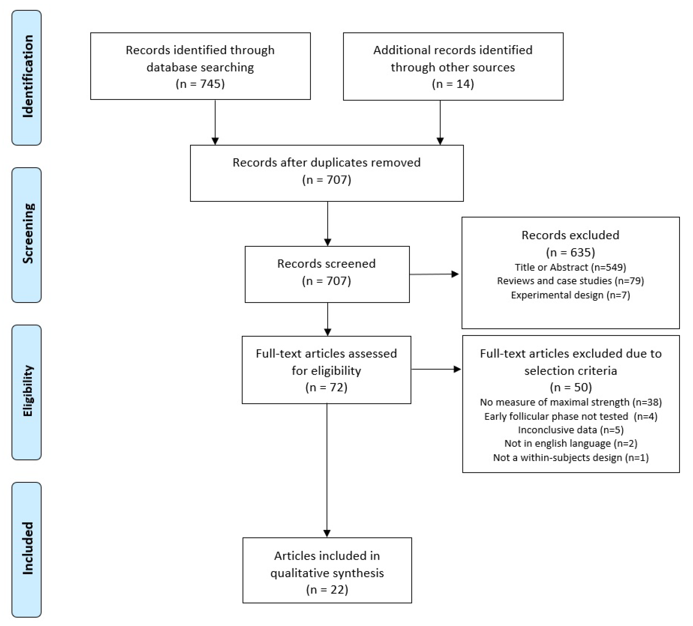
3.1. Selection of Studies

The process of the literature search is illustrated in Figure 1. The English PRISMA flow chart for systematic reviews [60] served as a template. The search yielded a total of 745 hits as well as 14 additional hits through a manual search of reference lists. After removing duplicates and excluding articles based on title or abstract as well as reviews, case studies, and articles with experimental study designs, 707 studies remained. Accordingly, seventy-two articles were subjected to a full-text search; of these articles, thirty-eight did not report a measurement of maximal strength, four did not test in the early follicular phase, five provided inclusive data, two were not written in English, and one did not use a within-subjects design.

3.2. Study Characteristics

Table 3 shows the characteristics of the studies analyzed. In the 22 included studies, a total of 433 subjects were tested for maximal strength performance in at least two phases of the menstrual cycle. The examinations comprised measurements taken during one to five phases of the menstrual cycle. Of the studies analyzed, all examined the early follicular phase, eleven the late follicular phase [6,28,31,32,71,72,73,74,75,76,77], twelve the ovulation phase [28,65,71,73,74,78,79,80,81,82,83,84], four the early luteal phase [28,72,73,85], and nineteen the mid-luteal phase [6,31,32,65,71,72,74,75,76,78,79,80,81,82,83,84,85,86,87]. The late luteal phase was mentioned in two studies [28,31]. Fifteen studies measured maximal isometric strength [28,32,65,72,73,74,75,77,80,81,82,83,84,85,86], eight maximal isokinetic strength [31,71,75,79,80,81,84,87], and three maximal dynamic strength [6,76,78]. Nine studies determined the menstrual cycle phase using blood samples [72,74,75,78,80,81,82,85,87], six using urine samples [6,65,71,73,83,86], three counting days [76,77,79], and one each using saliva samples [31], self-report [84], body temperature [28], or did not report the method [32].

3.3. Methodological Study Quality

Table S2 shows the results of the quality check using the adapted Downs and Black Checklist. A total of seven studies were rated as very low [28,32,73,76,77,79,84], eight as low [6,31,65,71,74,78,83,86], and six as moderate [72,75,80,81,82,87]. Only one of the studies was of high quality [85], and no study was of very high quality.

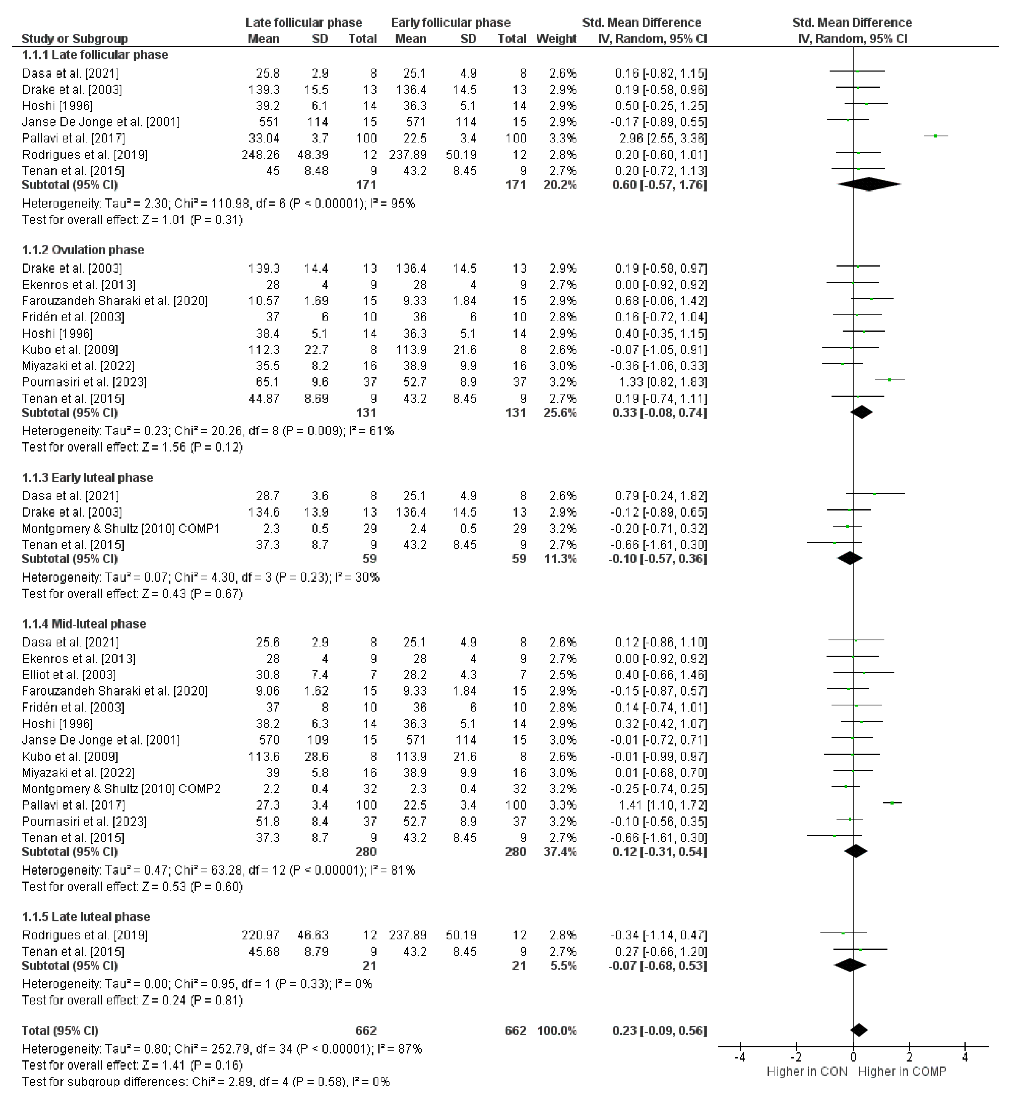
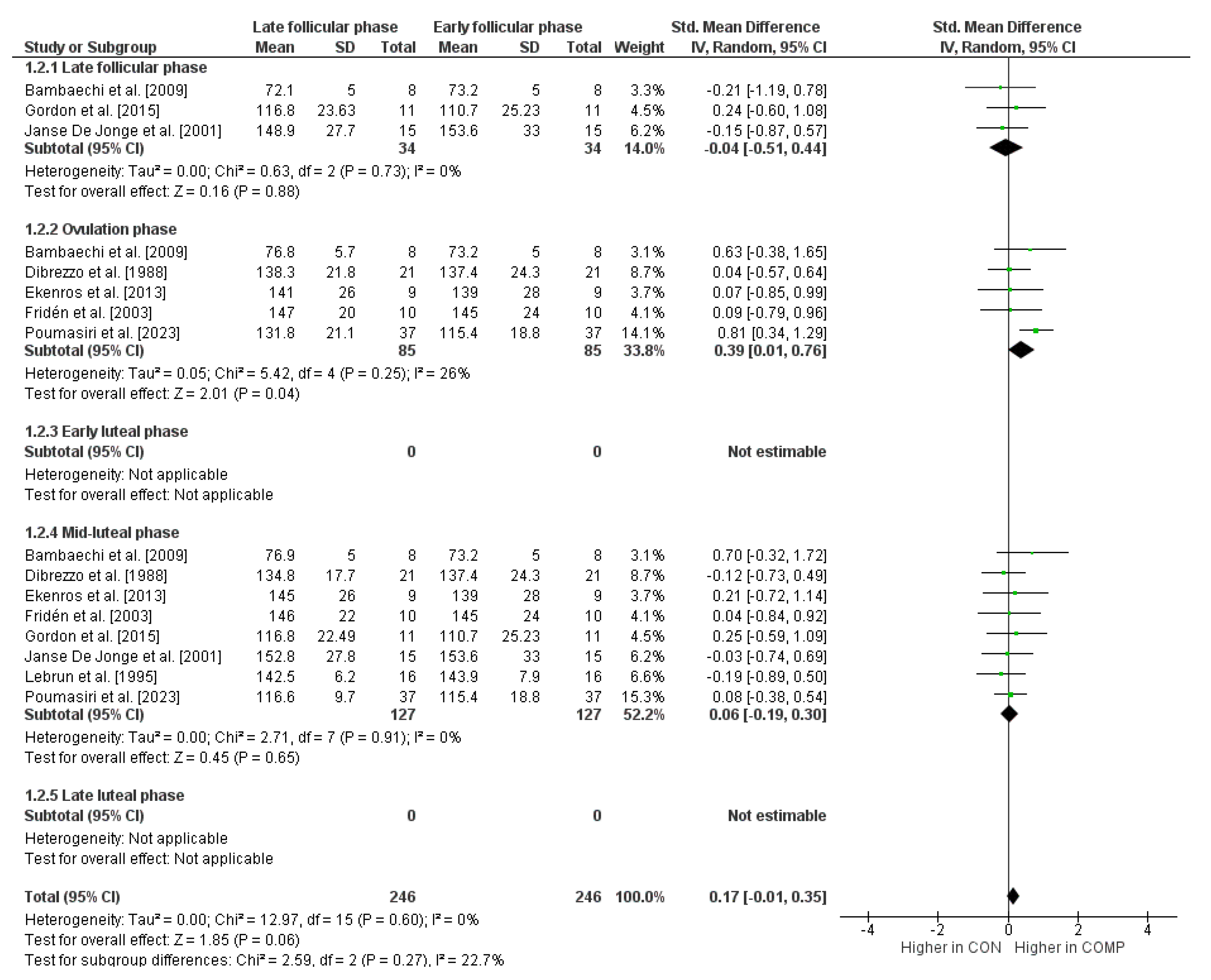
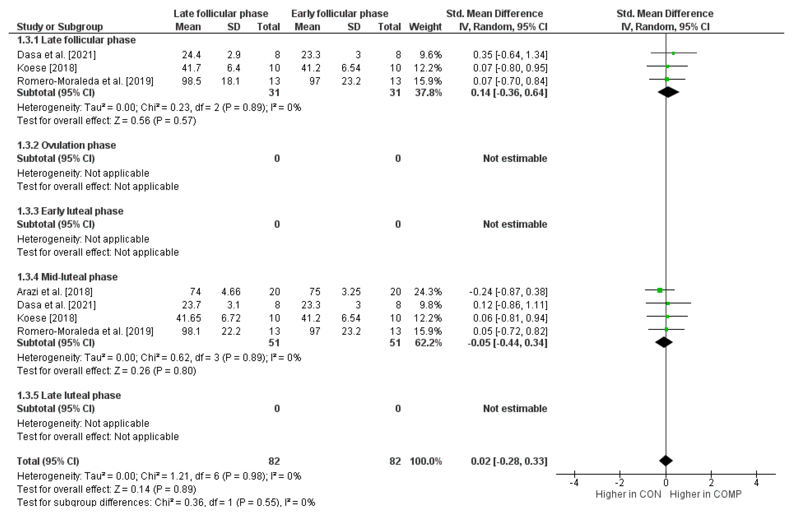
3.4. Influence of the Cycle Phases on Maximal Strength Performance

The maximal isometric strength performance for the early follicular phase compared to the other phases of the menstrual cycle is shown in Figure 2. The weighted mean SMD amounted to 0.60 when compared to the late follicular phase (I2 = 95%, Chi2 = 110.98, df = 6, p < 0.00001, seven studies), 0.33 for the ovulation phase (I2 = 61%, Chi2 = 20.26, df = 8, p = 0.009, nine studies), −0.10 for the early luteal phase (I2 = 30%, Chi2 = 4.30, df = 3, p = 0.23, four studies), 0.12 for the mid-luteal phase (I2 = 81%, Chi2 = 63.28, df = 12, p < 0.00001, thirteen studies), and −0.07 for the late luteal phase (I2 = 0%, Chi2 = 0.95, df = 1, p = 0.33, two studies), which is indicative of small to medium effects.
In terms of maximal isokinetic strength performance, the comparison of the early follicular phase with the other phases of the menstrual cycle is shown in Figure 3. The weighted mean SMD yielded −0.04 when compared to the late follicular phase (I2 = 0%, Chi2 = 0.63, df = 2, p = 0.73, three studies), 0.39 for the ovulation phase (I2 = 26%, Chi2 = 5.42, df = 4, p = 0.25, five studies), and 0.06 for the mid-luteal phase (I2 = 0%, Chi2 = 2.71, df = 7, p = 0.91, eight studies), which indicates small effects. No SMD values were calculated for the comparison of the early follicular phase with the early and late luteal phases since our search did not identify the required minimum of two studies.
Figure 4 shows maximal dynamic strength performance for the early follicular phase compared to the other phases of the menstrual cycle. The weighted mean SMD amounted to 0.14 when compared to the late follicular phase (I2 = 0%, Chi2 = 0.23, df = 2, p = 0.89, three studies) and −0.05 for the mid-luteal phase (I2 = 0%, Chi2 = 0.62, df = 3, p = 0.89, four studies), which is again indicative of small effects. For the comparison of the early follicular phase with the ovulation phase, the early luteal phase, and the late luteal phase, no SMD values were calculated since our search did not detect a minimum of two studies.

4. Discussion

This systematic review and meta-analysis was conducted to determine whether menstrual cycle hormone fluctuation influences maximal isometric, isokinetic, and dynamic strength. The 22 studies analyzed, encompassing a total of 433 women, showed heterogeneous results.
Most studies (86.4%) compared several phases (two or more) of the menstrual cycle, using the early follicular phase as a control condition to account for all relevant hormonal changes. This approach included comparisons of women’s performance during the early follicular phase with that of the late follicular, ovulation, early luteal, mid-luteal, and late luteal phases. However, at the same time, the relatively small sample sizes (N = 7–100) of the studies can be criticized because of the resulting small effects. Furthermore, only three studies (13.6%) qualified for a power analysis. These studies, however, only compared a maximum of three menstrual cycle phases. All the remaining studies did not include a power analysis, and their results therefore forfeit informative value.
Nevertheless, it can be summarized that regarding maximal isometric strength, 15 studies including 334 participants found that the late follicular phase (SMD = 0.60) followed by the ovulation phase (SMD = 0.33) showed increases in outcome measures compared to the early follicular phase, while the luteal phase resulted in decreased values (SMD values of −0.10, 0.12, and −0.07) and therefore seemed to be a more unsuitable point of time in the menstrual cycle for isometric strength assessment. This finding is in line with the hormone fluctuation during these phases, namely, the increased level of estrogen, which with its neuroexcitatory function and strength-enhancing effect influences force production in a positive manner, as well as the decrease in progesterone, which inhibits cortical excitability and therefore displays a negative influence in activity potential and performance [30,31,32,33]. During the late follicular phase (around day 11), the estrogen level reaches its peak value, while the progesterone level remains low. Around day 14, the progesterone level starts rising again until the mid-luteal phase (around day 23). At the same time, the estrogen level is low [27]. In the subsequent mid-luteal phase, both hormones rise to a high level. Here, the increased progesterone has a negative effect on the anabolic effect of estrogen and thus inhibits its effect [88], which might be a reason for the weak results during this phase measured in the included studies. Another factor not to be forgotten is the level of testosterone, due to its performance-enhancing character and its increased availability during the ovulation phase [38].
Regarding maximal isokinetic strength, which was investigated in eight studies including 127 participants, the ovulation phase (SMD = 0.39) followed by the mid-luteal phase (SMD = 0.06) seemed to be a more appropriate point of time in the menstrual cycle for improved performance, while the late follicular phase on the other hand did not seem to be suitable (SMD = −0.04). Taking a closer look at the hormone fluctuation and its consequences again, this finding is in accordance with Tenan et al. [47] regarding the luteal phase and its increased firing rate of motor units of the medial and oblique vastus, which increases the performance of explosive, maximal strength movements.
Considering maximal dynamic strength, only three studies including 43 participants assessed this outcome, with the result that the late follicular phase (SMD = 0.14) might be more suitable for this performance than the early follicular phase and the mid-luteal phase (SMD = −0.05). In the late follicular phase, high estrogen levels and low progesterone levels were found to increase muscle protein synthesis [89], promote muscle hypertrophy and muscle mass recovery [90], and increase type-II muscle fibers [91], which supports the findings of this meta-analysis. However, Thompson et al. [92] found that despite increased strength levels, explosive performance in the form of jumping and hopping decreases in the late follicular phase. The authors attributed this to other muscle and tendon characteristics such as reduced musculotendinous stiffness or increased joint laxity, which are associated with increased estrogen and decreased progesterone concentrations. Furthermore, the results regarding improved maximal strength in the late follicular phase partially contradict the most recent meta-analysis by Blagrove et al. [56], who found no differences related to the menstrual cycle phase. However, it should be noted that the authors of this study used a different phase model and only compared the early follicular, ovulatory, and mid-luteal phases, which means that differences in the late follicular phase were not taken into account.
It must be stated that the execution during the outcome measuring varied across the studies, for example regarding verbal encouragement or breaks between the attempts (20–180 s), which also might influence the results. Additionally, the participants were quite different and ranged from sedentary women to high-level athletes. This aspect should not be neglected, especially when it comes to psychological factors such as motivation, which is also influenced by the menstrual cycle [38] and is highly relevant for peak performance in sports. High-level athletes could be at an advantage here due to their experience and familiarity with testing situations.
Another strong point of criticism is the lack of blood samples to verify the menstrual cycle phases, which is the case in over half of the studies (54.5%). Furthermore, Allen et al. [93] showed that although the determination of cycle phases via blood samples is the most accurate, it also varies greatly depending on the frequency, timing, sensitivity, and type of analysis. These parameters appeared to be very heterogeneous within the studies. Although they all took measurements every morning [72,74,80,81,82,85,87], two were taken in the fasting state [75,78], two were non-fasted [72,80] and five did not specify further [74,81,82,85,87]. Six studies measured luteinizing (LH) and follicle-stimulating hormone (FSH) levels as well as estradiol and progesterone [72,74,75,80,81,82] levels, two only measured progesterone and estradiol levels [85,87], while one study only assessed LH and FSH levels for cycle phase determination [78]. The detection sensitivity (intra-assay / inter-assay coefficients of variation) ranged from 0.7 U/L (4.8%/9.4%) for LH, 0.1 U/L (5.4%/8.1%) for FSH, 2.9–55 pmol/L (5.2–10.6%/4.2–10.6%) for estradiol, and 0.16–0.6 nmol/L (4.1–10.3%/6.4–10%) for progesterone. However, four studies did not report on these measurement parameters [72,74,75,82].
In summary, the included studies are of low overall study quality due to the mostly imprecise determination of the cycle phases and should therefore be interpreted with caution. Out of twenty-two studies, fifteen achieved only low or very low study quality (68.2%) in the analysis using the adapted Downs and Black checklist, while six were moderate (27.3%), and only one of high quality (4.5%). As a result, the confidence in the evidence reported in the present systematic review with meta-analysis can be summarized as low. Thus, the results lose some level of meaning because the hormone levels present during the assessment are not clear. Comprehensive recommendations for action and comparisons between the studies become therefore unreliable. It is consequently mandatory to apply certain methodological requirements like menstrual cycle phase identification via blood samples, homogenous participant groups, outcomes, and consistent research designs to make studies more comparable as well as a priori sample size calculations to increase the study quality and therefore achieve significant and generalizable findings in future studies.

5. Conclusions

The current meta-analysis systematically evaluates the impact of hormone fluctuations during the menstrual cycle on maximal isometric, isokinetic, and dynamic strength in healthy women. Results show substantial variation between studies regarding the effects of hormone fluctuations on different maximal strength measures.
Overall, it can be concluded that the early follicular phase is unfavorable for all three tested maximal strength classes during the menstrual cycle. The late follicular phase is the optimal time for peak performance in isometric maximal strength, while the late luteal phase, likely due to low estrogen levels, is the least suitable. In contrast, isokinetic maximal strength is low during the late follicular phase, while the ovulatory phase is the best time according to the included studies, probably due to the increase in luteinizing hormone, which also stimulates testosterone availability. The late follicular phase appears to be the most conducive time for achieving dynamic maximal strength, possibly because of the high levels of estrogen present. Conversely, the mid-luteal phase is considered the least suitable time for optimal muscle function, due to decreased levels of estrogen (reduced neuromuscular facilitation) and increased levels of progesterone (decreased neuromuscular/synaptic transmission). This hormonal shift, as identified by Tenan et al. [47], suggests a potential reduction in maximal force generation during this phase, further influenced by the complex interplay with luteinizing hormone and its role in testosterone production.
The large variance among studies may be due to several methodological factors, the limited number of participants (Ø n = 19.7), and the associated high sample variance. Characteristics of participants, including sports history and related experience, may have also contributed to the significant heterogeneity observed between studies. On average, the studies investigated three conditions and involved fewer than 20 participants, leading to small effects.
The query arises as to who would benefit from discerning the nuanced variation in maximal strength performance during the menstrual cycle. In this regard, female competitive athletes are particularly pertinent. Thus, future studies should consider them as a distinct target population by incorporating more uniform participants to enhance result comparability. This review is the first to examine and identify differences in the three categories of maximal strength. However, none of the included studies examined the difference between all three categories, which could be a focus for future studies. Further attention should also be devoted to achieving high study quality, including implementing a suitable method for determining the correct menstrual cycle phase and conducting an a priori power analysis to optimize effect sizes and increase the informative value of the results. In particular, the late follicular phase and measurements of maximal dynamic strength should be given more attention in future studies. Due to the advantages in the hormonal regulation of this phase, especially during explosive movements, a transfer to sports-specific applications could be possible. The primary goal should be to obtain meaningful results that can inform practical recommendations, potentially also including those relevant to amateur athletes.

Supplementary Materials

The following supporting information can be downloaded at: https://www.mdpi.com/article/10.3390/sports12010031/s1, Table S1: PRISMA checklist: Transparent reporting of systematic reviews and meta-analyses; Table S2: Modified Downs and Black checklist.

Author Contributions

Conceptualization: M.N. and J.K.; methodology: M.N., O.T., and J.C.; formal analysis: M.N., J.S., and T.M.; writing—original draft preparation: M.N., N.W.-B., and T.M.; and writing—review and editing: M.N., N.W.-B., J.S., O.T., J.C., and T.M. All authors have read and agreed to the published version of the manuscript.

Funding

We acknowledge support from the Open Access Publication Fund of the University of Duisburg-Essen. Neither the Open Access Publication Fund of the University of Duisburg-Essen nor any of its employees were involved in the design of the study, the collection, analysis, and interpretation of data, or in writing the manuscript.

Data Availability Statement

The original contributions presented in the study are included in the article/Supplementary Materials, further inquiries can be directed to the corresponding author.

Conflicts of Interest

The authors declare no conflicts of interest.

References

  1. Castanier, C.; Bougault, V.; Teulier, C.; Jaffré, C.; Schiano-Lomoriello, S.; Vibarel-Rebot, N.; Villemain, A.; Rieth, N.; Le-Scanff, C.; Buisson, C.; et al. The Specificities of Elite Female Athletes: A Multidisciplinary Approach. Life 2021, 11, 622. [Google Scholar] [CrossRef] [PubMed]
  2. Janse De Jonge, X.; Thompson, B.; Han, A. Methodological Recommendations for Menstrual Cycle Research in Sports and Exercise. Med. Sci. Sports Exerc. 2019, 51, 2610–2617. [Google Scholar] [CrossRef] [PubMed]
  3. Sims, S.T.; Heather, A.K. Myths and Methodologies: Reducing Scientific Design Ambiguity in Studies Comparing Sexes and/or Menstrual Cycle Phases. Exp. Physiol. 2018, 103, 1309–1317. [Google Scholar] [CrossRef]
  4. Sundström Poromaa, I.; Gingnell, M. Menstrual Cycle Influence on Cognitive Function and Emotion Processing-from a Reproductive Perspective. Front. Neurosci. 2014, 8, 380. [Google Scholar] [CrossRef] [PubMed]
  5. Maïmoun, L.; Georgopoulos, N.A.; Sultan, C. Endocrine Disorders in Adolescent and Young Female Athletes: Impact on Growth, Menstrual Cycles, and Bone Mass Acquisition. J. Clin. Endocrinol. Metab. 2014, 99, 4037–4050. [Google Scholar] [CrossRef] [PubMed]
  6. Romero-Moraleda, B.; Coso, J.D.; Gutiérrez-Hellín, J.; Ruiz-Moreno, C.; Grgic, J.; Lara, B. The Influence of the Menstrual Cycle on Muscle Strength and Power Performance. J. Hum. Kinet. 2019, 68, 123–133. [Google Scholar] [CrossRef] [PubMed]
  7. Haff, G.; Triplett, N.T.; National Strength & Conditioning Association (U.S.) (Eds.) Essentials of Strength Training and Conditioning, 4th ed.; Human Kinetics: Champaign, IL, USA, 2016; ISBN 978-1-4925-0162-6. [Google Scholar]
  8. Zaciorskij, V.M.; Kraemer, W.J. Science and Practice of Strength Training, 2nd ed.; Human Kinetics: Champaign, IL, USA, 2006; ISBN 978-0-7360-5628-1. [Google Scholar]
  9. Maffiuletti, N.A.; Aagaard, P.; Blazevich, A.J.; Folland, J.; Tillin, N.; Duchateau, J. Rate of Force Development: Physiological and Methodological Considerations. Eur. J. Appl. Physiol. 2016, 116, 1091–1116. [Google Scholar] [CrossRef]
  10. Andrade, M.D.S.; De Lira, C.A.B.; Koffes, F.D.C.; Mascarin, N.C.; Benedito-Silva, A.A.; Da Silva, A.C. Isokinetic Hamstrings-to-Quadriceps Peak Torque Ratio: The Influence of Sport Modality, Gender, and Angular Velocity. J. Sports Sci. 2012, 30, 547–553. [Google Scholar] [CrossRef]
  11. Suchomel, T.J.; Nimphius, S.; Bellon, C.R.; Stone, M.H. The Importance of Muscular Strength: Training Considerations. Sports Med. 2018, 48, 765–785. [Google Scholar] [CrossRef]
  12. Chaudhari, A.M.W.; Zelman, E.A.; Flanigan, D.C.; Kaeding, C.C.; Nagaraja, H.N. Anterior Cruciate Ligament—Injured Subjects Have Smaller Anterior Cruciate Ligaments than Matched Controls: A Magnetic Resonance Imaging Study. Am. J. Sports Med. 2009, 37, 1282–1287. [Google Scholar] [CrossRef]
  13. Decker, M.J.; Torry, M.R.; Wyland, D.J.; Sterett, W.I.; Richard Steadman, J. Gender Differences in Lower Extremity Kinematics, Kinetics and Energy Absorption during Landing. Clin. Biomech. 2003, 18, 662–669. [Google Scholar] [CrossRef] [PubMed]
  14. Paterno, M.V.; Rauh, M.J.; Schmitt, L.C.; Ford, K.R.; Hewett, T.E. Incidence of Contralateral and Ipsilateral Anterior Cruciate Ligament (ACL) Injury after Primary ACL Reconstruction and Return to Sport. Clin. J. Sport. Med. 2012, 22, 116–121. [Google Scholar] [CrossRef] [PubMed]
  15. Elliott-Sale, K.J.; McNulty, K.L.; Ansdell, P.; Goodall, S.; Hicks, K.M.; Thomas, K.; Swinton, P.A.; Dolan, E. The Effects of Oral Contraceptives on Exercise Performance in Women: A Systematic Review and Meta-Analysis. Sports Med. 2020, 50, 1785–1812. [Google Scholar] [CrossRef]
  16. De Jonge, X.A.J. Effects of the Menstrual Cycle on Exercise Performance. Sports Med. 2003, 33, 833–851. [Google Scholar] [CrossRef]
  17. Sung, E.-S.; Kim, J.-H. The Difference Effect of Estrogen on Muscle Tone of Medial and Lateral Thigh Muscle during Ovulation. J. Exerc. Rehabil. 2018, 14, 419–423. [Google Scholar] [CrossRef] [PubMed]
  18. Yim, J.; Petrofsky, J.; Lee, H. Correlation between Mechanical Properties of the Ankle Muscles and Postural Sway during the Menstrual Cycle. Tohoku J. Exp. Med. 2018, 244, 201–207. [Google Scholar] [CrossRef] [PubMed]
  19. Kokkonen, J.; Nelson, A.G.; Cornwell, A. Acute Muscle Stretching Inhibits Maximal Strength Performance. Res. Q. Exerc. Sport 1998, 69, 411–415. [Google Scholar] [CrossRef]
  20. Lorimer, A.V.; Hume, P.A. Stiffness as a Risk Factor for Achilles Tendon Injury in Running Athletes. Sports Med. 2016, 46, 1921–1938. [Google Scholar] [CrossRef]
  21. Romero-Parra, N.; Cupeiro, R.; Alfaro-Magallanes, V.M.; Rael, B.; Rubio-Arias, J.Á.; Peinado, A.B.; Benito, P.J.; on behalf of the IronFEMME Study Group. Exercise-Induced Muscle Damage During the Menstrual Cycle: A Systematic Review and Meta-Analysis. J. Strength. Cond. Res. 2021, 35, 549–561. [Google Scholar] [CrossRef]
  22. Stachenfeld, N.S. Sex Hormone Effects on Body Fluid Regulation. Exerc. Sport. Sci. Rev. 2008, 36, 152–159. [Google Scholar] [CrossRef]
  23. Beynnon, B.D.; Bernstein, I.M.; Belisle, A.; Brattbakk, B.; Devanny, P.; Risinger, R.; Durant, D. The Effect of Estradiol and Progesterone on Knee and Ankle Joint Laxity. Am. J. Sports Med. 2005, 33, 1298–1304. [Google Scholar] [CrossRef] [PubMed]
  24. Herzberg, S.D.; Motu’apuaka, M.L.; Lambert, W.; Fu, R.; Brady, J.; Guise, J.-M. The Effect of Menstrual Cycle and Contraceptives on ACL Injuries and Laxity: A Systematic Review and Meta-Analysis. Orthop. J. Sports Med. 2017, 5, 2325967117718781. [Google Scholar] [CrossRef] [PubMed]
  25. Benton, M.J.; Hutchins, A.M.; Dawes, J.J. Effect of Menstrual Cycle on Resting Metabolism: A Systematic Review and Meta-Analysis. PLoS ONE 2020, 15, e0236025. [Google Scholar] [CrossRef] [PubMed]
  26. Oosthuyse, T.; Bosch, A.N. The Effect of the Menstrual Cycle on Exercise Metabolism: Implications for Exercise Performance in Eumenorrhoeic Women. Sports Med. 2010, 40, 207–227. [Google Scholar] [CrossRef] [PubMed]
  27. McNulty, K.L.; Elliott-Sale, K.J.; Dolan, E.; Swinton, P.A.; Ansdell, P.; Goodall, S.; Thomas, K.; Hicks, K.M. The Effects of Menstrual Cycle Phase on Exercise Performance in Eumenorrheic Women: A Systematic Review and Meta-Analysis. Sports Med. 2020, 50, 1813–1827. [Google Scholar] [CrossRef] [PubMed]
  28. Tenan, M.S.; Hackney, A.C.; Griffin, L. Maximal Force and Tremor Changes across the Menstrual Cycle. Eur. J. Appl. Physiol. 2016, 116, 153–160. [Google Scholar] [CrossRef]
  29. McClung, J.M.; Davis, J.M.; Wilson, M.A.; Goldsmith, E.C.; Carson, J.A. Estrogen Status and Skeletal Muscle Recovery from Disuse Atrophy. J. Appl. Physiol. 2006, 100, 2012–2023. [Google Scholar] [CrossRef]
  30. Smith, M.J.; Adams, L.F.; Schmidt, P.J.; Rubinow, D.R.; Wassermann, E.M. Effects of Ovarian Hormones on Human Cortical Excitability. Ann. Neurol. 2002, 51, 599–603. [Google Scholar] [CrossRef]
  31. Gordon, D.; Hughes, F.; Young, K.; Scruton, A.; Keiller, D.; Caddy, O.; Baker, J.; Barnes, R. The Effects of Menstrual Cycle Phase on the Development of Peak Torque under Isokinetic Conditions. IES 2013, 21, 285–291. [Google Scholar] [CrossRef]
  32. Pallavi, L.C.; Souza, D.U.J.; Shivaprakaash, G. Assessment of Musculoskeletal Strength and Levels of Fatigue during Different Phases of Menstrual Cycle in Young Adults. JCDR 2017, 11, CC11–CC13. [Google Scholar] [CrossRef]
  33. Phillips, S.K.; Sanderson, A.G.; Birch, K.; Bruce, S.A.; Woledge, R.C. Changes in Maximal Voluntary Force of Human Adductor Pollicis Muscle during the Menstrual Cycle. J. Physiol. 1996, 496, 551–557. [Google Scholar] [CrossRef]
  34. Lowe, D.A.; Baltgalvis, K.A.; Greising, S.M. Mechanisms Behind Estrogen’s Beneficial Effect on Muscle Strength in Females. Exerc. Sport Sci. Rev. 2010, 38, 61–67. [Google Scholar] [CrossRef]
  35. Baltgalvis, K.A.; Greising, S.M.; Warren, G.L.; Lowe, D.A. Estrogen Regulates Estrogen Receptors and Antioxidant Gene Expression in Mouse Skeletal Muscle. PLoS ONE 2010, 5, e10164. [Google Scholar] [CrossRef]
  36. Moran, A.L.; Warren, G.L.; Lowe, D.A. Removal of Ovarian Hormones from Mature Mice Detrimentally Affects Muscle Contractile Function and Myosin Structural Distribution. J. Appl. Physiol. 2006, 100, 548–559. [Google Scholar] [CrossRef] [PubMed]
  37. Crewther, B.T.; Cook, C.; Cardinale, M.; Weatherby, R.P.; Lowe, T. Two Emerging Concepts for Elite Athletes: The Short-Term Effects of Testosterone and Cortisol on the Neuromuscular System and the Dose-Response Training Role of These Endogenous Hormones. Sports Med. 2011, 41, 103–123. [Google Scholar] [CrossRef] [PubMed]
  38. Cook, C.J.; Kilduff, L.P.; Crewther, B.T. Basal and Stress-Induced Salivary Testosterone Variation across the Menstrual Cycle and Linkage to Motivation and Muscle Power. Scand. J. Med. Sci. Sports 2018, 28, 1345–1353. [Google Scholar] [CrossRef] [PubMed]
  39. Dougherty, D.; Bjork, J.; Moeller, F.; Swann, A. The Influence of Menstrual-Cycle Phase on the Relationship Between Testosterone and Aggression. Physiol. Behav. 1997, 62, 431–435. [Google Scholar] [CrossRef] [PubMed]
  40. Willett, H.N.; Koltun, K.J.; Hackney, A.C. Influence of Menstrual Cycle Estradiol-β-17 Fluctuations on Energy Substrate Utilization-Oxidation during Aerobic, Endurance Exercise. Int. J. Environ. Res. Public Health 2021, 18, 7209. [Google Scholar] [CrossRef]
  41. Tarnopolsky, M.A. Sex Differences in Exercise Metabolism and the Role of 17-Beta Estradiol. Med. Sci. Sports Exerc. 2008, 40, 648–654. [Google Scholar] [CrossRef]
  42. Lin, G.; Siddiqui, R.; Lin, Z.; Blodgett, J.M.; Patel, S.N.; Truong, K.N.; Mariakakis, A. Blood Glucose Variance Measured by Continuous Glucose Monitors across the Menstrual Cycle. npj Digit. Med. 2023, 6, 140. [Google Scholar] [CrossRef]
  43. Sylow, L.; Kleinert, M.; Richter, E.A.; Jensen, T.E. Exercise-Stimulated Glucose Uptake—Regulation and Implications for Glycaemic Control. Nat. Rev. Endocrinol. 2017, 13, 133–148. [Google Scholar] [CrossRef] [PubMed]
  44. Hummel, J.; Benkendorff, C.; Fritsche, L.; Prystupa, K.; Vosseler, A.; Gancheva, S.; Trenkamp, S.; Birkenfeld, A.L.; Preissl, H.; Roden, M.; et al. Brain Insulin Action on Peripheral Insulin Sensitivity in Women Depends on Menstrual Cycle Phase. Nat. Metab. 2023, 5, 1475–1482. [Google Scholar] [CrossRef] [PubMed]
  45. Justice, J.N.; Pierpoint, L.A.; Mani, D.; Schwartz, R.S.; Enoka, R.M. Motor Function Is Associated with 1,25(OH)2D and Indices of Insulin–Glucose Dynamics in Non-Diabetic Older Adults. Aging Clin. Exp. Res. 2014, 26, 249–254. [Google Scholar] [CrossRef]
  46. Del Vecchio, A.; Negro, F.; Holobar, A.; Casolo, A.; Folland, J.P.; Felici, F.; Farina, D. You Are as Fast as Your Motor Neurons: Speed of Recruitment and Maximal Discharge of Motor Neurons Determine the Maximal Rate of Force Development in Humans. J. Physiol. 2019, 597, 2445–2456. [Google Scholar] [CrossRef] [PubMed]
  47. Tenan, M.S.; Peng, Y.-L.; Hackney, A.C.; Griffin, L. Menstrual Cycle Mediates Vastus Medialis and Vastus Medialis Oblique Muscle Activity. Med. Sci. Sports Exerc. 2013, 45, 2151–2157. [Google Scholar] [CrossRef]
  48. Ansdell, P.; Brownstein, C.G.; Škarabot, J.; Hicks, K.M.; Simoes, D.C.M.; Thomas, K.; Howatson, G.; Hunter, S.K.; Goodall, S. Menstrual Cycle-Associated Modulations in Neuromuscular Function and Fatigability of the Knee Extensors in Eumenorrheic Women. J. Appl. Physiol. 2019, 126, 1701–1712. [Google Scholar] [CrossRef]
  49. Maciejczyk, M.; Wiecek, M.; Szymura, J.; Szygula, Z.; Brown, L.E. Influence of Increased Body Mass and Body Composition on Cycling Anaerobic Power. J. Strength. Cond. Res. 2015, 29, 58–65. [Google Scholar] [CrossRef] [PubMed]
  50. Maciejczyk, M.; Wiecek, M.; Szymura, J.; Cempla, J.; Wiecha, S.; Szygula, Z.; Brown, L.E. Effect of Body Composition on Respiratory Compensation Point During an Incremental Test. J. Strength. Cond. Res. 2014, 28, 2071–2077. [Google Scholar] [CrossRef]
  51. Maciejczyk, M.; Więcek, M.; Szymura, J.; Szyguła, Z.; Wiecha, S.; Cempla, J. The Influence of Increased Body Fat or Lean Body Mass on Aerobic Performance. PLoS ONE 2014, 9, e95797. [Google Scholar] [CrossRef]
  52. Fruzzetti, F.; Lello, S.; Lazzarini, V.; Fratta, S.; Orrù, M.; Sorge, R.; Minerba, L.; Ricci, C.; Genazzani, A.R.; Melis, G.B.; et al. The Oral Contraceptive Containing 30 Μg of Ethinylestradiol plus 3 Mg of Drospirenone Is Able to Antagonize the Increase of Extracellular Water Occurring in Healthy Young Women during the Luteal Phase of the Menstrual Cycle: An Observational Study. Contraception 2007, 75, 199–203. [Google Scholar] [CrossRef]
  53. Stachoń, A.J. Menstrual Changes in Body Composition of Female Athletes. Coll. Antropol. 2016, 40, 111–122. [Google Scholar]
  54. Akturk, M.; Toruner, F.; Aslan, S.; Altinova, A.E.; Cakir, N.; Elbeg, S.; Arslan, M. Circulating Insulin and Leptin in Women with and without Premenstrual Disphoric Disorder in the Menstrual Cycle. Gynecol. Endocrinol. 2013, 29, 465–469. [Google Scholar] [CrossRef] [PubMed]
  55. Szmuilowicz, E.D.; Adler, G.K.; Williams, J.S.; Green, D.E.; Yao, T.M.; Hopkins, P.N.; Seely, E.W. Relationship between Aldosterone and Progesterone in the Human Menstrual Cycle. J. Clin. Endocrinol. Metab. 2006, 91, 3981–3987. [Google Scholar] [CrossRef]
  56. Blagrove, R.C.; Bruinvels, G.; Pedlar, C.R. Variations in Strength-Related Measures during the Menstrual Cycle in Eumenorrheic Women: A Systematic Review and Meta-Analysis. J. Sci. Med. Sport. 2020, 23, 1220–1227. [Google Scholar] [CrossRef]
  57. Carmichael, M.A.; Thomson, R.L.; Moran, L.J.; Wycherley, T.P. The Impact of Menstrual Cycle Phase on Athletes’ Performance: A Narrative Review. Int. J. Env. Res. Public. Health 2021, 18, 1667. [Google Scholar] [CrossRef] [PubMed]
  58. Massulo, J.D.F.; Nascimento, M.A.D.; Nolêto, D.C.D.S.; Soares, E.D.S.; Lima, G.D.S.O.; Silva, L.P.D.; Coêlho, P.G.D.; Nolêto, V.L.; Silva, A.G.; Lourenço, B.D.S. Influence of the Phases of the Menstrual Cycle on Strength Capacity in Resistance Training. ISHR 2023, 2, 66–77. [Google Scholar] [CrossRef]
  59. Thompson, B.; Almarjawi, A.; Sculley, D.; Janse De Jonge, X. The Effect of the Menstrual Cycle and Oral Contraceptives on Acute Responses and Chronic Adaptations to Resistance Training: A Systematic Review of the Literature. Sports Med. 2020, 50, 171–185. [Google Scholar] [CrossRef] [PubMed]
  60. Page, M.J.; McKenzie, J.E.; Bossuyt, P.M.; Boutron, I.; Hoffmann, T.C.; Mulrow, C.D.; Shamseer, L.; Tetzlaff, J.M.; Akl, E.A.; Brennan, S.E.; et al. The PRISMA 2020 Statement: An Updated Guideline for Reporting Systematic Reviews. BMJ 2021, n71. [Google Scholar] [CrossRef]
  61. Guellich, A.; Schmidtbleicher, D. Struktur Der Kraftfähigkeiten Und Ihrer Trainingsmethoden. Dtsch. Z. Für Sportmed. 1999, 50, 223–234. [Google Scholar]
  62. Knowles, O.E.; Aisbett, B.; Main, L.C.; Drinkwater, E.J.; Orellana, L.; Lamon, S. Resistance Training and Skeletal Muscle Protein Metabolism in Eumenorrheic Females: Implications for Researchers and Practitioners. Sports Med. 2019, 49, 1637–1650. [Google Scholar] [CrossRef]
  63. Janicijevic, D.; Knezevic, O.M.; Garcia-Ramos, A.; Cvetic, D.; Mirkov, D.M. Isokinetic Testing: Sensitivity of the Force-Velocity Relationship Assessed through the Two-Point Method to Discriminate between Muscle Groups and Participants’ Physical Activity Levels. Int. J. Environ. Res. Public. Health 2020, 17, 8570. [Google Scholar] [CrossRef]
  64. Muehlbauer, T.; Granacher, U.; Borde, R.; Hortobágyi, T. Non-Discriminant Relationships between Leg Muscle Strength, Mass and Gait Performance in Healthy Young and Old Adults. Gerontology 2018, 64, 11–18. [Google Scholar] [CrossRef]
  65. Farouzandeh Shahraki, S.; Minoonejad, H.; Moghadas Tabrizi, Y. Comparison of Some Intrinsic Risk Factors of Shoulder Injury in Three Phases of Menstrual Cycle in Collegiate Female Athletes. Phys. Ther. Sport. 2020, 43, 195–203. [Google Scholar] [CrossRef] [PubMed]
  66. Croft, P.; Pope, D.; Zonca, M.; O’Neill, T.; Silman, A. Measurement of Shoulder Related Disability: Results of a Validation Study. Ann. Rheum. Dis. 1994, 53, 525–528. [Google Scholar] [CrossRef] [PubMed]
  67. Nodehi-Moghadam, A.; Nasrin, N.; Kharazmi, A.; Eskandari, Z. A Comparative Study on Shoulder Rotational Strength, Range of Motion and Proprioception between the Throwing Athletes and Non-Athletic Persons. Asian J. Sports Med. 2013, 4, 34–40. [Google Scholar] [CrossRef] [PubMed]
  68. Cohen, J. Statistical Power Analysis for the Behavioral Sciences, 2nd ed.; Routledge: New York, NY, USA, 1988; ISBN 978-0-203-77158-7. [Google Scholar]
  69. Deeks, J.J.; Higgins, J.P.; Altman, D.G.; on behalf of the Cochrane Statistical Methods Group. Analysing Data and Undertaking. Meta-analyses. In Cochrane Handbook for Systematic Reviews of Interventions; Higgins, J., Thomas, J., Chandler, J., Cumpston, M., Li, T., Page, M.J., Welch, V.A., Eds.; Wiley: New York, NY, USA, 2019; pp. 241–284. ISBN 978-1-119-53662-8. [Google Scholar]
  70. Higgins, J.P.T.; Thompson, S.G. Quantifying Heterogeneity in a Meta-Analysis. Statist. Med. 2002, 21, 1539–1558. [Google Scholar] [CrossRef] [PubMed]
  71. Bambaeichi, E.; Reilly, T.; Cable, N.T.; Giacomoni, M. The Isolated and Combined Effects of Menstrual Cycle Phase and Time-of-Day on Muscle Strength of Eumenorrheic Females. Chronobiol. Int. 2004, 21, 645–660. [Google Scholar] [CrossRef] [PubMed]
  72. Dasa, M.S.; Kristoffersen, M.; Ersvær, E.; Bovim, L.P.; Bjørkhaug, L.; Moe-Nilssen, R.; Sagen, J.V.; Haukenes, I. The Female Menstrual Cycles Effect on Strength and Power Parameters in High-Level Female Team Athletes. Front. Physiol. 2021, 12, 600668. [Google Scholar] [CrossRef]
  73. Drake, S.M.; Evetovich, T.; Eschbach, C.; Webster, M. A Pilot Study on the Effect of Oral Contraceptives on Electromyography and Mechanomyography during Isometric Muscle Actions. J. Electromyogr. Kinesiol. 2003, 13, 297–301. [Google Scholar] [CrossRef]
  74. Hoshi, A. Changes in Muscular Strength of Women in Different Phases of the Menstrual Cycle. Bull. Nippon. Dent. Univ. 1997, 26, 219–224. [Google Scholar]
  75. Janse De Jonge, X.A.; Boot, C.R.; Thom, J.M.; Ruell, P.A.; Thompson, M.W. The Influence of Menstrual Cycle Phase on Skeletal Muscle Contractile Characteristics in Humans. J. Physiol. 2001, 530, 161–166. [Google Scholar] [CrossRef] [PubMed]
  76. Koese, B. Analysis of the Effect of Menstrual Cycle Phases on Aerobic-Anaerobic Capacity and Muscle Strength. JETS 2018, 6, 23. [Google Scholar] [CrossRef]
  77. Rodrigues, P.; Correia, M.; Wharton, L. Effect of Menstrual Cycle on Muscle Strength. J. Exerc. Physiol. Online 2019, 22, 89–97. [Google Scholar]
  78. Arazi, H.; Nasiri, S.; Eghbali, E. Is There a Difference toward Strength, Muscular Endurance, Anaerobic Power and Hormonal Changes between the Three Phase of the Menstrual Cycle of Active Girls? Apunts. Med. De. L’esport 2019, 54, 65–72. [Google Scholar] [CrossRef]
  79. Dibrezzo, R.; Fort, I.L.; Brown, B. Dynamic Strength and Work Variations during Three Stages of the Menstrual Cycle. J. Orthop. Sports Phys. Ther. 1988, 10, 113–116. [Google Scholar] [CrossRef] [PubMed]
  80. Ekenros, L.; Hirschberg, A.L.; Heijne, A.; Fridén, C. Oral Contraceptives Do Not Affect Muscle Strength and Hop Performance in Active Women. Clin. J. Sport. Med. 2013, 23, 202–207. [Google Scholar] [CrossRef] [PubMed]
  81. Fridén, C.; Hirschberg, A.L.; Saartok, T. Muscle Strength and Endurance Do Not Significantly Vary Across 3 Phases of the Menstrual Cycle in Moderately Active Premenopausal Women. Clin. J. Sport. Med. 2003, 13, 238–241. [Google Scholar] [CrossRef]
  82. Kubo, K.; Miyamoto, M.; Tanaka, S.; Maki, A.; Tsunoda, N.; Kanehisa, H. Muscle and Tendon Properties during Menstrual Cycle. Int. J. Sports Med. 2009, 30, 139–143. [Google Scholar] [CrossRef]
  83. Miyazaki, M.; Maeda, S. Changes in Hamstring Flexibility and Muscle Strength during the Menstrual Cycle in Healthy Young Females. J. Phys. Ther. Sci. 2022, 34, 92–98. [Google Scholar] [CrossRef]
  84. Pournasiri, F.; Zarei, M.; Mainer-Pardos, E.; Rubio-Morales, A.; Nobari, H. Isometric and Isokinetic Strength of Lower-Limb Muscles in Female Athletes during Different Phases of Menstrual Cycle: A Causal-Comparative Study. BMC Women’s Health 2023, 23, 657. [Google Scholar] [CrossRef]
  85. Montgomery, M.M.; Shultz, S.J. Isometric Knee-Extension and Knee-Flexion Torque Production during Early Follicular and Postovulatory Phases in Recreationally Active Women. J. Athl. Train. 2010, 45, 586–593. [Google Scholar] [CrossRef] [PubMed]
  86. Elliott, K.J.; Cable, N.T.; Reilly, T.; Diver, M.J. Effect of Menstrual Cycle Phase on the Concentration of Bioavailable 17-Beta Oestradiol and Testosterone and Muscle Strength. Clin. Sci. 2003, 105, 663–669. [Google Scholar] [CrossRef]
  87. Lebrun, C.M.; McKenzie, D.C.; Prior, J.C.; Taunton, J.E. Effects of Menstrual Cycle Phase on Athletic Performance. Med. Sci. Sports Exerc. 1995, 27, 437–444. [Google Scholar] [CrossRef] [PubMed]
  88. Frankovich, R.J.; Lebrun, C.M. Menstrual Cycle, Contraception, And Performance. Clin. Sports Med. 2000, 19, 251–271. [Google Scholar] [CrossRef] [PubMed]
  89. Gharahdaghi, N.; Phillips, B.E.; Szewczyk, N.J.; Smith, K.; Wilkinson, D.J.; Atherton, P.J. Links Between Testosterone, Oestrogen, and the Growth Hormone/Insulin-Like Growth Factor Axis and Resistance Exercise Muscle Adaptations. Front. Physiol. 2021, 11, 621226. [Google Scholar] [CrossRef] [PubMed]
  90. Kissow, J.; Jacobsen, K.J.; Gunnarsson, T.P.; Jessen, S.; Hostrup, M. Effects of Follicular and Luteal Phase-Based Menstrual Cycle Resistance Training on Muscle Strength and Mass. Sports Med. 2022, 52, 2813–2819. [Google Scholar] [CrossRef]
  91. Sung, E.; Han, A.; Hinrichs, T.; Vorgerd, M.; Manchado, C.; Platen, P. Effects of Follicular versus Luteal Phase-Based Strength Training in Young Women. Springerplus 2014, 3, 668. [Google Scholar] [CrossRef]
  92. Thompson, B.M.; Drover, K.B.; Stellmaker, R.J.; Sculley, D.V.; Janse De Jonge, X.A.K. The Effect of the Menstrual Cycle and Oral Contraceptive Cycle on Muscle Performance and Perceptual Measures. Int. J. Environ. Res. Public Health 2021, 18, 10565. [Google Scholar] [CrossRef]
  93. Allen, A.M.; McRae-Clark, A.L.; Carlson, S.; Saladin, M.E.; Gray, K.M.; Wetherington, C.L.; McKee, S.A.; Allen, S.S. Determining Menstrual Phase in Human Biobehavioral Research: A Review with Recommendations. Exp. Clin. Psychopharmacol. 2016, 24, 1–11. [Google Scholar] [CrossRef]
Figure 1. Flowchart illustrating the different phases of the literature search and study selection.
Sports 12 00031 g001
Figure 2. Differences in measures of maximal isometric strength performance by menstrual cycle phase [32,47,65,72,73,74,75,77,80,81,82,83,84,85,86]. CI = confidence interval, COMP = comparator condition (i.e., at least one other cycle phase like late follicular phase, etc.), CON = control condition (i.e., early follicular phase), df = degrees of freedom, SE = standard error, and IV = inverse variance.
Sports 12 00031 g002
Figure 3. Differences in measures of maximal isokinetic strength performance by menstrual cycle phase [31,71,75,79,80,81,84,87]. CI = confidence interval, COMP = comparator condition (i.e., at least one other cycle phase like late follicular phase, etc.), CON = control condition (i.e., early follicular phase), df = degrees of freedom, SE = standard error, and IV = inverse variance.
Sports 12 00031 g003
Figure 4. Differences in measures of maximal dynamic strength performance by menstrual cycle phase [6,72,76,78]. CI = confidence interval, COMP = comparator condition (i.e., at least one other cycle phase like late follicular phase, etc.), CON = control condition (i.e., early follicular phase), df = degrees of freedom, SE = standard error, and IV = inverse variance.
Sports 12 00031 g004
Table 1. Overview of the applied inclusion and exclusion criteria.
CategoryInclusion CriteriaExclusion Criteria
Populationwomen with a regular menstrual cyclewomen taking hormonal contraceptives or who had menstrual cycle disorders
Interventionassessment of maximal muscle strength in a clearly defined cycle phaseno measurements of maximal muscle strength or unclearly defined cycle phase
Comparatorearly follicular phasestudies not including early follicular phase
Outcomemaximal isometric, isokinetic, or dynamic muscle strengthmuscle power or impulse
Study designlongitudinalbetween-subjects design, case studies, reviews
Table 2. Overview of the preferred and alternative outcome by category.
CategoryPreferred OutcomeAlternative Outcome
Isometric maximal strengthMVIC knee extension (n = 7)MVIC handgrip strength (n = 5)
MVIC FDI strength (n = 1)
MVIC leg press (n = 1)
MVIC shoulder internal rotation (n = 1)
Isokinetic maximal strengthPeak torque knee extension 60°/s (n = 4)Peak torque knee extension 120°/s (n = 2)
Peak torque knee extension 90°/s (n = 1)
Peak torque knee extension 30°/s (n = 1)
Dynamic maximal strength1RM half-squat (n = 1)1RM leg press (n = 1)
1RM bench press (n = 1)
MVIC = maximal voluntary isometric contraction; 1RM = one repetition maximum; FDI = first dorsal interosseus muscle.
Table 3. The influence of menstrual cycle phases on maximal strength performance (isometric strength/isokinetic strength/dynamic strength).
ReferencesNo. of Subjects (Sex); Activity; Age (Mean ± SD, or Range); Height (Mean ± SD, or Range); Mass (Mean ± SD, or Range); Determination of Cycle PhaseCompared Menstrual Cycle PhasesMuscle Groups or Function Examined; Method; SideTest Modality; Outcome MeasuresResults, Mean (SD)Level of Evidence
Arazi et al. [78]20 (F); recreationally active; 26.27 ± 2.75 yrs; 161 ± 4.25 cm; 60.14 ± 7.60 kg; blood sample COMP1: ovulation (days 12–14)
 
COMP2: mid-luteal (days 18–25)
 
CON: early follicular (days 3–8)
Leg press; dynamic strength; bilateralCalculation: 1RM with repetitions until exhaustion1RM leg press (kg)
COMP1: 72 (5.72)
COMP2: 74 (4.66)
CON: 75 (3.25)
Low
Bambaeichi et al. [71]8; sedentary/inactive; 30 ± 5 yrs; 163 ± 6 cm; 66.25 ± 4.6 kg; urine sampleCOMP1: late follicular (days 7–9)
 
COMP2: ovulation
 
COMP3: mid-luteal (days 19–21)
 
CON: early follicular (days 1–4)
Knee extension; isokinetic strength; dominant1 familiarization trial
 
Warm-up: 5 min cycle ergometer (80–120 W)
 
3 attempts; 180 s rest between; supine position; knee ROM 90°
Peak torque knee extension at 90°/s (Nm)
COMP1: 72.1 (5)
COMP2: 76.8 (5.7)
COMP3: 76.9 (5)
CON: 73.2 (5)
Low
Dasa et al. [72]8; high-level athletes; 22.5 ± 4.2 yrs; 168.8 ± 8.9 cm; 63.1 ± 9.3 kg; blood sampleCOMP1: late follicular (week 2)
 
COMP2: early luteal (week 3)
 
COMP3: mid-luteal (week 4)
 
CON: early follicular (week 1)
Handgrip; isometric strength; dominantWarm-up: 15 min cycle ergometer (100 W)
 
Handgrip: 2 attempts; seated; 90° elbow flexion; 3–5 s duration; 30 s rest between
MVIC handgrip strength (kg)
COMP1: 25.8 (2.9)
COMP2: 28.7 (3.6)
COMP3: 25.6 (2.9)
CON: 25.1 (4.9)
Moderate
Dibrezzo et al. [79]21; healthy; 18–36 yrs; NA; 65 ± 9 kg; counting daysCOMP1: ovulation (days 13–14)
 
COMP2: mid-luteal (day 10 from ovulation)
 
CON: early follicular (day 1)
Knee flexion and extension; isokinetic strength; dominant3 familiarization trials
 
4 attempts; 45 s rest between; velocity 60°/s and 180°/s
Peak torque knee extension at 60°/s (Nm)
COMP1: 138.3 (21.8)
COMP2: 134.8 (17.7)
CON: 137.4 (24.3)
Very low
Drake et al. [73]13; inactive; 24 ± 1 yrs; 1.68 ± 0.02 m; 70.97 ± 4.81 kg; urine sampleCOMP1: late follicular (days 9–11)
 
COMP2: ovulation
 
COMP3: early luteal (5 days after ovulation)
 
CON: early follicular (days 1–3)
Knee extension; isometric strength; dominant1 familiarization trial
 
2 max attempts; highest used for analysis; 3 s duration; 180 s rest between; 45° leg flexion
MVIC knee extension (Nm)
COMP1: 139.3 (15.5)
COMP2: 139.3 (14.4)
COMP3: 134.6 (13.9)
CON: 136.4 (14.5)
Very low
Ekenros et al. [80]9; recreationally active; 27.0 ± 4.8 yrs; 166.8 ± 5.2 cm; 61.8 ± 6.5 kg; blood sampleCOMP1: ovulation
 
COMP2: mid-luteal (days 7–8 after ovulation)
 
CON: early follicular (days 2–4)
Hand grip; isometric strength; dominant
 
Knee extension; isokinetic strength; right
3 familiarization trials
 
Warm-up: 10 min cycle ergometer (150 W)
Handgrip strength: 3 attempts; elbow extended, arm beside body
 
Knee extension: 5 attempts; back supporter in 85° angle; velocity 120°/s; knee ROM 90–10°
MVIC handgrip strength (kg)
COMP1: 28 (4)
COMP2: 28 (4)
CON: 28 (4)
 
Peak torque knee extension 120°/s (Nm)
COMP1: 141 (26)
COMP2: 145 (26)
CON: 139 (28)
Moderate
Elliot et al. [86]7; healthy; 25 ± 5 yrs; 1.6 ± 0.1 m; 62.1 ± 2.7 kg; urine sampleINT: mid-luteal (day 21)
 
CON: early follicular (day 2)
First dorsal interosseus muscle; isometric strength; dominant2 familiarization trials
 
Warm-up: hand and forearm with heating pad to 40 °C skin temperature
 
3 sub-max attempts; 180 s rest between; 3 max attempts for data; 60 s rest between
MVIC FDI strength (N)
INT: 30.8 (7.4)
CON: 28.2 (4.3)
Low
Farouzandeh Sharaki et al. [65]15; athletes; 23.27 ± 1.66 yrs; 167 ± 0.05 cm; 57.60 ± 6.75 kg; urine sampleCOMP1: ovulation (days 1–2 after ovulation)
 
COMP2: mid-luteal (day 7 after ovulation)
 
CON: early follicular (days 1–3)
Shoulder internal rotation; isometric strength; dominant90° angle in the elbow
Internal rotation in the supine position
MVIC shoulder internal rotation (kg)
COMP1: 10.57 (1.69)
COMP2: 9.06 (1.62)
CON: 9.33 (1.84)
Low
Fridén et al. [81]10; recreationally active; 25.3 ± 3.7 yrs; 171 ± 5 cm; 66 ± 7 kg; blood sampleCOMP1: ovulation
 
COMP2: mid-luteal (day 7 after ovulation)
 
CON: early follicular (days 3–5)
Handgrip; isometric strength; dominant
 
Knee extension; isokinetic strength; right
1 familiarization trial
 
Warm-up: 10 min cycle ergometer
 
Handgrip: 3 attempts; elbow extended, arm beside body
 
Knee extension: 5 attempts; back supporter in 85° angle; velocity 120°/s; knee ROM 90–10°
MVIC handgrip strength (kg)
COMP1: 37 (6)
COMP2: 37 (8)
CON: 36 (6)
Peak torque knee extension 120°/s (Nm)
COMP1: 147 (20)
COMP2: 146 (22)
CON: 145 (24)
Moderate
Gordon et al. [31]11; athletes; 20.7 ± 1.4 yrs; 166.8 ± 7.1 cm; 59.2 ± 6.9 kg; saliva sampleCOMP1: late follicular (days 9–11)
 
COMP2: mid-luteal (days 19–20)
 
COMP3: late luteal (days 27–28)
 
CON: early follicular (days 1–3)
Knee extension; isokinetic strength; NAWarm-up: 4 concentric extension/flexions at 60°/s, followed by a 30 s recovery period
 
5 attempts; 180 s rest between; velocity 60°/s
Peak torque knee extension at 60°/s(Nm)
COMP1: 116.8 (23.63)
COMP2: 116.8 (22.49)
COMP3: 116.2 (22.34)
CON: 110.7 (25.23)
Low
Hoshi [74]14; healthy; 19.6 ± 1 yrs; 162.1 ± 4.3 cm; 46.2 ± 5.5 kg; blood sampleCOMP1: late follicular (days 7–12)
 
COMP2: ovulation (days 1–2 after start ovulation)
 
COMP3: mid-luteal (days 7–12 after start of ovulation)
 
CON: early follicular (days 2–5)
Handgrip; isometric strength; NANo information providedMVIC handgrip strength (kg)
COMP1: 39.2 (6.1)
COMP2: 38.4 (5.1)
COMP3: 38.2 (6.3)
CON: 36.3 (5.1)
Low
Janse de Jonge et al. [75]15; healthy; 29.9 ± 8.0 yrs; 167 ± 7 cm; 61.4 ± 8.4 kg; blood sampleCOMP1: late follicular
 
COMP2: mid-luteal
 
CON: early follicular (days 1–3)
Knee flexion and extension; isometric and isokinetic strength; dominantWarm-up: 10 min cycle ergometer (50 W) with cadence of 60 rpm
 
Knee extension isometric: 5 attempts; 120 s rest between
 
Knee flexion isokinetic: 5 attempts; 120 s rest between; 60°/s and 240°/s
Knee extension isokinetic: 5 attempts; 120 s rest between; 60°/s and 240°/s
MVIC knee extension (N)
COMP1: 551 (114)
COMP2: 570 (109)
CON: 571 (114)
 
Peak torque knee extension at 60°/s (Nm)
COMP1: 148.9 (27.7)
COMP2: 152.8 (27.8)
CON: 153.6 (33.0)
Moderate
Koese [76]10; athletes; 21.4 ± 2.01 yrs; 169.6 ± 6.14 cm; 63.9 ± 5.76 kg; counting daysCOMP1: late follicular (days 8–9)
 
COMP2: mid-luteal (days 22–23)
 
CON: early follicular (days 2–3)
Bench press; dynamic strength; bilateral1RM test (until exhaustion with 65% of 1RM)1RM bench press (kg)
COMP1: 41.7 (6.4)
COMP2: 41.65 (6.72)
CON: 41.2 (6.54)
Very low
Kubo et al. [82]8; sedentary; 22.5 ± 0.9 yrs; 160.2 ± 5.1 cm; 56.4 ± 3.7 kg; blood sampleCOMP1: ovulation (days 1–2 after ovulation)
 
COMP2: mid-luteal (days 7–10 after ovulation)
 
CON: early follicular (days 1–3)
Knee extension; isometric strength; rightWarm-up: dynamic submaximal knee extension
 
3 attempts; 5 s duration; 180 s rest between; 90° knee flexion; strong verbal encouragement
MVIC knee extension (Nm)
COMP1: 112.3 (22.7)
COMP2: 113.6 (28.6)
CON: 113.9 (21.6)
Moderate
Lebrun et al. [87]16; athletes; 27.6 ± 3.8 yrs; 167.9 ± 5.3 cm; 59.6 ± 6.7 kg; blood sampleINT: mid-luteal (days 4–9 after ovulation)
 
CON: early follicular (days 3–8)
Knee flexion and extension; isokinetic; rightWarm-up: short and unspecific
 
3 attempts; seated
Peak torque knee extension at 30°/s (Nm)
INT: 142.5 (6.2)
CON: 143.9 (7.9)
Moderate
Miyazaki and Maeda [83]16; healthy; 21 ± 1 yrs; 159.5 ± 4.7 cm; 52.5 ± 5 kg; urine sampleCOMP1: ovulation (days 2–3 after ovulation)
 
COMP2: mid-luteal (days 20–22)
 
CON: early follicular (day 3)
Knee extension; isometric strength; right1 attempt; 3 s duration; prone position; 90° knee flexion; verbal encouragementMVIC knee extension (Nm)
COMP1: 35.3 (8.2)
COMP2: 39 (5.8)
CON: 38.9 (9.9)
Low
Montgomery & Shultz [85]61; recreationally active;
COMP1: 21.5 ± 2.7 yrs; 164.9 ± 7.2 cm; 61.7 ± 9.3 kg; Blood sample
COMP2: 21.2 ± 2.4 yrs; 164.0 ± 6.6 cm; 60.8 ± 9.1 kg; blood sample
COMP1 (n = 29): Early luteal (days 10–24)
 
CON1 (n = 29): Early follicular (days 1–3)
 
COMP2 (n = 32): Mid-luteal (days 15–28)
 
CON2 (n = 32): Early follicular (days 1–3)
Knee flexion and extension; isometric strength; dominantFamiliarization trials at 25, 50, 75, and 100% perceived maximal effort
 
Warm-up: 5 min cycle ergometer
 
3 attempts; 3 s duration; 30 s rest between; 20° knee flexion; strong verbal encouragement
MVIC knee extension (Nm/kg)
COMP1: 2.3 (0.5)
CON1: 2.4 (0.5)
COMP2: 2.2 (0.4)
CON2: 2.3 (0.4)
High
Pallavi et al. [32]100; healthy; 18.4 ± 0.7 yrs; 150 ± 6 cm; 50 ± 4.9 kg; NACOMP1: late follicular
 
COMP2: mid-luteal
 
CON: Early follicular (day 1)
Handgrip; isometric strength3 attempts; verbal encouragementMVIC handgrip strength (kg)
COMP1: 33.04 (3.7)
COMP2: 27.3 (3.4)
CON: 22.5 (3.4)
Very low
Poumasiri et al. [84]37; athletes; 21.65 ± 3.5 yrs; 171.14 ± 9.2 cm; 64.07 ± 4.9 kg; self-reportCOMP1: ovulation (days 10–14)
 
COMP2: mid-luteal (days 15–28)
 
CON: early follicular (days 1–9)
Knee flexion and extension; isokinetic and isometric strength; dominantWarm-up: 15 min cycle ergometer
 
3 attempts; 20 s rest between; back supporter in 70–85° angle; velocity 60°/s; knee ROM 0–90°
Peak torque knee extension at 60°/s (Nm)
COMP1: 131.8 (21.1
COMP2: 116.6 (9.7)
CON: 115.4 (18.8)
 
MVIC knee extension (Nm2)
COMP1: 65.1 (9.6)
COMP2: 51.8 (8.4)
CON: 52.7 (8.9)
Very low
Rodrigues et al. [77]12; recreationally active; 27.2 ± 3.4 yrs; 168.62 ± 5.19 cm; 61.37 ± 6.29 kg; counting daysCOMP1: late follicular (days 2–3)
 
COMP2: late luteal (days 2–3 before menstruation)
 
CON: early follicular (days 1–2 of menstruation)
Leg press; isometric strength; bilateral1 familiarization trial
 
Warm-up: 5 repetitions of leg press with 50% and then 70% 1RM with 2 min set break
 
3 attempts; 180 s rest between; 45° knee flexion (2–10 reps, with increments of 2, 3 or 4% with more than 10 possible reps
MVIC leg press (kg)
COMP1: 248.26 (48.39)
COMP2: 220.97 (46.63)
CON: 237.89 (50.19)
Very low
Romero-Moraleda et al. [6]13; athletes; 31.1 ± 5.5 yrs; 166 ± 6 cm; 58.6 ± 7.8 kg; urine sampleCOMP1: late follicular (days 8–14)
 
COMP2: mid-luteal (days 15–28)
 
CON: early follicular (days 1–7)
Half squat; dynamic strength; bilateral1 familiarization trial
 
1RM test with loads to 20, 40, 60, and 80% of 1RM, vertical guides regulate the barbell movement
1RM half-squat (kg)
COMP1: 98.5 (18.1)
COMP2: 98.1 (22.2)
CON: 97 (23.2)
Low
Tenan et al. [28]9; recreationally active; 24.7 ± 4.5 yrs; body temperatureCOMP1: late follicular (days 7–12)
 
COMP2: ovulation (days 13–15)
 
COMP3: early luteal (days 16–22)
 
COMP4: late luteal (days 22–28)
 
CON: early follicular (days 1–6)
Knee extension; isometric strength; dominantWarm-up: 12 dynamic submaximal knee extension without resistance
 
3 attempts; 3 s duration; 30 s rest between; seated; 90° knee flexion; strong verbal encouragement
MVIC knee extension (kg)
COMP1: 45.0 (8.48)
COMP2: 44.87 (8.69)
COMP3: 37.3 (8.7)
COMP4: 45.68 (8.79)
CON: 43.2 (8.45)
Very low
COMP = comparator condition (i.e., at least one other cycle phase like late follicular phase, etc.); CON = control condition (i.e., early follicular phase); MVIC = maximal voluntary isometric contraction; FDI = first dorsal interosseus muscle; 1RM = one repetition maximum; REP = repetition; rpm = rounds per minute.
Disclaimer/Publisher’s Note: The statements, opinions and data contained in all publications are solely those of the individual author(s) and contributor(s) and not of MDPI and/or the editor(s). MDPI and/or the editor(s) disclaim responsibility for any injury to people or property resulting from any ideas, methods, instructions or products referred to in the content.

