Review

Tripartite Interaction of Epigenetic Regulation, Brain Aging, and Neuroinflammation: Mechanistic Insights and Therapeutic Implications

Department of Stem Cell Biology and Medicine, Graduate School of Medical Sciences, Kyushu University, 3-1-1, Maidashi, Higashi-ku, Fukuoka 812-8582, Japan
*
Authors to whom correspondence should be addressed.
Epigenomes 2025, 9(4), 38; https://doi.org/10.3390/epigenomes9040038
Submission received: 31 August 2025 / Revised: 30 September 2025 / Accepted: 3 October 2025 / Published: 5 October 2025

Abstract

Aging of the central nervous system (CNS) involves widespread transcriptional and structural remodeling, prominently marked by synaptic loss, impaired neurogenesis, and glial dysfunction. While age-related gene expression changes have been documented for decades, recent genome-wide next-generation sequencing studies emphasize the importance of epigenetic mechanisms—such as DNA methylation and histone modification—in shaping these profiles. Notably, these modifications are potentially reversible, making them promising targets for therapeutic intervention. However, the mechanisms by which age-associated factors, such as inflammation and oxidative stress, orchestrate these epigenetic alterations across distinct CNS cell types remain poorly understood. In this review, we propose a framework for understanding how aging and neuroinflammation are regulated by epigenetic mechanisms, contributing to brain dysfunction and disease vulnerability.

1. Introduction

Since August Weismann’s seminal exploration into the nature of death in 1891 [1], our understanding of aging has progressively deepened alongside advances in experimental techniques and analytical tools. Aging is now widely characterized by several hallmarks: genomic instability, telomere attrition, epigenetic alterations, loss of proteostasis, impaired autophagy, deregulated nutrient sensing, mitochondrial dysfunction, cellular senescence, stem cell exhaustion, altered intercellular communication, chronic inflammation, and dysbiosis [2].
Brain aging is the primary risk factor for neurodegenerative diseases such as Alzheimer’s disease (AD) and Parkinson’s disease (PD); these disorders are characterized by chronic neuroinflammation, misfolded-protein aggregation (amyloid-β (Aβ)/tau in AD; α-synuclein (α-syn) in PD), progressive synaptic and circuit dysfunction, and ultimately selective neuronal loss.
In both normal brain aging and neurodegenerative diseases, numerous studies have reported the presence of neuroinflammation and specific epigenetic alterations. Epigenetics refers to the regulation of gene expression through modifications that do not alter the underlying DNA sequence. The major epigenetic mechanisms include DNA methylation, histone modifications, non-coding RNAs (ncRNAs) and chromatin remodeling. These modifications can either promote (e.g., histone acetylation) or repress gene expression (e.g., DNA methylation and microRNA (miRNA) mediated suppression of mRNA) (Figure 1).
While extensive efforts have been made to elucidate the relationship between epigenetic modifications and brain aging [3], as well as between neuroinflammation and aging [4], and between epigenetic regulation and neuroinflammatory responses [5], these factors are often examined in isolation. Relatively few studies have integrated all three—epigenetic alterations, brain aging, and neuroinflammation—into a unified framework.
In this review, we aim to bridge this gap by summarizing advances related to each of these processes and exploring their potential interplay. By doing so, we aim to provide new insights into how these interconnected mechanisms collectively shape the aging brain and to highlight their potential implications for therapeutic applications.

2. The Role of Epigenetics in Brain Aging

The adult CNS includes neurons, glial cells (astrocytes, oligodendrocytes, microglia), and neural stem cells (NSCs), which are confined to restricted niches. Initially, brain aging was primarily attributed to functional impairments of neurons, which were believed to underlie age-related cognitive deficits. However, recent studies have shown that brain aging is not limited to neurons; glial cells and NSCs also exhibit age-related changes that impair brain functions.
On the other hand, it has been known that cellular aging within the CNS is governed by both extrinsic factors (such as systemic or microenvironmental influences) and intrinsic mechanisms, including epigenetic alterations. In this section, we aim to summarize current knowledge on the roles of epigenetic alterations in the aging processes of each CNS cell type (Table 1).

2.1. Neural Stem Cell Aging

NSCs can give rise to neurons and glial cells (astrocytes and oligodendrocytes, but not microglia, which originate from yolk sac-derived hematopoietic progenitors) [17]. Once thought to be absent in the adult brain, NSCs were identified in the subgranular zone (SGZ) of the hippocampal dentate gyrus and subventricular zone (SVZ) of the lateral ventricles [18,19,20]. Subsequent studies have demonstrated that during aging and in various neurodegenerative diseases such as AD and PD, there is an accumulation of quiescent NSCs accompanied by a reduction in proliferative and neurogenic capacities [21]. These findings underscore the importance of maintaining cell cycle homeostasis of NSCs in preventing age-associated cognitive decline.
Epigenetic regulation plays a critical role in NSC maintenance and differentiation throughout life. Among these mechanisms, nuclear architecture alterations have emerged as key contributors to NSC aging. LaminB1, a major component of the nuclear lamina, anchors chromatin domains known as lamina-associated domains (LADs), which are typically enriched with repressive histone marks such as histone H3 Lysine 9 di- or tri-methylation (H3K9me2/3) and DNA methylation [22]. In aging hippocampal NSCs, LaminB1 expression progressively declines, leading to the disruption of LADs and global chromatin decondensation [6,7]. This structural alteration results in the silencing of neurogenic genes, thereby impairing NSC function to produce neurons. Importantly, ectopic expression of LaminB1 in aged NSCs has been shown to restore their proliferative and neurogenic capacity in vitro [6].
Histone methylation also plays a dynamic role in regulating the transition of NSCs between quiescent and activated states. It has been reported that loss of the H3K4 methyltransferase SET domain containing 1A (Setd1a) promotes NSC activation and enhances neurogenesis [8], whereas a decrease in SET domain containing 8 (Setd8) expression, the methyltransferase responsible for H4K20me1, leads to the upregulation of quiescence-associated genes, leading to NSC aging [9]. These findings suggest that histone modification patterns are not merely markers but active drivers of age-related transcriptional programs in NSCs.
Central to active DNA demethylation is the Ten-Eleven Translocation (TET) family of enzymes, which catalyze the conversion of 5-methylcytosine (5mC) to 5-hydroxymethylcytosine (5hmC). In the aged mouse hippocampal NSCs, Tet2 expression is significantly reduced, impairing adult neurogenesis, while its overexpression can restore cognitive function [10], probably due to the recovery of neurogenesis. These data indicate that TET-dependent demethylation functions as a mechanistic lever linking epigenome remodeling to NSC state and aging.
Overall, the current evidence supports the necessity of epigenetic regulation to enhance NSC activity as a potential strategy against aging and neurodegeneration.

2.2. Neuronal Aging

Neurons are the fundamental units of cognitive function in the brain, and their aging is considered a central driver of global brain aging and cognitive decline. Aged neurons exhibit multifaceted structural and functional deterioration, including impaired synaptic plasticity, reduced responsiveness to ion channels, and disrupted metabolic homeostasis [23]. One of the most striking morphological hallmarks is the loss of dendritic arbors and dendritic spines, which compromises integrative synaptic capacity and impairs learning and memory. These structural elements rely heavily on a well-organized cytoskeleton, particularly the microtubule and actin networks. During aging, α-tubulin undergoes hyperacetylation—a cytoskeletal modification associated with neuronal senescence [11]. While this phenomenon has been well documented, the upstream epigenetic mechanisms regulating cytoskeletal remodeling in aging neurons remain poorly understood and warrant further investigation.
At the molecular level, DNA methylation serves as a key marker of the epigenetic clock. The epigenetic clock model demonstrates a high concordance between DNA methylation age and chronological age in neural tissues, with aged brains frequently exhibiting accelerated epigenetic aging [24]. Notably, expression of calcium-signaling–related gene EF-Hand Calcium Binding Domain 5 (EFCAB5) in brain regions such as the cerebellum and prefrontal cortex is negatively correlated with epigenetic age acceleration, suggesting that methylation-mediated repression of functionally critical genes may contribute to cognitive decline [24].
Histone modifications, particularly acetylation, constitute another major axis of epigenetic regulation. Histone deacetylases (HDACs) remove acetyl groups from histone tails, promoting chromatin compaction and gene silencing. Increased expression of Hdac2 has been observed in aging neurons [25]. This upregulation represses the transcription of key synaptic genes, including Brain-derived neurotrophic factor (Bdnf) and Early growth response-1 (Egr1), thereby contributing to synaptic loss and reduced plasticity [12]. Notably, selective HDAC inhibitors—including SAHA (vorinostat) and sodium butyrate—can reverse these transcriptional deficits and ameliorate memory impairment. In parallel, Hdac1 plays a non-redundant role in maintaining genomic stability through DNA repair pathways, and its dysfunction has been linked to neurodegeneration [26].
In summary, DNA methylation and HDAC-driven histone deacetylation collectively form a core epigenetic network underpinning neuronal aging. These regulators not only reflect cellular aging status but also serve as potential therapeutic targets for mitigating neurodegeneration and cognitive decline.

2.3. Astrocytic Aging

As the most abundant glial cell type in the CNS, astrocytes play critical roles in supporting neuronal function through synaptic modulation, neurotransmitter recycling, metabolic coupling, and blood–brain barrier (BBB) regulation. With aging, astrocytes undergo functional changes, including reduced glutamate uptake, pro-inflammatory activation, and metabolic reprogramming [27]. These alterations not only reflect intrinsic cellular senescence but also actively contribute to age-related cognitive decline and neurodegeneration.
The role of epigenetic mechanisms in astrocyte differentiation has been extensively characterized. Specifically, DNA demethylation of astrocyte-specific genes is a critical regulatory switch for astrocyte lineage commitment during brain development [28,29,30]. By contrast, in the context of astrocyte aging, chromatin profiling indicates a shift toward a more repressive landscape, with a decrease in the active mark H3K4me3 and a concomitant increase in the repressive mark H3K9me3 at functionally relevant loci [31]. On the other hand, emerging evidence from AD models has implicated histone deacetylases in age-related astroglial dysfunction. For example, increased expression of HDAC7 has been observed in both AD patient brain tissue and PS19 tauopathy mouse models, where it contributes to tau accumulation and neurodegeneration [13]. Notably, targeted inhibition of Hdac7 restored cognitive function in PS19 mice, highlighting a potential therapeutic entry point for epigenetic modulation of aging astrocytes [13].
Taken together, elucidating the epigenetic alterations that underlie astrocyte senescence holds significant therapeutic promise. Decoding the regulatory layers that govern age-associated astroglial reprogramming may unveil novel molecular targets for mitigating astrocyte-driven neuroinflammatory cascades and cognitive decline during brain aging.

2.4. Oligodendrocytic Aging

Oligodendrocytes are the primary myelin-producing cells in the CNS, and their long-term function relies on stable epigenetic maintenance. A methylome analysis of human frontal white matter revealed a strong correlation between chronological and epigenetic age (R2 ≈ 0.99, p < 10−14) in both bulk tissue and directly converted oligodendrocytes [32], underscoring their dominant role in white matter methylome aging.
Epigenetic analysis of aged rodent brains revealed a marked decrease in transcriptional repression characterized by reduced histone methylation and deacetylation. Concurrently, enhanced histone acetylation and chromatin accessibility were observed [33]. These changes coincide with the downregulation of oligodendrocyte-related genes, including SRY-box transcription factor 10 (Sox10) and Oligodendrocyte transcription factor 2 (Olig2), and an aberrant increase in SRY-box transcription factor 2 (Sox2) positive cells within mature oligodendrocyte populations, indicative of lineage fluctuation [33]. In another transcriptomic study focused on oligodendrocyte precursor cells (OPCs), global DNA hypomethylation was observed in 16-month-old rats, which was associated with reduced expression of DNA methyltransferase 1 (Dnmt1) [14]. Collectively, these findings suggest that age-associated erosion of epigenetic memory impairs oligodendrocyte identity and compromises myelin maintenance.
These epigenetic alterations may contribute to white matter degeneration in aging and neurodegenerative conditions such as AD and multiple sclerosis. Recent findings demonstrate that pharmacological inhibition of epigenetic silencing using a small-molecule epigenetic-silencing-inhibitor (ESI1) significantly enhances remyelination and ameliorates cognitive deficits in aged mice [34]. These results underscore the therapeutic potential of reprogramming the oligodendroglial epigenetic landscape to rejuvenate regenerative capacity in the aging brain.

2.5. Microglial Aging

Age-associated epigenetic remodeling in microglia facilitates a phenotypic shift from a homeostatic state to a chronically primed, pro-inflammatory profile, marked by reduced activation thresholds and impaired resolution capacity. High-resolution Assay for Transposase-Accessible Chromatin using sequencing (ATAC-seq) profiling across young (3 months), middle-aged (14 months), and aged (24 months) mouse microglia revealed widespread alterations in chromatin accessibility during aging [35]. Promoter-proximal regions of positively age-dependent microglia genes, including AXL Receptor Tyrosine Kinase (Axl), Cluster of Differentiation 74 (Cd74), secreted phosphoprotein 1 (Spp1), and Cystatin F (Cst7), became increasingly accessible with age, suggesting epigenetic priming of inflammatory pathways; conversely, loci associated with negatively age-dependent microglia genes exhibited decreased accessibility [35]. Motif analysis further indicates an age-linked gain of CCAAT/enhancer binding protein beta (C/ebpβ) and a loss of Myocyte Enhancer Factor 2C (Mef2c) activity [35]. In parallel, an independent lifespan single-cell atlas corroborates age-linked state transitions and reduced network plasticity [36]. In line with the priming concept, longitudinal studies in the chronic unpredictable mild stress (CUMS) paradigm demonstrate that microglial activation follows state-dependent dynamics: within 2–4 weeks after CUMS initiation, re-exposure to acute stress modulates activation markers (enhancing or reversing them). In contrast, microglia in the primed state after 6 weeks become unresponsive, suggesting a transition from maladaptive hyperactivation to dysfunction [37]. This provides indirect evidence that microglial immune function becomes progressively dysregulated in a chronic inflammatory milieu.
Sirtuin 1 (Sirt1) expression is downregulated in aged microglia, leading to elevated Interleukin-1 beta (Il-1β) expression and exacerbation of age-related memory deficits in mouse models [15]. Mechanistically, because Sirt1 enhances Dnmt1 activity through deacetylation, Sirt1 reduction inactivates Dnmt1, promoting demethylation at the proximal promoter of Il-1β and thereby derepressing its transcription [15]. Notably, pharmacological blockade of Il-1β signaling ameliorates cognitive impairment in AD mouse models, emphasizing the pathological relevance of this epigenetically regulated inflammatory axis [38].
Beyond static remodeling, microglia acquire innate immune memory (training or tolerance) through epigenetic reprogramming that shapes subsequent responses to pathology [39]. Environment-dependent enhancer maps in human microglia and conserved epigenomic signals across mouse–human datasets link inflammatory activation to disease-relevant regulatory programs [40,41]. Functionally, aged brains accumulate lipid-droplet microglia with defective phagocytosis and heightened reactive oxygen species (ROS]/cytokine output, further coupling epigenetic drift to persistent inflammation [42]. DNA demethylation pathways (e.g., TET2) also tune microglial activation in neurodegenerative contexts, highlighting multi-layered epigenetic control over age-biased immune phenotypes [16].
Collectively, these data support a model in which aging-associated remodeling of microglial chromatin accessibility and histone/DNA-methylation pathways establishes transcriptional priming, reduces plasticity, and sustains neuroinflammation [43].

3. The Role of Epigenetics in Neuroinflammation

Neuroinflammation plays a dual role in maintaining brain health. Under physiological conditions, transient inflammatory responses orchestrated by microglia are essential for tissue repair, synaptic remodeling, and clearance of pathogens or damaged neurons. However, persistent or excessive activation of microglia shifts this protective response toward a chronic inflammatory state that contributes to synaptic dysfunction, neuronal loss, and ultimately cognitive decline [44,45]. This maladaptive transformation is increasingly recognized as a central driver of brain aging and neurodegenerative pathologies such as AD, in which aberrant tau accumulation and Aβ deposition both elicit sustained microglial activation and cytokine production [46].
Beyond intrinsic pathological protein aggregates, exogenous oxidative stress can destabilize the brain’s immune equilibrium and promote chronic neuroinflammation. As detailed in Section 3.1 (Oxidative Stress), these stimuli also induce widespread epigenetic alterations in neural cells, including DNA methylation reprogramming, histone acetylation/deacetylation dynamics, and chromatin remodeling, which modulate the inflammatory state of microglia. Furthermore, recent research findings indicate that aging and epigenetic alterations induce genomic DNA (gDNA) damage, release mitochondrial DNA (mtDNA), and generate cDNA transcribed from mRNAs containing retrotransposon elements (such as the long interspersed nuclear element-1 (L1) retrotransposon). These serve as endogenous “danger signals,” activating innate immune pathways through epigenetic regulatory mechanisms.
In this section, we systematically summarize how oxidative stress, endogenous DNA ligands, chromatin remodeling factors, ncRNAs and environmental, lifestyle factors influence neuroinflammation at the epigenetic level. We further address how these epigenetic modifications shape microglial phenotypes, thereby modulating the threshold and trajectory of neuroinflammatory responses in the aging brain.

3.1. Oxidative Stress

ROS, a byproduct of aerobic metabolism, must be tightly regulated to minimize cellular toxicity. Mitochondrial electron transport, nicotinamide adenine dinucleotide phosphate oxidase activation, and inflammatory signaling are major sources of ROS in cells. Under physiological conditions, antioxidants such as glutathione and superoxide dismutase maintain redox homeostasis. However, aging, mitochondrial dysfunction, and environmental stress factors such as radiation and heavy metal exposure can cause the accumulation of pathological ROS, leading to oxidative stress.
Beyond causing direct molecular damage, ROS act as potent modulators of the epigenome during neuroinflammation. Oxidative stress during neuroinflammation does not simply cause a global decrease in DNA methylation; rather, it reshapes methylation patterns in a locus- and region-specific manner, partly by modulating the expression and catalytic activity of Dnmts [47,48]. Simultaneously, oxidative stress skews the chromatin-acetylation equilibrium toward deacetylation. Acute ROS exposure increases class I/II HDAC activity and lowers bulk H3/H4 acetylation. In neurons, Hdac2 is recruited to stress-responsive loci and, in part through complex formation with forkhead box O3a (Foxo3a), enforces transcriptional repression programs that bias cells toward apoptosis [49]. Consistently, active histone marks—H3K9ac and, most notably, H4K12ac—decline in the aged hippocampus, indicating reduced transcriptional plasticity [50].
Furthermore, ROS-driven activation of Nuclear Factor-kappa B (NF-κB) and Mitogen-Activated Protein Kinase cascades sustain inflammatory gene expression in glia, particularly microglia [51]. This effect is compounded by redox-dependent inhibition of Sirt1: oxidative DNA damage activates Poly (ADP-ribose) polymerase 1(Parp-1), depleting nicotinamide adenine dinucleotide (NAD+) and thereby lowering the activity of the NAD+-dependent deacetylase Sirt1, which normally restrains NF-κB by deacetylating the p65 subunit. The resulting NAD+ loss and Sirt1 downregulation weaken this negative feedback and amplify cytokine production [52,53,54].
In glial cells, such ROS-linked epigenetic effects are closely tied to durable pro-inflammatory programming. Prolonged oxidative stress induces the expression of microRNAs—most notably miR-155, which targets Suppressor of cytokine signaling 1 and reinforces a self-perpetuating inflammatory state; by contrast, miR-146a is often co-induced as a context-dependent feedback regulator of Interleukin-1 Receptor-Associated Kinase 1/Tumor Necrosis Factor Receptor Associated Factor 6/NF-κB signaling in microglia and astrocytes [55,56].
Taken together, ROS acts not only as a mediator of oxidative injury but also as a key epigenetic regulator of neuroimmune activation. This ROS–epigenetic–inflammation axis likely drives the transition from acute responses to chronic neuroinflammation during brain aging and neurodegenerative disease.

3.2. Endogenous DNA Ligands

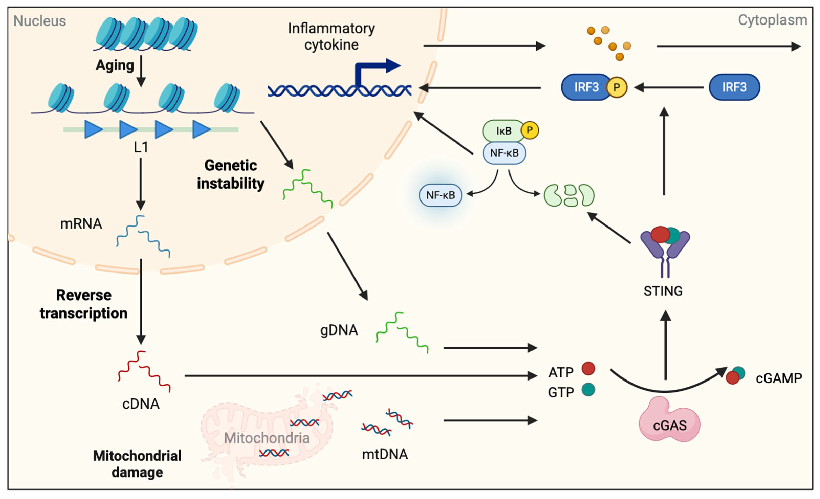
Major sources of endogenous DNA ligands include mitochondrial DNA leakage (for mtDNA), nuclear DNA damage (for gDNA), and retroelement activity (for L1 cDNA). Endogenous DNA ligands in cytosol can be a potent trigger of innate immune response in the CNS: their abnormal accumulation activates the cGAS–STING pathway, driving type-I interferon and NF-κB programs that promote microglial reactivity and neurodegeneration, especially in aging brains [57,58] (Figure 2). L1 expression is normally suppressed by DNA methylation and H3K9me3. However, in aged cells, loss of epigenetic silencing unlocks L1 transcription, leading to the accumulation of L1 cDNA in the cytoplasm following reverse transcription. This also activates the cGAS–STING pathway [59,60]. De Cecco et al. demonstrated that forkhead box A1 (FOXA1) is upregulated in human senescent cells and binds the 5′-UTR of L1, facilitating its reactivation and thereby triggering the cGAS–STING–IFN cascade [61].
Upon sensing endogenous DNA ligands, including L1 cDNA, cGAS synthesizes 2′3′-cGAMP to activate STING, which recruits TANK-binding kinase 1 and phosphorylates Interferon Regulatory Factor 3 (IRF3), while also engaging NF-κB; the result is sustained expression of Interferon beta (IFN-β) and inflammatory cytokines such as Tumor Necrosis Factor alpha (TNF-α) and Interleukin-6 (IL-6) [62]. On the other hand, our previous study has demonstrated that endogenous DNA ligands derived from degenerating neurons activate the Toll-like receptor 9 (TLR9) pathway in microglia within an epileptic mouse model, prompting these cells to release diverse inflammatory cytokines [63]. The effect of these inflammatory cytokines has been reported in brain aging and AD models, where STING activation drives reactive microglial states and cognitive decline, and pharmacological or genetic STING inhibition ameliorates pathology [64,65].
Collectively, an increase in endogenous DNA ligands and the sensing by cGAS–STING form a mechanistic bridge between cellular senescence, chromatin dysregulation, and innate immune activation in the aging brain.

3.3. Chromatin Remodeling Factors and ncRNAs

ncRNAs assist in the formation of chromatin structures and participate in the regulation of gene expression. Long ncRNAs (lncRNAs) recruit epigenetic enzymes (e.g., Polycomb repressive complex 2 (PRC2)) to specific DNA sites, altering local histone modifications [66,67,68]. Enhancer RNA (eRNA) promotes transcription by interacting with mediator and cohesin to help enhancers form loops and bind to promoters, thereby inducing the release of RNA polymerase II from its paused state [69,70,71]. miRNAs act indirectly: they lower or raise the expression levels of writers, erasers, and readers (e.g., Enhancer of zeste homolog 2 (EZH2), Lysine Demethylases (KDMs), HDACs, Bromodomain containing 4 (BRD4)) and shift transcription factor (TF) networks, which then affect chromatin openness [72].
These RNAs collectively establish an “epigenetic framework”—balancing DNA methylation, histone modifications, nucleosome placement, and enhancer activity in response to stimuli. They tip inflammatory genes toward either an “activated state” or an “suppressed state.” In microglia, disease- and age-associated lncRNAs (e.g., Nuclear Enriched Abundant Transcript 1 (NEAT1)) function with chromatin regulators of cytokine genes. miR-155 promotes inflammation, while miR-124 and miR-146a suppress it by negatively regulating NF-κB/Signal transducer and activator of transcription/Activator protein 1 signaling [73,74,75,76,77]. Furthermore, it has also been reported that, as a broad mechanism for enhancer activation, RNAPII binding and eRNA synthesis are crucial for gene expression programs responding to neuronal activity, thereby controlling neuronal function [78]. In parallel, CNS cell cultures revealed that Apolipoprotein E (APOE)-activated ncRNA (AANCR), which exhibits eRNA enhancement characteristics, can confer an inflammatory phenotype to astrocytes by influencing APOE expression [43]. In the future, the targeted inhibition of pro-inflammatory ncRNAs may emerge as a reliable therapeutic approach for controlling neuroinflammation [79].

3.4. Environmental Factors Affecting the Interaction of Epigenetic Alteration and Inflammation

Recent studies have indicated that environmental factors (such as air pollution, heavy metal contamination, chemical exposure, and maternal-fetal immune environments) and lifestyle habits (including circadian rhythms, exercise, and diet) also modulate neuroinflammation through epigenetic alterations. These findings suggest that the neuroinflammation is not only linked to intrinsic factors within the organism (such as oxidative stress) but also involves individual variations driven by external factors (such as lifestyle habits). In this section, we introduce circadian rhythm disruption, exposure to air pollution and drug (Valproic acid (VPA)) exposure as representative examples of how neuroinflammation is induced through epigenetic alterations.

3.4.1. Circadian Clock

Brain and Muscle ARNT-Like 1 (BMAL1) is a core component of the molecular circadian clock that orchestrates daily rhythms in physiology and behavior. Functioning as a transcription factor, BMAL1 forms a heterodimer with CLOCK, which binds to E-box elements in target gene promoters to drive rhythmic expression of core clock genes such as Period Circadian Regulator 1 (PER1), Cryptochrome Circadian Regulator 1 (CRY1) and Rev-Erb alpha (Rev-erbα). This transcriptional activation initiates the primary transcriptional–translational feedback loop underlying circadian oscillations. Beyond its canonical role in circadian regulation, BMAL1 exerts pleiotropic effects across multiple biological systems. It influences cellular metabolism, mitochondrial function, oxidative stress responses, and immune regulation. Loss of BMAL1 function in animal models disrupts circadian rhythmicity, accelerates aging phenotypes, and exacerbates neurodegenerative pathology. In the cerebral cortex of Bmal1 knockout mice, expression of the redox genes NAD(P)H dehydrogenase (quinone 1) (Nqo1) and Aldehyde dehydrogenase 2 family member (Aldh2) decreased, while the number of activated microglia was increased. This indicates that Bmal1 knockout leads to increased brain vulnerability to oxidative stress. Concurrently, Bmal1+/− heterozygous mice subjected to induced oxidative stress (3-NP treatment) exhibited neurodegenerative pathological features [80]. Furthermore, Bmal1–Rev-erbα axis is a central regulatory pathway of the circadian clock, and knockout of Rev-Erba revealed that the Bmal1–Rev-erbα axis critically regulates microglial activation via the NF-κB pathway [81]. Rev-ERBs are known to suppress transcription through multiple mechanisms, including DNA-binding domain (DBD)-dependent binding to Retinoic Acid-Related Orphan Receptors Response Elements (ROREs) [82], tissue-specific transcription factors [83], eRNA expression [84], and DBD-independent interactions regulating chromatin circulation [85]. Although the mechanism by which the Bmal1–Rev-erbα axis regulates transcription of NF-κB pathway-associated genes remains unclear, however, combined with findings from another experiment: 6 h of sleep deprivation affects chromatin accessibility in the mouse cerebral cortex, thereby exerting long-term effects on gene expression [86], it can be inferred that circadian rhythm balance plays a crucial role in regulating neuroinflammation.

3.4.2. PM2.5

Persistent epidemiological and experimental studies demonstrate that air pollution (e.g., particulate matter/ozone) is significantly associated with altered DNA methylation patterns, including differential methylation at inflammatory and stress response pathway gene loci [87,88,89]. Particulate matter 2.5 (PM2.5) refers to airborne particles with a diameter of less than 2.5 μm. Due to their small size, PM2.5 can penetrate deep into the respiratory tract, reach the alveoli, and even enter the bloodstream, thereby exerting systemic effects. In a study related to PM2.5, it was reported that inhaling PM2.5 (1 and 5 mg/kg bw) every other day for 4 weeks induced neuroinflammation in C57BL/6 mice, impaired synaptic integrity, and further reduced spatial learning and memory abilities [90]. Mechanistically, these effects are mediated via NF-κΒ p65–dependent downregulation of miR-574-5p, which targets β-secretase (Bace1). Notably, overexpression of miR-574-5p in the hippocampus reduced Bace1 expression, restored synaptic function, and improved spatial memory and learning following PM2.5 exposure [90]. Supporting a translational relevance to humans, genome-wide DNA methylation analysis of prefrontal cortex tissue from 159 AD patients identified 24 CpG sites mediating the association between PM2.5 exposure and neuropathological markers, several of which reside within genes associated with neuroinflammation [91]. Collectively, these findings reveal a previously unrecognized link between PM2.5-induced air pollution and neuroinflammation in AD, potentially mediated by epigenetic modifications, highlighting the environmental factor’s contributions to the pathogenesis of neurodegenerative diseases.

3.4.3. VPA

VPA is an antiepileptic drug; however also known as an HDAC inhibitor [92]. We have previously shown that VPA enhances neuronal differentiation of NSCs via HDAC inhibitory activity. Furthermore, we have revealed that VPA administration following NSC transplantation into spinal cord-injured mice increases the replenishment of newly generated neurons at the injury site, leading to dramatic recovery of hindlimb function [93]. However, VPA therapy also has adverse effects, and when administered to pregnant mothers, it increases the risk of developing autism spectrum disorder (ASD) [94]. Neuroinflammation is observed in the brains of ASD patients and model mice, and this inflammation is attributable, at least in part, to the pathogenesis of ASD, whereas the cause of the inflammation remains elusive. Nevertheless, it has recently been reported that L1 expression is upregulated in the brain of VPA-exposed ASD model mice compared to controls, probably due to the promotion of histone acetylation of L1 genes through the function of VPA as an HDAC inhibitor [95]. Moreover, VPA has been shown to induce cellular senescence [96], which may shift the epigenetic status of the cells toward aged states, leading to an increase in L1 expression. Therefore, this age-associated epigenetic state shift may also contribute to the enhanced expression of L1 in the VPA-exposed ASD model mice. As explained in Figure 2, L1 is reverse transcribed into cDNA, functioning as an endogenous DNA ligand for DNA sensors, e.g., cGAS and TLR9, inducing neuroinflammation. Taking these facts into consideration, reverse transcriptase inhibitors could be leveraged as therapeutic drugs for the treatment of ASD through the suppression of neuroinflammation.

4. The Tripartite Model: A Dynamic Feed-Forward Loop of Brain Aging, Neuroinflammation, and Epigenetics

Building upon the preceding chapters, we propose a tripartite model in which brain aging, neuroinflammation, and epigenetic remodeling interact in a dynamic, self-reinforcing feed-forward loop. This model goes beyond linear or pairwise interactions between these components and conceptualizes them as an interdependent regulatory triangle. Within this framework, each node—aging, inflammation, or epigenetic alteration—can function both as a driver and a consequence of the other two, collectively driving the progression of neurodegenerative processes.

4.1. Model Overview: Interdependency and Amplification

In the healthy brain, homeostatic control of inflammation, epigenetic plasticity, and age-associated transcriptional regulation maintains neuronal and glial function. However, with aging, epigenetic drift and loss of chromatin integrity predispose brain cells to aberrant gene expression patterns and increased vulnerability to inflammatory stimuli. In turn, chronic low-grade neuroinflammation further disrupts DNA methylation patterns, alters histone landscapes, particularly in glial cells such as microglia and astrocytes.
These inflammation-induced epigenetic alterations are not merely transient but often persistent, forming maladaptive transcriptional states that impair neurotrophic support and stress resilience. As a result, the accumulation of metabolic stress, ROS, and immune activation further accelerates brain aging. This vicious cycle thus connects the decline in transcriptional plasticity, heightened immune reactivity, and structural chromatin deterioration into a single progressive loop. Importantly, the feed-forward strength intensifies with age, pushing the nervous system from a state of resilience toward irreversible degeneration.

4.2. Case Example of the Network Disorder: Neurodegenerative Diseases

4.2.1. Alzheimer’s Disease (AD)

AD exemplifies the complex tripartite feed-forward loop between aging, neuroinflammation, and epigenetic dysregulation. While classical models emphasize Aβ plaques and tau tangles, convergent human and animal studies now show that glial activation, immune remodeling, and chromatin-level alterations are integral to disease progression.
It has been observed that the epigenetic clock is accelerated in AD contexts. In the 3xTg-AD model mouse, the cortex shows a stronger acceleration of the clock than the hippocampus, consistent with region-specific vulnerability [97]. At the DNA methylation level, genome-wide profiling of the superior temporal gyrus has identified hundreds of AD-associated differentially methylated regions, many overlapping poised promoters marked by H3K4me3 and H3K27me3, suggesting aberrant programming of neuronal and immune-associated genes [98]. Single-cell profiling revealed disease-associated microglia and related human microglial states with inflammatory/chemokine programs and Apolipoprotein E (Apo-E) upregulation. These states display chromatin remodeling at inflammatory loci and upregulation of cytokine/chemokine pathways (e.g., IL-1β, C-C motif chemokine ligand 2 (CCL2)), supporting an epigenetically primed innate immune response in AD [36,99,100,101].
At the chromatin level, AD brains show large-scale histone acetylation reconfiguration, including extensive H3K27ac changes at enhancers/promoters and loss of H4K16ac in the lateral temporal lobe; in parallel, Hdac2 upregulation is linked to repression of synaptic plasticity-related genes and memory impairment. Rather than a uniform global decrease, the acetylation landscape appears imbalanced and redistributed across disease-relevant regions [21,102,103].
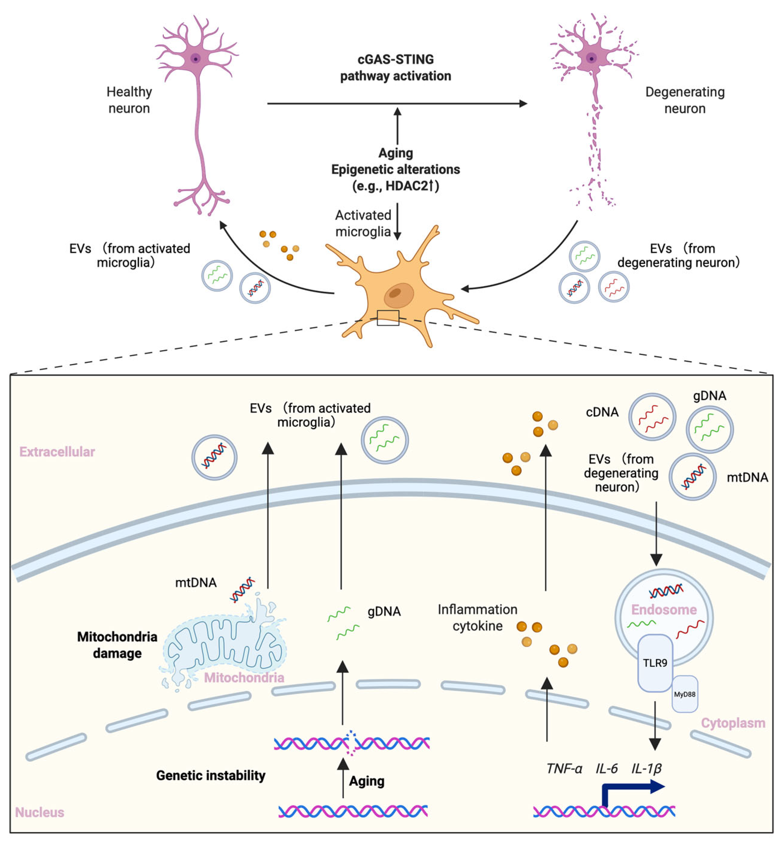
A recent study showed that, in mouse models, aging leads to the accumulation of endogenous DNA ligands in microglia (e.g., gDNA and mtDNA), which is transferred to neurons via extracellular vesicles and, by triggering the cGAS–STING pathway, elicits a widespread and sustained neuroinflammatory response that in turn induces neuronal degeneration [104]. This result provides a new possibility for triggering inflammation: endogenous genes generated by aging can not only activate inflammation-related pathways within the cytosol but may also be transferred to other types of cells to elicit inflammatory responses.
Integrating the pivotal role of epigenetic alterations in aging and neuroinflammation, we sought to explain part of AD pathogenesis with a feed-forward loop framework. As neurons age and undergo epigenetic alterations, gene expression becomes unstable; the resulting endogenous DNA ligands, such as gDNA, mtDNA and L1 cDNA, activate the cGAS–STING pathway, provoking neuroinflammation, neuronal degeneration, and microglial activation with release of inflammatory cytokines (e.g., TNFα, IL-6). Endogenous DNA ligands leaking from degenerating neurons further stimulate microglia, probably through TLR9 [63,105], to secrete cytokines, exacerbating the toxic inflammatory milieu in the brain. Conversely, aging and epigenetic alterations can also drive the accumulation of endogenous DNA ligands within microglia, which can be delivered to neurons via extracellular vesicles, thereby further engaging neuronal cGAS–STING and sustaining a broad inflammatory cascade (Figure 3). Under such chronic neuroinflammatory conditions, the genomic instability driven by aging and epigenetics is further exacerbated, and microglial immune functions become dysregulated, diminishing their protective role against neuroinflammation. On this basis—combined with other pathological processes such as tau protein deposition—irreversible neurodegenerative decline in brain function ultimately occurs.
Together, our model frames AD not merely as a disorder of peptide aggregation, but as a systems level collapse of immune epigenetic homeostasis, wherein aging related epigenetic drift, glial inflammatory priming, and chromatin remodeling act in concert to mutually reinforce disease progression.

4.2.2. Parkinson’s Disease (PD)

Supportive evidence for the tripartite loop model we propose has been confirmed in other neurodegenerative diseases (such as PD and amyotrophic lateral sclerosis (ALS), in addition to AD. Of note is the increase in endogenous DNA ligands induced by aging and epigenetic dysregulation, which activates the cGAS-STING pathway and triggers neuroinflammation.
In PD, misfolded α-syn aggregates in Lewy bodies and neurites, impairing synaptic vesicle trafficking and mitochondrial quality control. The oligomeric/fibrillar species of α-syn disseminate in a prion-like manner, imposing oxidative and genotoxic stress on microglia, which subsequently reprogram astrocytes into neurotoxic states [106,107,108,109]. In addition, aging induces epigenetic alterations (DNA demethylation) at PD risk gene loci (most prominently Synuclein alpha (SNCA) and Parkin (PARK2)), resulting in the upregulation of their expression, which further promotes the accumulation of α-syn. Moreover, aging-induced impairment of mitophagy–lysosomal flux and of genome maintenance increase endogenous DNA ligands (e.g., mtDNA, gDNA and L1 cDNA) in the cytosol that triggers cGAS–STING pathway activation [110,111,112,113,114,115,116]. It has been also reported that aggregated α-syn induces an increase of endogenous DNA ligands (e.g., mtDNA and gDNA), activating the cGAS–STING pathway [117]. This pathway activation results in the microglia–astrocyte inflammatory cascade and accelerates nigral degeneration, whereas genetic or pharmacologic attenuation of cGAS–STING signaling has been shown to mitigate the neuroinflammation and pathology of a PD mouse model [118,119,120,121]. Taken together, in PD, the following axis is assumed to exist: “Aging → Epigenetic dysregulation → α-syn aggregation → Organellar and genome stress → Neuroinflammation via cGAS–STING pathway activation → Degeneration of dopaminergic neurons” [110,118,119,120,121,122]. Therefore, the components in this axis could be a promising therapeutic target for the treatment of PD.

4.2.3. Amyotrophic Lateral Sclerosis (ALS)

In the dominant histopathology of ALS, that is, in TAR DNA-binding protein 43 (TDP-43) proteinopathy, TDP-43 plays a crucial role in the induction of genome maintenance defects and mitochondrial stress, which activate innate immune signaling. Loss or abnormal localization of TDP-43 disrupts nuclear RNA/DNA homeostasis and chromatin structure, relaxing heterochromatin around L1 genes to increase their expression while broadly reducing the expression of DNA damage and repair-related genes [123,124]. In parallel, TDP-43 dysfunction drives mtDNA release through the 1-methyl-4-phenyl-1,2,3,6-tetrahydropyridine (MPTP), which is sensed by cGAS to activate STING and type-I interferon programs. This mechanism has been demonstrated in patient-derived iPSC motor neurons and mouse models and is mitigated by genetic or pharmacological pathway inhibition [125,126,127]. In addition, it was previously reported that de-repression of retrotransposons contributes to neurodegeneration even in a Drosophila TDP-43 model, supporting a feed-forward loop between genome instability and neuroinflammation [128,129,130]. Together, these data outline an ALS pathway logic: Nuclear depletion of TDP-43 → chromatin/retroelement instability and mtDNA leakage → cGAS–STING–driven inflammation → motor-neuron degeneration.
Taken all together, the body of evidence spanning AD, PD, and ALS argues strongly for the broad relevance of our proposed tripartite feed-forward loop to brain aging and neurodegeneration. Regarding the putative route in which aging-associated L1 upregulation drives cGAS–STING pathway activation, findings from animal models and human specimens provide substantial indirect support that this mechanism likely operates across multiple neurodegenerative conditions (Table 2). However, to the best of our knowledge, no study in PD or ALS has yet shown that selective suppression of L1 expression directly attenuates cGAS–STING activation in the same model. Establishing this causal linkage—in vivo or in rigorously humanized systems—thus represents an urgent experimental priority for the field.

5. Epigenetics-Related Therapeutic Interventions

During brain aging, the interplay among epigenetic reprogramming, innate immune activation, and cellular senescence defines a central axis of brain functional vulnerability. We have outlined how intrinsic and extrinsic stressors initiate and propagate this axis through convergent mechanisms. The tripartite model proposed here emphasizes that aging, inflammation, and epigenetic dysregulation form a mutually reinforcing chain that accelerates cognitive decline and heightens susceptibility to neurodegenerative diseases (Figure 4).
Within our tripartite framework, two axes are important. Axis 1 links environmental exposures and lifestyle factors with chronic neuroinflammation to drive epigenetic age acceleration and DNA damage, which induces functional compromise of neural cell types (most notably microglial immune dysregulation) and diminishes cellular resilience to aging. These changes, in turn, accelerate brain senescence and increase vulnerability to neurodegeneration. Axis 2 links aging- and disease-associated gene instability to proteinopathy. Accompanied by increased endogenous DNA ligands (e.g., gDNA, L1 cDNA and mtDNA), cGAS–STING pathway is engaged to amplify neuroinflammation. Targeted interventions along these two axes may therefore provide a rationale for anti-aging and disease-modifying strategies in neurodegenerative disorders.
The timing of the intervention is also important. To determine the optimal timing, it is crucial to leverage cutting-edge technologies such as AI-assisted high-dimensional single-cell profiling (scRNA-seq, scATAC-seq, single-cell methylome) to learn and detect epigenetic and inflammatory markers within the feed-forward loop (e.g., L1 cDNA). This enables early prediction of brain aging trajectories and neurodegenerative progression, contributing to the design of intervention strategies (e.g., modulation of key epigenetic writers/erasers, restoration of redox balance, adjustment of neuroimmune tone). Animal studies indicated that overexpression of Tet2 or inhibition of Hdac2 can enhance NSC activity and improve the functions of the aged brain [16,143]. In parallel, given that loss of epigenetic silencing elevates L1 mRNA expression, which is reverse-transcribed into cDNA and can trigger cGAS–STING pathway activation, to prevent cDNA production by reverse-transcriptase inhibitors represents a feasible therapeutic measure [144].
Despite abundant animal data and a small number of early clinical observations supporting our proposed model, deeper validation using human cells and tissues is necessary to account for interspecies differences. We believe there are two paths we can take to achieve this. First is to leverage patient-derived iPSC brain organoids that can recapitulate human-specific phenotypes attributable to human genetic backgrounds [145,146,147]. Second is to construct spatiotemporal atlases of human brain aging–inflammation–epigenetic alterations—analogous to recent works in human brain development [148]. Although conventional single-cell assays have not been able to present information of histone modification changes at single-cell level, emerging platforms provide a new possibility: The Epitope-based Time-of-Flight Mass Spectrometry (EpiTOF) method simultaneously quantifies multiple histone modifications/epigenetic enzymes and cell lineage markers at single-cell resolution to derive cell type-specific “epigenetic fingerprints” [149]. Spatial-DMT (DNA Methylome and Transcriptome) simultaneously profiles DNA methylation and transcription across tissue sections, mapping genome-wide DNA methylation and elucidating the spatial coupling of epigenetic states and transcription [150].
However, several clear limitations still constrain the application of these two technologies. EpiTOF depends heavily on antibody specificity, limiting cross-laboratory comparability. Spatial-DMT currently operates at pixel, rather than true single-nucleus resolution. Accordingly, optimizing assay process and improving quantitative precision, while establishing standardized workflows and reference datasets, will be decisive for building reliable, human-anchored spatiotemporal maps of aging, inflammation, and epigenetic regulation.
Suppression of neuroinflammation is increasingly recognized as an effective anti-aging strategy [151]. Given that our above-mentioned Axis 2 converges on cGAS–STING pathway as a principal route to neuroinflammation, we view pharmacologic inhibition of the cGAS–STING pathway as a practical therapeutic direction. To that end, we summarize in Table 2 preclinical and clinical attempts to dampen cGAS–STING signaling across aging, AD, PD, ALS, and other neurodegenerative disorders. Although human data on cGAS–STING inhibition remain limited and largely in early-phase, encouragingly, open-label phase IIa pilot trial (NCT04552795) enrolling participants with MCI/early AD, oral lamivudine (3TC, a reverse transcriptase inhibitor) administration for 24 weeks was safe and well tolerated and was associated with reductions in multiple CSF biomarkers of neurodegeneration and neuroinflammation. An increase in the plasma Aβ 42/40 ratio—typically interpreted as consistent with lower amyloid burden was observed, although the study did not directly quantify brain plaque load [135]. Looking forward, we anticipate and encourage more clinical evaluations of cGAS–STING pathway suppression, pathway modulation, and disease modification.
Ultimately, current findings on neurodegeneration and anti-aging support the validity of our proposed tripartite model, in which epigenetic alterations mediate the crosstalk between neuroinflammation and brain aging. Further exploration of this model—and elucidation of the mutual influences among its three components—will have substantial research and translational value for therapies targeting neurodegenerative diseases and brain aging.

Author Contributions

Conceptualization, S.M., H.N. and K.N.; writing—original draft preparation, S.M., H.N. and K.N.; writing—review and editing, S.M., H.N. and K.N.; supervision, K.N.; funding acquisition, H.N. and K.N. All authors have read and agreed to the published version of the manuscript.

Funding

This work was funded by the Japan Society for the Promotion of Science (JSPS) KAKENHI (JP23H00391, JP25H01319), and AMED (JP20gm1310008, JP24wm0625306) to K.N.; JSPS KAKENHI (JP25H01866) and AMED (JP23ek0109640) to H.N.

Institutional Review Board Statement

Not applicable.

Informed Consent Statement

Not applicable.

Data Availability Statement

Not applicable.

Acknowledgments

We thank all of the members of the Department of Stem Cell Biology and Medicine, Kyushu University, for valuable comments.

Conflicts of Interest

The authors declare no conflicts of interest.

References

  1. Weismann, A. Essays Upon Heredity; Clarendon Press: Oxford, UK, 1891. [Google Scholar]
  2. López-Otín, C.; Blasco, M.A.; Partridge, L.; Serrano, M.; Kroemer, G. Hallmarks of aging: An expanding universe. Cell 2023, 186, 243–278. [Google Scholar] [CrossRef] [PubMed]
  3. Barter, J.D.; Foster, T.C. Aging in the Brain: New Roles of Epigenetics in Cognitive Decline. Neuroscience 2018, 24, 516–525. [Google Scholar] [CrossRef]
  4. Jurcau, M.C.; Jurcau, A.; Cristian, A.; Hogea, V.O.; Diaconu, R.G.; Nunkoo, V.S. Inflammaging and Brain Aging. Int. J. Mol. Sci. 2024, 25, 10535. [Google Scholar] [CrossRef]
  5. Giallongo, S.; Longhitano, L.; Denaro, S.; D’aprile, S.; Torrisi, F.; La Spina, E.; Giallongo, C.; Mannino, G.; Furno, D.L.; Zappalà, A.; et al. The Role of Epigenetics in Neuroinflammatory-Driven Diseases. Int. J. Mol. Sci. 2022, 23, 15218. [Google Scholar] [CrossRef]
  6. Bin Imtiaz, M.K.; Jaeger, B.N.; Bottes, S.; Machado, R.A.; Vidmar, M.; Moore, D.L.; Jessberger, S. Declining lamin B1 expression mediates age-dependent decreases of hippocampal stem cell activity. Cell Stem Cell 2021, 28, 967–977.e8. [Google Scholar] [CrossRef]
  7. Bedrosian, T.A.; Houtman, J.; Eguiguren, J.S.; Ghassemzadeh, S.; Rund, N.; Novaresi, N.M.; Hu, L.; Parylak, S.L.; Denli, A.M.; Randolph-Moore, L.; et al. Lamin B1 decline underlies age-related loss of adult hippocampal neurogenesis. EMBO J. 2021, 40, 967–977. [Google Scholar] [CrossRef]
  8. Zhao, T.; Hong, Y.; Yan, B.; Huang, S.; Ming, G.-L.; Song, H. Epigenetic maintenance of adult neural stem cell quiescence in the mouse hippocampus via Setd1a. Nat. Commun. 2024, 15, 5674. [Google Scholar] [CrossRef]
  9. Matsubara, S.; Matsuda-Ito, K.; Sekiryu, H.; Doi, H.; Nakagawa, T.; Murao, N.; Oda, H.; Nakashima, K.; Matsuda, T. Epigenetic regulation of neural stem cell aging in the mouse hippocampus by Setd8 downregulation. EMBO J. 2025, 44, 3645–3668. [Google Scholar] [CrossRef] [PubMed]
  10. Gontier, G.; Iyer, M.; Shea, J.M.; Bieri, G.; Wheatley, E.G.; Ramalho-Santos, M.; Villeda, S.A. Tet2 Rescues Age-Related Regenerative Decline and Enhances Cognitive Function in the Adult Mouse Brain. Cell Rep. 2018, 22, 1974–1981. [Google Scholar] [CrossRef]
  11. Janke, C.; Montagnac, G. Causes and Consequences of Microtubule Acetylation. Curr. Biol. 2017, 27, R1287–R1292. [Google Scholar] [CrossRef] [PubMed]
  12. Guan, J.-S.; Haggarty, S.J.; Giacometti, E.; Dannenberg, J.-H.; Joseph, N.; Gao, J.; Nieland, T.J.F.; Zhou, Y.; Wang, X.; Mazitschek, R.; et al. HDAC2 negatively regulates memory formation and synaptic plasticity. Nature 2009, 459, 55–60. [Google Scholar] [CrossRef] [PubMed]
  13. Ye, J.; Zhong, S.; Wan, H.; Guo, X.; Yao, X.; Liu, Q.; Chen, L.; Wang, J.-Z.; Xiao, S. Upregulated astrocyte HDAC7 induces Alzheimer-like tau pathologies via deacetylating transcription factor-EB and inhibiting lysosome biogenesis. Mol. Neurodegener. 2025, 20, 5. [Google Scholar] [CrossRef]
  14. Zhou, J.; Wu, Y.-C.; Xiao, B.-J.; Guo, X.-D.; Zheng, Q.-X.; Wu, B. Age-related Changes in the Global DNA Methylation Profile of Oligodendrocyte Progenitor Cells Derived from Rat Spinal Cords. Curr. Med. Sci. 2019, 39, 67–74. [Google Scholar] [CrossRef]
  15. Cho, S.-H.; Chen, J.A.; Sayed, F.; Ward, M.E.; Gao, F.; Nguyen, T.A.; Krabbe, G.; Sohn, P.D.; Lo, I.; Minami, S.; et al. SIRT1 Deficiency in Microglia Contributes to Cognitive Decline in Aging and Neurodegeneration via Epigenetic Regulation of IL-1β. J. Neurosci. 2015, 35, 807–818. [Google Scholar] [CrossRef]
  16. Carrillo-Jimenez, A.; Deniz, Ö.; Niklison-Chirou, M.V.; Ruiz, R.; Bezerra-Salomão, K.; Stratoulias, V.; Amouroux, R.; Yip, P.K.; Vilalta, A.; Cheray, M.; et al. TET2 Regulates the Neuroinflammatory Response in Microglia. Cell Rep. 2019, 29, 697–713.e8. [Google Scholar] [CrossRef]
  17. Ginhoux, F.; Greter, M.; Leboeuf, M.; Nandi, S.; See, P.; Gokhan, S.; Mehler, M.F.; Conway, S.J.; Ng, L.G.; Stanley, E.R.; et al. Fate mapping analysis reveals that adult microglia derive from primitive macrophages. Science 2010, 330, 841–845. [Google Scholar] [CrossRef]
  18. Eriksson, P.S.; Perfilieva, E.; Björk-Eriksson, T.; Alborn, A.M.; Nordborg, C.; Peterson, D.A.; Gage, F.H. Neurogenesis in the adult human hippocampus. Nat. Med. 1998, 4, 1313–1317. [Google Scholar] [CrossRef]
  19. Gage, F.H. Mammalian neural stem cells. Science 2000, 287, 1433–1438. [Google Scholar] [CrossRef] [PubMed]
  20. Dumitru, I.; Paterlini, M.; Zamboni, M.; Ziegenhain, C.; Giatrellis, S.; Saghaleyni, R.; Björklund, Å.; Alkass, K.; Tata, M.; Druid, H.; et al. Identification of proliferating neural progenitors in the adult human hippocampus. Science 2025, 389, 58–63. [Google Scholar] [CrossRef] [PubMed]
  21. Moreno-Jiménez, E.P.; Flor-García, M.; Terreros-Roncal, J.; Rábano, A.; Cafini, F.; Pallas-Bazarra, N.; Ávila, J.; Llorens-Martín, M. Adult hippocampal neurogenesis is abundant in neurologically healthy subjects and drops sharply in patients with Alzheimer’s disease. Nat. Med. 2019, 25, 554–560. [Google Scholar] [CrossRef]
  22. Van Steensel, B.; Belmont, A.S. Lamina-Associated Domains: Links with Chromosome Architecture, Heterochromatin, and Gene Repression. Cell 2017, 169, 780–791. [Google Scholar] [CrossRef] [PubMed]
  23. Rizzo, V.; Richman, J.; Puthanveettil, S.V. Dissecting mechanisms of brain aging by studying the intrinsic excitability of neurons. Front. Aging Neurosci. 2015, 6, 337. [Google Scholar] [CrossRef] [PubMed]
  24. Lu, A.T.; Hannon, E.; Levine, M.E.; Crimmins, E.M.; Lunnon, K.; Mill, J.; Geschwind, D.H.; Horvath, S. Genetic architecture of epigenetic and neuronal ageing rates in human brain regions. Nat. Commun. 2017, 8, 15353. [Google Scholar] [CrossRef]
  25. Singh, P.; Thakur, M.K. Histone Deacetylase 2 Inhibition Attenuates Downregulation of Hippocampal Plasticity Gene Expression during Aging. Mol. Neurobiol. 2018, 55, 2432–2442. [Google Scholar] [CrossRef] [PubMed]
  26. Pao, P.-C.; Patnaik, D.; Watson, L.A.; Gao, F.; Pan, L.; Wang, J.; Adaikkan, C.; Penney, J.; Cam, H.P.; Huang, W.-C.; et al. HDAC1 modulates OGG1-initiated oxidative DNA damage repair in the aging brain and Alzheimer’s disease. Nat. Commun. 2020, 11, 2484. [Google Scholar] [CrossRef]
  27. Labarta-Bajo, L.; Allen, N.J. Astrocytes in aging. Neuron 2025, 113, 109–126. [Google Scholar] [CrossRef]
  28. Namihira, M.; Kohyama, J.; Semi, K.; Sanosaka, T.; Deneen, B.; Taga, T.; Nakashima, K. Committed Neuronal Precursors Confer Astrocytic Potential on Residual Neural Precursor Cells. Dev. Cell 2009, 16, 245–255. [Google Scholar] [CrossRef]
  29. Sanosaka, T.; Imamura, T.; Hamazaki, N.; Chai, M.; Igarashi, K.; Ideta-Otsuka, M.; Miura, F.; Ito, T.; Fujii, N.; Ikeo, K.; et al. DNA Methylome Analysis Identifies Transcription Factor-Based Epigenomic Signatures of Multilineage Competence in Neural Stem/Progenitor Cells. Cell Rep. 2017, 20, 2992–3003. [Google Scholar] [CrossRef]
  30. Takizawa, T.; Nakashima, K.; Namihira, M.; Ochiai, W.; Uemura, A.; Yanagisawa, M.; Fujita, N.; Nakao, M.; Taga, T. DNA Methylation Is a Critical Cell-Intrinsic Determinant of Astrocyte Differentiation in the Fetal Brain. Dev. Cell 2001, 1, 749–758. [Google Scholar] [CrossRef]
  31. Chisholm, N.C.; Henderson, M.L.; Selvamani, A.; Park, M.J.; Dindot, S.; Miranda, R.C.; Sohrabji, F. Histone methylation patterns in astrocytes are influenced by age following ischemia. Epigenetics 2015, 10, 142–152. [Google Scholar] [CrossRef]
  32. Windener, F.; Grewing, L.; Thomas, C.; Dorion, M.-F.; Otteken, M.; Kular, L.; Jagodic, M.; Antel, J.; Albrecht, S.; Kuhlmann, T. Physiological aging and inflammation-induced cellular senescence may contribute to oligodendroglial dysfunction in MS. Acta Neuropathol. 2024, 147, 82. [Google Scholar] [CrossRef]
  33. Shen, S.; Liu, A.; Li, J.; Wolubah, C.; Casaccia-Bonnefil, P. Epigenetic memory loss in aging oligodendrocytes in the corpus callosum. Neurobiol. Aging 2008, 29, 452–463. [Google Scholar] [CrossRef]
  34. Liu, X.; Xin, D.E.; Zhong, X.; Zhao, C.; Li, Z.; Zhang, L.; Dourson, A.J.; Lee, L.; Mishra, S.; Bayat, A.E.; et al. Small-molecule-induced epigenetic rejuvenation promotes SREBP condensation and overcomes barriers to CNS myelin regeneration. Cell 2024, 187, 2465–2484.e22. [Google Scholar] [CrossRef]
  35. Li, X.; Li, Y.; Jin, Y.; Zhang, Y.; Wu, J.; Xu, Z.; Huang, Y.; Cai, L.; Gao, S.; Liu, T.; et al. Publisher Correction: Transcriptional and epigenetic decoding of the microglial aging process. Nat. Aging 2023, 4, 276. [Google Scholar] [CrossRef] [PubMed]
  36. Hammond, T.R.; Dufort, C.; Dissing-Olesen, L.; Giera, S.; Young, A.; Wysoker, A.; Walker, A.J.; Gergits, F.; Segel, M.; Nemesh, J.; et al. Single-Cell RNA Sequencing of Microglia throughout the Mouse Lifespan and in the Injured Brain Reveals Complex Cell-State Changes. Immunity 2019, 50, 253–271.e6. [Google Scholar] [CrossRef]
  37. Chen, J.; Huang, J.; Han, T.; Kojima, N. Chronic Stress Modulates Microglial Activation Dynamics, Shaping Priming Responses to Subsequent Stress. Brain Sci. 2025, 15, 534. [Google Scholar] [CrossRef] [PubMed]
  38. Kitazawa, M.; Cheng, D.; Tsukamoto, M.R.; A Koike, M.; Wes, P.D.; Vasilevko, V.; Cribbs, D.H.; LaFerla, F.M. Blocking IL-1 Signaling Rescues Cognition, Attenuates Tau Pathology, and Restores Neuronal β-Catenin Pathway Function in an Alzheimer’s Disease Model. J. Immunol. 2011, 187, 6539–6549. [Google Scholar] [CrossRef]
  39. Wendeln, A.-C.; Degenhardt, K.; Kaurani, L.; Gertig, M.; Ulas, T.; Jain, G.; Wagner, J.; Häsler, L.M.; Wild, K.; Skodras, A.; et al. Innate immune memory in the brain shapes neurological disease hallmarks. Nature 2018, 556, 332–338. [Google Scholar] [CrossRef]
  40. Gosselin, D.; Skola, D.; Coufal, N.G.; Holtman, I.R.; Schlachetzki, J.C.M.; Sajti, E.; Jaeger, B.N.; O’Connor, C.; Fitzpatrick, C.; Pasillas, M.P.; et al. An environment-dependent transcriptional network specifies human microglia identity. Science 2017, 356, eaal3222. [Google Scholar] [CrossRef] [PubMed]
  41. Gjoneska, E.; Pfenning, A.R.; Mathys, H.; Quon, G.; Kundaje, A.; Tsai, L.-H.; Kellis, M. Conserved epigenomic signals in mice and humans reveal immune basis of Alzheimer’s disease. Nature 2015, 518, 365–369. [Google Scholar] [CrossRef]
  42. Marschallinger, J.; Iram, T.; Zardeneta, M.; Lee, S.E.; Lehallier, B.; Haney, M.S.; Pluvinage, J.V.; Mathur, V.; Hahn, O.; Morgens, D.W.; et al. Lipid-droplet-accumulating microglia represent a dysfunctional and proinflammatory state in the aging brain. Nat. Neurosci. 2020, 23, 194–208. [Google Scholar] [CrossRef]
  43. Wan, M.; Liu, Y.; Li, D.; Snyder, R.J.; Elkin, L.B.; Day, C.R.; Rodriguez, J.; Grunseich, C.; Mahley, R.W.; Watts, J.A.; et al. The enhancer RNA, AANCR, regulates APOE expression in astrocytes and microglia. Nucleic Acids Res. 2024, 52, 10235–10254. [Google Scholar] [CrossRef]
  44. Niraula, A.; Sheridan, J.F.; Godbout, J.P. Microglia Priming with Aging and Stress. Neuropsychopharmacology 2017, 42, 318–333. [Google Scholar] [CrossRef]
  45. Perry, V.H.; Holmes, C. Microglial priming in neurodegenerative disease. Nat. Rev. Neurol. 2014, 10, 217–224. [Google Scholar] [CrossRef]
  46. Sarlus, H.; Heneka, M.T. Microglia in Alzheimer’s disease. J. Clin. Invest. 2017, 217, 3240–3249. [Google Scholar] [CrossRef] [PubMed]
  47. Matt, S.M.; Zimmerman, J.D.; Lawson, M.A.; Bustamante, A.C.; Uddin, M.; Johnson, R.W. Inhibition of DNA Methylation With Zebularine Alters Lipopolysaccharide-Induced Sickness Behavior and Neuroinflammation in Mice. Front. Neurosci. 2018, 12, 636. [Google Scholar] [CrossRef] [PubMed]
  48. Seddon, A.R.; MacArthur, C.P.; Hampton, M.B.; Stevens, A.J. Inflammation and DNA methylation in Alzheimer’s disease: Mechanisms of epigenetic remodelling by immune cell oxidants in the ageing brain. Redox Rep. 2024, 29, 2428152. [Google Scholar] [CrossRef]
  49. Peng, S.; Zhao, S.; Yan, F.; Cheng, J.; Huang, L.; Chen, H.; Liu, Q.; Ji, X.; Yuan, Z. HDAC2 Selectively Regulates FOXO3a-Mediated Gene Transcription during Oxidative Stress-Induced Neuronal Cell Death. J. Neurosci. 2015, 35, 1250–1259. [Google Scholar] [CrossRef]
  50. Peleg, S.; Sananbenesi, F.; Zovoilis, A.; Burkhardt, S.; Bahari-Javan, S.; Agis-Balboa, R.C.; Cota, P.; Wittnam, J.L.; Gogol-Doering, A.; Opitz, L.; et al. Altered Histone Acetylation Is Associated with Age-Dependent Memory Impairment in Mice. Science 2010, 328, 753–756. [Google Scholar] [CrossRef]
  51. Simpson, D.S.A.; Oliver, P.L. ROS Generation in Microglia: Understanding Oxidative Stress and Inflammation in Neurodegenerative Disease. Antioxidants 2020, 9, 743. [Google Scholar] [CrossRef] [PubMed]
  52. Luna, A.; I Aladjem, M.; Kohn, K.W. SIRT1/PARP1 crosstalk: Connecting DNA damage and metabolism. Genome Integr. 2013, 4, 6. [Google Scholar] [CrossRef]
  53. Kauppinen, T.M.; Gan, L.; Swanson, R.A. Poly(ADP-ribose) polymerase-1-induced NAD+ depletion promotes nuclear factor-κB transcriptional activity by preventing p65 de-acetylation. Biochim. Biophys. Acta 2013, 1833, 1985–1991. [Google Scholar] [CrossRef]
  54. Wu, Q.-J.; Zhang, T.-N.; Chen, H.-H.; Yu, X.-F.; Lv, J.-L.; Liu, Y.-Y.; Liu, Y.-S.; Zheng, G.; Zhao, J.-Q.; Wei, Y.-F.; et al. The sirtuin family in health and disease. Signal Transduct. Target. Ther. 2022, 7, 402. [Google Scholar] [CrossRef]
  55. Cardoso, A.L.; Guedes, J.R.; Pereira de Almeida, L.; Pedroso de Lima, M.C. miR-155 modulates microglia-mediated immune re-sponse by down-regulating SOCS-1 and promoting cytokine and nitric oxide production. Immunology 2012, 135, 73–88. [Google Scholar] [CrossRef]
  56. Liang, Y.; Wang, L. Inflamma-MicroRNAs in Alzheimer’s Disease: From Disease Pathogenesis to Therapeutic Potentials. Front. Cell Neurosci. 2021, 15, 785433. [Google Scholar] [CrossRef]
  57. Gulen, M.F.; Samson, N.; Keller, A.; Schwabenland, M.; Liu, C.; Glück, S.; Thacker, V.V.; Favre, L.; Mangeat, B.; Kroese, L.J.; et al. cGAS-STING drives ageing-related inflammation and neurodegeneration. Nature 2023, 620, 374–380. [Google Scholar] [CrossRef] [PubMed]
  58. Govindarajulu, M.; Ramesh, S.; Beasley, M.; Lynn, G.; Wallace, C.; Labeau, S.; Pathak, S.; Nadar, R.; Moore, T.; Dhanasekaran, M. Role of cGAS-Sting Signaling in Alzheimer’s Disease. Int. J. Mol. Sci. 2023, 24, 8151. [Google Scholar] [CrossRef] [PubMed]
  59. Fukuda, K.; Shinkai, Y. SETDB1-Mediated Silencing of Retroelements. Viruses 2020, 12, 596. [Google Scholar] [CrossRef]
  60. Padeken, J.; Methot, S.P.; Gasser, S.M. Establishment of H3K9-methylated heterochromatin and its functions in tissue differentia-tion and maintenance. Nat. Rev. Mol. Cell Biol. 2022, 23, 623–640. [Google Scholar] [CrossRef] [PubMed]
  61. De Cecco, M.; Ito, T.; Petrashen, A.P.; Elias, A.E.; Skvir, N.J.; Criscione, S.W.; Caligiana, A.; Brocculi, G.; Adney, E.M.; Boeke, J.D.; et al. L1 drives IFN in senescent cells and promotes age-associated inflammation. Nature 2019, 566, 73–78. [Google Scholar] [CrossRef]
  62. Zhang, X.; Bai, X.-C.; Chen, Z.J. Structures and Mechanisms in the cGAS-STING Innate Immunity Pathway. Immunity 2020, 53, 43–53. [Google Scholar] [CrossRef]
  63. Matsuda, T.; Murao, N.; Katano, Y.; Juliandi, B.; Kohyama, J.; Akira, S.; Kawai, T.; Nakashima, K. TLR9 signalling in microglia attenuates seizure-induced aberrant neurogenesis in the adult hippocampus. Nat. Commun. 2015, 6, 6514. [Google Scholar] [CrossRef] [PubMed]
  64. Paul, B.D.; Snyder, S.H.; Bohr, V.A. Signaling by cGAS–STING in Neurodegeneration, Neuroinflammation, and Aging. Trends Neurosci. 2021, 44, 83–96. [Google Scholar] [CrossRef] [PubMed]
  65. Udeochu, J.C.; Amin, S.; Huang, Y.; Fan, L.; Torres, E.R.S.; Carling, G.K.; Liu, B.; McGurran, H.; Coronas-Samano, G.; Kauwe, G.; et al. Tau activation of microglial cGAS-IFN reduces MEF2C-mediated cognitive resilience. Nat. Neurosci. 2023, 26, 737–750. [Google Scholar] [CrossRef] [PubMed]
  66. Zhao, J.; Sun, B.K.; Erwin, J.A.; Song, J.-J.; Lee, J.T. Polycomb Proteins Targeted by a Short Repeat RNA to the Mouse X Chromosome. Science 2008, 322, 750–756. [Google Scholar] [CrossRef]
  67. Kaneko, S.; Bonasio, R.; Saldaña-Meyer, R.; Yoshida, T.; Son, J.; Nishino, K.; Umezawa, A.; Reinberg, D. Interactions between JARID2 and Noncoding RNAs Regulate PRC2 Recruitment to Chromatin. Mol. Cell 2014, 53, 290–300. [Google Scholar] [CrossRef]
  68. Rinn, J.L.; Chang, H.Y. Genome regulation by long noncoding RNAs. Annu. Rev. Biochem. 2012, 81, 145–166. [Google Scholar] [CrossRef]
  69. Kagey, M.H.; Newman, J.J.; Bilodeau, S.; Zhan, Y.; Orlando, D.A.; Van Berkum, N.L.; Ebmeier, C.C.; Goossens, J.; Rahl, P.B.; Levine, S.S.; et al. Mediator and cohesin connect gene expression and chromatin architecture. Nature 2010, 467, 430–435. [Google Scholar] [CrossRef]
  70. Schaukowitch, K.; Joo, J.-Y.; Liu, X.; Watts, J.K.; Martinez, C.; Kim, T.-K. Enhancer RNA Facilitates NELF Release from Immediate Early Genes. Mol. Cell 2014, 56, 29–42. [Google Scholar] [CrossRef]
  71. Tsai, P.-F.; Dell’oRso, S.; Rodriguez, J.; Vivanco, K.O.; Ko, K.-D.; Jiang, K.; Juan, A.H.; Sarshad, A.A.; Vian, L.; Tran, M.; et al. A Muscle-Specific Enhancer RNA Mediates Cohesin Recruitment and Regulates Transcription In trans. Mol. Cell 2018, 71, 129–141.e8. [Google Scholar] [CrossRef]
  72. Varambally, S.; Cao, Q.; Mani, R.S.; Shankar, S.; Wang, X.; Ateeq, B.; Laxman, B.; Cao, X.; Jing, X.; Ramnarayanan, K.; et al. Genomic loss of microRNA-101 leads to overexpression of histone methyltransferase EZH2 in cancer. Science 2008, 322, 1695–1699. [Google Scholar] [CrossRef]
  73. Thome, A.D.; Harms, A.S.; Volpicelli-Daley, L.A.; Standaert, D.G. microRNA-155 Regulates Alpha-Synuclein-Induced Inflammatory Responses in Models of Parkinson’s Disease. J. Neurosci. 2016, 36, 2383–2390. [Google Scholar] [CrossRef]
  74. Ponomarev, E.D.; Veremeyko, T.; Barteneva, N.; Krichevsky, A.M.; Weiner, H.L. MicroRNA-124 promotes microglia quiescence and suppresses EAE by deactivating macrophages via the C/EBP-α–PU.1 pathway. Nat. Med. 2011, 17, 64–70. [Google Scholar] [CrossRef]
  75. Taganov, K.D.; Boldin, M.P.; Chang, K.-J.; Baltimore, D. NF-κB-dependent induction of microRNA miR-146, an inhibitor targeted to signaling proteins of innate immune responses. Proc. Natl. Acad. Sci. USA 2006, 103, 12481–12486. [Google Scholar] [CrossRef] [PubMed]
  76. Saba, R.; Sorensen, D.L.; Booth, S.A. MicroRNA-146a: A Dominant, Negative Regulator of the Innate Immune Response. Front. Immunol. 2014, 5, 578. [Google Scholar] [CrossRef] [PubMed]
  77. Zhao, J.L.; Rao, D.S.; Boldin, M.P.; Taganov, K.D.; O’COnnell, R.M.; Baltimore, D. NF-κB dysregulation in microRNA-146a–deficient mice drives the development of myeloid malignancies. Proc. Natl. Acad. Sci. USA 2011, 108, 9184–9189. [Google Scholar] [CrossRef]
  78. Kim, T.-K.; Hemberg, M.; Gray, J.M.; Costa, A.M.; Bear, D.M.; Wu, J.; Harmin, D.A.; Laptewicz, M.; Barbara-Haley, K.; Kuersten, S.; et al. Widespread transcription at neuronal activity-regulated enhancers. Nature 2010, 465, 182–187. [Google Scholar] [CrossRef] [PubMed]
  79. He, C.; Li, Z.; Yang, M.; Yu, W.; Luo, R.; Zhou, J.; He, J.; Chen, Q.; Song, Z.; Cheng, S. Non-Coding RNA in Microglia Activation and Neuroinflammation in Alzheimer’s Disease. J. Inflamm. Res. 2023, 16, 4165–4211. [Google Scholar] [CrossRef]
  80. Musiek, E.S.; Xiong, D.D.; Holtzman, D.M. Sleep, circadian rhythms, and the pathogenesis of Alzheimer Disease. Exp. Mol. Med. 2015, 47, e148. [Google Scholar] [CrossRef]
  81. Griffin, P.; Dimitry, J.M.; Sheehan, P.W.; Lananna, B.V.; Guo, C.J.; Robinette, M.L.; Hayes, M.E.; Cedeño, M.R.; Nadarajah, C.; Ezerskiy, L.A.; et al. Circadian clock protein Rev-erbα regulates neuroinflammation. Proc. Natl. Acad. Sci. USA 2019, 116, 5102–5107. [Google Scholar] [CrossRef]
  82. Guillaumond, F.; Dardente, H.; Giguère, V.; Cermakian, N. Differential Control of Bmal1 Circadian Transcription by REV-ERB and ROR Nuclear Receptors. J. Biol. Rhythm. 2005, 20, 391–403. [Google Scholar] [CrossRef] [PubMed]
  83. Zhang, Y.; Fang, B.; Damle, M.; Guan, D.; Li, Z.; Kim, Y.H.; Gannon, M.; Lazar, M.A. HNF6 and Rev-erbα integrate hepatic lipid metabolism by overlapping and distinct transcriptional mechanisms. Genes Dev. 2016, 30, 1636–1644. [Google Scholar] [CrossRef] [PubMed]
  84. Lam, M.T.Y.; Cho, H.; Lesch, H.P.; Gosselin, D.; Heinz, S.; Tanaka-Oishi, Y.; Benner, C.; Kaikkonen, M.U.; Kim, A.S.; Kosaka, M.; et al. Rev-Erbs repress macrophage gene expression by inhibiting enhancer-directed transcription. Nature 2013, 498, 511–515. [Google Scholar] [CrossRef]
  85. Kim, Y.H.; Marhon, S.A.; Zhang, Y.; Emmett, M.J.; Damle, M.; Kyoung-Jae, W.; Lazar, M.A. Rev-erbα dynamically modulates chromatin looping to control circadian gene transcription. Science 2018, 359, 1274–1277. [Google Scholar] [CrossRef]
  86. Hor, C.N.; Yeung, J.; Jan, M.; Emmenegger, Y.; Hubbard, J.; Xenarios, I.; Naef, F.; Franken, P. Sleep–wake-driven and circadian contributions to daily rhythms in gene expression and chromatin accessibility in the murine cortex. Proc. Natl. Acad. Sci. USA 2019, 116, 25773–25783. [Google Scholar] [CrossRef]
  87. Rider, C.F.; Carlsten, C. Air pollution and DNA methylation: Effects of exposure in humans. Clin. Epigenetics 2019, 11, 131. [Google Scholar] [CrossRef]
  88. Wu, Y.; Qie, R.; Cheng, M.; Zeng, Y.; Huang, S.; Guo, C.; Zhou, Q.; Li, Q.; Tian, G.; Han, M.; et al. Air pollution and DNA methylation in adults: A systematic review and meta-analysis of observational studies. Environ. Pollut. 2021, 284, 117152. [Google Scholar] [CrossRef]
  89. Prunicki, M.; Cauwenberghs, N.; Lee, J.; Zhou, X.; Movassagh, H.; Noth, E.; Lurmann, F.; Hammond, S.K.; Balmes, J.R.; Desai, M.; et al. Air pollution exposure is linked with methylation of immunoregulatory genes, altered immune cell profiles, and increased blood pressure in children. Sci. Rep. 2021, 11, 7043. [Google Scholar] [CrossRef]
  90. Ku, T.; Li, B.; Gao, R.; Zhang, Y.; Yan, W.; Ji, X.; Li, G.; Sang, N. NF-κB-regulated microRNA-574-5p underlies synaptic and cognitive impairment in response to atmospheric PM2.5 aspiration. Part Fibre Toxicol. 2017, 14, 34. [Google Scholar] [CrossRef]
  91. Li, Z.; Liang, D.; Ebelt, S.; Gearing, M.; Kobor, M.S.; Konwar, C.; Maclsaac, J.L.; Dever, K.; Wingo, A.P.; Levey, A.A.; et al. Differential DNA methylation in the brain as potential mediator of the association between traffic-related PM2.5 and neuro-pathology markers of Alzheimer’s disease. Alzheimers Dement. 2024, 20, 2538–2551. [Google Scholar] [CrossRef] [PubMed]
  92. Hsieh, J.; Nakashima, K.; Kuwabara, T.; Mejia, E.; Gage, F.H. Histone deacetylase inhibition-mediated neuronal differentiation of multipotent adult neural progenitor cells. Proc. Natl. Acad. Sci. USA 2004, 101, 16659–16664. [Google Scholar] [CrossRef]
  93. Abematsu, M.; Tsujimura, K.; Yamano, M.; Saito, M.; Kohno, K.; Kohyama, J.; Namihira, M.; Komiya, S.; Nakashima, K. Neurons derived from transplanted neural stem cells restore disrupted neuronal circuitry in a mouse model of spinal cord injury. J. Clin. Investig. 2010, 120, 3255–3266. [Google Scholar] [CrossRef]
  94. Christensen, J.; Grønborg, T.K.; Sørensen, M.J.; Schendel, D.; Parner, E.T.; Pedersen, L.H.; Vestergaard, M. Prenatal valproate exposure and risk of autism spectrum disorders and childhood autism. JAMA 2013, 309, 1696–1703. [Google Scholar] [CrossRef] [PubMed]
  95. Garcia-Perez, J.L.; Morell, M.; Scheys, J.O.; Kulpa, D.A.; Morell, S.; Carter, C.C.; Hammer, G.D.; Collins, K.L.; O’sHea, K.S.; Menendez, P.; et al. Epigenetic silencing of engineered L1 retrotransposition events in human embryonic carcinoma cells. Nature 2010, 466, 769–773. [Google Scholar] [CrossRef] [PubMed]
  96. Rhinn, M.; Zapata-Bodalo, I.; Klein, A.; Plassat, J.-L.; Knauer-Meyer, T.; Keyes, W.M. Aberrant induction of p19Arf-mediated cellular senescence contributes to neurodevelopmental defects. PLOS Biol. 2022, 20, e3001664. [Google Scholar] [CrossRef]
  97. Coninx, E.; Chew, Y.C.; Yang, X.; Guo, W.; Coolkens, A.; Baatout, S.; Moons, L.; Verslegers, M.; Quintens, R. Hippocampal and cortical tissue-specific epigenetic clocks indicate an increased epigenetic age in a mouse model for Alzheimer’s disease. Aging 2020, 12, 20817–20834. [Google Scholar] [CrossRef]
  98. Watson, C.T.; Roussos, P.; Garg, P.; Ho, D.J.; Azam, N.; Katsel, P.L.; Haroutunian, V.; Sharp, A.J. Genome-wide DNA methylation profiling in the superior temporal gyrus reveals epigenetic signatures associated with Alzheimer’s disease. Genome Med. 2016, 8, 5. [Google Scholar] [CrossRef] [PubMed]
  99. Keren-Shaul, H.; Spinrad, A.; Weiner, A.; Matcovitch-Natan, O.; Dvir-Szternfeld, R.; Ulland, T.K.; David, E.; Baruch, K.; Lara-Astaiso, D.; Toth, B.; et al. A Unique Microglia Type Associated with Restricting Development of Alzheimer’s Disease. Cell 2017, 169, 1276–1290. [Google Scholar] [CrossRef]
  100. Mancuso, R.; Fattorelli, N.; Martinez-Muriana, A.; Davis, E.; Wolfs, L.; Van Den Daele, J.; Geric, I.; Premereur, J.; Polanco, P.; Bijnens, B.; et al. Xenografted human microglia display diverse transcriptomic states in response to Alzheimer’s disease-related amyloid-b pathology. Nat. Neurosci. 2024, 27, 886–900. [Google Scholar] [CrossRef]
  101. Mathys, H.; Davila-Velderrain, J.; Peng, Z.; Gao, F.; Mohammadi, S.; Young, J.Z.; Menon, M.; He, L.; Abdurrob, F.; Jiang, X.; et al. Single-cell transcriptomic analysis of Alzheimer’s disease. Nature 2019, 570, 332–337. [Google Scholar] [CrossRef]
  102. Marzi, S.J.; Leung, S.K.; Ribarska, T.; Hannon, E.; Smith, A.R.; Pishva, E.; Poschmann, J.; Moore, K.; Troakes, C.; Al-Sarraj, S.; et al. A histone acetylome-wide association study of Alzheimer’s disease identifies disease-associated H3K27ac differences in the entorhinal cortex. Nat. Neurosci. 2018, 21, 1618–1627. [Google Scholar] [CrossRef] [PubMed]
  103. Nativio, R.; Donahue, G.; Berson, A.; Lan, Y.; Amlie-Wolf, A.; Tuzer, F.; Toledo, J.B.; Gosai, S.J.; Gregory, B.D.; Torres, C.; et al. Dysregulation of the epigenetic landscape of normal aging in Alzheimer’s disease. Nat. Neurosci. 2018, 21, 497–505. [Google Scholar] [CrossRef]
  104. Arvanitaki, E.S.; Goulielmaki, E.; Gkirtzimanaki, K.; Niotis, G.; Tsakani, E.; Nenedaki, E.; Rouska, I.; Kefalogianni, M.; Xydias, D.; Kalafatakis, I.; et al. Microglia-derived extracellular vesicles trigger age-related neurodegeneration upon DNA damage. Proc. Natl. Acad. Sci. USA 2024, 121, e2317402121. [Google Scholar] [CrossRef]
  105. Heikenwalder, M.; Polymenidou, M.; Junt, T.; Sigurdson, C.; Wagner, H.; Akira, S.; Zinkernagel, R.; Aguzzi, A. Lymphoid follicle destruction and immunosuppression after repeated CpG oligodeoxynucleotide administration. Nat. Med. 2004, 10, 187–192. [Google Scholar] [CrossRef] [PubMed]
  106. Chou, T.-W.; Chang, N.P.; Krishnagiri, M.; Patel, A.P.; Lindman, M.; Angel, J.P.; Kung, P.-L.; Atkins, C.; Daniels, B.P. Fibrillar α-synuclein induces neurotoxic astrocyte activation via RIP kinase signaling and NF-κB. Cell Death Dis. 2021, 12, 756. [Google Scholar] [CrossRef] [PubMed]
  107. Yun, S.P.; Kam, T.I.; Panicker, N.; Kim, S.; Oh, Y.; Park, J.S.; Kwon, S.H.; Park, Y.J.; Karuppagounder, S.S.; Park, H.; et al. Block of A1 astrocyte conversion by microglia is neuroprotective in models of Parkinson’s disease. Nat. Med. 2018, 24, 931–938. [Google Scholar] [CrossRef]
  108. Song, N.; Chen, L.; Xie, J. Alpha-Synuclein Handling by Microglia: Activating, Combating, and Worsening. Neurosci. Bull. 2021, 37, 751–753. [Google Scholar] [CrossRef]
  109. Rostami, J.; Mothes, T.; Kolahdouzan, M.; Eriksson, O.; Moslem, M.; Bergström, J.; Ingelsson, M.; O’Callaghan, P.; Healy, L.M.; Falk, A.; et al. Crosstalk between astrocytes and microglia results in increased degradation of α-synuclein aggregates. J. Neu-roinflamm. 2021, 18, 124. [Google Scholar] [CrossRef]
  110. Matsumoto, L.; Takuma, H.; Tamaoka, A.; Kurisaki, H.; Date, H.; Tsuji, S.; Iwata, A. CpG Demethylation Enhances Alpha-Synuclein Expression and Affects the Pathogenesis of Parkinson’s Disease. PLoS ONE 2010, 5, e15522. [Google Scholar] [CrossRef]
  111. De Boni, L.; Riedel, L.; Schmitt, I.; Kraus, T.F.; Kaut, O.; Piston, D.; Akbarian, S.; Wüllner, U. DNA methylation levels of α-synuclein intron 1 in the aging brain. Neurobiol. Aging 2015, 36, 3334.e7–3334.e11. [Google Scholar] [CrossRef]
  112. Gu, J.; Barrera, J.; Yun, Y.; Murphy, S.K.; Beach, T.G.; Woltjer, R.L.; Serrano, G.E.; Kantor, B.; Chiba-Falek, O. Cell-Type Specific Changes in DNA Methylation of SNCA Intron 1 in Synucleinopathy Brains. Front. Neurosci. 2021, 15, 652226. [Google Scholar] [CrossRef] [PubMed]
  113. Miranda-Morales, E.; Meier, K.; Sandoval-Carrillo, A.; Salas-Pacheco, J.; Vázquez-Cárdenas, P.; Arias-Carrión, O. Implications of DNA methylation in parkinson’s disease. Front. Mol. Neurosci. 2017, 10, 225. [Google Scholar] [CrossRef] [PubMed]
  114. Jiménez-Loygorri, J.I.; Villarejo-Zori, B.; Viedma-Poyatos, Á.; Zapata-Muñoz, J.; Benítez-Fernández, R.; Frutos-Lisón, M.D.; Tomás-Barberán, F.A.; Espín, J.C.; Area-Gómez, E.; Gomez-Duran, A.; et al. Mitophagy curtails cytosolic mtDNA-dependent activation of cGAS/STING inflammation during aging. Nat. Commun. 2024, 15, 830. [Google Scholar] [CrossRef]
  115. Hinkle, J.T.; Patel, J.; Panicker, N.; Karuppagounder, S.S.; Biswas, D.; Belingon, B.; Chen, R.; Brahmachari, S.; Pletnikova, O.; Troncoso, J.C.; et al. STING mediates neurodegeneration and neuroinflammation in nigrostriatal α-synucleinopathy. Proc. Natl. Acad. Sci. USA 2022, 119, e2118819119. [Google Scholar] [CrossRef]
  116. Standaert, D.G.; Childers, G.M. Alpha-synuclein–mediated DNA damage, STING activation, and neuroinflammation in Parkinson’s disease. Proc. Natl. Acad. Sci. USA 2022, 119, e2204058119. [Google Scholar] [CrossRef]
  117. Huang, Y.; Liu, B.; Sinha, S.C.; Amin, S.; Gan, L. Mechanism and therapeutic potential of targeting cGAS-STING signaling in neuro-logical disorders. Mol. Neurodegener. 2023, 18, 79. [Google Scholar] [CrossRef]
  118. Jiang, S.-Y.; Tian, T.; Yao, H.; Xia, X.-M.; Wang, C.; Cao, L.; Hu, G.; Du, R.-H.; Lu, M. The cGAS-STING-YY1 axis accelerates progression of neurodegeneration in a mouse model of Parkinson’s disease via LCN2-dependent astrocyte senescence. Cell Death Differ. 2023, 30, 2280–2292. [Google Scholar] [CrossRef]
  119. Szego, E.M.; Malz, L.; Bernhardt, N.; Rösen-Wolff, A.; Falkenburger, B.H.; Luksch, H. Constitutively active STING causes neuroinflammation and degeneration of dopaminergic neurons in mice. Elife 2022, 11, e81943. [Google Scholar] [CrossRef]
  120. Wang, B.; Wang, Y.; Qiu, J.; Gao, S.; Yu, S.; Sun, D.; Lou, H. The STING inhibitor C-176 attenuates MPTP-induced neuroinflammation and neurodegeneration in mouse parkinsonian models. Int. Immunopharmacol. 2023, 124, 110827. [Google Scholar] [CrossRef] [PubMed]
  121. Zhang, Y.; Zou, M.; Wu, H.; Zhu, J.; Jin, T. The cGAS-STING pathway drives neuroinflammation and neurodegeneration via cellular and molecular mechanisms in neurodegenerative diseases. Neurobiol. Dis. 2024, 202, 106710. [Google Scholar] [CrossRef]
  122. Fedotova, E.Y.; Iakovenko, E.V.; Abramycheva, N.Y.; Illarioshkin, S.N. SNCA Gene Methylation in Parkinson’s Disease and Multiple System Atrophy. Epigenomes 2023, 7, 5. [Google Scholar] [CrossRef]
  123. Yu, C.-H.; Davidson, S.; Harapas, C.R.; Hilton, J.B.; Mlodzianoski, M.J.; Laohamonthonkul, P.; Louis, C.; Low, R.R.J.; Moecking, J.; De Nardo, D.; et al. TDP-43 Triggers Mitochondrial DNA Release via mPTP to Activate cGAS/STING in ALS. Cell 2020, 183, 636–649.e18. [Google Scholar] [CrossRef]
  124. Marques, C.; Held, A.; Dorfman, K.; Sung, J.; Song, C.; Kavuturu, A.S.; Aguilar, C.; Russo, T.; Oakley, D.H.; Albers, M.W.; et al. Neuronal STING activation in amyotrophic lateral sclerosis and frontotemporal dementia. Acta Neuropathol. 2024, 147, 56. [Google Scholar] [CrossRef]
  125. Tan, H.Y.; Yong, Y.K.; Xue, Y.C.; Liu, H.; Furihata, T.; Shankar, E.M.; Ng, C.S. cGAS and DDX41-STING mediated intrinsic immunity spreads intercellularly to promote neuroinflammation in SOD1 ALS model. iScience 2022, 25, 104404. [Google Scholar] [CrossRef]
  126. Krug, L.; Chatterjee, N.; Borges-Monroy, R.; Hearn, S.; Liao, W.-W.; Morrill, K.; Prazak, L.; Rozhkov, N.; Theodorou, D.; Hammell, M.; et al. Retrotransposon activation contributes to neurodegeneration in a Drosophila TDP-43 model of ALS. PLOS Genet. 2017, 13, e1006635. [Google Scholar] [CrossRef]
  127. Tam, O.H.; Rozhkov, N.V.; Shaw, R.; Kim, D.; Hubbard, I.; Fennessey, S.; Propp, N.; NYGC ALS Consortium; Fagegaltier, D.; Harris, B.T.; et al. Postmortem Cortex Samples Identify Distinct Molecular Sub-types of ALS: Retrotransposon Activation, Oxidative Stress, and Activated Glia. Cell Rep. 2019, 29, 1164–1177.e5. [Google Scholar] [CrossRef]
  128. Pereira, G.C.; Sanchez, L.; Schaughency, P.M.; Rubio-Roldán, A.; Choi, J.A.; Planet, E.; Batra, R.; Turelli, P.; Trono, D.; Ostrow, L.W.; et al. Properties of LINE-1 proteins and repeat element expression in the context of amyotrophic lateral sclerosis. Mob. DNA 2018, 9, 35. [Google Scholar] [CrossRef]
  129. Chang, Y.-H.; Dubnau, J. Endogenous retroviruses and TDP-43 proteinopathy form a sustaining feedback driving intercellular spread of Drosophila neurodegeneration. Nat. Commun. 2023, 14, 966. [Google Scholar] [CrossRef] [PubMed]
  130. Guo, X.; Yang, L.; Wang, J.; Wu, Y.; Li, Y.; Du, L.; Li, L.; Fang, Z.; Zhang, X. The cytosolic DNA-sensing cGAS–STING pathway in neurodegeneration. CNS Neurosci. Ther. 2024, 30, e14671. [Google Scholar] [CrossRef] [PubMed]
  131. Hou, Y.; Wei, Y.; Lautrup, S.; Yang, B.; Wang, Y.; Cordonnier, S.; Mattson, M.P.; Croteau, D.L.; Bohr, V.A. NAD+ supplementation reduces neuroinflammation and cell senescence in a transgenic mouse model of Alzheimer’s disease via cGAS–STING. Proc. Natl. Acad. Sci. USA 2021, 118, e2011226118. [Google Scholar] [CrossRef] [PubMed]
  132. Chung, S.; Jeong, J.-H.; Park, J.-C.; Han, J.W.; Lee, Y.; Kim, J.-I.; Mook-Jung, I. Blockade of STING activation alleviates microglial dysfunction and a broad spectrum of Alzheimer’s disease pathologies. Exp. Mol. Med. 2024, 56, 1936–1951. [Google Scholar] [CrossRef]
  133. Xie, X.; Ma, G.; Li, X.; Zhao, J.; Zhao, Z.; Zeng, J. Activation of innate immune cGAS-STING pathway contributes to Alzheimer’s pathogenesis in 5×FAD mice. Nat. Aging 2023, 3, 202–212. [Google Scholar] [CrossRef] [PubMed]
  134. Van Acker, Z.P.; Perdok, A.; Hellemans, R.; North, K.; Vorsters, I.; Cappel, C.; Dehairs, J.; Swinnen, J.V.; Sannerud, R.; Bretou, M.; et al. Phospholipase D3 degrades mitochondrial DNA to regulate nucleotide signaling and APP metabolism. Nat. Commun. 2023, 14, 2847. [Google Scholar] [CrossRef]
  135. Sullivan, A.C.; Zuniga, G.; Ramirez, P.; Fernandez, R.; Wang, C.-P.; Li, J.; Davila, L.; Pelton, K.; Gomez, S.; Sohn, C.; et al. A Phase IIa clinical trial to evaluate the effects of anti-retroviral therapy in Alzheimer’s disease (ART-AD). NPJ Dement. 2025, 1, 2. [Google Scholar] [CrossRef]
  136. Sun, Z.; Kwon, J.-S.; Ren, Y.; Chen, S.; Walker, C.K.; Lu, X.; Cates, K.; Karahan, H.; Sviben, S.; Fitzpatrick, J.A.J.; et al. Modeling late-onset Alzheimer’s disease neuropathology via direct neuronal reprogramming. Science 2024, 385, adl2992. [Google Scholar] [CrossRef]
  137. Vallés-Saiz, L.; Ávila, J.; Hernández, F. Lamivudine (3TC), a Nucleoside Reverse Transcriptase Inhibitor, Prevents the Neuropathological Alterations Present in Mutant Tau Transgenic Mice. Int. J. Mol. Sci. 2023, 24, 11144. [Google Scholar] [CrossRef]
  138. Wahl, D.; Smith, M.E.; McEntee, C.M.; Cavalier, A.N.; Osburn, S.C.; Burke, S.D.; Grant, R.A.; Nerguizian, D.; Lark, D.S.; Link, C.D.; et al. The reverse transcriptase inhibitor 3TC protects against age-related cognitive dysfunction. Aging Cell 2023, 22, e13798. [Google Scholar] [CrossRef]
  139. Carling, G.K.; Fan, L.; Foxe, N.R.; Norman, K.; Wong, M.Y.; Zhu, D.; Corona, C.; Razzoli, A.; Yu, F.; Yarahmady, A.; et al. Alzheimer’s disease-linked risk alleles elevate microglial cGAS-associated senescence and neurodegeneration in a tauopathy model. Neuron 2024, 112, 3877–3896.e8. [Google Scholar] [CrossRef]
  140. He, S.; Li, X.; Mittra, N.; Bhattacharjee, A.; Wang, H.; Song, S.; Zhao, S.; Liu, F.; Han, X. Microglial cGAS Deletion Preserves Intercellular Communication and Alleviates Amyloid-β-Induced Pathogenesis of Alzheimer’s Disease. Adv. Sci. 2025, 12, e2410910. [Google Scholar] [CrossRef] [PubMed]
  141. Ma, C.; Liu, Y.; Li, S.; Ma, C.; Huang, J.; Wen, S.; Yang, S.; Wang, B. Microglial cGAS drives neuroinflammation in the MPTP mouse models of Parkinson’s disease. CNS Neurosci. Ther. 2023, 29, 2018–2035. [Google Scholar] [CrossRef] [PubMed]
  142. McCauley, M.E.; O’rOurke, J.G.; Yáñez, A.; Markman, J.L.; Ho, R.; Wang, X.; Chen, S.; Lall, D.; Jin, M.; Muhammad, A.K.M.G.; et al. C9orf72 in myeloid cells suppresses STING-induced inflammation. Nature 2020, 585, 96–101. [Google Scholar] [CrossRef]
  143. Gräff, J.; Rei, D.; Guan, J.-S.; Wang, W.-Y.; Seo, J.; Hennig, K.M.; Nieland, T.J.F.; Fass, D.M.; Kao, P.F.; Kahn, M.; et al. An epigenetic blockade of cognitive functions in the neurodegenerating brain. Nature 2012, 483, 222–226. [Google Scholar] [CrossRef]
  144. Simon, M.; Van Meter, M.; Ablaeva, J.; Ke, Z.; Gonzalez, R.S.; Taguchi, T.; De Cecco, M.; Leonova, K.I.; Kogan, V.; Helfand, S.L.; et al. LINE1 Derepression in Aged Wild-Type and SIRT6-Deficient Mice Drives Inflammation. Cell Metab. 2019, 29, 871–885.e5. [Google Scholar] [CrossRef]
  145. Eichmüller, O.L.; Knoblich, J.A. Human cerebral organoids—A new tool for clinical neurology research. Nat. Rev. Neurol. 2022, 18, 661–680. [Google Scholar] [CrossRef]
  146. Qian, X.; Nguyen, H.N.; Song, M.M.; Hadiono, C.; Ogden, S.C.; Hammack, C.; Yao, B.; Hamersky, G.R.; Jacob, F.; Zhong, C.; et al. Brain-Region-Specific Organoids Using Mini-bioreactors for Modeling ZIKV Exposure. Cell 2016, 165, 1238–1254. [Google Scholar] [CrossRef]
  147. Qian, X.; Song, H.; Ming, G.-L. Brain organoids: Advances, applications and challenges. Development 2019, 146, dev166074. [Google Scholar] [CrossRef] [PubMed]
  148. Qian, X.; Coleman, K.; Jiang, S.; Kriz, A.J.; Marciano, J.H.; Luo, C.; Cai, C.; Manam, M.D.; Caglayan, E.; Lai, A.; et al. Spatial transcriptomics reveals human cortical layer and area specification. Nature 2025, 644, 153–163. [Google Scholar] [CrossRef] [PubMed]
  149. Ditzer, N.; Senoglu, E.; Kolodziejczyk, A.; Schütze, T.M.; Nikolaidi, A.; Küster, K.; Sameith, K.; Dietz, S.; Derihaci, R.P.; Birdir, C.; et al. Epigenome profiling identifies H3K27me3 regulation of extracellular matrix composition in human corticogenesis. Neuron 2025, 113, 2927–2944.e10. [Google Scholar] [CrossRef]
  150. Lee, C.N.; Fu, H.; Cardilla, A.; Zhou, W.; Deng, Y. Spatial joint profiling of DNA methylome and transcriptome in tissues. Nature 2025. [Google Scholar] [CrossRef] [PubMed]
  151. Li, X.; Li, C.; Zhang, W.; Wang, Y.; Qian, P.; Huang, H. Inflammation and aging: Signaling pathways and intervention therapies. Signal Transduct. Target. Ther. 2023, 8, 239. [Google Scholar] [CrossRef]
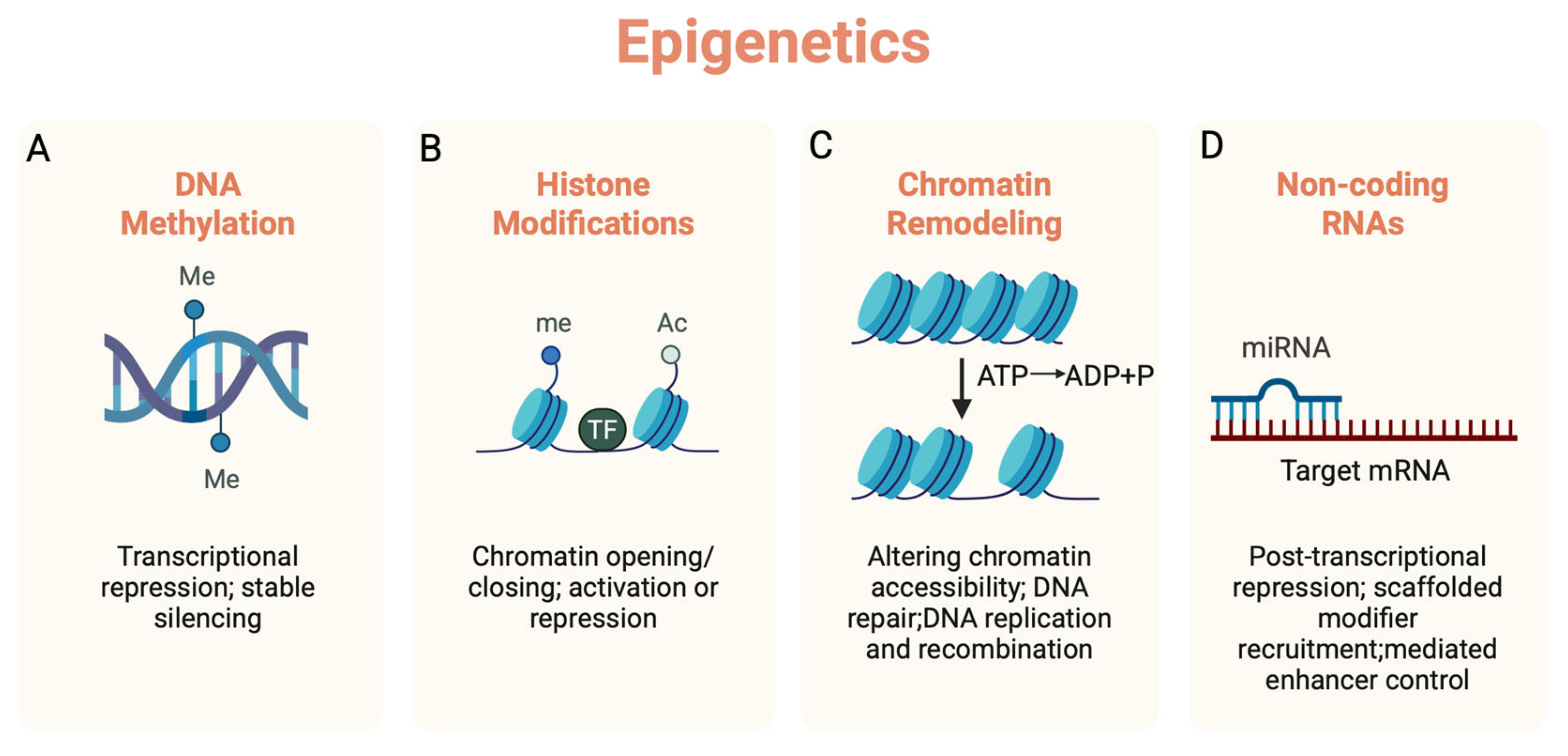
Figure 1. Four major epigenetic mechanisms regulating gene expression. (A) DNA methylation at CpG sites generally leads to transcriptional repression and stable silencing of genes. (B) Histone modifications (e.g., acetylation and methylation) modulate the opening/closing of nucleosomes, resulting in the activation or repression of genes. (C) Regulation of gene expression by altering the position and structure of nucleosomes and the covalent modifications of histones. This facilitates the binding of transcription factors to DNA, thereby promoting or inhibiting gene transcription. (D) non-coding RNAs (e.g., miRNAs) impose post-transcriptional repression, driving mRNA decay/translational inhibition. Me: DNA methylation, Ac: Histone acetylation, me: Histone methylation. Created with BioRender.com.
Epigenomes 09 00038 g001
Figure 2. Aging- and epigenetic alteration-induced endogenous DNA fragments trigger cGAS-STING pathway activation. With aging, there is a global increase in histone deacetylation, loss of stability in gene expression, and gDNA damages. Small, damaged fragments of gDNA leak from the nucleus into the cytosol. At the same time, elevated L1 expression leads to an increase in reverse-transcribed DNA from L1 mRNA, resulting in the accumulation of L1 cDNA in the cytosol. Aging-related mitochondrial damage causes mtDNA to leak into the cytosol. These endogenous DNAs activate the cGAS–STING pathway, producing cGAMP. In turn, cGAMP activates the transcription factors IRF3 and NF-κB—via phosphorylation and degradation of IκB, respectively. The activated transcription factors enter the nucleus to drive the transcription of inflammatory cytokines, thereby triggering neuroinflammation. mRNA: messenger RNA. cDNA: complementary DNA. gDNA: genomic DNA. mtDNA: mitochondrial DNA. Created with BioRender.com.
Epigenomes 09 00038 g002
Figure 3. Feed-forward loop linking microglial activation, epigenetic alterations, and neurodegeneration in AD. In AD, the activation of the cGAS-STING pathway driven by aging- and epigenetics-related changes leads to neuroinflammation and neurodegeneration. Endogenous DNA ligands (gDNA, mtDNA, L1 cDNA) leaked from degenerating neurons activate microglia via TLR9 pathway activation, promoting the release of inflammatory cytokines from microglia. Meanwhile, aging and epigenetic alterations also induce the generation of endogenous DNA ligands within microglia, which can be transferred to neurons via extracellular vesicles. This bidirectional exchange exacerbates the inflammatory milieu, further drives neuronal degeneration, and establishes a self-perpetuating feed-forward loop. mRNA: messenger RNA. cDNA: complementary DNA. gDNA: genomic DNA. mtDNA: mitochondrial DNA. EVs: Extracellular vesicles. Created with BioRender.com.
Epigenomes 09 00038 g003
Figure 4. A tripartite feed-forward loop linking aging, neuroinflammation, and epigenetic remodeling. Each node (aging, neuroinflammation, epigenetic alterations) acts both as driver and consequence of the others. Aging induces epigenetic drift and chromatin imbalance that lower inflammatory thresholds; inflammatory signaling remodels enhancer landscapes and histone/DNA marks; epigenetic alterations, including enhancer priming and endogenous DNA ligands/cGAS–STING activation, sustain neuroinflammatory tone. This framework suggests that single-target interventions may be insufficient and supports mechanism-aligned strategies that concurrently dampen inflammatory tone and restore epigenetic plasticity. Created with BioRender.com.
Epigenomes 09 00038 g004
Table 1. Epigenetic alterations in aging of CNS cells.
Cell TypeEpigenetic AlterationsOutcomeReferences
NSCLaminB1 expression progressively declinesSilencing of neurogenic genes[6,7]
Loss of Setd1a (H3K4me)Promotes NSC activation[8]
Downregulation of Setd8 expressionUpregulation of quiescence-associated genes, inhibition of NSC proliferation[9]
Downregulation of Tet2 expressionImpairing adult neurogenesis[10]
Neuronα-tubulin undergoes hyperacetylationLoss of dendritic arbors and spines[11]
Increased expression of Hdac2Synaptic loss and reduced plasticity[12]
AstrocyteIncreased expression of HDAC7Contributes to tau accumulation and degeneration of neurons[13]
Oligodendrocyte/OPCReduced expression of Dnmt1Global DNA hypomethylation was observed, reduced remyelination efficiency and impaired OPC differentiation[14]
MicrogliaDownregulation of Sirt1 expressionUpregulation of Il-1β expression[15]
Increased expression of TET2Microglial activation[16]
Table 2. Studies of cGAS-STING pathway inhibitor in aging and neurodegenerative diseases.
DiseaseConclusionsModelsStrategyReference
AgingReduced inflammation associated with aging.
Resulting in improved tissue function.
Aging miceH-151, si-STING[56]
ADMicroglia transition from a harmful phenotype to a protective phenotype.
Decreased expression of IL-6, Il-1β, and Tnf-α.
Increased expression of Arg1 and Fizz1.
Reduced degree of aging.
APP/PS1 miceH-151, si-STING[131]
Reduced brain inflammation and microglial synaptic phagocytosis.
Significantly improved Aβ burden, tau phosphorylation, and cognitive impairment.
AppNL-G-F/hTau-double KI miceH-151[132]
Increased Aβ clearance.
Suppression of neurotoxic A1 astrocytes.
Decreased expression of Ifn-β, Il-6, Tnf-α, and Il-1α.
Enhanced phagocytic activity of microglia.
Alleviation of cognitive impairment and Aβ pathological changes.
5×FAD miceH-151[133]
Reduced of cleaved caspase 3. PLD3 knockout SH-SY5Y cells H-151[134]
Decreased GFAP in cerebrospinal fluid [reduced neuroinflammation).
Elevated Ab42/40 ratio in plasma (reduced plaque burden in the brain).
HumanLamivudine (3TC)[135]
Reduced expression of single-stranded DNA.
Reduced DNA damage.
Reduced neuronal death.
Increased expression of PI16 and ADAMDEC1.
reduction of Aβ deposition p-tau and K63-linked ubiquitin-positive tau
3D neural spheroids generated from late-onset AD patient fibroblasts via direct neural reprogramming technology3TC[136]
Reduced tau phosphorylation, inflammation, neuronal death, and hippocampal atrophy.
Alleviated motor deficits (Rotarod test) and improved short-term memory (Y-maze test).
Inhibited the insertion of L1.
P301S mice3TC[137]
Improved cognitive function.
Reduced inflammation and anxiety.
Reduced Iba1 and GFAP expression.
rTg4510 mice3TC[138]
Enhanced the neuronal Mef2c transcriptional network.
Restored synaptic integrity, plasticity, and memory.
P301S transgenic miceTDI-6570 [65]
Reduced of p21 positive cells.Mouse microglia cellsTDI-6570[139]
Restricted Aβ deposition.
Alleviated neuroinflammation.
Reduced neuronal damage.
Improved cognitive behavioral.
5xFAD; cGASfl/fl; Cx3cr1+/− miceSTING deficiency[140]
PDReduced interferon expression in the striatum.
Treatment of motor dysfunction, pathological α-synuclein deposition, and dopaminergic neuron loss.
Stinggt mice STING deficiency[115]
Attenuated PD-associated behavioral phenotypes.
Reduced loss of TH-positive neurons.
Decreased the number of activated microglia.
Lowered levels of factors associated with cGAS-dependent inflammation.
MPTP PD miceRU.521[141]
Reduced expression of Ifn-β, Tnf-α, Il-1β, and Il-6.
Decreased expression of Nlpr3 and caspase-1.
Alleviated neuroinflammation associated with MPTP neurotoxicity.
Protected substantia nigra striatal dopaminergic neurons from degeneration.
Lowered levels of reactive astrocytes.
MPTP PD miceC-176[120]
ALSReduced of IFN-I, ISGs, pro-inflammatory, and chemokines genes.
Improved motor functions.
SOD1 ALS miceC-176, H-151, RU.521[125]
Reduced the expression of Il-1β, Il-6, Tnf, Mx1, Infb 1.
Decreased neuronal loss.
TDP-43 mutant miceH-151[123]
Preventing motor neuron death.Human iPSC-derived motor neurons
Reduced levels of TNF-α, IL-6 and CXCL10.
Suppressed inflammatory responses.
Human iPSC-derived neuronsH-151, RU.521[124]
C9orf72 mice
Reduced of ISGs expressionC9orf72 knockout miceH-151[142]
Arg1: Arginase 1; Fizz1: Resistin like beta; GFAP: Glial fibrillary acidic protein; PI16: Peptidase inhibitor 16 ADAMDEC1: ADAM like decysin 1; Nlpr3: NLR family pyrin domain containing 3; IFN-I: Interferon type I; ISGs: Interferon stimulated genes; Mx1: Interferon-induced GTP-binding protein Mx1; Infb 1: Interferon beta 1; CXCL10: CXC Motif chemokine ligand 10.
Disclaimer/Publisher’s Note: The statements, opinions and data contained in all publications are solely those of the individual author(s) and contributor(s) and not of MDPI and/or the editor(s). MDPI and/or the editor(s) disclaim responsibility for any injury to people or property resulting from any ideas, methods, instructions or products referred to in the content.