Article

Male and Female Cat Fleas (Ctenocephalides felis) Differ in Bacterial Control, Survival, and Circulating Hemocyte Responses Following Systemic Challenge

Department of Biology, Georgia Southern University, 4324 Old Register Rd., Statesboro, GA 30460, USA
*
Author to whom correspondence should be addressed.
Insects 2026, 17(9), 952; https://doi.org/10.3390/insects17090952
Submission received: 10 August 2026 / Revised: 10 September 2026 / Accepted: 10 September 2026 / Published: 12 September 2026
(This article belongs to the Special Issue Insect Vector Physiology and Competence: Interactions and Adaptations)

Simple Summary

Fleas transmit several microorganisms that cause disease in humans and animals, yet relatively little is known about how their immune systems respond to infection. Previous studies of cat flea immunity have focused primarily on females, leaving unanswered the question of whether males and females respond similarly to invading microorganisms. In this study, we compared the responses of male and female cat fleas after bacteria were experimentally introduced directly into their body cavity. Female fleas maintained lower levels of bacterial infection and survived better than males. Females also had higher circulating immune cell counts and increased these counts following injury and bacterial challenge, whereas males did not. In addition, a greater proportion of female immune cells captured and consumed bacteria, although individual immune cells from males and females consumed similar numbers of bacteria. These findings demonstrate that male and female cat fleas differ in their responses to experimental bacterial infection. Understanding variation in immune function between the sexes provides new insight into flea biology and establishes a foundation for future studies examining whether similar differences occur when fleas encounter disease-causing microorganisms through natural routes of infection.

Abstract

Although both male and female cat fleas (Ctenocephalides felis) consume vertebrate blood throughout adulthood, previous studies of flea immunity have focused primarily on females, leaving whether systemic immune responses differ between the sexes largely unknown. To address this knowledge gap, we compared bacterial persistence, survival, circulating hemocyte abundance, and phagocytic activity in male and female C. felis following microinjection of Escherichia coli into the hemocoel. Females exhibited significantly lower infection prevalence and bacterial burdens than males 24 h post-challenge. Consistent with these differences, bacterial infection reduced survival in both sexes but had a substantially greater effect on males, while sterile injury alone significantly reduced male survival but had little effect on females. Across all treatment groups, females possessed significantly higher circulating hemocyte abundance than males and were the only sex to exhibit an increase in circulating hemocyte numbers following injury or bacterial challenge. Additionally, a significantly greater proportion of female hemocytes participated in phagocytosis, although the number of bacteria internalized per hemocyte did not differ between the sexes. Together, these findings demonstrate sex-specific differences in the systemic antibacterial responses of C. felis, with females exhibiting lower bacterial burdens, greater survival, higher circulating hemocyte abundance, and a greater proportion of phagocytic hemocytes than males. These results identify sex as a biological variable influencing systemic immune responses in cat fleas and provide a foundation for determining whether similar differences occur following natural routes of microbial exposure.

1. Introduction

Despite their importance as vectors of numerous zoonotic pathogens, the mechanisms by which fleas (Order Siphonaptera) respond to microbial infection remain poorly understood compared with those of other hematophagous arthropods [1,2]. Like all insects, fleas rely exclusively on innate immunity, which consists of coordinated cellular and humoral responses that detect and combat invading microorganisms [3,4,5]. Cellular immunity is mediated primarily by hemocytes, which eliminate pathogens through phagocytosis, encapsulation, and nodulation, while humoral defenses involve activation of conserved immune signaling pathways that stimulate the production of antimicrobial peptides, reactive oxygen species, and other soluble immune effectors. Much of the current understanding of these defenses comes from studies of Drosophila melanogaster, where genetic and molecular approaches have provided a framework for investigating innate immunity in less well-characterized insects [5]. Recent studies have begun to characterize flea immune defenses, including the involvement of conserved immune signaling pathways such as IMD in antibacterial responses [6,7,8,9,10,11,12,13,14,15], yet the biological factors that influence infection outcomes remain largely unexplored.
Sex-specific differences in immune function have been documented across a broad range of animal taxa and are thought to contribute to variation in susceptibility and resistance to infection [16]. Traditionally, females have been considered the more immunocompetent sex because selection favors prolonged survival and sustained reproductive output, whereas males are expected to allocate proportionally more resources toward reproductive traits than immune defense [17,18,19,20]. However, accumulating evidence indicates that immune sexual dimorphism is neither universal nor consistently female-biased. In insects, patterns of sex-biased immunity are often condition- and pathogen-dependent, and the mechanisms responsible remain poorly understood (extensively reviewed in [16,21]). One factor proposed to influence these differences is feeding ecology, as males and females of many insect species exploit different nutritional resources and consequently experience distinct microbial environments and immune challenges [22]. This hypothesis is particularly relevant in hematophagous insects, where sex-specific blood-feeding behavior varies considerably among taxa. For example, in mosquitoes (Aedes aegypti and Anopheles aquasalis) and sand flies (Lutzomyia longipalpis), where males feed exclusively on plant nectar and females feed on both plant nectar and vertebrate blood, males exhibit higher basal expression of several conserved immune genes involved in systemic humoral immunity [23]. Conversely, in the kissing bug Rhodnius prolixus, where both sexes are obligate blood feeders, females exhibit higher basal expression of these same immune genes than males [23]. These contrasting patterns suggest that differences in feeding ecology may contribute to the evolution of immune sexual dimorphism.
To date, systemic innate immune responses to bacterial infection have been investigated in only one flea species, the cat flea, Ctenocephalides felis. Using model Gram-positive and Gram-negative bacteria, Muñoz et al. [10] demonstrated that both cellular and humoral immune defenses contribute to antibacterial immunity, with microbial challenge eliciting increased circulating hemocyte abundance and robust phagocytic activity. Moreover, experimental impairment of phagocytosis led to significantly higher bacterial burdens, thereby establishing hemocyte-mediated phagocytosis as an important component of the systemic antibacterial response. However, this study was conducted exclusively on female fleas, leaving it unknown whether males and females differ in their systemic immune responses. Therefore, this study aimed to determine whether male and female C. felis differ in their responses to systemic bacterial challenge.

2. Materials and Methods

2.1. Flea Maintenance

Adult cat fleas (C. felis) were obtained from a laboratory colony maintained at Georgia Southern University or purchased from Ecto Services, Inc. (Henderson, NC, USA). Fleas were maintained on defibrinated bovine blood (HemoStat Laboratories, Dixon, CA, USA) using an artificial membrane feeding system as previously described [6]. All experiments were performed using blood-fed adult fleas between 2 and 3 days post-emergence. Male and female fleas were distinguished by external reproductive morphology under a dissecting microscope based on the terminal abdominal segments and genitalia [24]. Males and females were housed together and provided blood ad libitum, except when separated by sex for experimental manipulation and sample collection.

2.2. Bacterial Culture and Flea Infection

Ampicillin-resistant, green fluorescent protein (GFP)-expressing Escherichia coli (ATCC® 25922GFP, Manassas, VA, USA; E. coli) was cultured and prepared for flea infection as previously described by Muñoz et al. [10]. Briefly, bacterial cultures were grown overnight in tryptic soy broth (TSB) supplemented with ampicillin (100 µg/mL) at 37 °C with shaking (350 rpm) and standardized to an optical density of OD600 = 5.0 using a BioPhotometer D30 (Eppendorf AG, Hamburg, Germany) prior to injection.
Fleas were cold-anesthetized and injected into the thorax using a Nanoject III Auto-Nanoliter Injector (Drummond Scientific Company, Broomall, PA, USA). Each flea received 69 nL of either E. coli suspended in TSB or sterile TSB alone as an injury control. Additional fleas were cold-anesthetized but not injected to serve as naïve controls. Fleas that died within 1 h of treatment were excluded from the analysis to minimize mortality associated with handling or injection. Sample sizes reported for each experiment represent the number of surviving individuals in the experimental cohort after the 1 h exclusion period. The number of individuals excluded during this initial period was not recorded. To verify the inoculation dose, a subset of fleas was homogenized immediately after injection, plated on tryptic soy agar (TSA) containing ampicillin, incubated overnight at 37 °C, and colony-forming units (CFUs) were enumerated. Each experimental endpoint was evaluated in separate experiments using independently prepared cohorts of fleas and bacterial cultures.

2.3. Bacterial Persistence

To quantify bacterial persistence, male and female fleas were injected with E. coli as described above. Twenty-four hours after infection, individual fleas were homogenized in 200 µL phosphate-buffered saline (PBS), serially diluted, and plated on TSA containing ampicillin. Following overnight incubation at 37 °C, CFUs were enumerated. Infection was quantified as (1) prevalence, defined as the proportion of fleas yielding detectable E. coli, and (2) intensity, defined as the number of CFUs recovered from infected individuals. Three independent biological replicates were performed, each consisting of 30 females and 30 males (n = 90 fleas per sex).

2.4. Survival Analysis

To determine whether bacterial infection differentially affected survival, male and female fleas were assigned to one of three treatment groups: naïve, TSB-injected (injury), or E. coli-injected. Following treatment, male and female fleas were maintained together in the same feeding chambers under standard colony conditions and provided defibrinated bovine blood ad libitum using the artificial membrane feeding system described above. Survival was monitored daily for 5 days by recording the number of live and dead fleas in each treatment group, and dead fleas were removed after each daily observation. Three independent biological replicates were conducted, each consisting of 30 females and 30 males per treatment (n = 90 per sex per treatment).

2.5. Circulating Hemocyte Counts

Male and female fleas were assigned to the naïve, TSB-injected, or E. coli-injected treatment groups. Twenty-four hours after treatment, circulating hemocytes were collected by perfusion as previously described by Muñoz et al. [10]. Briefly, an incision was made across the penultimate abdominal segment, and approximately 200 μL of PBS was injected into the thorax. Although the osmolarity of C. felis hemolymph has not been characterized and PBS was not independently validated for hemocyte viability in the present study, this method previously yielded morphologically intact flea hemocytes suitable for quantitative and functional analyses [10]. Hemolymph exiting through the abdominal incision was collected on glass slides, allowed to adhere for 20 min at room temperature, fixed, and stained with Hema 3 (Fisher Scientific, Pittsburgh, PA, USA). Total circulating hemocytes were counted under bright-field illumination at 400× magnification. Three independent biological replicates were performed, each consisting of 20 females and 20 males per treatment (n = 60 fleas per sex per treatment).

2.6. Phagocytosis Assay

To evaluate phagocytic activity, male and female fleas were injected with E. coli as described above. One hour after infection, circulating hemocytes were collected by perfusion and stained with Hema 3 [10]. For each flea, the first 100 hemocytes observed were examined microscopically, and the number of intracellular bacteria was recorded. These observations were used to calculate the phagocytic index (percentage of hemocytes containing bacteria) and phagocytic capacity (mean number of bacteria per hemocyte). Three independent biological replicates were performed, each consisting of 10 females and 10 males (n = 30 fleas per sex).

2.7. Statistical Analysis

Data from independent biological replicates were pooled for analysis in GraphPad Prism version 10 (GraphPad Software, San Diego, CA, USA). Differences in bacterial prevalence, infection intensity, and phagocytic capacity were analyzed using the Mann–Whitney test. The phagocytic index was compared using Welch’s t-test. Circulating hemocyte counts were analyzed by two-way ANOVA with treatment and sex as fixed factors, followed by Tukey’s multiple-comparison test. Survival curves were compared using the Logrank (Mantel–Cox) test. Statistical significance was defined as p ≤ 0.05. For survival analyses, Bonferroni correction was applied to account for multiple pairwise comparisons, resulting in an adjusted significance threshold of p < 0.0167.

3. Results

3.1. Females Exhibit Lower Bacterial Prevalence and Burden than Males Following Systemic Challenge

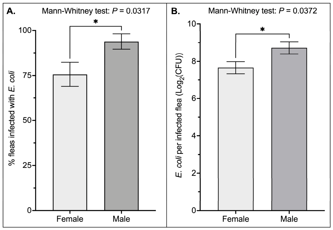
To determine whether bacterial persistence differed between the sexes, male and female fleas were injected with E. coli into the hemocoel, and bacterial infection was quantified 24 h post-injection by measuring infection prevalence (the proportion of infected fleas) and infection intensity (the mean number of bacteria per infected flea). Males exhibited significantly greater infection prevalence than females (Figure 1A; Mann–Whitney, p = 0.0317), with 24% more males remaining infected 24 h after challenge. Likewise, infection intensity was significantly greater in males than females (Figure 1B; Mann–Whitney, p = 0.0372), with infected males harboring 12% more bacteria than infected females. Collectively, these findings demonstrate that female fleas exhibit lower bacterial prevalence and bacterial burdens than males 24 h after systemic challenge.

3.2. Females Exhibit Greater Survival Following Systemic Bacterial Infection than Males

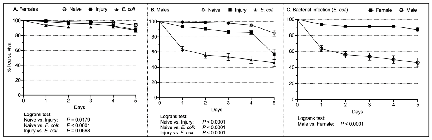
To determine whether bacterial infection differentially affected survival, male and female fleas were assigned to one of three treatment groups (naïve, injury, or E. coli) and survival was monitored for 5 days. Relative to naïve controls, E. coli infection significantly reduced survival in both females (Figure 2A; Logrank test, p < 0.0001) and males (Figure 2B; Logrank test, p < 0.0001). In females, survival did not differ between the naïve and injury control groups (Figure 2A; Logrank test, p = 0.0179) or between the injury control and E. coli-infected groups (Figure 2A; Logrank test, p = 0.0668). In contrast, sterile injury alone significantly reduced survival in males compared with naïve controls (Figure 2B; Logrank test, p < 0.0001), although E. coli infection caused the greatest reduction in survival (Figure 2B; Logrank test, p < 0.0001).
Direct comparison of survival following E. coli infection revealed that females survived significantly longer than males (Figure 2C; Logrank test, p < 0.0001). By day 5 post-infection, 87% of E. coli-infected females remained alive compared with only 46% of infected males. In contrast, survival of naïve fleas was 94% in females and 85% in males, while sterile injury reduced male survival to 57% but had little effect on female survival (88%). Collectively, these findings demonstrate that systemic bacterial infection has a substantially greater impact on male survival than female survival.

3.3. Females Have Higher Circulating Hemocyte Counts than Males, and Only Females Increase Hemocyte Abundance Following Injury and Bacterial Challenge

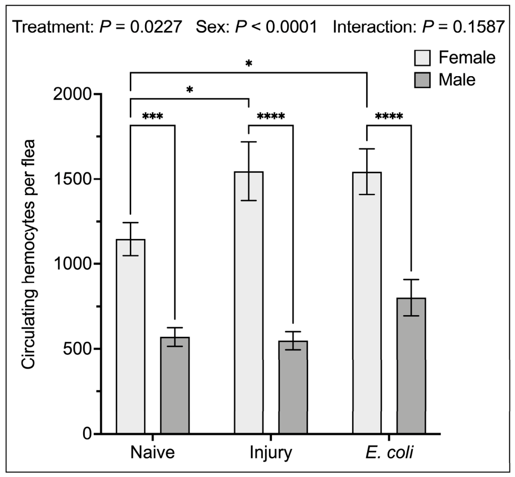
Circulating hemocytes are a major component of the insect cellular immune response and can influence the ability to eliminate invading microorganisms. To determine whether sex-specific differences in systemic antibacterial responses were associated with differences in circulating hemocyte abundance, male and female fleas were assigned to naïve, injury, or E. coli-infected treatment groups, and circulating hemocytes were quantified 24 h later. Across all treatment groups, females exhibited significantly higher circulating hemocyte counts than males (Figure 3; Naïve: Female vs. Male, Tukey’s, p = 0.0001; Injury: Female vs. Male, Tukey’s, p < 0.0001; E. coli: Female vs. Male, Tukey’s, p < 0.0001). In particular, naïve and E. coli-infected females possessed 101% and 93% more circulating hemocytes, respectively, than their male counterparts. Within females, both sterile injury and E. coli infection significantly increased circulating hemocyte counts relative to naïve controls (Figure 3; Naïve vs. Injury, Tukey’s, p = 0.0265; Naïve vs. E. coli, Tukey’s, p = 0.0343), corresponding to a 35% increase in circulating hemocyte abundance following either treatment. In contrast, circulating hemocyte counts did not differ among treatment groups in males. Collectively, these findings demonstrate that female fleas maintain a larger circulating hemocyte population than males and increase circulating hemocyte abundance following injury and bacterial challenge, whereas males exhibit no detectable change.

3.4. Females Exhibit a Higher Proportion of Phagocytic Hemocytes than Males, Whereas Bacterial Uptake per Hemocyte Is Comparable Between the Sexes

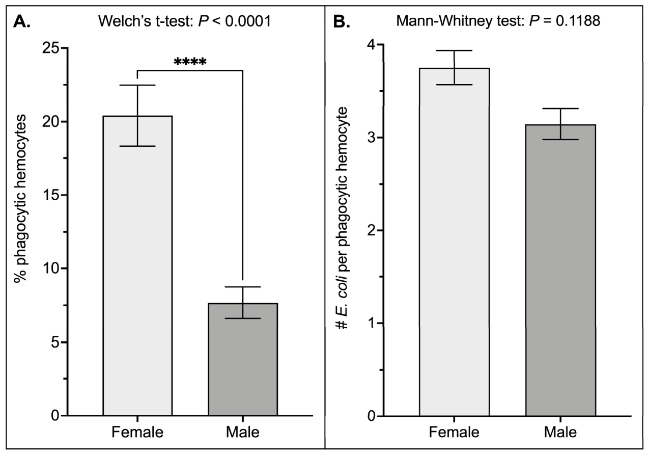
Because hemocytes mediate bacterial burden through phagocytosis, sex-specific differences in systemic antibacterial responses may result not only from variation in circulating hemocyte abundance but also from differences in the proportion or activity of hemocytes participating in phagocytosis. To determine whether phagocytic activity differs between male and female fleas, individuals of both sexes were injected with E. coli into the hemocoel, and phagocytosis was quantified 1 h later. The phagocytic index, defined as the percentage of hemocytes containing internalized bacteria, was significantly greater in females than in males (Figure 4A; Welch’s t-test, p < 0.0001). Specifically, females exhibited a 150% higher phagocytic index than males. In contrast, phagocytic capacity, defined as the number of bacteria internalized per hemocyte, did not differ between the sexes (Figure 4B; Mann–Whitney test, p = 0.1188). Individual hemocytes internalized between 1 and 18 E. coli cells in females and between 1 and 12 cells in males. Collectively, these findings indicate that enhanced bacterial uptake by female fleas is driven by a greater proportion of hemocytes participating in phagocytosis rather than by increased phagocytic capacity of individual hemocytes.

4. Discussion

The present study demonstrates that male and female C. felis differ in their responses to experimental systemic bacterial challenge. Following introduction of an equivalent E. coli inoculum directly into the hemocoel, females exhibited lower bacterial prevalence and burdens, greater survival, higher circulating hemocyte abundance, and a greater proportion of phagocytic hemocytes than males. These findings provide evidence of sex-specific variation in systemic antibacterial responses in C. felis and lay the groundwork for future studies examining whether comparable differences occur when flea-borne pathogens are acquired via an infectious blood meal.
One of the clearest differences between male and female C. felis was the lower prevalence and intensity of bacterial infection observed in females 24 h after systemic challenge. Because males and females received identical doses of E. coli, these differences reflect greater control of the experimentally introduced bacteria in females rather than differences in initial bacterial exposure. Comparable studies using systemic bacterial challenge have likewise identified sex-specific differences in antibacterial immunity among hematophagous insects. Following bacterial injection into the hemocoel, female Anopheles gambiae cleared bacteria more efficiently than males [25], whereas male Ae. aegypti exhibited greater bacterial clearance than females under similar experimental conditions [23]. Similar patterns have also been reported in the non-hematophagous insect D. melanogaster, where males were more susceptible to systemic Staphylococcus aureus infection and harbored higher bacterial loads than females [26]. Together, these studies suggest that sex-specific differences in systemic antibacterial immunity may be common among insects, although the direction of these differences varies among species. The present study extends these observations to fleas by demonstrating that female C. felis has greater control of systemic bacterial infection compared with males.
The reduced bacterial burden observed in females was accompanied by a marked survival advantage following systemic infection, further supporting the conclusion that females possess more effective systemic immune defenses than males. While E. coli infection reduced survival in both sexes, the effect was substantially greater in males, with infected females nearly twice as likely to survive the 5-day observation period. Interestingly, sterile injury alone significantly reduced male survival but had little effect on females, suggesting that males are less capable of tolerating or recovering from disruption of the cuticle and entry into the hemocoel, even in the absence of bacterial challenge. This sex-specific effect of sterile injection on survival also highlights a limitation of the experimental model, as the procedure itself affected males and females differently. Additionally, because the flea cuticle was not surface sterilized prior to injection, the possibility that cuticle-associated microorganisms entered the hemocoel during injection and contributed to the response observed in the sterile-injury group cannot be excluded. Sex-biased survival following bacterial infection has been documented across insects, although the direction of this bias varies considerably depending on the host species, bacterial pathogen, host genotype, mating status, and environmental conditions [27,28,29,30,31,32,33,34,35]. Among hematophagous insects, Barletta Ferreira et al. [23] reported that female Ae. aegypti survived systemic bacterial infection better than males despite maintaining higher bacterial burdens, suggesting that female survival was associated with greater tolerance to infection rather than more efficient bacterial control. In contrast, female C. felis exhibited both lower bacterial burdens and greater survival, indicating that enhanced survival in fleas is associated with more effective control of systemic bacterial infection. Together, these findings demonstrate that the relationship between systemic infection and survival differs among hematophagous insects.
The greater ability of female C. felis to limit bacterial burdens within the hemocoel was accompanied by marked differences in circulating hemocyte abundance, suggesting that sex-specific variation in cellular immunity contributes to the enhanced antibacterial defenses observed in females. Regardless of treatment, females maintained nearly twice as many circulating hemocytes as males, indicating that sexual dimorphism in hemocyte abundance exists even in the absence of immune challenge. Similar female-biased differences have been reported in mosquitoes [25,36], scorpionflies [37], moths [38], and beetles [39]; however, studies in D. melanogaster have reported female-biased, male-biased, or no differences in adult hemocyte abundance [27,30,40], suggesting that baseline sexual dimorphism in circulating hemocyte populations varies among insect taxa. Moreover, female fleas responded to both sterile injury and bacterial infection by significantly increasing the number of circulating hemocytes, whereas males exhibited no detectable change. Similar increases in circulating hemocyte abundance following systemic bacterial challenge have been reported previously in female C. felis [10], but the present study demonstrates that this inducible response is largely restricted to females. Although few studies have examined sex-specific changes in circulating hemocyte abundance following infection, several studies demonstrate that hemocyte regulation differs between male and female insects. In the firebug Pyrrhocoris apterus, females increased circulating hemocyte numbers in response to changes in photoperiod, whereas males did not [41]. Likewise, hemocyte concentrations increased with age in the European earwig, although the increase was more pronounced in males than females [42]. Comparable differences in inducible hemocyte responses have also been observed among mosquitoes, where systemic bacterial infection increased circulating hemocyte abundance in An. gambiae but not in Ae. aegypti; however, these comparisons were made between species rather than between sexes [43]. Importantly, the perfusion method used in the present study quantified circulating hemocytes but did not measure sessile hemocytes associated with tissues or the body wall. Thus, the observed increase in circulating hemocyte abundance cannot be assumed to represent an increase in the total number of hemocytes within the flea. The mechanisms responsible for the increased number of circulating hemocytes remain unclear and may include shifts in the distribution between sessile and circulating hemocytes, as well as proliferation of circulating cells. Mitosis of circulating hemocytes was visually documented in female C. felis by Muñoz et al. [10], while mobilization of sessile hemocytes into the circulation has been described in mosquitoes following systemic bacterial infection [44,45]. Collectively, these findings demonstrate that female C. felis maintain more hemocytes in circulation and exhibit a greater increase in circulating hemocyte abundance following systemic challenge than males, although whether these differences reflect changes in total hemocyte number, hemocyte distribution, or both remains unknown.
In addition to maintaining more circulating hemocytes, females also exhibited a significantly greater proportion of cells participating in phagocytosis, further strengthening their cellular immune response. Importantly, phagocytic activity was assessed by evaluating the first 100 hemocytes encountered from each flea, thereby standardizing comparisons between sexes regardless of differences in total circulating hemocyte abundance. In contrast, individual hemocytes internalized similar numbers of bacteria in both sexes, indicating that the phagocytic capacity of individual cells does not differ between male and female fleas. Similar variability in phagocytic responses has been reported in other insects. In scorpionflies, females exhibited greater overall phagocytic activity than males because they possessed more circulating hemocytes and a higher proportion of cells engaged in phagocytosis, whereas the phagocytic capacity of individual hemocytes did not differ between the sexes [37]. This pattern closely parallels the results of the present study, in which females exhibited a greater proportion of phagocytic hemocytes despite no difference in the number of E. coli ingested per phagocytic cell. By comparison, male D. melanogaster hemocytes exhibit a slightly higher ex vivo phagocytic index than those of females [30], whereas adult female An. gambiae hemocytes carry a greater phagocytic burden than larval hemocytes, demonstrating that phagocytic activity can also vary across developmental stages [46]. Collectively, the lower bacterial burdens observed in female C. felis are associated with both a larger circulating hemocyte population and a greater proportion of hemocytes participating in phagocytosis, rather than greater bacterial uptake by individual phagocytic cells.
One factor that may contribute to the higher baseline hemocyte abundance observed in females is the tendency for female fleas to be larger than males [47]. Because females generally possess a greater body size and, presumably, a larger hemolymph volume, they may support a larger circulating hemocyte population; however, these factors were not evaluated in the present study. Even so, body size alone is unlikely to explain the observed sex-specific immune responses. Females not only maintained a larger circulating hemocyte population under naïve conditions but also uniquely increased circulating hemocyte abundance following injury and bacterial challenge while exhibiting a greater proportion of hemocytes participating in phagocytosis. Collectively, these findings indicate that although body size may contribute to baseline differences in circulating hemocyte abundance, it is unlikely to fully account for the sex-specific differences in cellular immune responses observed following challenge.
An additional factor that may have influenced immune responses is the reproductive status of the fleas. Adult C. felis begin feeding immediately after acquiring a host, mate shortly thereafter, and females become reproductively active within the first few days [48,49]. Consequently, the two- to three-day-old fleas used in this study were likely reproductively active during the experiments. Reproductive status has been shown to influence immune function in insects, although the magnitude and direction of these effects vary considerably among species and immune traits [27,34,35,42,50,51,52,53,54,55]. Whether reproductive status contributes to the sex-specific differences in systemic immunity observed in C. felis remains unknown and warrants further investigation.
In conclusion, this study demonstrates that male and female C. felis differ in their responses to experimental systemic bacterial challenge. Female fleas exhibited lower bacterial prevalence and burdens, greater survival, higher circulating hemocyte abundance, and a greater proportion of phagocytic hemocytes than males. These findings identify sex as a biological variable associated with variation in systemic antibacterial responses in cat fleas. Because bacteria were introduced directly into the hemocoel, the present study does not determine whether comparable differences occur following natural acquisition of flea-borne microorganisms. Future studies comparing male and female fleas following exposure through an infectious blood meal will be necessary to determine whether sex influences pathogen establishment, dissemination, or transmission.

Author Contributions

A.L.: investigation, writing—original draft; M.M.: investigation, writing—review & editing; Y.P.: investigation, writing—review & editing; L.D.B.: conceptualization, formal analysis, funding acquisition, methodology, project administration, resources, supervision, visualization, writing—review & editing. All authors have read and agreed to the published version of the manuscript.

Funding

This research was funded by the National Institutes of Health/National Institute for Allergy and Infectious Diseases award number 1R16 AI170530. The National Science Foundation’s Research Experience for Undergraduates (NSF DBI award #2244232) and Research and Mentoring for Postbaccalaureates (NSF DBI award #2319755) to Georgia Southern University provided support for MM and YP, respectively, to contribute to this project.

Data Availability Statement

The data supporting the findings of this study are available within the article. Raw data are available from the corresponding author upon request.

Conflicts of Interest

The authors declare that they have no competing interests.

Abbreviations

CFUColony Forming Units
GFPGreen Fluorescent Protein
PBSPhosphate-Buffered Saline
SEMStandard Error of the Mean
TSATryptic Soy Agar
TSBTryptic Soy Broth

References

  1. Brown, L.D. Immunity of fleas (order Siphonaptera). Dev. Comp. Immunol. 2019, 98, 76–79. [Google Scholar] [CrossRef] [Scilit] [PubMed]
  2. Mehlhorn, H. Fleas as Underestimated Vectors of Agents of Diseases. In Arthropods as Vectors of Emerging Diseases; Mehlhorn, H., Ed.; Springer: Berlin/Heidelberg, Germany, 2012; pp. 301–328. [Google Scholar]
  3. Makkar, P.; Singh, C.K.; Nitin, V.; Narang, P.K.; Sodhi, K.K. Insect immunity unveiled: Exploring the molecular and cellular defenses against microbial threats. Arch. Microbiol. 2026, 208, 379. [Google Scholar] [CrossRef] [Scilit] [PubMed]
  4. Hillyer, J.F. Insect immunology and hematopoiesis. Dev. Comp. Immunol. 2016, 58, 102–118. [Google Scholar] [CrossRef] [Scilit] [PubMed]
  5. Lemaitre, B.; Hoffmann, J. The host defense of Drosophila melanogaster. Annu. Rev. Immunol. 2007, 25, 697–743. [Google Scholar] [CrossRef] [Scilit] [PubMed]
  6. Brown, L.D.; Maness, R.; Hall, C.; Gibson, J.D. Reactive oxygen species-mediated immunity against bacterial infection in the gut of cat fleas (Ctenocephalides felis). Insect Biochem. Mol. Biol. 2021, 136, 103620. [Google Scholar] [CrossRef] [Scilit] [PubMed]
  7. Weber, K.; Karnik, D.; Brown, L.D. Transcriptional induction of the IMD signaling pathway and associated antibacterial activity in the digestive tract of cat fleas (Ctenocephalides felis). Parasites Vectors 2024, 17, 546. [Google Scholar] [CrossRef] [Scilit] [PubMed]
  8. Zellner, P.N.; Njoroge, E.; Lynch, A.; Brown, L.D. Physiology of the cat flea (Ctenocephalides felis) gut: Serine protease activity, midgut pH, and intestinal barrier integrity. J. Med. Entomol. 2026, 63, tjag037. [Google Scholar] [CrossRef] [Scilit] [PubMed]
  9. Zhou, W.; Russell, C.W.; Johnson, K.L.; Mortensen, R.D.; Erickson, D.L. Gene expression analysis of Xenopsylla cheopis (Siphonaptera: Pulicidae) suggests a role for reactive oxygen species in response to Yersinia pestis infection. J. Med. Entomol. 2012, 49, 364–370. [Google Scholar] [CrossRef] [Scilit] [PubMed]
  10. Muñoz, M.; Lin, N.; Lin, R.; King, B.; Brown, L.D. Immune defense mechanisms against a systemic bacterial infection in the cat flea (Ctenocephalides felis). J. Invertebr. Pathol. 2022, 195, 107850. [Google Scholar] [CrossRef] [Scilit] [PubMed]
  11. Bland, D.M.; Martens, C.A.; Virtaneva, K.; Kanakabandi, K.; Long, D.; Rosenke, R.; Saturday, G.A.; Hoyt, F.H.; Bruno, D.P.; Ribeiro, J.M.; et al. Transcriptomic profiling of the digestive tract of the rat flea, Xenopsylla cheopis, following blood feeding and infection with Yersinia pestis. PLoS Negl. Trop. Dis. 2020, 14, e0008688. [Google Scholar] [CrossRef] [Scilit] [PubMed]
  12. Danchenko, M.; Laukaitis, H.J.; Macaluso, K.R. Dynamic gene expression in salivary glands of the cat flea during Rickettsia felis infection. Pathog. Dis. 2021, 79, ftab020. [Google Scholar] [CrossRef] [Scilit] [PubMed]
  13. Rennoll, S.A.; Rennoll-Bankert, K.E.; Guillotte, M.L.; Lehman, S.S.; Driscoll, T.P.; Beier-Sexton, M.; Rahman, M.S.; Gillespie, J.J.; Azad, A.F. The cat flea (Ctenocephalides felis) immune deficiency signaling pathway regulates Rickettsia typhi infection. Infect. Immun. 2018, 86, 10-1128. [Google Scholar] [CrossRef] [Scilit] [PubMed]
  14. Dreher-Lesnick, S.; Ceraul, S.; Lesnick, S.; Gillespie, J.; Anderson, J.; Jochim, R.; Valenzuela, J.; Azad, A. Analysis of Rickettsia typhi-infected and uninfected cat flea (Ctenocephalides felis) midgut cDNA libraries: Deciphering molecular pathways involved in host response to R. typhi infection. Insect Mol. Biol. 2010, 19, 229–241. [Google Scholar] [CrossRef] [Scilit] [PubMed]
  15. Greene, W.K.; Macnish, M.G.; Rice, K.L.; Thompson, R.A. Identification of genes associated with blood feeding in the cat flea, Ctenocephalides felis. Parasites Vectors 2015, 8, 368. [Google Scholar] [CrossRef] [Scilit] [PubMed]
  16. Nunn, C.L.; Lindenfors, P.; Pursall, E.R.; Rolff, J. On sexual dimorphism in immune function. Philos. Trans. R. Soc. Lond. B Biol. Sci. 2009, 364, 61–69. [Google Scholar] [CrossRef] [Scilit] [PubMed]
  17. Zuk, M.; Stoehr, A.M. Immune defense and host life history. Am. Nat. 2002, 160, S9–S22. [Google Scholar] [CrossRef] [PubMed]
  18. Williams, G.C. Adaptation and Natural Selection: A Critique of Some Current Evolutionary Thought; Princeton University Press: Princeton, NJ, USA, 1966; p. 336. [Google Scholar]
  19. Bateman, A.J. Intra-sexual selection in Drosophila. Heredity 1948, 2, 349–368. [Google Scholar] [CrossRef] [Scilit] [PubMed]
  20. Rolff, J. Bateman’s principle and immunity. Proc. R. Soc. Lond. B Biol. Sci. 2002, 269, 867–872. [Google Scholar] [CrossRef] [Scilit] [PubMed]
  21. Belmonte, R.L.; Corbally, M.-K.; Duneau, D.F.; Regan, J.C. Sexual dimorphisms in innate immunity and responses to infection in Drosophila melanogaster. Front. Immunol. 2020, 10, 3075. [Google Scholar] [CrossRef] [Scilit] [PubMed]
  22. Fanson, B.G.; Fanson, K.V.; Taylor, P.W. Sex differences in insect immune function: A consequence of diet choice? Evol. Ecol. 2013, 27, 937–947. [Google Scholar] [CrossRef] [Scilit]
  23. Barletta Ferreira, A.B.; Bahia, A.C.; Pitaluga, A.N.; Barros, E.; Gama dos Santos, D.; Bottino-Rojas, V.; Kubota, M.S.; Oliveira, P.L.d.; Pimenta, P.F.P.; Traub-Csekö, Y.M.; et al. Sexual dimorphism in immune responses and infection resistance in Aedes aegypti and other hematophagous insect vectors. Front. Trop. Dis. 2022, 3, 847109. [Google Scholar] [CrossRef] [Scilit]
  24. Krämer, F.; Mencke, N. General Morphology. In Flea Biology and Control: The Biology of the Cat Flea Control and Prevention with Imidacloprid in Small Animals; Krämer, F., Mencke, N., Eds.; Springer: Berlin/Heidelberg, Germany, 2001; pp. 3–4. [Google Scholar]
  25. Brown, L.D.; Shapiro, L.L.M.; Thompson, G.A.; Estévez-Lao, T.Y.; Hillyer, J.F. Transstadial immune activation in a mosquito: Adults that emerge from infected larvae have stronger antibacterial activity in their hemocoel yet increased susceptibility to malaria infection. Ecol. Evol. 2019, 9, 6082–6095. [Google Scholar] [CrossRef] [Scilit] [PubMed]
  26. Aleksandrowicz, J. Sexual Dimorphism in hemocyte Responses to Staphylococcus aureus Infection in Drosophila melanogaster. Master’s Thesis, The University of Edinburgh, Edinburgh, UK, 2024. [Google Scholar]
  27. Duneau, D.F.; Kondolf, H.C.; Im, J.H.; Ortiz, G.A.; Chow, C.; Fox, M.A.; Eugénio, A.T.; Revah, J.; Buchon, N.; Lazzaro, B.P. The Toll pathway underlies host sexual dimorphism in resistance to both Gram-negative and Gram-positive bacteria in mated Drosophila. BMC Biol. 2017, 15, 124. [Google Scholar] [CrossRef] [Scilit] [PubMed]
  28. Buchanan, J.L.; Meiklejohn, C.D.; Montooth, K.L. Mitochondrial dysfunction and infection generate immunity–fecundity tradeoffs in Drosophila. Integr. Comp. Biol. 2018, 58, 591–603. [Google Scholar] [CrossRef] [Scilit] [PubMed]
  29. Bastos, R.G.; Howard, Z.P.; Hiroyasu, A.; Goodman, A.G. Host and bacterial factors control susceptibility of Drosophila melanogaster to Coxiella burnetii infection. Infect. Immun. 2017, 85, 10-1128. [Google Scholar] [CrossRef] [Scilit] [PubMed]
  30. Leech, T.; Evison, S.E.; Armitage, S.A.; Sait, S.M.; Bretman, A. Interactive effects of social environment, age and sex on immune responses in Drosophila melanogaster. J. Evol. Biol. 2019, 32, 1082–1092. [Google Scholar] [CrossRef] [Scilit] [PubMed]
  31. Kutch, I.C.; Fedorka, K.M. A test for Y-linked additive and epistatic effects on surviving bacterial infections in Drosophila melanogaster. J. Evol. Biol. 2017, 30, 1400–1408. [Google Scholar] [CrossRef] [Scilit] [PubMed]
  32. Vincent, C.M.; Sharp, N.P. Sexual antagonism for resistance and tolerance to infection in Drosophila melanogaster. Proc. R. Soc. Lond. B Biol. Sci. 2014, 281, 20140987. [Google Scholar] [CrossRef] [Scilit] [PubMed]
  33. Chowdhury, M.; Li, C.-F.; He, Z.; Lu, Y.; Liu, X.-S.; Wang, Y.-F.; Ip, Y.T.; Strand, M.R.; Yu, X.-Q. Toll family members bind multiple Spätzle proteins and activate antimicrobial peptide gene expression in Drosophila. J. Biol. Chem. 2019, 294, 10172–10181. [Google Scholar] [CrossRef] [Scilit] [PubMed]
  34. Short, S.M.; Lazzaro, B.P. Female and male genetic contributions to post-mating immune defence in female Drosophila melanogaster. Proc. R. Soc. Lond. B Biol. Sci. 2010, 277, 3649. [Google Scholar] [CrossRef] [Scilit] [PubMed]
  35. Schwenke, R.A.; Lazzaro, B.P. Juvenile hormone suppresses resistance to infection in mated female Drosophila melanogaster. Curr. Biol. 2017, 27, 596–601. [Google Scholar] [CrossRef] [Scilit] [PubMed]
  36. Castillo, J.; Robertson, A.; Strand, M. Characterization of hemocytes from the mosquitoes Anopheles gambiae and Aedes aegypti. Insect Biochem. Mol. Biol. 2006, 36, 891–903. [Google Scholar] [CrossRef] [Scilit] [PubMed]
  37. Kurtz, J.; Wiesner, A.; Götz, P.; Sauer, K.P. Gender differences and individual variation in the immune system of the scorpionfly Panorpa vulgaris (Insecta: Mecoptera). Dev. Comp. Immunol. 2000, 24, 1–12. [Google Scholar] [CrossRef] [Scilit] [PubMed]
  38. Beetz, S.; Holthusen, T.K.; Koolman, J.; Trenczek, T. Correlation of hemocyte counts with different developmental parameters during the last larval instar of the tobacco hornworm, Manduca sexta. Arch. Insect Biochem. Physiol. 2008, 67, 63–75. [Google Scholar] [CrossRef] [Scilit] [PubMed]
  39. Rolff, J.; Armitage, S.A.; Coltman, D.W. Genetic constraints and sexual dimorphism in immune defense. Evolution 2005, 59, 1844–1850. [Google Scholar] [CrossRef] [Scilit]
  40. Mackenzie, D.K.; Bussière, L.F.; Tinsley, M.C. Senescence of the cellular immune response in Drosophila melanogaster. Exp. Gerontol. 2011, 46, 853–859. [Google Scholar] [CrossRef] [Scilit] [PubMed]
  41. Bajgar, A.; Krejčová, G.; Smýkal, V.; Doležel, D. Photoperiod induces sex-specific immune priming in Pyrrhocoris apterus. Insect Biochem. Mol. Biol. 2026, 192, 104589. [Google Scholar] [CrossRef] [Scilit] [PubMed]
  42. Vogelweith, F.; Foitzik, S.; Meunier, J. Age, sex, mating status, but not social isolation interact to shape basal immunity in a group-living insect. J. Insect Physiol. 2017, 103, 64–70. [Google Scholar] [CrossRef] [Scilit] [PubMed]
  43. Coggins, S.A.; Estévez-Lao, T.Y.; Hillyer, J.F. Increased survivorship following bacterial infection by the mosquito Aedes aegypti as compared to Anopheles gambiae correlates with increased transcriptional induction of antimicrobial peptides. Dev. Comp. Immunol. 2012, 37, 390–401. [Google Scholar] [CrossRef] [Scilit] [PubMed]
  44. King, J.G.; Hillyer, J.F. Spatial and temporal in vivo analysis of circulating and sessile immune cells in mosquitoes: Hemocyte mitosis following infection. BMC Biol. 2013, 11, 55. [Google Scholar] [CrossRef] [Scilit] [PubMed]
  45. Sigle, L.T.; Hillyer, J.F. Mosquito hemocytes associate with circulatory structures that support intracardiac retrograde hemolymph flow. Front. Physiol. 2018, 9, 1187. [Google Scholar] [CrossRef] [Scilit] [PubMed]
  46. League, G.P.; Estévez-Lao, T.Y.; Yan, Y.; Garcia-Lopez, V.A.; Hillyer, J.F. Anopheles gambiae larvae mount stronger immune responses against bacterial infection than adults: Evidence of adaptive decoupling in mosquitoes. Parasites Vectors 2017, 10, 367. [Google Scholar] [CrossRef] [Scilit] [PubMed]
  47. Krasnov, B.R. Ecology of sexual dimorphism, gender differences and sex ratio. In Functional and Evolutionary Ecology of Fleas: A Model for Ecological Parasitology; Cambridge University Press: Cambridge, UK, 2008; pp. 79–102. [Google Scholar]
  48. Dryden, M.W.; Gaafar, S.M. Blood consumption by the cat flea, Ctenocephalides felis (Siphonaptera: Pulicidae). J. Med. Entomol. 1991, 28, 394–400. [Google Scholar] [CrossRef] [Scilit] [PubMed]
  49. Dryden, M.W. Host association, on-host longevity and egg production of Ctenocephalides felis felis. Vet. Parasitol. 1989, 34, 117–122. [Google Scholar] [CrossRef] [Scilit] [PubMed]
  50. Winterhalter, W.E.; Fedorka, K.M. Sex-specific variation in the emphasis, inducibility and timing of the post-mating immune response in Drosophila melanogaster. Proc. R. Soc. Lond. B Biol. Sci. 2009, 276, 1109–1117. [Google Scholar] [CrossRef] [Scilit] [PubMed]
  51. Peng, J.; Zipperlen, P.; Kubli, E. Drosophila sex-peptide stimulates female innate immune system after mating via the Toll and Imd pathways. Curr. Biol. 2005, 15, 1690–1694. [Google Scholar] [CrossRef] [Scilit] [PubMed]
  52. Rolff, J.; Siva-Jothy, M.T. Copulation corrupts immunity: A mechanism for a cost of mating in insects. Proc. Natl. Acad. Sci. USA 2002, 99, 9916–9918. [Google Scholar] [CrossRef] [Scilit] [PubMed]
  53. Schwenke, R.A.; Lazzaro, B.P.; Wolfner, M.F. Reproduction–immunity trade-offs in insects. Annu. Rev. Entomol. 2016, 61, 239–256. [Google Scholar] [CrossRef] [Scilit] [PubMed]
  54. Kelly, B.J.; Joseph, N.C.; Magistrado, D.; Short, S.M. The effects of mating and blood feeding on the immune defense of female Aedes aegypti mosquitoes. PLoS Negl. Trop. Dis. 2025, 19, e0013542. [Google Scholar] [CrossRef] [Scilit] [PubMed]
  55. Shoemaker, K.L.; Parsons, N.M.; Adamo, S.A. Mating enhances parasite resistance in the cricket Gryllus texensis. Anim. Behav. 2006, 71, 371–380. [Google Scholar] [CrossRef] [Scilit]
Figure 1. Measurement of bacterial infection prevalence and intensity in male and female Ctenocephalides felis following systemic E. coli infection at 24 h post-infection. (A) Mean percentage of fleas infected with E. coli (infection prevalence). (B) Mean number of E. coli per infected flea (infection intensity). Error bars represent the standard error of the mean (SEM). Three independent experiments were performed, each containing 30 females and 30 males (n = 90 fleas per sex). Statistical comparisons were performed using the Mann–Whitney test, with significance defined as p ≤ 0.05. Asterisks denote statistical significance: * p ≤ 0.05.
Insects 17 00952 g001
Figure 2. Survival of female and male Ctenocephalides felis. Fleas were left unmanipulated (naïve), injured, or infected with Escherichia coli, and survival was monitored daily for 5 days. (A) Survival of female fleas. (B) Survival of male fleas. (C) Direct comparison of E. coli-infected female and male fleas. Data from three independent experiments were combined, and error bars represent the standard error of the mean (SEM). Three independent biological replicates were conducted, each consisting of 30 females and 30 males per treatment (n = 90 per sex per treatment). Statistical comparisons were performed using the log-rank (Mantel–Cox) test. Because panels A and B each compared three treatment groups (naïve, injury, and E. coli), a Bonferroni correction for multiple comparisons was applied, with statistical significance defined as p ≤ 0.0167. Panel C compared only two groups (E. coli-infected females and males) and was considered significant at p ≤ 0.05.
Insects 17 00952 g002
Figure 3. Total number of circulating hemocytes in female and male Ctenocephalides felis. Fleas were left unmanipulated (naïve), injured, or infected with Escherichia coli, and circulating hemocytes were counted 24 h later. Column heights represent the mean number of circulating hemocytes from three independent experiments, and error bars represent the standard error of the mean (SEM). Three independent experiments were performed, each containing 20 females and 20 males per treatment (n = 60 fleas per sex per treatment). Data were analyzed by two-way ANOVA followed by Tukey’s multiple comparisons test. Differences were considered significant at p ≤ 0.05. Asterisks denote statistical significance: * p ≤ 0.05, *** p ≤ 0.001, **** p ≤ 0.0001.
Insects 17 00952 g003
Figure 4. Phagocytic activity of circulating hemocytes in female and male Ctenocephalides felis. (A) Mean percentage of circulating hemocytes that engaged in phagocytosis. (B) Mean number of Escherichia coli per phagocytic hemocyte (phagocytic capacity). Error bars represent the standard error of the mean (SEM). Three independent experiments were performed, each containing 10 females and 10 males (n = 30 fleas per sex). Data were analyzed using Welch’s t-test (A) and the Mann–Whitney test (B). Differences were considered significant at p ≤ 0.05. Asterisks denote statistical significance: **** p ≤ 0.0001.
Insects 17 00952 g004
Disclaimer/Publisher’s Note: The statements, opinions and data contained in all publications are solely those of the individual author(s) and contributor(s) and not of MDPI and/or the editor(s). MDPI and/or the editor(s) disclaim responsibility for any injury to people or property resulting from any ideas, methods, instructions or products referred to in the content.