Article

Vitellogenin-3-like and Vitellogenin receptor Genes Involved in the Regulation of Ovarian Development and Oviposition in Diaphorina citri

1
Guangxi Key Laboratory of Polysaccharide Materials and Modifcation, School of Marine Sciences and Biotechnology, Guangxi Minzu University, Nanning 530008, China
2
Guangxi Key Laboratory of Agrio-Environment and Agric-Product Safety, College of Agriculture, Guangxi University, Nanning 530004, China
*
Author to whom correspondence should be addressed.
These authors contributed equally to this work.
Insects 2026, 17(6), 562; https://doi.org/10.3390/insects17060562
Submission received: 11 April 2026 / Revised: 23 May 2026 / Accepted: 27 May 2026 / Published: 29 May 2026
(This article belongs to the Section Insect Physiology, Reproduction and Development)

Simple Summary

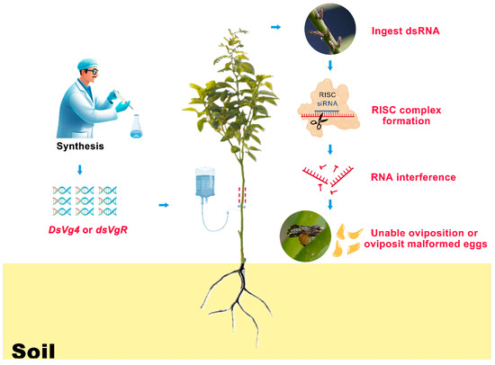
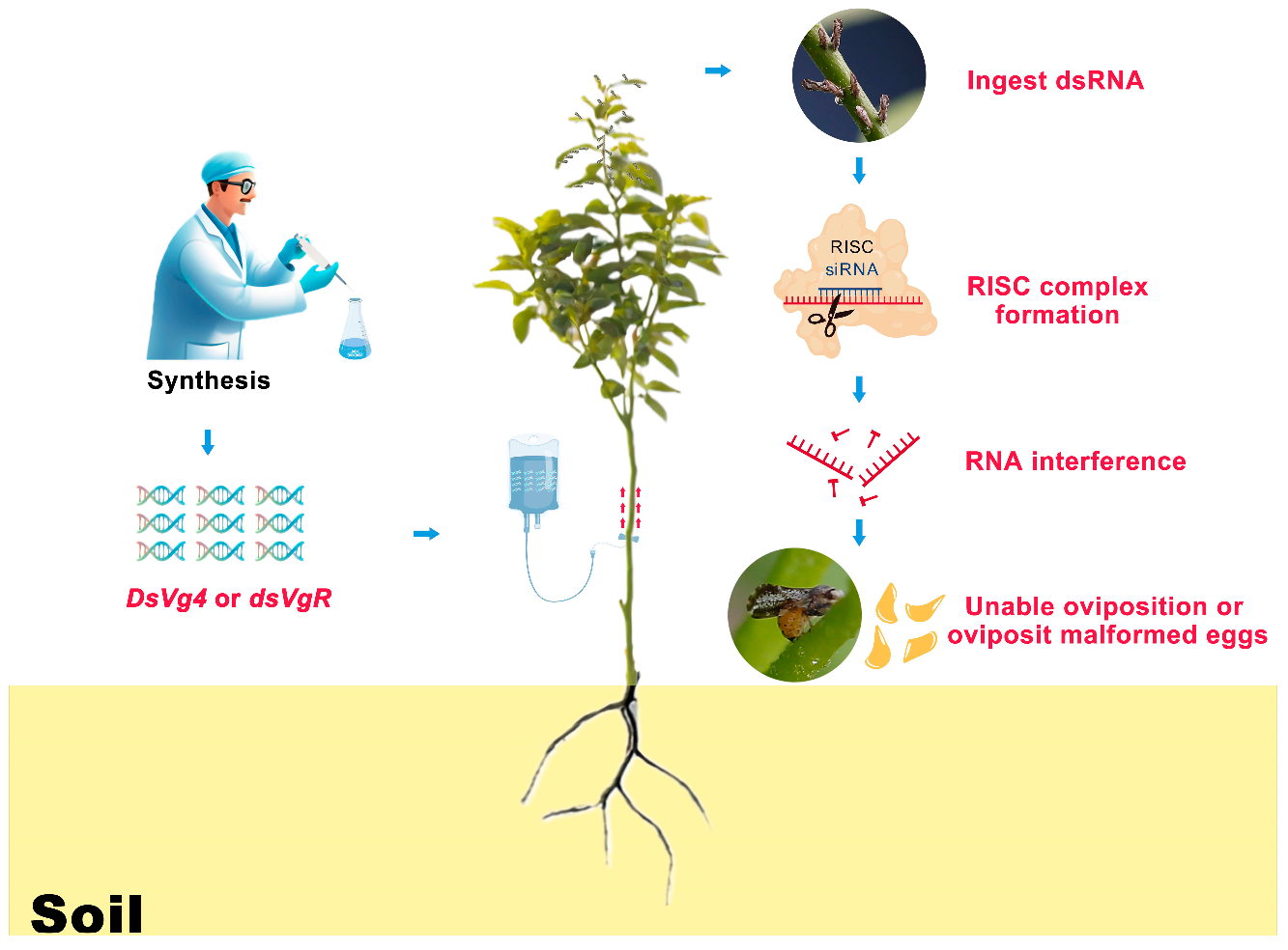
Diaphorina citri (Kuwayama) female ovary development and oviposition behavior are dependent on yolk accumulation, which is defined by vitellogenin protein synthesis in the fat body, blood transport to the ovaries, and entry into the oocytes via vitellogenin receptor-mediated endocytosis to form yolk. We created expression profiles of ovarian Vg and VgR gene family members over the 1- to 30-day developmental stage and then utilized the IPS approach to apply RNAi interference to Vg4 and VgR gene expression. The inhibiting Vg4 and VgR gene expression inhibited ovarian development and oviposition behavior. Our findings suggest that the Vg4 and VgR genes play critical roles in ovarian development and oviposition behavior.

Abstract

Diaphorina citri (Kuwayama) is one of the primary vectors of Citrus Huanglongbing (HLB), which poses a danger to the long-term development of the citrus industry. In this work, the spatiotemporal expression processes of the genes encoding vitellogenin-3-like (Vg4) and vitellogenin receptor (VgR) in D. citri females during the 1- to 30-day developmental stage of ovarian development and oviposition behavior were investigated. Vg4 and VgR gene expression largely supports oocyte maturation in D. citri females at the 1- to 15-day developmental stage. During the 15- to 30-day developmental stage, the expression of the Vg4 and VgR genes predominantly promotes the progression of oviposition behavior. Comparisons of ovarian development and oviposition behavior revealed that in the 15- to 30-day developmental stage, female egg production accounted for 75–85% of total oviposition, while mature oocyte formation occurred largely after the 15th day of ovarian development. The findings of this study provide fresh theoretical support and insights into the use of RNA interference (RNAi) technology to reduce the damage caused by D. citri populations.

Graphical Abstract

1. Introduction

Vitellogenin (Vg) is an essential insect glycoprotein that controls ovarian development, oocyte production, and oviposition behavior [1,2]. The vitellogenin receptor (VgR) is a key carrier protein that controls the entry of Vg proteins into oocytes; VgR activities include transporting Vg and regulating ovarian development and signal transduction, with a specific focus on controlling ovarian maturation in female insects [3,4]. Therefore, understanding how the vitellogenin (Vg) and vitellogenin receptor (VgR) genes govern ovarian development and oviposition could provide a theoretical basis to develop molecular pesticides.
Insect Vg and VgR genes play important roles in ovarian development by mediating nutrient accumulation and maturation in oocytes as well as influencing the oviposition [5,6,7]. Previous studies illustrated that Vg proteins are generated in fat bodies and their synthesis is affected by hormones such as ecdysone and juvenile hormone (JH) [8,9]. Following synthesis, Vg proteins are transported via the bloodstream to the ovaries, where they accumulate outside the oocytes. After binding to the VgR receptor, expressed specifically on the oocyte membrane, Vg proteins enter the oocyte interior via VgR-mediated endocytosis to form the yolk sac that provides nutrients for embryonic development [10]. These results indicate that the coexpression of Vg and VgR genes is critical for ovarian development and suggest that RNA interference (RNAi) targeting either the Vg or VgR gene could result in abnormal ovarian development, oocyte degeneration, or a significant reduction in egg production [11,12,13]. For instance, interfering with the expression of the Vg and VgR genes in Lasioderma serricorne (Fabricius) resulted in considerably shorter average female oocyte lengths (203.7 µm; 228.9 µm; 226.5 µm) than those in the control check (CK) (416.5 µm) [14]. Although previous research has revealed partial functions of Vg and VgR genes using microinjection and RNAi technologies, the processes through which these genes influence ovarian development and oviposition behavior are long-term [15,16,17], especially for insect species with long longevity. Taking Helicoverpa armigera, Apis mellifera, Leptopilina boulardi, and Bactrocera dorsalis as an example, short-term RNAi of these insects Vg and VgR genes via microinjection may be insufficient to fully understand the intricate activities of these genes in regulating ovarian development and oviposition [18,19,20,21]. Given the complexity of temporal and spatial expression of the Vg and VgR gene family, more studies focused on the regulatory mechanisms that govern ovarian development and oviposition are needed.
Diaphorina citri (Kuwayama) is one of the primary vectors of Citrus Huanglongbing (HLB) [22,23]. The rapid spread of HLB in the world is due primarily to the high reproductive capacity and rapid spread of D. citri causing extremely serious economic losses worldwide annually [24,25,26,27,28,29,30,31]. Currently, one of the effective measures to minimize the impact of HLB has been based on area-wide suppression of D. citri population reproduction and dispersal [32]. However, D. citri population has a high reproductive capacity and significant overlap between generations, and, due to the heavy use of pesticides, D. citri has strong pesticide resistance, meaning that long-term management of this pest has yet to be established [33,34,35,36]. For example, partial D. citri females can live for 40 days [37], resulting in a higher egg production (312.50 ± 4.92 eggs per female) to increase the extent of its devastation [38]. Therefore, suppression of oviposition may be an effective technology to manage the D. citri population. Our preliminary research conducted high-throughput transcriptome sequencing on the abdomens of D. citri female, obtaining the vitellogenin-1-like-1 (Vg1), vitellogenin-1-like-2 (Vg2), vitellogenin-2-like (Vg3), vitellogenin-3-like (Vg4), vitellogenin-like (Vg5), and vitellogenin receptor (VgR) genes in the abdomens of females [39]. We further discovered that the Vg4 protein, corresponding to the Vg4 gene, had the most complex amino acid structure in the Vg gene family and was highly expressed in the female abdomen [39]. This finding suggests that the Vg4 gene may play a crucial role in egg formation physiology. RNA interference (RNAi) research revealed that the IPS (in-plant-system) technique for the delivery of double-stranded vitellogenin-3-like (dsVg4) and double-stranded vitellogenin receptor (dsVgR) strongly inhibited oviposition in D. citri females, and female Vg4 and VgR genes were interfered with, resulting in incomplete eggs and nymphal abnormalities [13,40], which indicates that the Vg and VgR gene families are excellent candidates for critical target genes to decrease female oviposition. However, the D. citri Vg4 and VgR genes regulating ovarian development and oviposition mechanisms remain unclear.
In this study, we used qRT–PCR to investigate the spatiotemporal expression patterns of Vg and VgR gene family members in D. citri female ovaries during the 1- to 30-day developmental stage. Furthermore, we used RNAi technology to investigate the functions of D. citri Vg4 and VgR genes in ovarian development and oviposition. Our findings could provide a solid theoretical foundation to understand how the Vg and VgR genes influence ovarian development and oviposition and to develop novel techniques to achieve the sustainable management of D. citri.

2. Materials and Methods

2.1. Rearing and Collection

The population of D. citri was collected on a planting base (Murraya exotica L.) in Guangxi Minzu University (Guangxi Zhuang Autonomous Region, China). Adults were fed with M. exotica and stably passaged for more than three generations in an artificial climate chamber at 25 ± 1 °C with 75 ± 5% relative humidity and a light/dark (L:D) cycle = 14:10 h. Male and female adults emerging on the same day were randomly selected for the following experiments. For the RNAi assay, dsRNA was dissolved in water, added to the tube, and then transported to the tender shoot through the xylem of M. exotica for feeding the females. Oviposition among the females in the rearing container was observed and recorded.
Females in different treatment groups were reared in plastic containers. For the fabrication and usage of plastic containers, refer to [13]. For the RNAi assay, dsRNA was dissolved in water, added to the tube, and then transported to the tender shoot through the xylem of M. exotica for feeding the females. Oviposition of adults in the rearing container were observed and recorded.

2.2. DsRNA Synthesis

Ovaries of D. citri were dissected as described above and immediately frozen in liquid nitrogen in a 1.5 mL Eppendorf tube for total RNA extraction using TransZol Up (TransGen Biotech, Beijing, China). Afterward, cDNA synthesis was carried out using the PrimescriptTM RT reagent kit with gDNA eraser (perfect real time) (TaKaRa, Shiga, Japan) following the manufacturer’s instructions: (1) prepare the following mixture in an RNase-free EP tube: RNase-free ddH2O (up to 16 µL), 4 µL of 4 × 2-Step Gdna Erase-Out Mix, total RNA (1 pg–1 µg), 42 °C reaction for 2 min; (2) add 4 µL of 5×ToloScript qRT EasyMix directly to the reaction solution in step 1, and conduct reverse transcription, 37 °C for 15 min, 85 °C for 5 s. Vg4 and VgR gene interference fragments were designed using SnapDragon-dsRNA Design https://www.flyrnai.org (accessed on 25 June 2022) (DRSC/TRiP Functional Genomics Resources & DRSC-BTRR, Harvard Medical School). Using the nucleotide sequences of the Vg4 and VgR genes, primers were designed for PCR, quantitative real-time PCR (qRT–PCR) and synthetic dsRNA primers from the National Center for Biotechnology Information (NCBI, Bethesda, MD, USA) (Table 1). Vg4 (477 bp) cDNA fragments were generated with the primer pair Vg4 F3/R3 using 2×FastPfu Premix (TOLOBIO, Central lslip, NY, USA). The purified PCR-generated Vg4 fragments were subsequently cloned and inserted into the pMD18T vector (DH5α; Thermo Fisher Scientific, Waltham, MA, USA). The resulting Vg4 plasmids were used as templates to generate dsVg4 with a dsRNA synthesis kit (T7 RiboMAXTM Express RNAi System, Beijing, China; Supplementary Materials). The synthesis method is described in the manufacturer’s instructions: preparation of dsRNA synthesis (RiboMAXTM Express T7 2X Buffer*, 10.0 μL; transcriptional template, 6.0 μL; DEPC water, 2.0 μL; Enzyme Mix T7 Express, 2.0 μL), in vitro transcription, reaction at 37 °C for 30 min; after holding at 70 °C for 10 min, hold at room temperature for 20 min; after standing at 37 °C for 30 min, place on ice for 5 min; and centrifuge for 10 min and anneal to obtain dsRNA. The synthesized dsRNA was quantified using an N80 Touch nanophotometer (Implen, München, Germany) at 260 nm, and the integrity was analyzed by agarose gel electrophoresis. The syntheses of dsVgR (346 bp) and dsGFP (726 bp) were carried out as described for dsVg4; the fragments amplified by the qRT-PCR primers used for the Vg4 and VgR genes are located outside the RNAi target sequences, making them well-suited for evaluating the effectiveness of RNAi interference. The gene qRT-PCR amplification fragments can be found in the Supplementary Materials.

2.3. Spatiotemporal Expression

Seven rearing containers were prepared, with each rearing container holding 15 pairs of one-day-old females and males; the specific specifications and characteristics of the rearing containers have been described by [13]. All females from one container were selected every 5 days at 8 am and dissected for ovary sampling, while M. exotica tender shoots (Height 8 cm) and sterile water (500 μL) were replaced in other containers. The 15 female-dissected ovaries were first photographed with a microscopic measurement system coupled to a stereoscope (SMZ800N, Nikon, Tokyo, Japan) for further mature oocyte measurements, and then we merged 15 female-dissected ovaries to be sampled for RNA extraction. For the anatomical and measurement methods for the ovary, refer to [13]: measure 10 oocytes from the per-female ovaries, and record the mature oocyte number, as well as oocyte length and width (µm). In this way, ovaries were collected from 1-, 5-, 10-, 15-, 20-, 25-, and 30-day-old females. The experimental procedure was repeated three times. A total of 315 pairs of adult males and females are required. The cDNA from these samples was prepared as previously described and used for Vg1, Vg2, Vg3, Vg4, Vg5, VgR gene qRT–PCR assays (Supplementary Materials). qRT–PCR was carried out using 2 × Q3 SYBR qPCR Master Mix (High Rox) (TOLOBIO, Shanghai, China) following the manufacturer’s instructions: (1) prepare the following mixture in an RNase-free Eppendorf tube: 10 μL of 2 × Q3 SYBR qPCR Master Mix, 0.4 μL of primer 1 (10 µM), 0.4 μL of primer 2 (10 µM), 1 μL of template cDNA, and 8.2 μL of ddH2O; (2) place the prepared solution into 96-well plates and perform qRT–PCR on a qRT–PCR machine. The qRT–PCR conditions were as follows: predenaturation, cycle 1 at 95 °C for 30 s; circular reaction, cycle 40 at 95 °C for 10 s; melting curves, cycle 1 at 95 °C for 15 s, 60 °C for 60 s, and 95 °C for 15 s. All reactions were performed with the QuantStudioTM Real-Time PCR system (Applied Biosystems, Waltham, MA, USA) using the primers listed in Table 1. Relative gene expression data of all treatment groups from ovaries qRT–PCR were analyzed using the dual internal reference gene Actin and Elongation Factor 1-alpha (EF1a) 2−ΔΔCT method. QRT-PCR was performed for each gene with three biological replicates and three technical replicates (Supplementary Materials).

2.4. RNAi for Ovarian Development

Six rearing containers were prepared, with each rearing container holding 15 pairs of one-day-old females and males. M. exotica tender shoots and 300 μL dsVg4 solution (1 ng/μL) were placed in containers, while 15 females from one container were selected every 5 days and dissected for ovary sampling, and the solution and M. exotica tender shoots were replaced. For the anatomical and measurement methods for the ovary, refer to [13]. Refer to the method for the experiment for the spatiotemporal expression of Vg and VgR genes for photographing and measuring the ovaries. Then, 15 female ovaries were sampled for RNA extraction. In this way, ovaries were collected from 5-, 10-, 15-, 20-, 25-, and 30-day-old females. The experimental procedure was repeated three times. A total of 270 pairs of adult males and females are required. The dsVgR and dsGFP treatment groups were handled identically, and the solution was replaced according to different treatment processes. The cDNA from these samples was prepared as previously described and used for ovary qRT–PCR assays. QRT-PCR was performed for each gene with three biological replicates and three technical replicates (Supplementary Materials).

2.5. RNAi for Oviposition Behavior

Ten newly emerged, unmated females were randomly selected, fed dsVg4 (1 ng/μL, 300 μL), and paired with ten 7- to 12-day-old unmated males. A pair with a male and a female was placed in each rearing container. The dsVgR, dsVg4 + VgR, blank control (sterile water) and dsGFP treatments were carried out as described for the dsVg4 treatment. Each day at 8 am, M. exotica shoots were replaced when eggs were detected on the shoots. Change the solution in the rearing container once every 5 days. The replaced M. exotica shoots were stored separately after the number of eggs was counted. The number of nymphs was also counted after the eggs hatched. The preoviposition, fecundity, oviposition rhythm, egg hatchability, deformity rate of the eggs and nymphs, and female survival rate were assessed for 30 days. The experimental procedure was repeated five times. In total, 50 pairs of adult males and females are required.

2.6. Statistical Analysis

Data analysis was performed using SPSS 25.0 (IBM Corp., Armonk, New York, NY, USA) and GraphPad Prism 10 (GraphPad Software, San Diego, CA, USA). Data on the spatiotemporal expression of the Vg and VgR gene families were analyzed using Tukey’s honestly significant difference (HSD) multiple tests. Expression levels of the Vg4 and VgR gene RNAi were analyzed using an independent samples t test. Number of mature eggs and egg length and width were analyzed using Tukey’s HSD. Data on the preoviposition, daily oviposition, number of eggs per female, egg hatchability, and deformity rates of eggs and nymphs were analyzed using one-way analysis of variance (ANOVA). Analysis of female survival rate between the interference group and the control group in the RNAi for oviposition behavior experiment used the log-rank (Mantel–Cox) test. The difference was considered to be statistically significant at the 5% level (p < 0.05).

3. Results

3.1. Spatiotemporal Expression of the Vg and VgR Genes

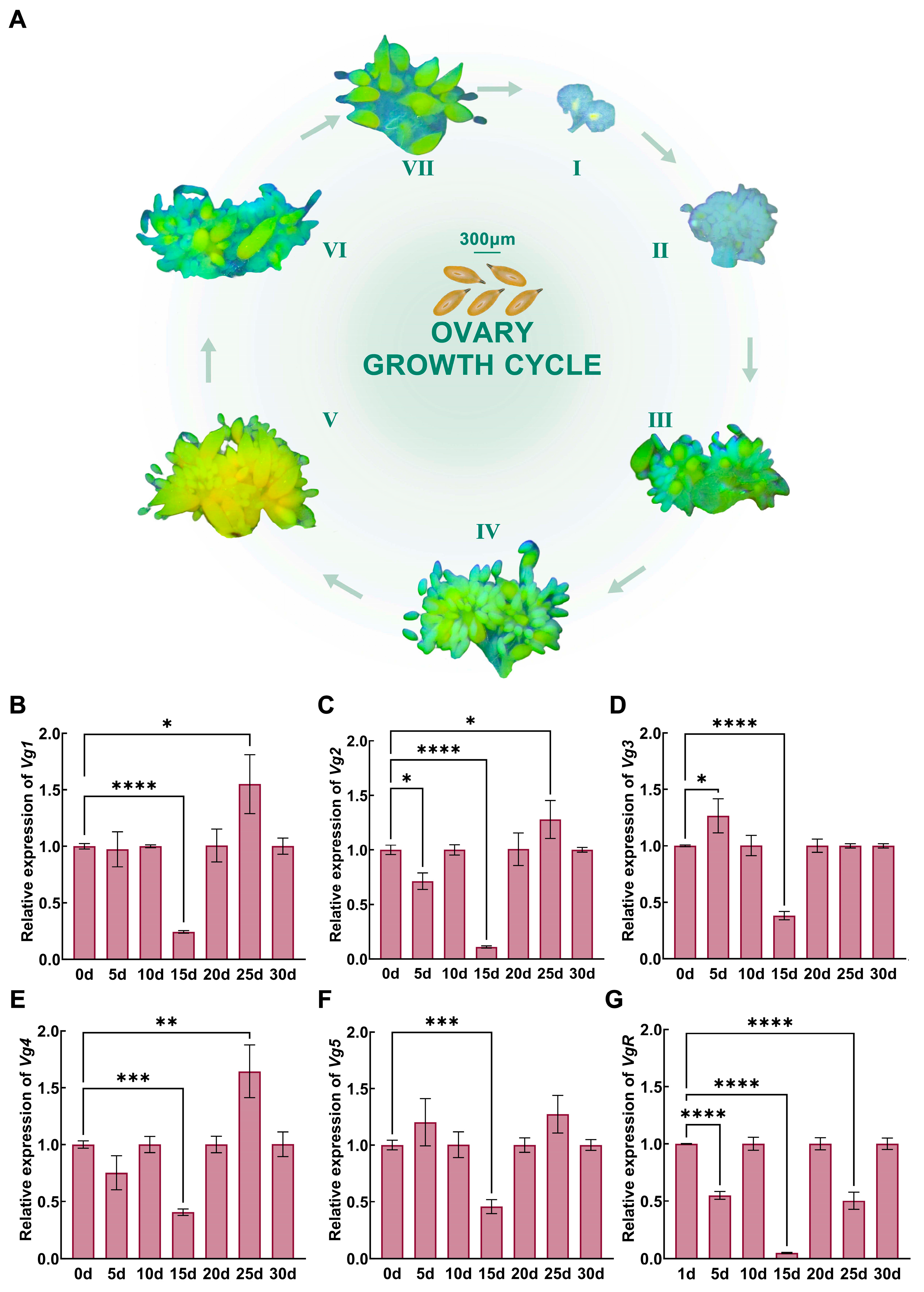
Female ovaries were dissected at the 1- to 30-day developmental stage, and the expression levels of ovarian Vg and VgR gene family members were detected using qRT–PCR technology (Figure 1; Supplementary Materials). The results indicate that during the 1- to 15-day developmental stage, oocyte development is characterized by a gradual increase in volume; during the 20- to 30-day stage, no significant differences in changes in the length or width of the oocytes were observed. The stage with the maximum length and width of the oocytes occurred at 25 days (length: 290.60 ± 2.10 µm; width: 119.46 ± 3.35 µm) (Figure 1A). Vg and VgR gene family members were expressed in the 1- to 30-day female adults. Compared with the expression levels of newly emerged females (1-day), the expression of vitellogenin-1-like-1 (Vg1), vitellogenin-1-like-2 (Vg2), vitellogenin-2-like (Vg3), vitellogenin-3-like (Vg4), vitellogenin-like (Vg5) and vitellogenin receptor (VgR) showed the most significant decrease at 15 days (Vg1, t = 47.68, p < 0.0001; Vg2, t = 34.70, p < 0.0001; Vg3, t = 28.42, p < 0.0001; Vg4, t = 24.37, p < 0.001; Vg5, t = 12.50, p < 0.001; VgR, t = 297.90, p < 0.0001; Figure 1B–G), whereas the expression of the five genes showed the most significant increase at 25 days (Vg1, t = 3.64, p < 0.05), 25 days (Vg2, t = 2.69, p < 0.05), 5 days (Vg3, t = 3.05, p < 0.05), and 25 days (Vg4, t = 4.75, p < 0.01), respectively (Figure 1B–E). However, there was no significant increase in Vg5 and VgR in comparison in newly emerged females (Figure 1F,G).

3.2. Effects of Vg4 and VgR Gene RNAi on Ovarian Development

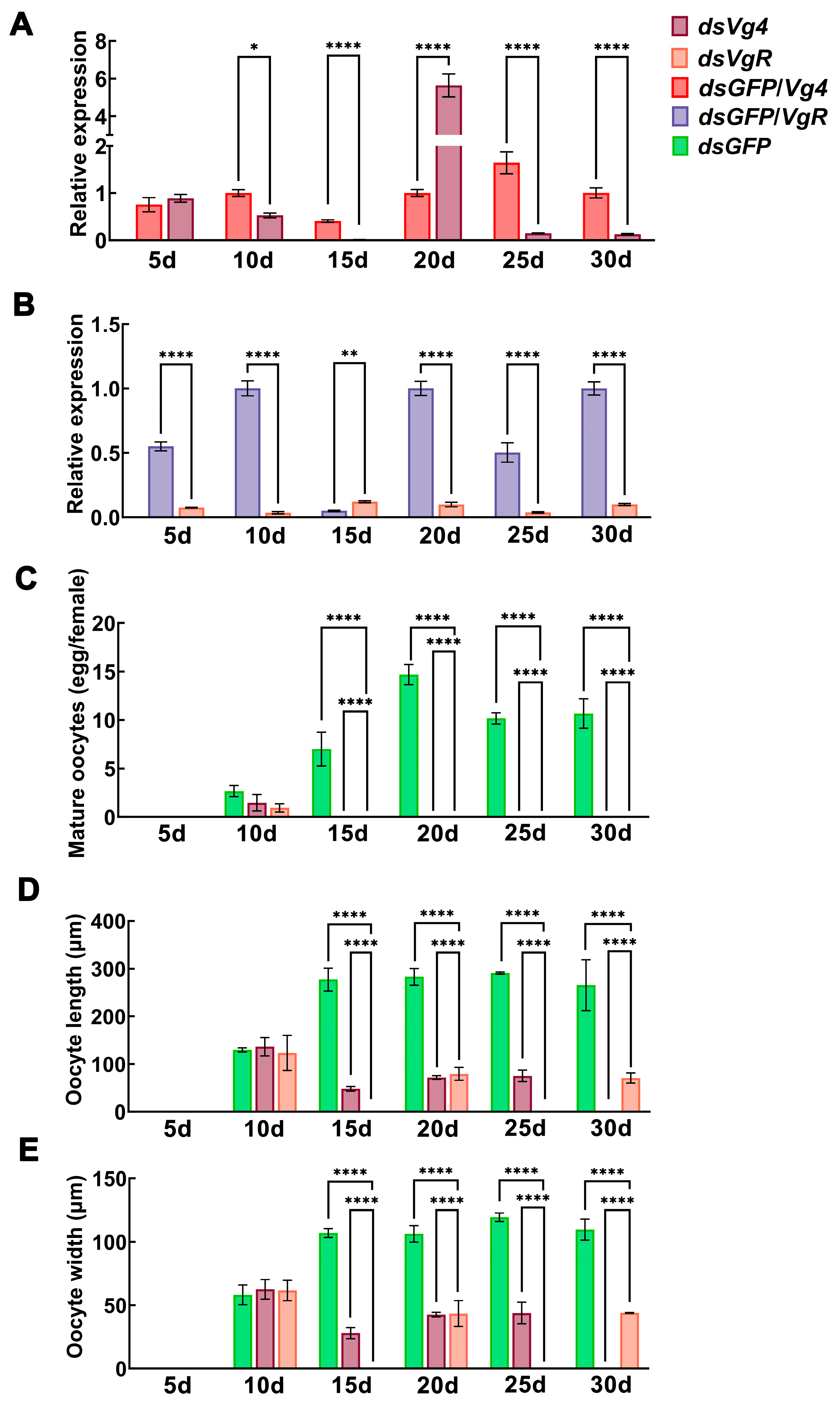
Following continuous feeding on dsVg4 and dsVgR during the 1- to 30-day ovarian development stage in D. citri females, both Vg4 and VgR gene expression and ovarian development were effectively disrupted, with significant downregulation of Vg4 and VgR gene expression in the females (Figure 2; Supplementary Materials). Compared with the dsGFP negative control, the dsVg4 interference group exhibited the most significant decrease in Vg4 gene expression at 15 days, with a relative expression reduction of 97.69% (p < 0.0001). However, at the 20-day ovarian development stage, the relative expression of the Vg4 gene abnormally increased by 462.37% (p < 0.0001) (Figure 2A). In the dsVgR interference group, VgR gene expression decreased most significantly at 10 days, with a relative expression reduction of 96.59% (p < 0.0001). However, at the 15-day ovarian development stage, the relative expression of the VgR gene abnormally increased by 146.22% (p < 0.01) (Figure 2B).
An analysis of the mature oocyte number in the ovaries, as well as oocyte length and width, using Tukey’s HSD test revealed dsVg4 and dsVgR interference groups. The mature oocyte number during the ovarian development stages from 15 to 30 days was 0.00 ± 0.00 per female (Figure 2C), which was significantly lower than that of the dsGFP negative control at 15 days (7.00 ± 1.73 mature oocytes per female; dsVg4, t = 7.00, p < 0.01; dsVgR, t = 7.00, p < 0.01), 20 days (14.67 ± 1.04 mature oocytes per female; dsVg4, t = 24.41, p < 0.001; dsVgR, t = 24.41, p < 0.001), 25 days (10.17 ± 0.58 mature oocytes per female; dsVg4, t = 30.50, p < 0.001; dsVgR, t = 30.50, p < 0.001), and 30 days (10.67 ± 1.53 mature oocytes per female; dsVg4, t = 12.09, p < 0.001; dsVgR, t = 12.09, p < 0.001). Interference with the expression of the Vg4 and VgR genes effectively suppressed female mature oocyte formation. The average dimensions of the dsVg4 interference group of oocytes were as follows: 15 days (length, 47.99 ± 4.89 µm; width, 28.02 ± 4.34 µm), 20 days (length, 71.81 ± 4.10 µm; width, 42.62 ± 1.77 µm), 25 days (length, 75.17 ± 12.13 µm; width, 43.79 ± 8.56 µm), and 30 days (length, 0.00 ± 0.00 µm; width, 0.00 ± 0.00 µm); the average dimensions of the dsVgR interference group of oocytes were 15 days (length, 0.00 ± 0.00 µm; width, 0.00 ± 0.00 µm), 20 days (length, 79.53 ± 13.57 µm; width, 43.46 ± 10.22 µm), 25 days (length, 0.00 ± 0.00 µm; width, 0.00 ± 0.00 µm), and 30 days (length, 70.64 ± 10.86 µm; width, 43.96 ± 0.29 µm) (Figure 2D,E). These values were significantly lower than those of the dsGFP negative control at 15 days (length 269.80 ± 28.47 µm; width 107.05 ± 3.54 µm), 20 days (length 282.89 ± 17.47 µm; width 106.38 ± 6.47 µm), 25 days (length 290.60 ± 2.10 µm; width 119.46 ± 3.35 µm), and 30 days (length 265.27 ± 53.22 µm; width 109.73 ± 8.32 µm) (Figure 2D,E). These results indicate that the Vg4 and VgR genes are key genes for ovarian development and oocyte formation in D. citri.

3.3. Effects of Vg4 and VgR Gene RNAi on the Preoviposition Period and Oviposition Rhythms

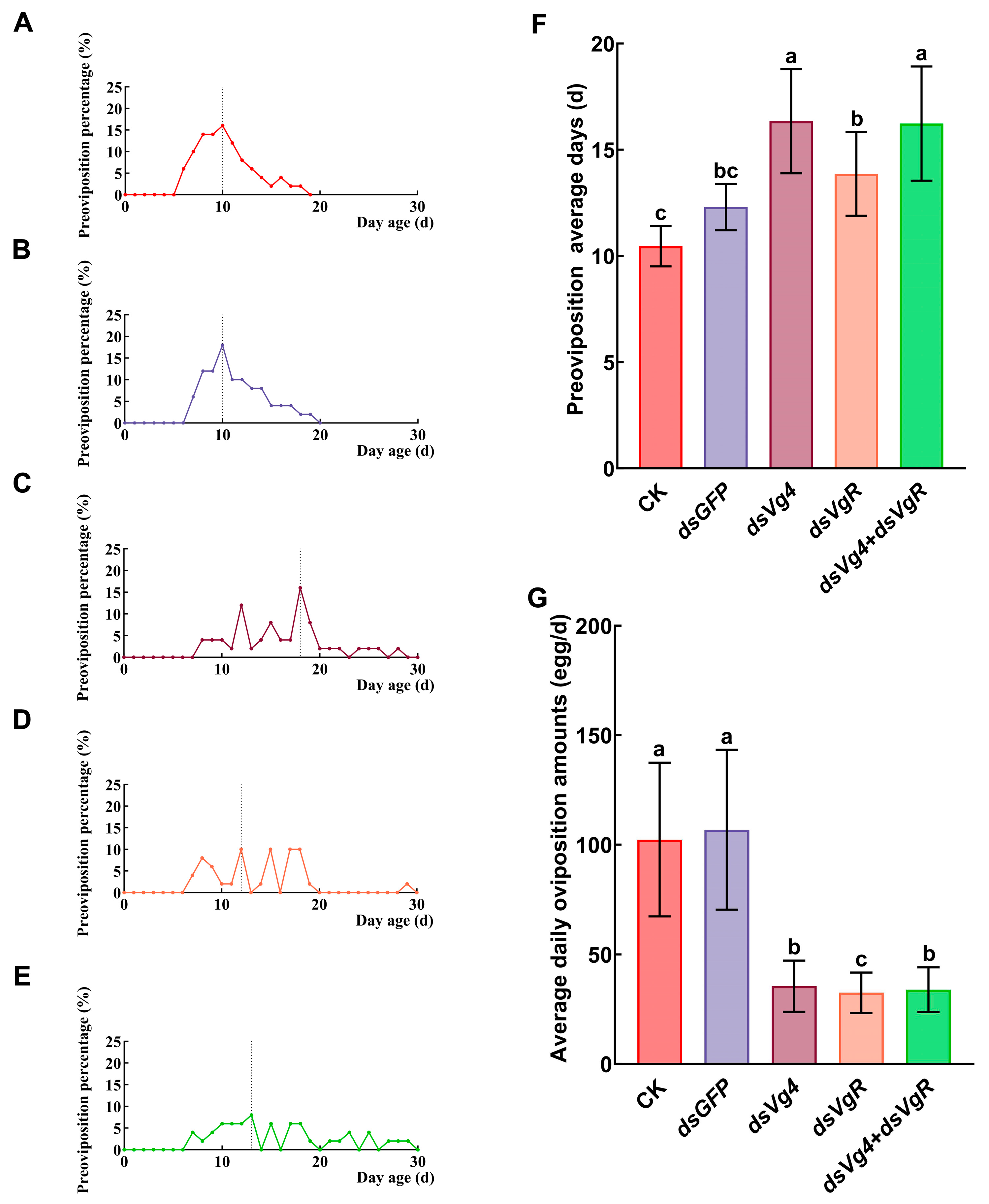
During the 1- to 30-day ovarian development stage, we observed the effects of continuous feeding on dsVg4, dsVgR, and dsVg4 + dsVgR during the preoviposition period and oviposition rhythm in D. citri females (Figure 3). The longest average preoviposition period was observed in the dsVg4 interference group; females required 7 days of feeding posteclosion to reach the oviposition stage, with an average preoviposition period of 16.34 ± 2.45 days. We checked the 50-female oviposition every day (total eggs: 1065) and found average daily oviposition amounts of 35.50 ± 11.67 eggs per day (Figure 3C,F,G). The lowest average daily oviposition amount was observed in the dsVgR interference group; females required 6 days of feeding posteclosion to reach the oviposition stage, with an average preoviposition period of 13.86 ± 1.97 days. We checked the 50-female oviposition every day (total eggs 976) and found average daily oviposition amounts of 32.53 ± 9.21 eggs per day (Figure 3D,F,G).
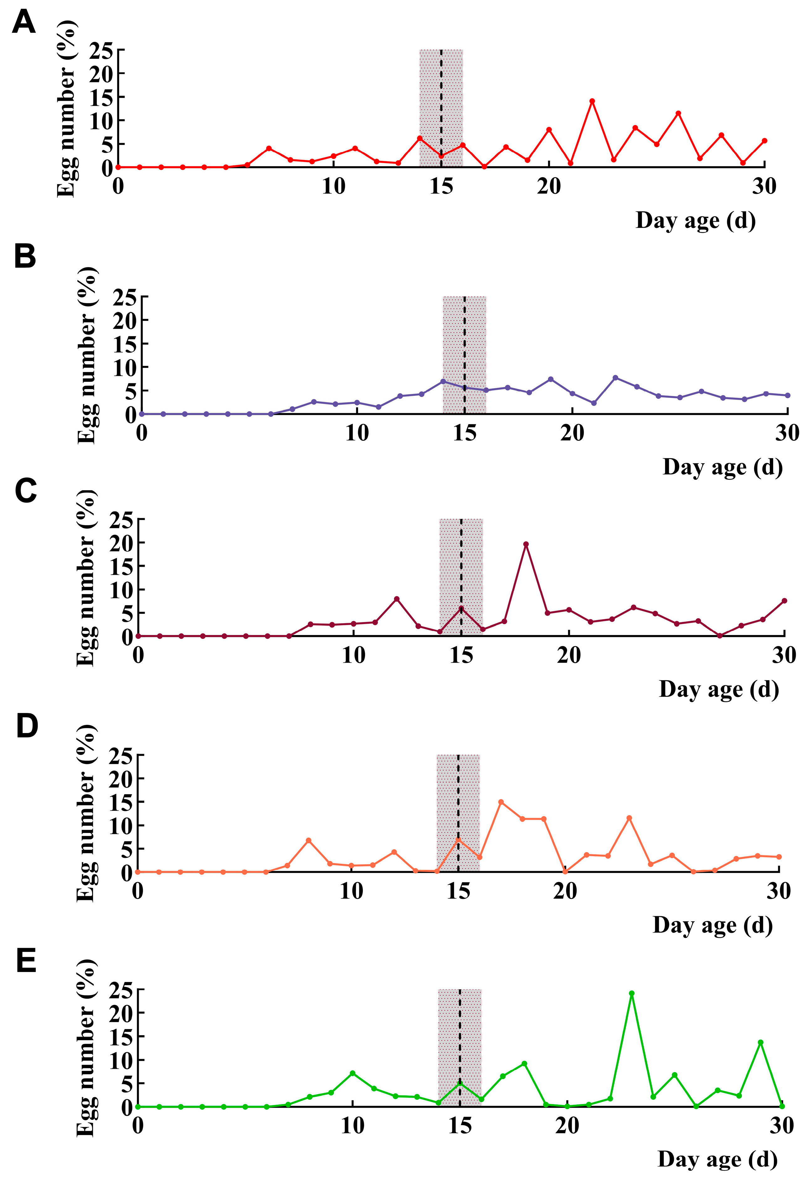
The average preoviposition period of the interference group was generally greater than that of the control group, while the average daily oviposition amount was significantly lower than that of the control group. Comparisons of the oviposition rhythm data revealed that the female oviposition rhythm in the interference group significantly fluctuated, with weaker persistence of oviposition activity in the interference group than in the control group. The female oviposition amount in the dsVg4, dsVgR and dsVg4 + dsVgR interference groups during the 15- to 30-day ovarian development stage accounted for 78.15%, 82.17% and 78.13% of the total oviposition amount, respectively (Figure 4C–E). The females in the CK and dsGFP groups exhibited highly sustained oviposition activity, with periodic fluctuations in daily peaks, though overall showing an increasing-to-decreasing trend; however, overall, the oviposition amount contributed 77.92% and 75.44% of the total oviposition amount during the 15- to 30-day ovarian development stage, respectively (Figure 4A,B).

3.4. Effects of Vg4 and VgR Gene RNAi on Female Reproductive Capacity and Survival Rate

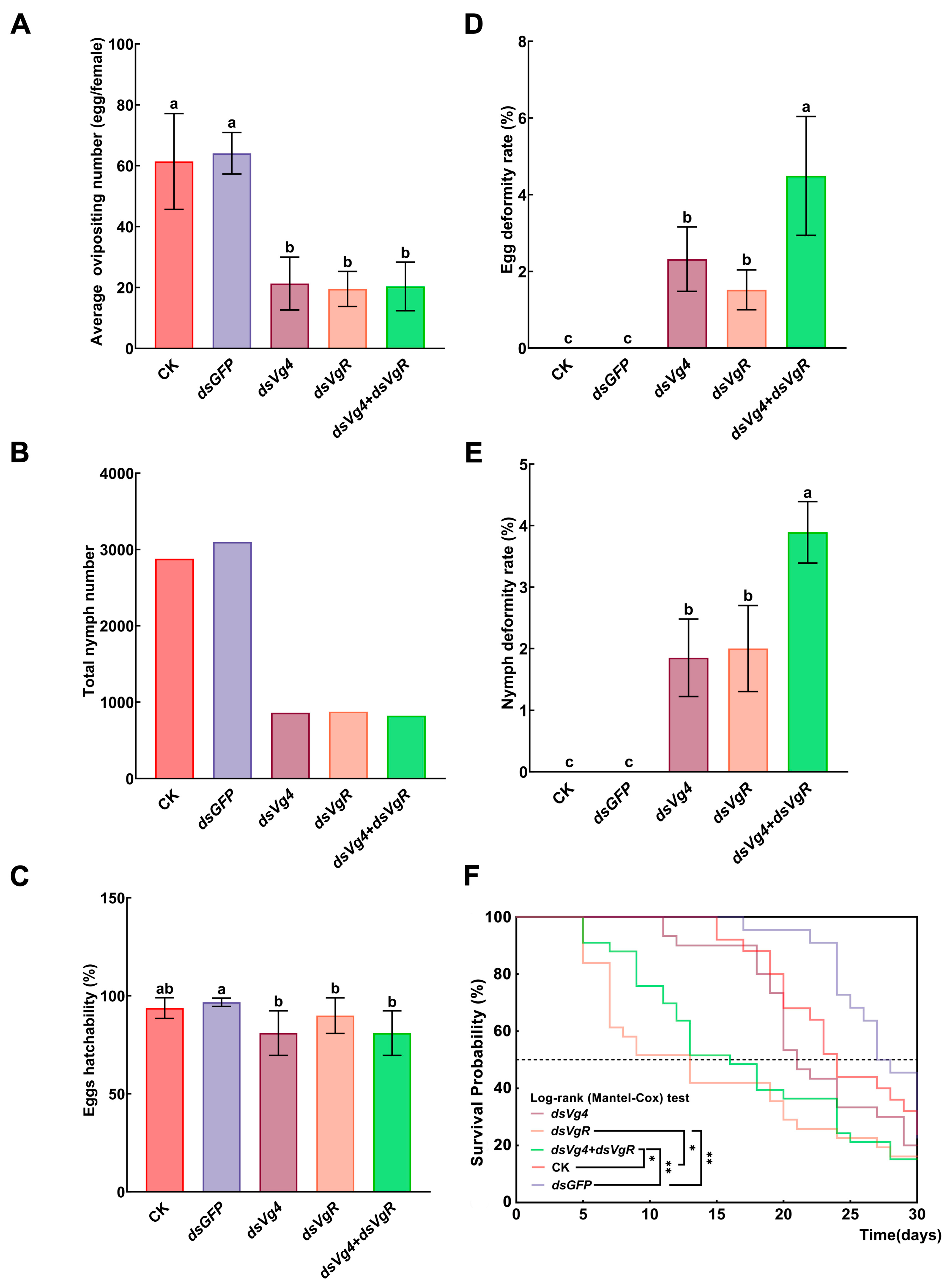
A comparison of the reproductive capacity and survival rates of D. citri females in the 1- to 30-day ovarian development stage between the interference and control groups revealed that the total oviposition amount per female in the dsVg4, dsVgR, and dsVg4 + dsVgR interference groups was significantly lower than that in the control group. Females in the dsVgR interference group presented the lowest average total oviposition amount per female (19.52 ± 5.76 eggs per female; total eggs 976), whereas those in the dsVg4 + dsVgR interference group hatched the fewest total nymphs (hatched nymphs 670) (Figure 5A,B). The lowest egg hatching rate was observed in the dsVg4 interference group; all the treatment groups presented egg hatching rates above 80%, and the majority of the eggs successfully hatched (Figure 5C). The highest rates of egg and nymph deformities were observed in the dsVg4 + dsVgR interference group, with an egg deformity rate of 4.49 ± 1.55% and a nymph deformity rate of 3.89 ± 0.50% (Figure 5D,E). A comparison of the survival rate data from the log-rank (Mantel–Cox) test revealed that compared with those in the other treatment groups, the lifespan of females in the dsVgR and dsVg4 + dsVgR interference groups was significantly shorter during the 1- to 30-day ovarian development stage. In the DsVgR interference group compared with CK, the median survival (time taken to reach a survival of 50%) of CK females was 24 (95% CI of ratio: 1.02–3.34) days compared to 13 (95% CI of ratio: 0.30 to 0.98) days for dsVgR interference group females; this translates to a 45.83% decrease in longevity in dsVgR interference group females. In the dsVgR interference group compared with the dsGFP negative control, the median survival (time taken to reach a survival of 50%) of dsGFP females was 27.5 (95% CI of ratio: 1.15–3.90) days compared to 13 (95% CI of ratio: 0.26 to 0.87) days for dsVgR interference group females; this translates to a 52.73% decrease in longevity in dsVgR interference group females (Figure 5F). In the DsVg4 + dsVgR interference group compared with CK, the median survival (time taken to reach a survival of 50%) of CK females was 24 (95% CI of ratio: 0.84–2.69) days compared to 16 (95% CI of ratio: 0.37 to 1.19) days for dsVg4 + dsVgR interference group females; this translates to a 33.33% decrease in longevity in dsVg4 + dsVgR interference group females. In the dsVg4 + dsVgR interference group compared with the dsGFP negative control, the median survival (time taken to reach a survival of 50%) of dsGFP females was 27.5 (95% CI of ratio: 0.94–3.14) days compared to 16 (95% CI of ratio: 0.32 to 1.06) days for dsVg4 + dsVgR interference group females; this translates to a 41.82% decrease in longevity in DsVgR interference group females (Figure 5F).

4. Discussion

This study investigated the spatiotemporal expression mechanisms of the Vg4 and VgR genes in D. citri females during ovarian development and oviposition behavior. Both the Vg4 and VgR genes govern ovarian development and control oviposition behavior progression. A spatiotemporal expression study using qRT–PCR and ovarian dissection revealed that 15 days was the time point at which the lowest expression levels of the Vg and VgR gene families occurred, which coincided with the period of rapid oocyte volume expansion. D. citri females go through developmental stages from 1 to 15 days, in which expression of the Vg and VgR genes primarily promotes oocyte maturation, and from 15 to 30 days, in which expression of these genes primarily promotes the progression of oviposition behavior. It is worth noting that the relative expression of the VgR and Vg4 genes was significantly elevated at the 15- and 20-day developmental stages (Figure 2). This phenomenon may be attributed to the regulation of cellular immunity by dsRNA. Previous studies have shown that dsRNA-degrading enzymes (dsRNases) are key factors in reducing various insect species the efficiency of RNA interference and may lead to increased gene expression, but the related molecular mechanism is not clear; further research is needed [41,42]. Therefore, the expression of egg-laying-related gene pathways in the citrus psyllid at the 15- and 20-day developmental stages warrants further investigation. Comparisons of oviposition rhythm data revealed that during the 15- to 30-day developmental stage, females in the interference and control groups accounted for 75–85% of the total oviposition amount, respectively. Oocyte maturation predominantly occurred after 15 days of ovarian development. D. citri female oocyte development and oviposition behavior are phased, reflecting the staged expression of the Vg and VgR genes. Their peak expression time is strongly linked to ovarian developmental phases, making them useful molecular markers for analyzing D. citri female reproductive patterns [43]. However, research into the gene networks underlying the staged expression of the Vg and VgR genes in D. citri females is lacking, as well as into how these genes interact with other hormone signaling pathways or regulatory networks to regulate these processes, requiring further investigation.
According to previous studies, Vg and VgR genes show low expression during the egg or larval stages in some insects, with significant upregulation occurring during the pupal stage. Using Tomicus yunnanensis as an example, the peak expression levels of the Vg and VgR genes in T. yunnanensis females can exceed pupal levels by more than twice [44]. This finding demonstrates that the high expression phenomenon of the Vg and VgR genes does not persist throughout the life cycle of the insect. Instead, it has a three-phase rhythm: expression during adulthood, a peak specific to ovarian development, and a quick decrease following yolk filling [45,46]. The expression levels of the Vg and VgR genes in D. citri females decrease rapidly at the 15-day developmental stage, which could be attributable to the particular suppression of Vg and VgR gene expression by upstream or downstream regulatory genes after the peak accumulation of yolk in the ovaries. However, more research is needed on the underlying molecular pathways that govern this phenomenon.
Recent research suggests that essential gene pathways controlling insect oviposition involve several hormones signaling and dietary regulation networks [47,48,49,50]. Among them, the juvenile hormone (JH) pathway may be one of the primary regulatory systems that controls the expression of Vg and VgR genes [51]. The JH pathway influences insect oviposition by activating Vg protein synthesis via the methoprene-tolerant/Taiman (Met/Tai) receptor complex, which regulates lipocyte polyploidization and follicle cell channel opening, promoting Vg protein entry into the oocyte [52]. Using Diaphorina citri (Kuwayama) as an example, disrupting female JH gene expression resulted in impaired ovarian development and a significant reduction in reproductive capacity in females (interference group: 136.61 eggs per female; CK: 236.29 eggs per female), demonstrating the certain critical role of JH in female oviposition [52]. Methoprene-tolerant (Met), Juvenile hormone (JH), Taiman (Tai), Vitellogenin (Vg), and Vitellogenin receptor (VgR) are some key genes for regulating insect oviposition. The JH, Met, and Tai genes are upstream regulators of Vg and VgR gene expression and may serve as ideal RNAi targets for D. citri regulation research, but more research is needed to verify this [53,54,55,56]. The JH pathway regulates Vg and VgR gene expression, providing vital insights into the relationships and regulatory processes among the Vg, VgR, JH, Met, and Tai gene families in female D. citri. In future studies, nanomaterials (chitosan) or pesticides (metaflumizone) at different concentrations could be used as co-delivery systems for dsRNA to enhance the interference effect of dsRNA targeting JH pathway genes, thereby further elucidating the mechanisms by which the JH pathway regulates oviposition in D. citri [57,58].

5. Conclusions

In conclusion, during the 1- to 15-day developmental stage, the expression of the Vg and VgR genes predominantly promotes oocyte maturation; during the 15- to 30-day developmental stage, the expression of these genes mostly promotes oviposition behavior progression within the ovaries. RNAi interference tests revealed that the Vg4 and VgR genes play critical roles in ovarian development and oviposition behavior in D. citri females. Future studies will focus on three main areas. First, we will perform in-depth studies on the pathway expression mechanisms of the Vg and VgR genes, with a focus on their connections with the JH signaling pathway. Second, we will construct dsVg4- and dsVgR-related bacterial vectors to improve their stability. Third, using the Vg and VgR genes as key network nodes, we will investigate the pathway expression mechanisms that control genes linked with oviposition behavior.

Supplementary Materials

The following supporting information can be downloaded at https://www.mdpi.com/article/10.3390/insects17060562/s1. Figure S1: Vg1 gene qRT-PCR amplification, melting curve and standard curve; Figure S2: Vg2 gene qRT-PCR amplification, melting curve and standard curve; Figure S3: Vg3 gene qRT-PCR amplification, melting curve and standard curve; Figure S4: Vg4 gene qRT-PCR amplification, melting curve and standard curve; Figure S5: Vg5 gene qRT-PCR amplification, melting curve and standard curve; Figure S6: VgR gene qRT-PCR amplification, melting curve and standard curve; Figure S7: Actin gene qRT-PCR amplification, melting curve and standard curve; Figure S8: EF1a gene qRT-PCR amplification, melting curve and standard curve; Figure S9: Ovaries at the newly eclosion stage (0d); Figure S10: Ovaries at the 5-day ovarian development stage; Figure S11: Ovaries at the 10-day ovarian development stage; Figure S12: Ovaries at the 15-day ovarian development stage; Figure S13: Ovaries at the 20-day ovarian development stage; Figure S14: Ovaries at the 25-day ovarian development stage; Figure S15: Ovaries at the 30-day ovarian development stage; Figure S16: DsVg4, dsVgR, and dsGFP were absorbed by tender M. odorifera shoots for six days, and gel electrophoresis was used to detect the persistence of dsVg4, dsVgR, and dsGFP after each day. The gel electrophoresis bands of dsVg4 were relatively clear at 1–6 d; the gel electrophoresis bands of dsVgR were relatively clear at 1–2 d, but blurry at 3–6 d; the gel electrophoresis bands of dsGFP were relatively clear at 1–6 d. The dsVg4-1, 2, 3, 4, 5, 6/dsVgR-1, 2, 3, 4, 5, 6/dsGFP-1, 2, 3, 4, 5, 6 represent the six replicates for each treatment; Figure S17: After M. odorifera shoots total RNA of absorbing dsVg4 and dsVgR were extracted, the sequencing results of Vg4 and VgR gene interference fragments were cloned using cDNA as template. (A) Base sequence and base feasibility of Vg4 gene, sequence alignment between the Vg4 sequencing sequence and the original sequence. (B) Base sequence and base feasibility of VgR gene, sequence alignment between the VgR sequencing sequence and the original sequence; Table S1: Gene qRT-PCR CT value; Table S2: Gene RNAi qRT-PCR CT value.

Author Contributions

Conceptualization, F.W. and H.L. (Hailin Li); methodology, H.L. (Hailin Li); software, X.W.; validation, F.W., X.Z. and H.L. (Hailin Li); formal analysis, Z.H.; investigation, J.W.; resources, H.L. (Hailin Li); data curation, X.D. and H.L. (Hongcun Liu); writing—original draft preparation, F.W.; writing—review and editing, H.L. (Hailin Li); visualization, F.W.; supervision, H.L. (Hailin Li); project administration, H.L. (Hailin Li); funding acquisition, H.L. (Hailin Li). All authors have read and agreed to the published version of the manuscript.

Funding

This work was financially supported by the Guangxi Natural Science Foundation (No. 2026GXNSFBA00640007), the Second Batch of Inclusive Support Policies for Young Talents (Natural Science Project), Introduction of Talents and Launch of Scientific Research Projects (No. 2023KJQD22), and the Guangxi Natural Science Foundation (No. 2023GXNSFBA026131).

Data Availability Statement

The original contributions presented in this study are included in the article/Supplementary Materials. Further inquiries can be directed to the corresponding author.

Acknowledgments

This work is supported by the Guangxi Key Laboratory of Polysaccharide Materials and Modification.

Conflicts of Interest

The authors declare no conflicts of interest.

References

  1. Shen, G.W.; Liu, H.L.; Lin, Y.; Xing, D.X.; Zhang, Y.J.; Xia, Q.Y. Effects of certain cis-regulatory elements on stage-specific vitellogenin expression in the Bombyx mori (Lepidoptera: Bombycidae). J. Insect Sci. 2020, 20, 18. [Google Scholar] [CrossRef]
  2. Rasool, K.G.; Husain, M.; Mehmood, K.; Tufail, M.; Alwaneen, W.S.; Aldawood, A.S. Impact of vitellogenin based dsRNA feeding on reproductive biology of red palm weevil, Rhynchophorus ferrugineus (Olivier), (Coleoptera: Dryophthoridae) under laboratory conditions. J. King Saud. Univ. Sci. 2023, 35, 102701. [Google Scholar] [CrossRef]
  3. Deng, S.F.; Wang, J.X.; Ma, E.B.; Zhang, J.Z.; Xing, S.P. TDRD5 is required for spermatogenesis and oogenesis in Locusta migratoria. Insects 2022, 13, 227. [Google Scholar] [CrossRef]
  4. Lian, Y.Y.; Peng, S.H.; Jia, J.J.; Li, J.L.; Wang, A.Q.; Yang, S.Y.; Zheng, R.J.; Yang, X.F.; Zhou, S.H. Function of Vitellogenin receptor gene in reproductive regulation of Zeugodacus cucurbitae (Coquillett) after short-term high-temperature treatment. Front. Physiol. 2022, 13, 995004. [Google Scholar] [CrossRef]
  5. Li, L.L.; Fu, J.H.; Dai, C.G.; Zhou, Y.H.; Hu, Y.; Li, H.B. Disruption of a microvitellogenin gene impairs eggshell formation in Mythimna separata. J. Integr. Agric. 2024, 23, 3801–3811. [Google Scholar] [CrossRef]
  6. Xu, C.Y.; Li, F.; Hou, M.L.; Liu, Y.D. Molecular characterization of Vitellogenin-like1 gene in Sogatella furcifera (Hemiptera: Delphacidae), and its function on reproduction. J. Insect Sci. 2024, 24, 17. [Google Scholar] [CrossRef]
  7. Yang, W.J.; Yan, X.; Han, P.; Wang, M.H.; Zhang, C.; Song, J.H.; Zhang, G.F.; Zhang, Y.B.; Wan, F.H. Ovarian development and role of vitellogenin gene in reproduction of the tomato leaf miner Tuta absoluta. Entomol. Gen. 2024, 44, 423–432. [Google Scholar] [CrossRef]
  8. Maharaj, S.; Ekoka, E.; Erlank, E.; Nardini, L.; Reader, J.; Birkholtz, L.M.; Koekemoer, L.L. The ecdysone receptor regulates several key physiological factors in Anopheles funestus. Malaria J. 2022, 21, 97. [Google Scholar] [CrossRef]
  9. Wang, X.D.; Bi, S.Y.; Tang, Y.H.; Zhang, G.F.; Huang, C.; Wan, F.H.; Lu, Z.C.; Liu, W.X. Krüppel-homologue 1 regulates the development of Tuta absoluta and its cascade regulation pattern in the juvenile hormone signalling pathway. Open Biol. 2023, 13, 220372. [Google Scholar] [CrossRef]
  10. Huo, Y.; Yu, Y.L.; Liu, Q.; Liu, D.; Zhang, M.T.; Liang, J.N.; Chen, X.Y.; Zhang, L.L.; Fang, R.X. Rice stripe virus hitchhikes the vector insect vitellogenin ligand-receptor pathway for ovary entry. Phil. Trans. R. Soc. B 2019, 374, 20180312. [Google Scholar] [CrossRef] [PubMed]
  11. Du, L.; Wang, M.M.; Li, J.L.; He, S.Y.; Huang, J.X.; Wu, J. Characterization of a Vitellogenin Receptor in the Bumblebee, Bombus lantschouensis (Hymenoptera, Apidae). Insects 2019, 10, 445. [Google Scholar] [CrossRef]
  12. Chen, E.X.; Chen, Z.W.; Li, S.L.; Xing, D.X.; Guo, H.Z.; Liu, J.Q.; Ji, X.C.; Lin, Y.; Liu, S.P.; Xia, Q.Y. Bmo-miR-2739 and the novel microRNA miR-167 coordinately regulate the expression of the vitellogenin receptor in Bombyx mori oogenesis. Devel 2020, 147, dev183723. [Google Scholar] [CrossRef]
  13. Li, H.L.; Mo, J.L.; Wang, X.Y.; Pan, B.Q.; Xu, S.; Li, S.R.; Zheng, X.L.; Lu, W. IPS (In-Plant System) delivery of double-stranded vitellogenin and vitellogenin receptor via hydroponics for pest control in Diaphorina citri Kuwayama (Hemiptera: Psyllidae). Int. J. Mol. Sci. 2023, 24, 9497. [Google Scholar] [CrossRef]
  14. Guo, Q.; Zu, M.X.; Liu, D.Q.; Yan, Y.; Yang, W.J.; Xu, K.K. Roles of vitellogenin and its receptor genes in female reproduction of the cigarette beetle, Lasioderma serricorne. Insects 2025, 16, 175. [Google Scholar] [CrossRef] [PubMed]
  15. He, K.; Lin, K.J.; Ding, S.M.; Wang, G.R.; Li, F. The vitellogenin receptor has an essential role in vertical transmission of rice stripe virus during oogenesis in the small brown plant hopper. Pest Manag. Sci. 2019, 75, 1370–1382. [Google Scholar] [CrossRef] [PubMed]
  16. Husain, M.; Rasool, K.G.; Tufail, M.; Aldawood, A.S. Molecular characterization, expression pattern and RNAi-mediated silencing of vitellogenin receptor gene in almond moth, Cadra cautella. Insect Mol. Biol. 2020, 29, 417–430. [Google Scholar] [CrossRef]
  17. Wang, C.X.; Bao, H.Q.; Yan, Z.C.; Wang, J.; Wang, S.; Li, Y.X. Knockdown of vitellogenin receptor based on minute insect RNA interference methods affects the initial mature egg load in the pest natural enemy Trichogramma dendrolimi. Insect Sci. 2025, 32, 487–500. [Google Scholar] [CrossRef] [PubMed]
  18. Zhang, W.N.; Ma, L.; Xiao, H.J.; Xie, B.T.; Smagghe, G.; Guo, Y.Y.; Liang, G.M. Molecular characterization and function analysis of the vitellogenin receptor from the cotton bollworm, Helicoverpa armigera (Hubner) (Lepidoptera, Noctuidae). PLoS ONE 2016, 11, e0155785. [Google Scholar] [CrossRef]
  19. Dohanik, V.T.; Goncalves, W.G.; Oliveira, L.L.; Zanuncio, J.C.; Serrao, J.E. Vitellogenin transcytosis in follicular cells of the honeybee Apis mellifera and the wasp Polistes simillimus. Protoplasma 2018, 255, 1703–1712. [Google Scholar] [CrossRef]
  20. Sheng, Y.F.; Chen, J.; Jiang, H.Y.; Lu, Y.Q.; Dong, Z.; Pang, L.; Zhang, J.W.; Wang, Y.; Chen, X.X.; Huang, J.H. The vitellogenin receptor gene contributes to mating and host-searching behaviors in parasitoid wasps. iScience 2023, 26, 106298. [Google Scholar] [CrossRef]
  21. Xie, Q.P.; Wang, B.Y.; Dou, W.; Smagghe, G.; Zhang, Q.; Wang, J.J. CRISPR/Cas9-mediated vitellogenin receptor knockout impairs vitellogenin uptake and reproduction in Bactrocera dorsalis. Pest Manag. Sci. 2025, 81, 5043–5051. [Google Scholar] [CrossRef]
  22. Li, R.M.; Chen, Y.; Zhou, Y.; Shan, S.J. Molecular mechanism of huanglongbing interfering with the biological process of leaf senescence in Citrus sinensi. Plant Health Med. 2025, 4, 12–19. [Google Scholar]
  23. Pérez-Hedo, M.; Hoddle, M.S.; Alferez, F.; Tena, A.; Wade, T.; Chakravarty, S. Huanglongbing (HLB) and its vectors: Recent research advances and future challenges. Entomol. Gen. 2025, 45, 17–35. [Google Scholar] [CrossRef]
  24. Wang, N. The citrus Huanglongbing crisis and potential solutions. Mol. Plant 2019, 12, 607–609. [Google Scholar] [CrossRef]
  25. Singerman, A.; Rogers, M.E. The economic challenges of dealing with citrus greening: The case of Florida. J. Integr. Pest Manag. 2020, 11, 3. [Google Scholar] [CrossRef]
  26. Fan, J.Y.; Pan, H.M.; Yuan, C.Y.; Liu, T.Y.; Shang, F.; Zhou, Y.X.; Gao, Y.F.; Yi, L.; Wang, J.J.; Dou, W. The relationship between the Huanglongbing pathogen and Diaphorina citri with body color polyphenism: Field dynamics, acquisition efficiency, and immunity response. Pest Manag. Sci. 2025, 81, 3750–3761. [Google Scholar] [CrossRef] [PubMed]
  27. Galdeano, D.M.; Rawat, T.; Ingram, W.; Carlson, C.R.; Alves, G.R.; Kuo, Y.W. Biological properties and vector competence of Diaphorina citri for Candidatus Liberibacter asiaticus modulated by an insect-specific virus. iScience 2025, 28, 112982. [Google Scholar] [CrossRef]
  28. Shangguan, C.Z.; Kuang, Y.H.; Zhong, L.Q.; Gao, L.W.; Yu, X.D. Screening and functional analysis of a novel salivary effector DcE13 from the asian citrus psyllid, Diaphorina citri. J. Agric. Food. Chem. 2025, 73, 8897–8906. [Google Scholar] [CrossRef]
  29. Hu, S.; Xiao, J.G.; Huang, B.Y.; Sun, H.G.; Lan, Y.B.; Xu, R.; Deng, X.L. A deep survival analysis approach for citrus huanglongbing prognosis. Smart Agric. Technol. 2026, 13, 101828. [Google Scholar] [CrossRef]
  30. Yuan, C.Y.; Liu, T.Y.; Fan, J.Y.; Zhou, Y.X.; Yuan, Y.Z.; Yi, L.; Dou, W.; Wang, J.J. ATPSyn-β in Diaphorina citri facilitates the transmission of Candidatus Liberibacter asiaticus by interacting with its outer membrane protein A. Pest Manag. Sci. 2025, 81, 3685–3694. [Google Scholar] [CrossRef] [PubMed]
  31. Xu, S.G.; Zheng, Z.; Deng, X.L. Research progress on the pathogen control of citrus huanglongbing. Acta Hortic. Sin. 2025, 52, 2059–2080. [Google Scholar]
  32. Bassanezi, R.B.; Montesino, L.H.; Gimenes-Fernandes, N.; Yamamoto, P.T.; Gottwald, T.R.; Amorim, L.; Bergamin, A. Efficacy of area-wide inoculum reduction and vector control on temporal progress of Huanglongbing in young sweet orange plantings. Plant Dis. 2013, 97, 789–796. [Google Scholar] [CrossRef]
  33. Wang, Z.B.; Tian, F.J.; Cai, L.J.; Zhang, J.; Liu, J.L.; Zeng, X.N. Identification of candidate ATP-binding cassette transporter gene family members in Diaphorina citri (Hemiptera: Psyllidae) via adult tissues transcriptome analysis. Sci. Rep. 2019, 9, 15842. [Google Scholar] [CrossRef] [PubMed]
  34. Cui, L.; Deng, G.Y.; Wu, J.H.; Ding, F.; Wang, W.J.; Yu, H.Y.; Song, Z.Y.; Rui, C.H.; Han, H.Y.; Yuan, H.Z. Fabrication of nanogels to improve the toxicity and persistence of cycloxaprid against Diaphorina citri, the vector of citrus huanglongbing. J. Adv. Res. 2025, 73, 105–116. [Google Scholar] [CrossRef] [PubMed]
  35. Yue, L.; Hu, Q.T.; Mo, Z.H.; Li, C.M.; Liu, K. Integrated transcriptomic and small RNA sequencing reveal miR-11-3p/UGT383A3 axis mediates thiamethoxam susceptibility in Diaphorina citri. J. Agric. Food Chem. 2026, 74, 12475–12488. [Google Scholar] [CrossRef]
  36. Gao, J.; Tao, T.L.; Arthurs, S.P.; Ye, F.X.; An, X.C.; Hussain, M.; Mao, R.Q. Plant jasmonic acid mediated contrasting effects of two citrus aphid species on Diaphorina citri Kuwayama. Pest Manag. Sci. 2023, 79, 811–820. [Google Scholar] [CrossRef]
  37. Santos-Ortega, Y.; Killiny, N. Silencing of Aquaporin Homologue Accumulates Uric Acid and Decreases the Lifespan of the Asian Citrus Psyllid, Diaphorina citri (Hemiptera: Liviidae). Insects 2021, 12, 931. [Google Scholar] [CrossRef]
  38. Wang, Y.F.; Liu, X.T.; Zhang, Z.Y.; Zhang, H.Y.; Zheng, W.W. Effects of temperature on the growth, development and reproduction of Diaphorina citri. Plant Prot. 2025, 51, 182–193. [Google Scholar]
  39. Li, H.L.; Wang, X.Y.; Zheng, X.L.; Dong, Z.S.; Yi, X.L.; Lu, W. Transcriptome and proteome analysis of oviposition- and spermatogenesis-related genes of Diaphorina citri. Anim. Gene 2022, 23, 200120. [Google Scholar] [CrossRef]
  40. Andrade, E.C.; Hunter, W.B. RNAi feeding bioassay: Development of a non-transgenic approach to control Asian citrus psyllid and other hemipterans. Entomol. Exp. Appl. 2016, 162, 389–396. [Google Scholar] [CrossRef]
  41. Garbutt, J.S.; Bellés, X.; Richards, E.H.; Reynolds, S.E. Persistence of double-stranded RNA in insect hemolymph as a potential determiner of RNA interference success: Evidence from Manduca sexta and Blattella germanica. J. Insect Physiol. 2013, 59, 171–178. [Google Scholar] [CrossRef] [PubMed]
  42. Chen, J.Z.; Jiang, Y.X.; Li, M.W.; Li, J.W.; Zha, B.H.; Yang, G. Double-stranded RNA-degrading enzymes reduce the efficiency of RNA interference in Plutella xylostella. Insects 2021, 12, 712. [Google Scholar] [CrossRef]
  43. Chen, D.; Han, H.L.; Li, W.J.; Wang, J.J.; Wei, D. Expression and role of vitellogenin genes in ovarian development of Zeugodacus cucurbitae. Insects 2022, 13, 452. [Google Scholar] [CrossRef]
  44. Wang, Y.Q.; Zhao, Y.M.; Zhang, X.M.; Zhao, N.; Zhu, J.Y.; Yang, B. Influence of volatiles from leaves of Celtis kunmingensis and Alnus nepalensis on gene expression of vitellogenin and its receptor of Tomicus yunnanensis. J. S. W. For. Univ. 2023, 43, 62–68. [Google Scholar]
  45. Yu, S.S.; Qin, J.; Wang, P.; Wang, J.; Liu, T.; Liu, T.T.; Wang, Y. Vitellogenin and its regulatory signaling pathway in mosquito. China Trop. Med. 2020, 20, 897–903. [Google Scholar]
  46. Wu, Z.X.; Yang, L.B.; He, Q.G.; Zhou, S.T. Regulatory mechanisms of vitellogenesis in insects. Front. Cell Dev. Biol. 2021, 8, 1–11. [Google Scholar] [CrossRef] [PubMed]
  47. Hu, Y.; Yang, Z.Z.; Gong, C.; Wu, Q.J.; Wang, S.L.; Li, C.R.; Zhang, Y.J.; Guo, Z.J. Molecular characterization of insulin-like peptide genes and their important roles in Bemisia tabaci reproduction. Entomol. Gen. 2022, 42, 977–986. [Google Scholar] [CrossRef]
  48. Pan, X.; Pei, Y.F.; Zhang, C.C.; Huang, Y.L.; Chen, L.; Wei, L.Q.; Li, C.R.; Dong, X.L.; Chen, X. Effect of insulin receptor on juvenile hormone signal and fecundity in Spodoptera litura (F.). Insects 2022, 13, 701. [Google Scholar] [CrossRef]
  49. Benrabaa, S.; Orchard, I.; Lange, A.B. A critical role for ecdysone response genes in regulating egg production in adult female Rhodnius prolixus. PLoS ONE 2023, 18, e0283286. [Google Scholar] [CrossRef]
  50. Zhang, X.Q.; Jin, L.; Li, G.Q. RNAi-mediated functional analysis reveals the regulation of oocyte vitellogenesis by ecdysone signaling in two coleoptera species. Biol. Biol. 2023, 12, 1284. [Google Scholar] [CrossRef]
  51. Li, L.J.; Yang, L.B.; Yu, G.Z.; Guo, M.F.; Liu, L.J.; Xu, M.R.; Fu, X.Y.; Zhang, L.J.; Jing, Y.P. Juvenile hormone interacts with insulin and potentiates the ERK signaling pathway for female reproduction in locusts. Entomol. Gen. 2025, 45, 547–556. [Google Scholar] [CrossRef]
  52. Nian, X.G.; Wang, B.; Holford, P.; Beattie, G.A.C.; Tan, S.J.; Yuan, W.W.; Cen, Y.J.; He, Y.R.; Zhang, S.D. Neuropeptide ecdysis-triggering hormone and its receptor mediate the fecundity improvement of ‘Candidatus Liberibacter Asiaticus’-infected Diaphorina citri females and CLas proliferation. Adv. Sci. 2025, 12, e2412384. [Google Scholar] [CrossRef]
  53. Liu, P.C.; Fu, X.N.; Zhu, J.S. Juvenile hormone-regulated alternative splicing of the taiman gene primes the ecdysteroid response in adult mosquitoes. Proc. Natl. Acad. Sci. USA 2018, 115, 7738–7747. [Google Scholar]
  54. Kuang, S.J.; Tang, Y.; Gao, Q.; He, H.L.; Ding, W.B.; Xue, J.; Li, Y.Z.; Qiu, L. Identification of potential target transcription factor genes regulated by Krüppel Homolog 1 in Chilo suppressalis (Lepidoptera: Crambidae)1. J. Entomol. Sci. 2023, 58, 318–334. [Google Scholar] [CrossRef]
  55. Wu, S.J.; Li, J.Y.; Tan, S.J.; He, J.L.; Wang, D.S.; Cen, Y.J.; He, Y.R.; Nian, X.G. Methoprene-tolerant and Krüppel-homologue 1 are involved in the fecundity improvement of Diaphorina citri induced by Cordyceps fumosorosea. Entomol. Gen. 2023, 43, 1127–1137. [Google Scholar] [CrossRef]
  56. Li, Z.Y.; Liu, Y.M.; Liang, Y.Q.; Pan, T.Q.; Liu, J.S. RNA interference-based pesticides: Mechanism, application, and commercialization in sustainable pest management. Pestic. Biochem. Physiol. 2026, 219, 107034. [Google Scholar] [CrossRef] [PubMed]
  57. Liu, J.S.; He, Q.Y.; Lin, X.F.; Smagghe, G. Recent progress in nanoparticle-mediated RNA interference in insects: Unveiling new frontiers in pest control. J. Insect Physiol. 2025, 167, 104884. [Google Scholar] [CrossRef]
  58. Qiao, H.; Zong, S.M.; Zhao, J.; Xiao, L.B.; Zhu-Salzman, K.; Xu, D.J.; Xu, G.C.; Tan, Y.A. High-efficiency green management to Spodoptera frugiperda by synergistic control of pH-responsive chitosan nanoparticle-mediated RNAi and pesticide. Chem. Eng. J. 2025, 528, 172175. [Google Scholar] [CrossRef]
Figure 1. Morphological characteristics of ovarian development in D. citri females during the 1–30 d developmental stage, along with the spatiotemporal expression profiles of the Vg and VgR genes. (A) Ovarian development characteristics. (BG) The relative expression levels of Vg1, Vg2, Vg3, Vg4, Vg5, and VgR, respectively, on the 1st, 5th, 10th, 15th, 20th, 25th, and 30th days. Data are shown as the mean  ±  SD, and p values are based on Tukey’s HSD multiple tests: **** p  <  0.0001; *** p  <  0.001; ** p  <  0.01; * p  <  0.05.
Insects 17 00562 g001
Figure 2. Interference effect of dsVg4 and dsVgR on Vg4 and VgR expression as well as ovary developmental and morphological characteristics in D. citri females during the 1–30 d developmental stage. (A,B) The relative expression levels of Vg4 and VgR were analyzed using independent samples t test. (CE) The number of mature oocytes, mature oocyte length, and mature oocyte width, its were analyzed using Tukey’s HSD, respectively. Data are shown as the mean  ±  SD; p values are based on the independent samples t test and Tukey’s HSD multiple tests: **** p  <  0.0001; ** p  <  0.01; * p  <  0.05.
Insects 17 00562 g002
Figure 3. Comparison of the females’ first oviposition rate at different days of age, preoviposition period average days and average daily oviposition amounts in D. citri females across the 1–30 d developmental stage. (AE) The females’ first oviposition rate at different days age of the control check (CK), dsGFP negative control, dsVg4-treated, dsVgR-treated and dsVg4 + dsVgR-treated, respectively. (F,G) The preoviposition period average days and average daily oviposition amounts, respectively. Data are shown as the means  ±  SDs, and different lowercase letters indicate significant differences based on the ANOVA (p < 0.05).
Insects 17 00562 g003
Figure 4. Oviposition daily rhythm of D. citri females across 1–30 d developmental stage. (AE) The daily oviposition rhythms of control check (CK), dsGFP negative control, dsVg4-treated, dsVgR-treated and dsVg4 + dsVgR-treated, respectively.
Insects 17 00562 g004
Figure 5. Comparison of reproductive capacity and survival rate in D. citri females across 1–30 d developmental stages. (AF) The average oviposition number, total nymph number, egg hatchability, egg deformity rate, nymph deformity rate, and survival probability, respectively. (A,CE) Results were analyzed using ANOVA and are shown as the means  ±  SDs; different lowercase letters indicate significant differences based on the ANOVA (p < 0.05). (F) Results analyzed using log-rank (Mantel–Cox) test: ** p  <  0.01; * p  <  0.05.
Insects 17 00562 g005
Table 1. Oligonucleotide primer pairs used in this study.
GenePrimer NameSequences of Primers (5′ → 3′)Application
Vg1Vg1 F1TACGCTGGATTTGCTTqRT-PCR
Vg1 R1TTGACGGATTTGTGGT
Vg2Vg2 F1CCACCTACTCCTTGTCCqRT-PCR
Vg2 R1ATCGTTTGGCGTCAGC
Vg3Vg3 F1TACGGAGAATCCAGCACqRT-PCR
Vg3 R1GGCGTAGGAGGTAAGG
Vg4Vg4 F1ATGGCCATGAAACAATGGATPCR
Vg4 R1AAGACGTTGGAAGTTGGTGG
Vg4T7 F1TAATACGACTCACTATAGGG
ATGGCCATGAAACAATGGAT
dsRNA synthesis
Vg4T7 R1TAATACGACTCACTATAGGG
AAGACGTTGGAAGTTGGTGG
Vg4 F2GAAAGATACATCCCATACTqRT-PCR; RNAi fragment qRT-PCR
Vg4 R2GAATACAGCAGCACAAC
Vg5Vg5 F1AAGACACCGTCACTGGAqRT-PCR
Vg5 R1GGAAGTCCGAAGTGGTA
VgRVgR F1ACGCTCACATGGGACCTAACPCR
VgR R1GACGTCCAATACATTCGCCT
VgRT7 F1TAATACGACTCACTATAGGG
ACGCTCACATGGGACCTAAC
dsRNA synthesis
VgRT7 R1TAATACGACTCACTATAGGG
GACGTCCAATACATTCGCCT
VgR F2TGATGGCAATGATGACqRT-PCR; RNAi fragment qRT-PCR
VgR R2GGCTGGGTAGTGTAGAA
GFPGFP F1ATGGTGAGCAAGGGCGAGGAGPCR
GFP R1CTTGTACAGCTCGTCCATGCCG
GFPT7 F1TAATACGACTCACTATAGGG
ATGGTGAGCAAGGGCGAGGAG
dsRNA synthesis
GFPT7 R1TAATACGACTCACTATAGGG
CTTGTACAGCTCGTCCATGCCG
ActinActin FTGTGACGAAGAAGTTGCTGCqRT-PCR
Actin RTGGGGTATTTCAGGGTCAGG
EF1aEF1a FGCCAACCTCACCACTGqRT-PCR
EF1a RGCGACGAAACCACGAC
The T7 RNA polymerase promoter is underlined.
Disclaimer/Publisher’s Note: The statements, opinions and data contained in all publications are solely those of the individual author(s) and contributor(s) and not of MDPI and/or the editor(s). MDPI and/or the editor(s) disclaim responsibility for any injury to people or property resulting from any ideas, methods, instructions or products referred to in the content.