Article

NPF-Driven Gart Expression Fuels Gut Absorption and Modulates Feeding via a Negative Feedback Loop


1
Shanxi Key Lab Nucl Acid Biopesticides, Institute of Applied Biology, Shanxi University, Taiyuan 030006, China
2
School of Life Sciences, Shanxi University, Taiyuan 030006, China
*
Authors to whom correspondence should be addressed.
These authors contributed equally to this work.
Insects 2026, 17(5), 528; https://doi.org/10.3390/insects17050528
Submission received: 21 April 2026 / Revised: 18 May 2026 / Accepted: 19 May 2026 / Published: 21 May 2026
(This article belongs to the Section Insect Behavior and Pathology)

Simple Summary

To maintain normal survival, animals must balance the energy they take in from food with what they use. The brain sends hunger signals to encourage eating, but how those signals actually help the body absorb nutrients from food is not well understood. Using Drosophila, we discovered a new communication pathway between the brain and the gut. We found that a feeding-promoting signal called NPF works directly on the gut to turn on a key metabolic enzyme named GART. This enzyme helps the gut absorb nutrients more efficiently. Surprisingly, once absorption happens, the gut sends a signal back to reduce the original brain signal, creating a self-balancing loop. This prevents overeating and ensures energy is taken up correctly. Our findings show that hunger signals do more than just make us want to eat—they also fine-tune how well the digestive system works. Understanding this loop may help people develop better strategies for managing conditions linked to eating, such as obesity or malnutrition, in humans.

Abstract

Energy homeostasis requires precise coordination between brain-derived appetitive signals and peripheral nutrient-handling mechanisms. Although Neuropeptide F (NPF) and its mammalian homolog NPY are well-established central stimulators of feeding, whether and how they regulate nutrient assimilation in the gut remains unknown. Here, using Drosophila, we identify a previously unrecognized transcriptional circuit between NPF and the purine synthesis enzyme GART trifunctional enzyme (Gart) that governs feeding by controlling gut absorptive efficiency. We show that NPF signaling acts via its receptor NPFR to positively regulate Gart expression specifically within the intestine. Conversely, Gart activity exerts negative feedback on NPF expression, forming a reciprocal regulatory loop. Functionally, gut-specific, but not glial or fat body-specific, Gart is necessary and sufficient for promoting food absorption and consumption. Genetic epistasis experiments demonstrate that Gart acts downstream of NPF to execute its function. Strikingly, peripheral NPF from the fat body and gut, rather than brain-derived NPF, serves as the primary systemic signal driving this loop. Our findings reveal a gut-centered homeostatic module where NPF activates Gart to boost nutrient absorption, while the resultant feeding activity in turn curbs the signal, ensuring calibrated energy intake. This work redefines a canonical neuropeptide’s role from a pure behavioral driver to a key regulator of peripheral metabolic efficiency, and establishes a novel framework for understanding gut–brain communication in energy balance.

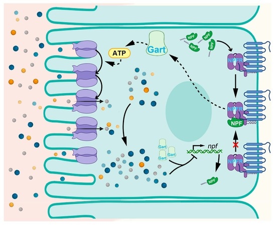
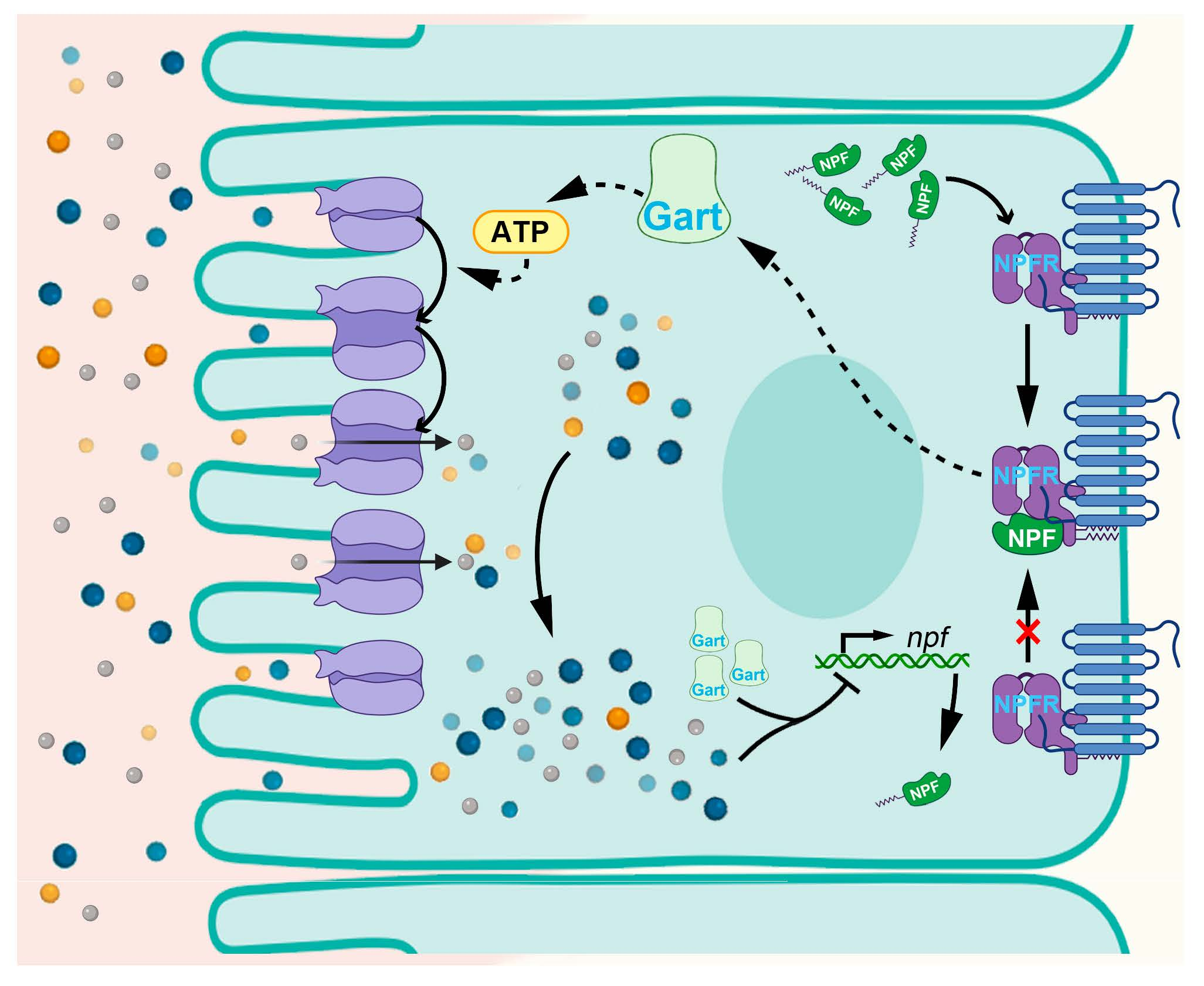
Graphical Abstract

1. Introduction

The maintenance of energy homeostasis is a fundamental biological challenge, requiring organisms to precisely balance nutrient intake with expenditure. This balance is governed by a complex interplay between central neural circuits that drive motivated behaviors, such as foraging and feeding, and peripheral metabolic organs that execute nutrient acquisition, processing, and storage [1,2,3]. Although significant progress has been made in decoding the brain-derived signals that stimulate or terminate eating, the molecular mechanisms by which these signals orchestrate adaptive changes in peripheral nutrient assimilation remain less understood.
The neuropeptide system is a phylogenetically conserved master regulator of energy balance. In mammals, NPY is a potent orexigenic (appetite-stimulating) signal released from the hypothalamus during states of energy deficit [4,5,6]. Its functional homolog in Drosophila, Neuropeptide F (NPF), similarly promotes feeding behaviors, especially under conditions of hunger or stress [7,8,9,10,11]. NPF signaling plays an important role in coordinating feeding behavior with metabolic need. The canonical view posits that NPF peptides primarily act on central circuits to modulate motivational drive [12]. However, emerging evidence suggests these peptides also exert direct effects on peripheral tissues, influencing glucose and lipid metabolism [13,14,15]. Although the intestine is the ultimate site of nutrient uptake, the pathways through which systemic NPF signals, integrated to fine-tune its absorptive capacity in response to whole-body energy demands, are entirely unknown.
GART trifunctional enzyme (Gart) is a critical component of the de novo purine synthesis pathway [16,17,18,19]. Purines are essential not only as building blocks for DNA and RNA but also as central components of cellular energy currency (ATP) and signaling molecules [20,21,22]. As such, Gart activity is intrinsically linked to cellular proliferation, bioenergetic capacity, and overall metabolic flux. Beyond its well-established role in development, we have previously shown that Gart operates in multiple peripheral tissues, including glia, the fat body, and gut, to regulate feeding rhythm and to control the storage and mobilization of energy reserves (glycogen and triglycerides), thereby contributing to systemic energy homeostasis [23]. These findings position Gart as a peripheral metabolic integrator to coordinate feeding behavior with energy needs.
Both NPF and Gart are involved in the regulation of feeding and energy homeostasis. Here, using Drosophila melanogaster as a model, we detected and discovered a previously uncharacterized regulatory circuit that operates within the gut to control nutrient absorptive efficiency through the gut–brain axis regulated by the NPF–Gart pathway. Our findings shift the paradigm of how a conserved neuropeptide regulates feeding, from a purely central driver of motivation to a peripheral coordinator of metabolic efficiency, and reveal a novel homeostatic feedback loop that ensures calibrated energy intake.

2. Materials and Methods

2.1. Fly Strains and Rearing

Flies were raised at 25 °C and 65% humidity under a 12 h light:12 h dark cycle. Three-to-five-day-old flies were used for experiments.
The [T2A-Gal4]Gart/cyo and 10 × UAS-Gart-V5 was generated by CRIPSPR-Cas9 [23]; the repo-Gal4/TM3 (BDSC: 7415), nsyb-Gal4 (BDSC: 51635) and nsyb-Gal80 (BDSC: 92154) were purchased from the Bloomington Drosophila Stock Center (Bloomington, IN USA); the CG-Gal4 was obtained from Junzheng Zhang’s lab (Beijing, China); the UAS-NPF-RNAi (THU2569) and UAS-NPFR-RNAi (THU2116) were purchased from the TsingHua Fly Center (Beijing, China); the MyoIA-Gal4 was obtained from Yi Rao’s lab (Beijing, China); the UAS-Gart-RNAi (VDRC: 46293) was purchased from the VDRC stock center (Wien, Austria). All lines in this study were outcrossed with w1118 for at least 6 generations. All experiments were conducted using male flies.

2.2. RNA Isolation, RT—PCR and qRT—PCR

The brain, fat body and gut of 50 flies or whole bodies of 30 flies were dissected on ice, and were collected by liquid nitrogen (Taineng Gas Co., Ltd., Taiyuan, China) for RNA preparation. The protocols for RNA extraction, cDNA synthesis and qRT-PCR are described by He et al. [23]. The results were conducted according to threshold cycle (Ct) value based on the 2−ΔΔCT method [24]. The primers are shown in Table S1.

2.3. Feeding Assay

The feeding assay was modified from our previous publication [23]. Briefly, male flies aged 3–5 days were entrained at 25 °C in LD for 3 days. The flies were switched from normal food to blue-dye food (100 mL of normal food (0.8% agar, 3.32% yeast, 3.16% sucrose, 6.32% glucose, 7.77% corn meal, 0.5% propionic acid, and distilled water) containing 2.5 g of FD&C Blue No. 1 [McCormick] (ROHA Dyechem (SHANGHAI) Co., Ltd., Shanghai, China)) for 2 h at different time points, after which they were frozen in liquid nitrogen, and decapitated. The samples were homogenized in 1000 μL ddH2O (Beijing Solarbio Science & Technology Co., Ltd., Beijing, China) and centrifuged (13,000 rpm) for 15 min. The supernatant was passed through a 0.22 μm syringe filter (Tianjin Keyilong Lab Equipment Co., Ltd., Tianjin, China) to remove debris and lipids and transferred to a new tube. The absorbance at 625 nm was measured by SpectraMax i3x (Meigu Biotechnology (Zhejiang) Co., Ltd., Wenzhou, China). The feeding levels were also normalized to the absorbance value/25 flies. The value of OD625 represents the intake of food.

2.4. Food Absorption Rate

Newly eclosed flies were maintained in a 25 °C LD cycle for 3 days and then transferred to diet containing blue dye for 24 h. Twenty-five flies were immediately frozen in liquid nitrogen as the pre-feeding baseline (recorded as A1). Another 25 flies were transferred to transparent diet (Beijing Solarbio Science & Technology Co., Ltd., Beijing, China) (containing 1% agar and 5% sucrose), fed freely for 6 h, then they were frozen in liquid nitrogen, decapitated, and collected as the post-feeding sample (recorded as A2). All samples were homogenized in 1000 μL ddH2O and centrifuged (13,000 rpm) for 15 min. The supernatant was passed through a 0.22 μm syringe filter to remove debris and lipids and transferred to a new tube. Absorbance at 625 nm was measured using a SpectraMax i3x microplate reader. Food absorption was calculated as described previously [25].
Absorption rate = (A1 − A2)/A1 × 100%

2.5. Quantification and Statistical Analysis

Analyses were performed using Microsoft Excel or Prism 10.4.0 (GraphPad), and graphs were plotted using Prism 10.4.0. For comparison among different genotypes or treatment groups, pairwise analyses were conducted by unpaired t test for two groups and one-way ANOVA for multiple groups of data. Data are represented as the mean ± SEM unless otherwise noted. Significance is indicated in the figure legends.

3. Results

3.1. Gut-Specific Gart Positively Regulates Feeding Through Absorption Efficiency

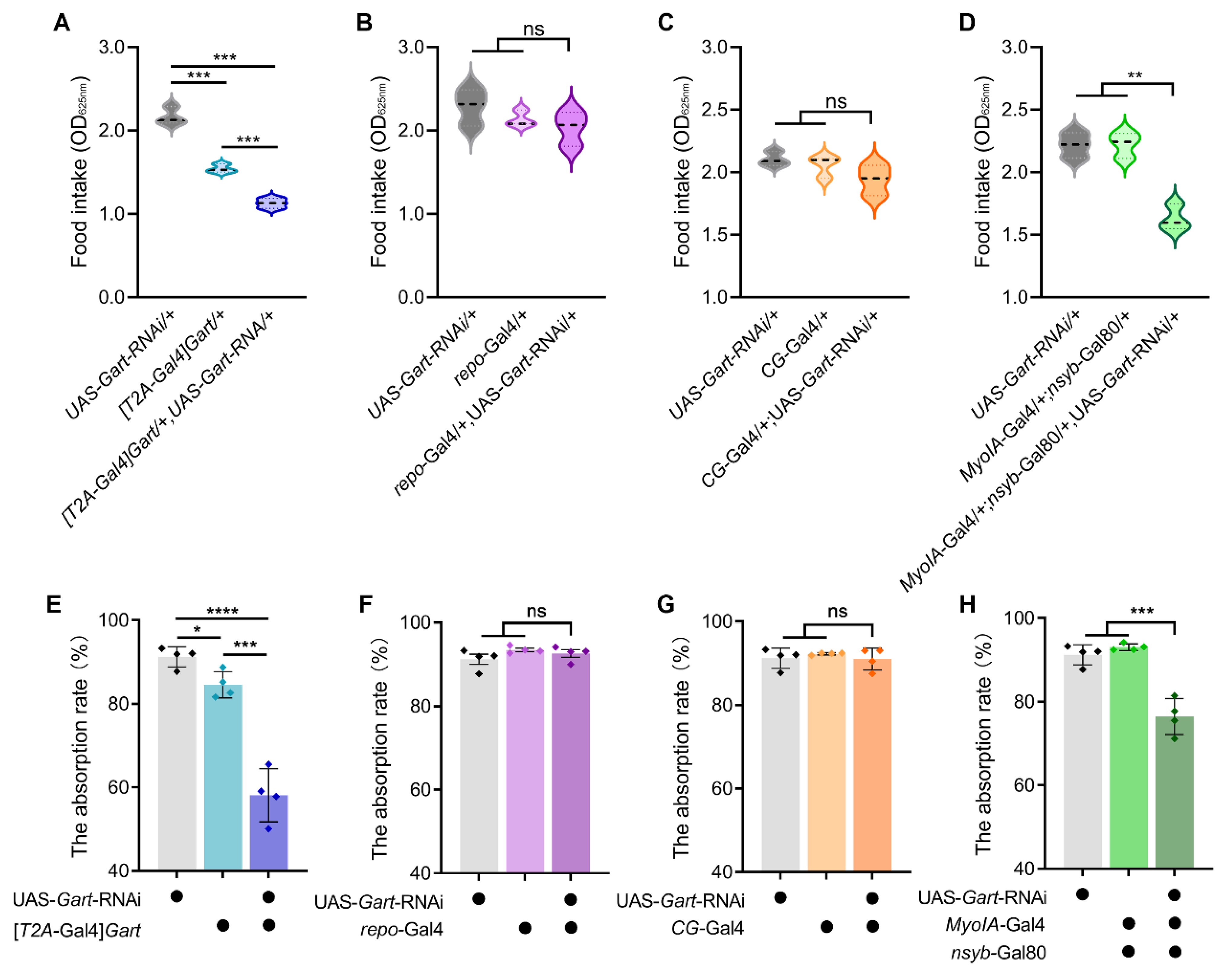
To determine whether and how Gart regulates feeding behavior, we first examined feeding of a heterozygous Gart mutant ([T2A-Gal4]Gart/+) (homozygous Gart knockout is lethal) and flies with further downregulation of Gart expression in heterozygous Gart mutant background ([T2A-Gal4]Gart/+; UAS-Gart-RNAi/+). Quantification of food intake revealed a significant decrease in feeding concomitant with reduced Gart expression (Figure 1A), indicating that Gart promotes feeding. Given the broad expression of Gart, we next sought to identify the tissue responsible for this metabolic function. Using tissue-specific RNAi drivers (repo-Gal4 for glia, CG-Gal4 for fat body, and MyoIA-Gal4; nsyb-Gal80 for the gut), we knocked down Gart in distinct tissues. Downregulation of Gart in either glia or the fat body had no discernible effect on feeding (Figure 1B,C). In striking contrast, gut-specific Gart knockdown recapitulated the low food intake phenotype of the Gart deficiency mutant (Figure 1D). These results indicate Gart predominantly functions in the gut.
We reasoned that intestinal Gart might modulate feeding by altering nutrient assimilation. To test this, we measured the food absorption rate. Consistent with the feeding phenotype, systemic reduction in Gart significantly impaired absorption efficiency (Figure 1E). Tissue-specific analysis demonstrated that this defect was exclusively attributable to the loss of Gart in the gut, while its knockdown in glia or the fat body was without any effects (Figure 1F–H). Collectively, these findings demonstrate that Gart promotes feeding primarily by enhancing nutrient absorption in the gut, positioning gut-specific Gart as a key determinant of feeding efficiency.

3.2. Peripheral NPF Is the Main Signal Source That Activates the Expression of Gart

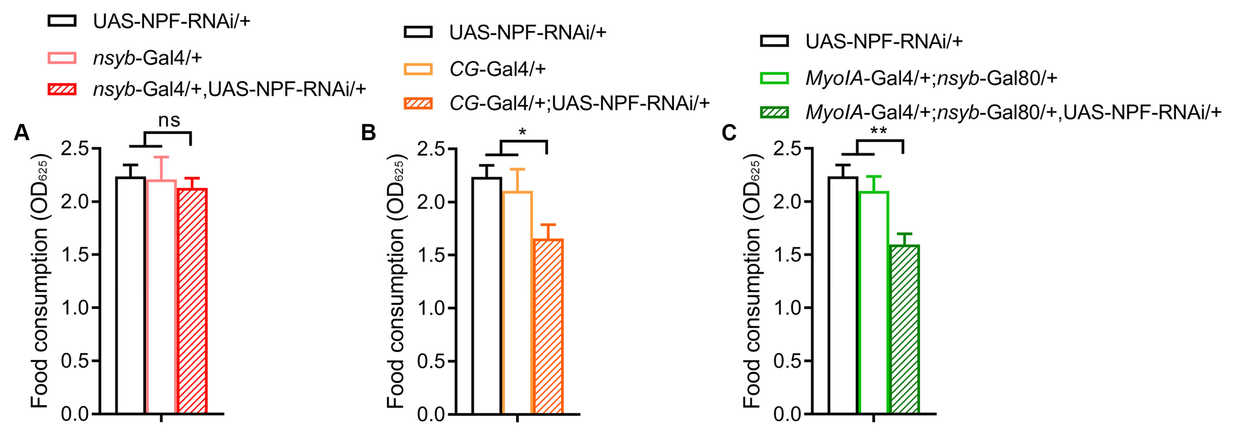
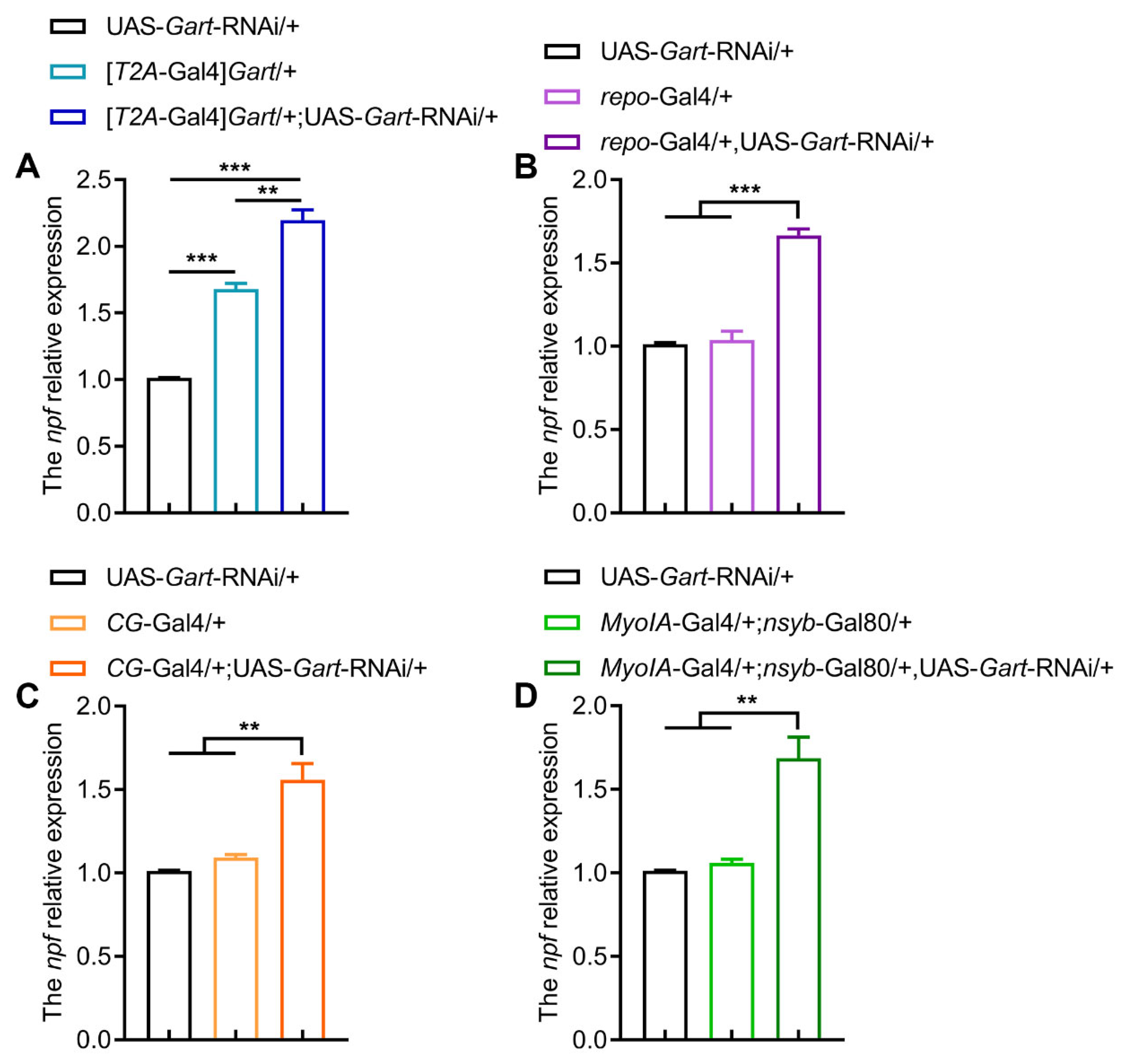
To delineate the relationship between NPF and Gart, we first assessed their tissue-specific contributions to feeding. Strikingly, the brain-specific knockdown of NPF (nsyb-Gal4) had no effect on food intake, while knockdown of NPF in either the fat body (CG-Gal4) or the gut (MyoIA-Gal4; nsyb-Gal80) significantly reduced feeding (Figure 2A–C), indicating that peripheral (fat body and gut) NPF is a key driver of feeding behavior. Subsequently, we explored the molecular interplay between NPF and Gart. In flies with globally reduced Gart expression, we observed a corresponding upregulation of NPF transcript levels (Figure 3A). This negative feedback was recapitulated by tissue-specific Gart knockdown in glia, fat body and gut (Figure 3B–D), indicating that Gart represses NPF expression in multiple tissues.
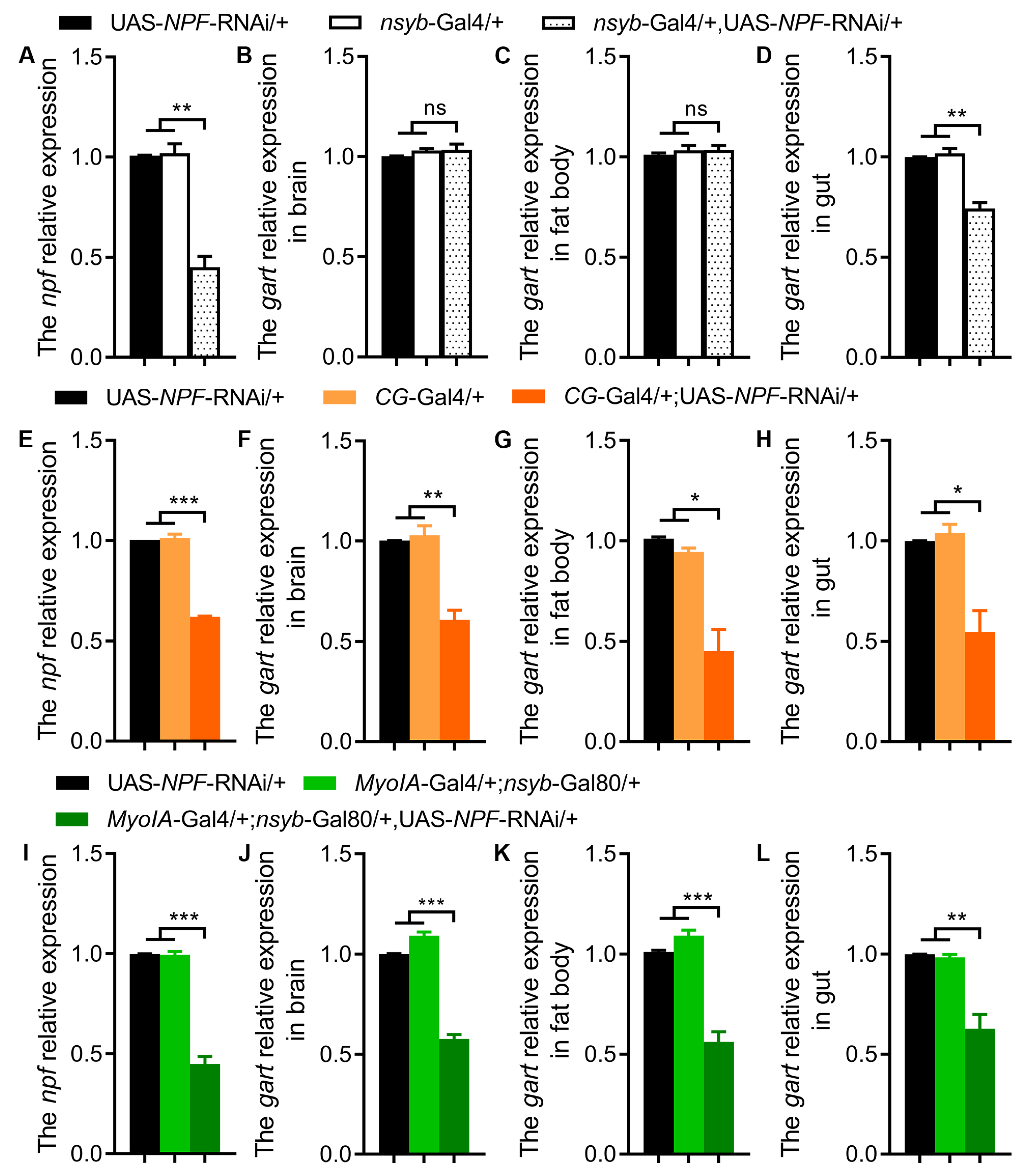
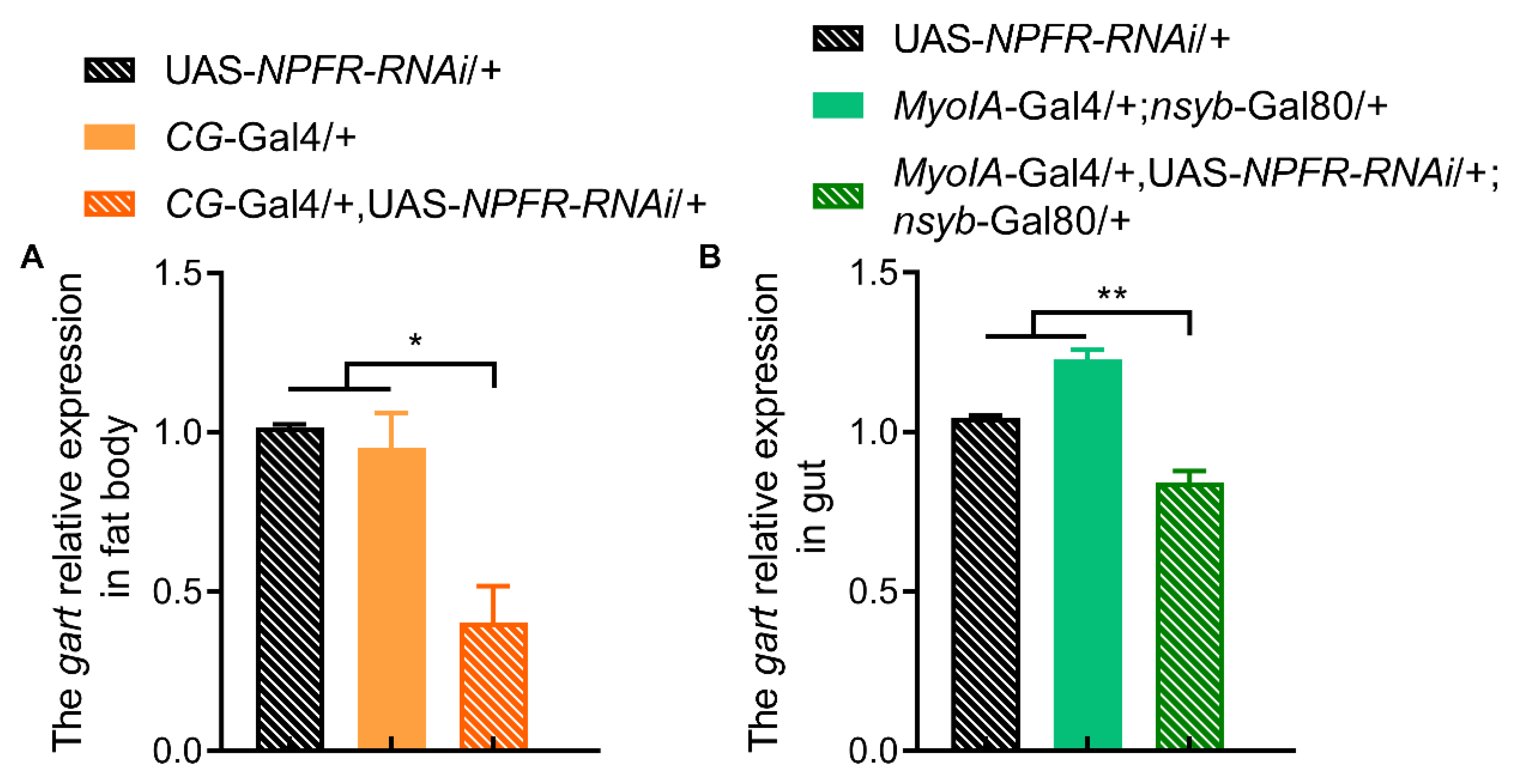
Conversely, we investigated whether NPF regulates Gart. Brain-specific NPF knockdown selectively reduced gut-specific Gart expression without affecting its levels in the brain or fat body (Figure 4A–D). In contrast, NPF knockdown in the fat body or gut led to a pronounced downregulation of Gart not only in the targeted tissue but also systemically, including in the brain (Figure 4E–L). This demonstrates that peripheral NPF is the primary positive regulator of Gart expression across tissues. Furthermore, fat body- or gut-specific knockdown of the NPF receptor (NPFR) similarly reduced Gart levels (Figure 5A,B), confirming that NPF signals through NPFR to maintain Gart expression.
Collectively, these data reveal a core peripheral regulatory axis: NPF/NPFR signaling positively regulates Gart expression, while Gart, in turn, provides negative feedback on NPF. This reciprocal relationship is central to feeding control; furthermore, peripheral NPF serves as the dominant source for activating systemic Gart expression.

3.3. Gart Acts as the Essential Metabolic Effector of NPF in Feeding Regulation

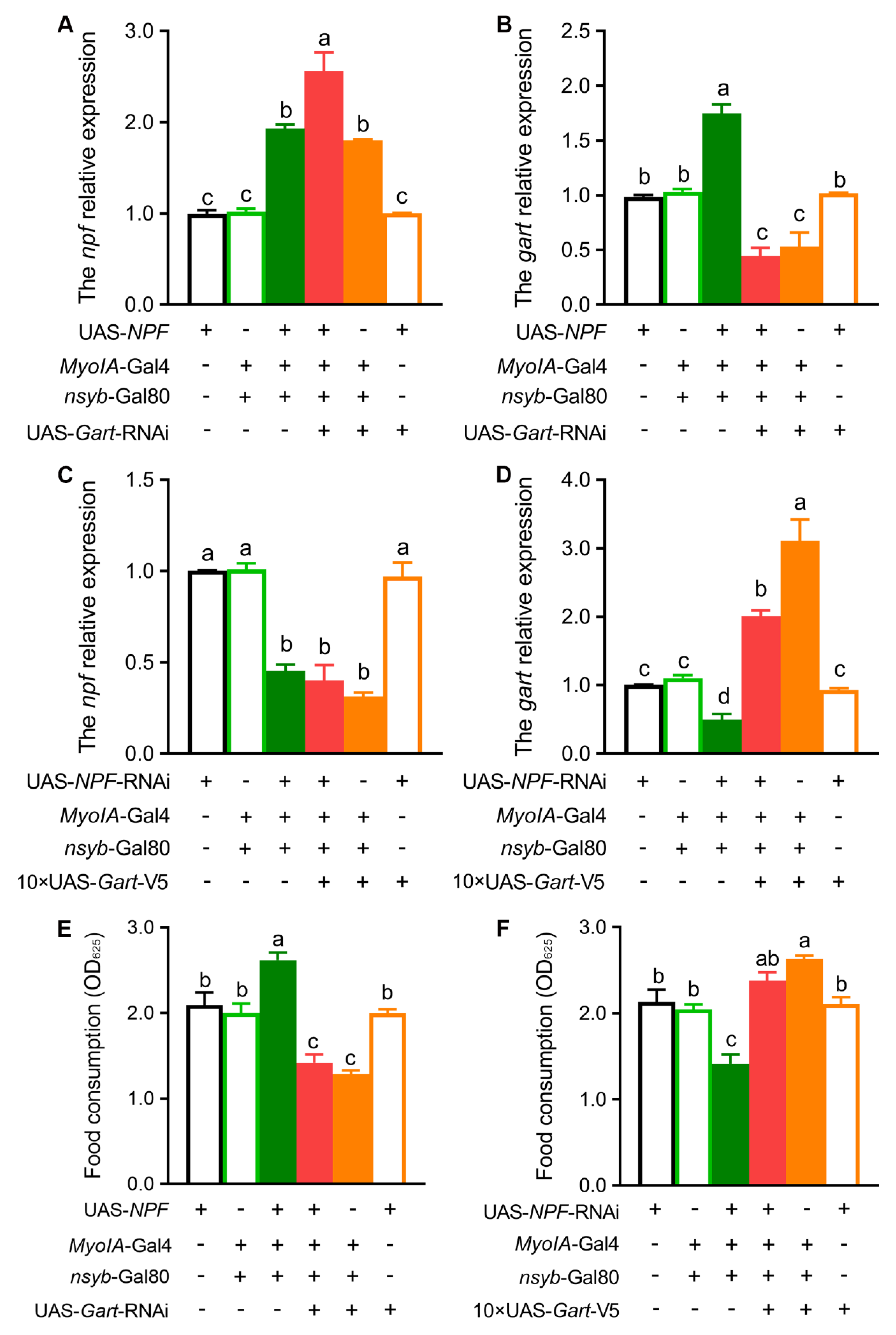
To definitively establish the genetic hierarchy between NPF and Gart, we performed a series of rescue experiments at the transcriptional and functional levels. First, we manipulated their expression in opposite directions. As expected, overexpression of NPF led to a significant increase in Gart transcript levels (Figure 6A,B). Crucially, when Gart was simultaneously knocked down in this NPF-overexpression background, the Gart mRNA level was reduced (Figure 6B), confirming that NPF acts upstream to promote Gart expression. Conversely, in a background of NPF knockdown, overexpression of Gart successfully elevated Gart mRNA without rescuing the low NPF levels (Figure 6C,D), consistent with Gart functioning downstream. Subsequently, we asked whether Gart is required for the physiological function of NPF. Overexpression of NPF robustly increased food intake, as anticipated (Figure 6E). Strikingly, this high feeding phenotype was completely abolished when Gart was knocked down concurrently, and feeding levels significantly decreased further (Figure 6E). Reciprocally, the decrease in feeding caused by NPF knockdown was fully reversed by co-overexpression of Gart (Figure 6F). These behavioral rescue experiments showed that Gart is a downstream effector of NPF. Collectively, these genetic and behavioral assays demonstrated that Gart is both sufficient and necessary to mediate the effects of NPF on feeding.

3.4. Gart Rescues the Impact of NPF Deficiency on Absorption Rate

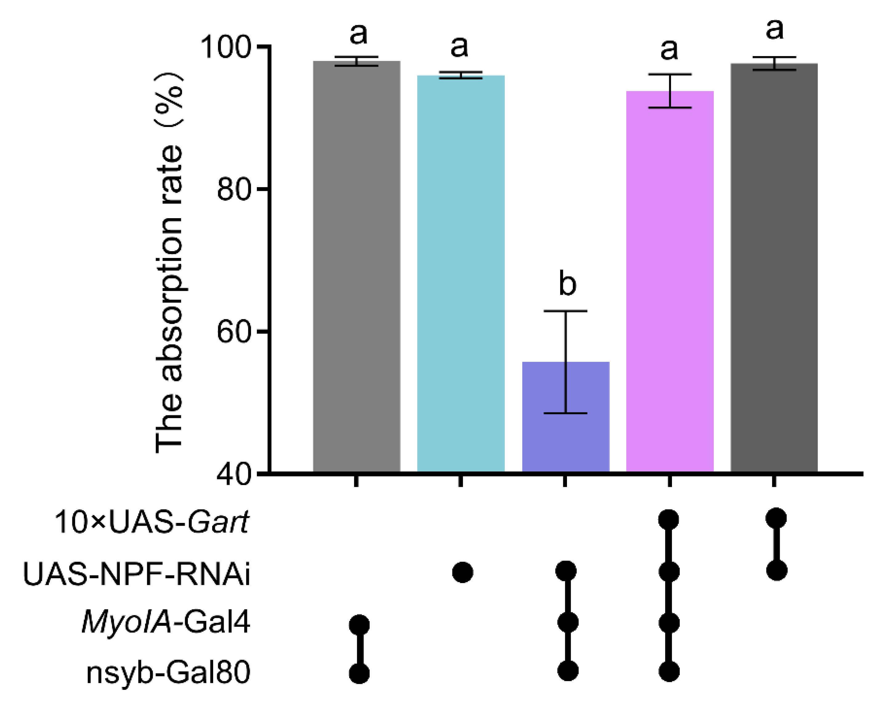
To directly test whether the NPF-Gart axis converges on nutrient absorption, the mechanistic basis of feeding regulation, we measured the food absorption rate under genetic perturbations of this pathway. Consistent with the low feeding phenotype, knockdown of NPF significantly reduced the food absorption rate (Figure 7). Critically, this absorptive defect was fully rescued by concurrent overexpression of Gart (Figure 7). This rescue experiment provides definitive functional evidence that Gart acts downstream of NPF to modulate intestinal absorption efficiency. Thus, NPF promotes feeding primarily by upregulating gut-specific Gart, which in turn enhances nutrient assimilation.
Collectively, these findings establish a novel gut-centric homeostatic module wherein feeding is controlled through the precise regulation of nutrient absorption. We demonstrate that the purine synthesis enzyme, Gart, acts specifically within the intestine to enhance food absorption, thereby promoting feeding. This key metabolic effector is regulated by a reciprocal transcriptional loop with the neuropeptide NPF: peripheral NPF signaling, originating dominantly from the fat body and gut, acts through its receptor NPFR to upregulate Gart expression systemically, while Gart in turn provides negative feedback on NPF levels. Genetic epistasis analyses confirm that Gart is both necessary and sufficient to execute the effects of NPF on feeding and absorption, placing it as the essential downstream effector. Thus, our work redefines a conserved hunger signal from a pure behavioral driver to a coordinator of peripheral metabolic efficiency, revealing a dynamic feedback circuit that calibrates energy intake by modulating gut absorptive capacity.

4. Discussion

Feeding behavior represents the primary determinant of energy intake, serving as the fundamental behavioral interface between an organism and its environment [23,26,27]. Through foraging and consumption, animals secure the caloric resources necessary for survival, growth and reproduction [28]. However, the act of feeding alone does not guarantee energy acquisition; rather, it is nutrient absorption within the gut that constitutes the true gateway through which ingested food is converted into bioavailable energy [29]. The efficiency of this absorptive process ultimately dictates how much of the consumed nutrients are assimilated to meet systemic metabolic demands [30]. Despite its centrality, the molecular mechanisms by which absorptive efficiency is dynamically regulated to match whole-body energy needs have remained poorly understood. This study establishes a previously unrecognized homeostatic circuit that directly links a conserved orexigenic signal to the control of gut absorptive capacity. We demonstrate that NPF and Gart form a reciprocal transcriptional loop within peripheral tissues that governs feeding by tuning the efficiency of nutrient absorption in the gut. We therefore propose a dynamic working model. Under energy demand, peripheral NPF signaling is induced, which positively drives Gart expression in the gut via its receptor NPFR. Elevated Gart boosts purine metabolic flux, potentially enhancing enterocyte energetics (e.g., ATP levels) and/or nutrient transporter capacity, thereby increasing nutrient absorption. As absorption proceeds and energy status improves, accumulated Gart or its metabolites transcriptionally repress NPF, which reduces feeding by preventing over-activation of the absorptive program. Although our findings demonstrate that NPF in peripheral tissues (gut) modulates feeding behavior by affecting gut absorption capacity, NPF expressed in the central nervous system plays a more critical role in feeding regulation, possibly by integrating gustatory signals or coordinating energy homeostasis. Future studies using tissue-specific knockdown of NPF in the CNS will help dissect its distinct roles relative to peripheral NPF. This “feed-forward-driven, feedback-braked” loop enables an automatic transition from “accelerated absorption” to “homeostatic maintenance,” ensuring precise coupling between energy harvest and physiological need. This model carries significant physiological and evolutionary implications. It represents a strategy of “precision nutrition,” where organisms adapt to environmental flux not only by modulating “how much they want to eat” (appetite) but also by dynamically tuning “how much they can absorb” (efficiency). Such a reciprocal “neuropeptide–core metabolic enzyme” circuit may represent an evolutionarily conserved homeostatic principle. Analogous push–pull systems exist in mammals, where hunger signals like ghrelin and satiety signals like leptin/peptide YY (PYY) interact [31,32,33]. Our NPF-Gart module provides a simpler, potentially more ancient, cellular/organ-level analog for rapid metabolic tuning. Placing a fundamental housekeeping enzyme like Gart under dynamic transcriptional control by a neuropeptide offers a fresh perspective on metabolic adaptation. In addition, this work establishes gut absorptive efficiency as a critical and tunable regulatory dimension of energy homeostasis, and reveals how a neuropeptide signal can orchestrate metabolic output at the level of the absorbing organ itself.
Notably, our work fundamentally shifts the focus of feeding regulation from the central nervous system to the gut—the ultimate site of nutrient assimilation. Despite being broadly expressed across multiple tissues, Gart functions in a highly tissue-specific manner: only its loss in the gut, but not in glia or the fat body, leads to a concurrent reduction in both food intake and absorptive efficiency. This tissue-specific requirement suggests that gut-specific Gart governs feeding behavior by modulating nutrient absorption, thereby generating a peripheral “satiety” or “metabolic repletion” signal that is relayed to the brain to curtail further food consumption. These findings establish the gut as a key decision-making node in energy homeostasis and reveal that absorptive efficiency itself can serve as a feedback signal to regulate central feeding circuits. This mechanism operates in complementary parallel to central hunger/satiety circuits. In addition, a key finding is the dominant role of peripheral NPF in initiating this loop. NPF derived from the fat body and gut, rather than the brain, serves as the primary systemic signal to activate Gart expression across tissues, including the brain. This is consistent with research in Ostrinia furnacalis, where gut-derived NPF is the central regulator of feeding, operating through the activation of the PI3K signaling pathway [13]. Functionally, peripheral NPF serves as a systemic “metabolic accelerator” that drives nutrient absorption, whereas the Gart-mediated negative feedback acts as a “brake” to prevent excessive energy intake. This locally established gut-centric regulatory loop likely influences central feeding decisions through absorbed nutrients or gut-derived humoral signals, thereby constituting an integrated model of “peripheral metabolic circuit-to-central behavioral output” that orchestrates systemic energy homeostasis.
In summary, this study delineates a novel and physiologically coherent gut–brain circuit for feeding regulation, centered on a peripheral NPF–Gart reciprocal transcriptional loop that directly governs intestinal absorptive efficiency. This work fundamentally repositions the intestine as a primary executive target of the conserved orexigenic signal NPF, moving beyond its canonical central role. This research provides the first mechanistic evidence of a direct transcriptional feedback loop linking a neuropeptide to a core metabolic enzyme, thereby forging a definitive molecular bridge between behavioral drive and cellular energetics. Furthermore, this finding conceptually establishes “nutrient absorptive efficiency” as a critical, tunable dimension of feeding control, revealing a refined physiological strategy for adaptive energy harvest. Collectively, these findings offer a new molecular framework and a dynamic homeostatic model for understanding how inter-organ communication precisely calibrates energy balance. However, several important questions remain. First, what is the precise mechanistic link between Gart and absorption? How does peripheral NPF signaling communicate with the brain to downregulate central Gart? Is it via a humoral factor or a vagal-like neural pathway? Addressing these questions will not only refine this axis but may also offer novel insights into metabolic diseases, such as whether dysregulation of the human NPF/NPY system similarly disrupts gut function to contribute to obesity or malnutrition.

Supplementary Materials

The following supporting information can be downloaded at: https://www.mdpi.com/article/10.3390/insects17050528/s1, Table S1: Primer sequences used in this study.

Author Contributions

L.H. and Z.Z. conceived the paper. L.H. designed the experiments. L.H., Y.G., Q.L. and Q.W. performed the experiments. L.H., Y.G. and Q.W. analyzed the data. L.H. and Q.W. wrote the manuscript. L.H. and Z.Z. revised the paper. All authors have read and agreed to the published version of the manuscript.

Funding

This research was funded by the National Natural Science Foundation of China grant number 32500404.

Institutional Review Board Statement

This manuscript has not included any experimental data or material and ethics issues.

Informed Consent Statement

All the authors reviewed and approved the final manuscript and consented to its publication.

Data Availability Statement

The original contributions presented in this study are included in the article/Supplementary Materials. Further inquiries can be directed to the corresponding authors.

Acknowledgments

We thank Yi Rao (Peking University), Sheng Li (South China Normal University) and JunZheng Zhang (China Agricultural University) for providing our Drosophila line. The work was supported by the National Natural Science Foundation Youth Project of China Grants (32500404 to L.H.).

Conflicts of Interest

The authors declare no conflicts of interest.

References

  1. Grosjean, E.; Simonneaux, V.; Challet, E. Reciprocal interactions between circadian clocks, food intake, and energy metabolism. Biology 2023, 12, 539. [Google Scholar] [CrossRef] [PubMed]
  2. Wu, C.S.; Bongmba, O.Y.N.; Lee, J.H.; Tuchaai, E.; Zhou, Y.; Li, D.P.; Xue, B.; Chen, Z.; Sun, Y. Ghrelin receptor in agouti-related peptide neurons regulates metabolic adaptation to calorie restriction. J. Neuroendocrinol. 2019, 31, e12763. [Google Scholar] [CrossRef] [PubMed]
  3. Hurtado del Pozo, C.; Ruiz, H.H.; Arivazhagan, L.; Aranda, J.F.; Shim, C.; Daya, P.; Derk, J.; MacLean, M.; He, M.; Frye, L.; et al. A receptor of the immunoglobulin superfamily regulates adaptive thermogenesis. Cell Rep. 2019, 28, 773–791.e7. [Google Scholar] [CrossRef] [PubMed]
  4. Chee, M.J.S.; Myers, M.G.; Price, C.J.; Colmers, W.F. Neuropeptide Y suppresses anorexigenic output from the ventromedial nucleus of the hypothalamus. J. Neurosci. 2010, 30, 3380. [Google Scholar] [CrossRef]
  5. Hökfelt, T.; Stanic, D.; Sanford, S.D.; Gatlin, J.C.; Nilsson, I.; Paratcha, G.; Ledda, F.; Fetissov, S.; Lindfors, C.; Herzog, H. NPY and its involvement in axon guidance, neurogenesis, and feeding. Nutrition 2008, 24, 860–868. [Google Scholar] [CrossRef]
  6. Chee, M.J.S.; Colmers, W.F. Y eat? Nutrition 2008, 24, 869–877. [Google Scholar] [CrossRef]
  7. Wu, Q.; Wen, T.; Lee, G.; Park, J.H.; Cai, H.N.; Shen, P. Developmental control of foraging and social behavior by the Drosophila neuropeptide Y-like system. Neuron 2003, 39, 147–161. [Google Scholar] [CrossRef]
  8. Wu, Q.; Zhao, Z.; Shen, P. Regulation of aversion to noxious food by Drosophila neuropeptide Y– and insulin-like systems. Nat. Neurosci. 2005, 8, 1350–1355. [Google Scholar] [CrossRef]
  9. Wu, Q.; Zhang, Y.; Xu, H.; Shen, P. Regulation of hunger-driven behaviors by neural ribosomal S6 kinase in Drosophila. Proc. Natl. Acad. Sci. USA 2005, 102, 13289–13294. [Google Scholar]
  10. Beshel, J.; Zhong, Y. Graded encoding of food odor value in the Drosophila brain. J. Neurosci. 2013, 33, 15693. [Google Scholar] [CrossRef]
  11. Chung, B.Y.; Ro, J.; Hutter, S.A.; Miller, K.M.; Guduguntla, L.S.; Kondo, S.; Pletcher, S.D. Drosophila neuropeptide F signaling independently regulates feeding and sleep-wake behavior. Cell Rep. 2017, 19, 2441–2450. [Google Scholar] [CrossRef]
  12. Zhao, X.L.; Campos, A.R. Insulin signalling in mushroom body neurons regulates feeding behaviour in Drosophila larvae. J. Exp. Biol. 2012, 215, 2696–2702. [Google Scholar] [CrossRef] [PubMed]
  13. Zhao, J.J.; Song, Y.; Jiang, X.M.; He, L.; Wei, L.Y.; Zhao, Z.W. Synergism of feeding and digestion regulated by the neuropeptide F system in Ostrinia furnacalis larvae. Cells 2023, 12, 194. [Google Scholar] [CrossRef] [PubMed]
  14. Zhao, J.J.; Yan, S.; Li, M.S.; Sun, L.N.S.; Dong, M.; Yin, M.Z.; Shen, J.; Zhao, Z.W. NPFR regulates the synthesis and metabolism of lipids and glycogen via AMPK: Novel targets for efficient corn borer management. Int. J. Biol. Macromol. 2023, 247, 125816. [Google Scholar] [CrossRef] [PubMed]
  15. Yu, Z.F.; Shi, J.; Jiang, X.M.; Song, Y.; Du, J.; Zhao, Z.W. Neuropeptide F regulates feeding via the juvenile hormone pathway in Ostrinia furnacalis larvae. Pest Manag. Sci. 2023, 79, 1193–1203. [Google Scholar] [CrossRef]
  16. Cipolletti, M.; Leone, S.; Bartoloni, S.; Acconcia, F. A functional genetic screen for metabolic proteins unveils GART and the de novo purine biosynthetic pathway as novel targets for the treatment of luminal A ERalpha expressing primary and metastatic invasive ductal carcinoma. Front. Endocrinol. 2023, 14, 1129162. [Google Scholar] [CrossRef]
  17. Cong, X.; Lu, C.; Huang, X.; Yang, D.; Cui, X.; Cai, J.; Lv, L.; He, S.; Zhang, Y.; Ni, R. Increased expression of glycinamide ribonucleotide transformylase is associated with a poor prognosis in hepatocellular carcinoma, and it promotes liver cancer cell proliferation. Hum. Pathol. 2014, 45, 1370–1378. [Google Scholar] [CrossRef]
  18. Mazzarino, R.C.; Baresova, V.; Zikanova, M.; Duval, N.; Wilkinson, T.G.; Patterson, D.; Vacano, G.N. Transcriptome and metabolome analysis of crGART, a novel cell model of de novo purine synthesis deficiency: Alterations in CD36 expression and activity. PLoS ONE 2021, 16, e0247227. [Google Scholar] [CrossRef]
  19. Ng, A.; Uribe, R.A.; Yieh, L.; Nuckels, R.; Gross, J.M. Zebrafish mutations in gart and paics identify crucial roles for de novo purine synthesis in vertebrate pigmentation and ocular development. Development 2009, 136, 2601–2611. [Google Scholar] [CrossRef]
  20. Mazzarino, R.C. Targeting future pandemics, a case for de novo purine synthesis and basic research. Front. immunol. 2021, 12, 694300. [Google Scholar] [CrossRef]
  21. Schuldt, M.; Driel, B.V.; Algül, S.; Parbhudayal, R.Y.; Barge-Schaapveld, D.Q.C.M.; Güçlü, A.; Jansen, M.; Michels, M.; Baas, A.F.; Wiel, M.A.V.D. Distinct metabolomic signatures in preclinical and obstructive hypertrophic cardiomyopathy. Cells 2021, 10, 2950. [Google Scholar] [CrossRef] [PubMed]
  22. Naffouje, R.; Grover, P.; Yu, H.; Sendilnathan, A.; Wolfe, K.; Majd, N.; Smith, E.P.; Takeuchi, K.; Senda, T.; Kofuji, S.; et al. Anti-tumor potential of IMP dehydrogenase inhibitors: A century-long story. Cancers 2019, 11, 1346. [Google Scholar] [CrossRef]
  23. He, L.; Wu, B.B.; Shi, J.; Du, J.; Zhao, Z.W. Regulation of feeding and energy homeostasis by clock-mediated Gart in Drosophila. Cell Rep. 2023, 42, 112912. [Google Scholar] [CrossRef]
  24. Livak, K.J.; Schmittgen, T.D. Analysis of relative gene expression data using real-time quantitative PCR and the 2(-Delta Delta C(T)) Method. Methods 2001, 25, 402–408. [Google Scholar] [CrossRef] [PubMed]
  25. Titos, I.; Juginovic, A.; Vaccaro, A.; Nambara, K.; Gorelik, P.; Mazor, O.; Rogulja, D. A gut-secreted peptide suppresses arousability from sleep. Cell 2023, 186, 1382–1397. [Google Scholar] [CrossRef] [PubMed]
  26. Wang, J.; Liu, Z.; Lin, H.; Jiao, H.; Zhao, J.; Ma, B.; Wang, Y.; He, S.; Wang, X. Daily feeding frequency affects feed intake and body weight management of growing layers. Poult. Sci. 2024, 103, 103748. [Google Scholar] [CrossRef]
  27. Grmai, L.; Michaca, M.; Lackner, E.; Nampoothiri, V.P.N.; Vasudevan, D. Integrated stress response signaling acts as a metabolic sensor in fat tissues to regulate oocyte maturation and ovulation. Cell Rep. 2024, 43, 113863. [Google Scholar] [CrossRef]
  28. Di Francesco, A.; Di Germanio, C.; Bernier, M.; de Cabo, R. A time to fast. Science 2018, 362, 770–775. [Google Scholar] [CrossRef]
  29. Mao, J.; Hu, X.; Xiao, Y.; Yang, C.; Ding, Y.; Hou, N.; Wang, J.; Cheng, H.; Zhang, X. Overnutrition stimulates intestinal epithelium proliferation through β-catenin signaling in obese mice. Diabetes 2013, 62, 3736–3746. [Google Scholar] [CrossRef]
  30. Kotrschal, A.; Szidat, S.; Taborsky, B. Developmental plasticity of growth and digestive efficiency in dependence of early-life food availability. Funct. Ecol. 2014, 28, 878–885. [Google Scholar] [CrossRef]
  31. Mittelman, S.D.; Klier, K.; Braun, S.; Azen, C.; Geffner, M.E.; Buchanan, T.A. Obese Adolescents show impaired meal responses of the appetite-regulating hormones ghrelin and PYY. Obesity 2010, 18, 918–925. [Google Scholar] [CrossRef]
  32. Barham, P.; Skibsted, L.H.; Bredie, W.L.P.; Bom Frøst, M.; Møller, P.; Risbo, J.; Snitkjær, P.; Mortensen, L.M. Molecular gastronomy: A new emerging scientific discipline. Chem. Rev. 2010, 110, 2313–2365. [Google Scholar] [CrossRef]
  33. Hernando-Redondo, J.; Toloba, A.; Benaiges, D.; Salas-Salvadó, J.; Martínez-Gonzalez, M.; Corella, D.; Estruch, R.; Tinahones, F.; Ros, E.; Goday, A.; et al. Mid- and long-term changes in satiety-related hormones, lipid and glucose metabolism, and inflammation after a mediterranean diet intervention with the goal of losing weight: A randomized, clinical trial. Front. Nutr. 2022, 9, 950900. [Google Scholar] [CrossRef]
Figure 1. The effect of Gart deficiency on feeding behavior and food absorption rate of Drosophila melanogaster. (AD) The food intake of flies with downregulated Gart in whole body (A), glial cells (B), fat body (C) and gut (D). (EH) The food absorption rate of flies with downregulated Gart in whole body (E), glial cells (F), fat body (G) and gut (H). Black dots indicate the presence of this component in flies. The graphs show the mean ± SEM (t test; * p < 0.05, ** p < 0.01, *** p < 0.001, **** p < 0.0001; ns not significant). N = 3 biological replicates (25 ± 5 flies/repeat).
Insects 17 00528 g001
Figure 2. The effect of npf deficiency on feeding behavior of Drosophila melanogaster. (AC) The food intake of flies with downregulated npf in brain (A), fat body (B) and gut (C). The graphs show the mean ± SEM (t test; * p < 0.05, ** p < 0.01; ns not significant). N = 3 biological replicates (25 ± 5 flies/repeat).
Insects 17 00528 g002
Figure 3. The effect of Gart deficiency on NPF expression levels. (AD) The NPF expression levels of flies with downregulated Gart in whole body (A), glial cells (B), fat body (C) and gut (D). The graphs show the mean ± SEM (t test; ** p < 0.01, *** p < 0.001). N = 3 biological replicates (20 ± 5 flies/repeat).
Insects 17 00528 g003
Figure 4. The effect of NPF deficiency on Gart expression levels. (A) The NPF expression levels of flies with downregulated npf in brain. (BD) The Gart expression levels in brain (B), fat body (C) and gut (D) of flies lacking npf. (E) The NPF expression levels of flies with downregulated npf in fat body. (FH) The Gart expression levels in brain (F), fat body (G) and gut (H) of flies lacking npf. (I) The NPF expression levels of flies with downregulated npf in gut. (JL) The Gart expression levels in brain (J), fat body (K) and gut (L) of flies lacking npf. The graphs show the mean ± SEM (t test; * p < 0.05, ** p < 0.01, *** p < 0.001; ns not significant). N = 3 biological replicates (20 ± 5 flies/repeat).
Insects 17 00528 g004
Figure 5. The effect of NPFR deficiency on Gart expression levels. (A,B) The Gart expression levels in fat body (A) and gut (B) of flies lacking npfr. The graphs show the mean ± SEM (t test; * p < 0.05, ** p < 0.01). N = 3 biological replicates (20 ± 5 flies/repeat).
Insects 17 00528 g005
Figure 6. Gart rescues the impact of NPF deficiency on fruit fly feeding. (A,B) In the background of NPF overexpression, downregulation of Gart results in the expression levels of NPF (A) and Gart (B). (C,D) In the background of NPF downregulation, overexpression of Gart results in the expression levels of NPF (C) and Gart (D). (E) In the background of NPF overexpression, downregulation of Gart results in the food intake. (F) In the background of NPF downregulation, overexpression of Gart results in food intake. The graphs show the mean ± SEM (one-way ANOVA; the data with different lowercase letters in the column show significant differences). N = 3 biological replicates (25 ± 5 flies/repeat).
Insects 17 00528 g006
Figure 7. Gart rescues the impact of NPF deficiency on fly food absorption rate. The graphs show the mean ± SEM (one-way ANOVA; the data with different lowercase letters in the column show significant differences). N = 5 biological replicates (25 ± 5 flies/repeat).
Insects 17 00528 g007
Disclaimer/Publisher’s Note: The statements, opinions and data contained in all publications are solely those of the individual author(s) and contributor(s) and not of MDPI and/or the editor(s). MDPI and/or the editor(s) disclaim responsibility for any injury to people or property resulting from any ideas, methods, instructions or products referred to in the content.