Article

Molecular Composition and Ligand Binding Characteristics of Native Ionotropic GABA Receptors in Rice Stem Borer, Chilo suppressalis


1
Key Laboratory of Integrated Pest Management on Crops in East China, Ministry of Agriculture and Rural Affair, College of Plant Protection, Nanjing Agricultural University, Nanjing 211800, China
2
Plant Protection, Quarantine and Cultivated Land & Fertilizer Management Station, Huzhou Municipal Bureau of Agriculture and Rural Affairs, Huzhou 313000, China
3
State Key Laboratory of Agricultural and Forestry Biosecurity, College of Plant Protection, Nanjing Agricultural University, Nanjing 211800, China
*
Author to whom correspondence should be addressed.
Insects 2026, 17(5), 477; https://doi.org/10.3390/insects17050477
Submission received: 12 March 2026 / Revised: 23 April 2026 / Accepted: 28 April 2026 / Published: 6 May 2026

Simple Summary

Ionotropic γ-aminobutyric acid receptor (iGABAR) is one of the key molecular targets for developing insecticides. However, the molecular composition and ligand binding characteristics of native iGABARs have not been characterized in insects. In this study, native iGABARs composed of CsRDL1, CsRDL2 or CsLCCH3 were identified from postsynaptic membranes of rice stem borer (RSB). N-terminal-truncated transcripts and proteins of CsRDL2 (∆N-CsRDL2) and CsLCCH3 (∆N-CsLCCH3), as well as CsRDL1ad, CsRDL1bd and post-translationally modified CsRDL1, were detected in native iGABARs. In addition, CsRDL1, CsRDL2 and CsLCCH3 were distributed in the same region in the adult head. Notably, the binding affinities of desmethyl-broflanilide (DMBF) to heteromeric iGABARs assembled by CsRDL1, ∆N-CsRDL2, and ∆N-CsLCCH3 were higher than those of DMBF to homomeric iGABAR assembled by CsRDL1. Overall, our findings indicated that ∆N-CsRDL2 and ∆N-CsLCCH3 could compose native iGABARs with CsRDL1, and they attenuate DMBF binding to iGABARs in the RSB. These results provide new insights into the molecular constituents of postsynaptic ion channels and support the rational design of novel insecticides.

Abstract

The ionotropic γ-aminobutyric acid receptor (iGABAR) is an important insecticidal molecular target. However, the native iGABARs composition remains unknown in insect. Here, CsRdl1, truncated transcripts of CsRdl2 and CsLcch3 were obtained in the rice stem borer (RSB), Chilo suppressalis Walker. The N-terminal-truncated CsRDL2 (∆N-CsRDL2) and N-terminal-truncated CsLCCH3 (∆N-CsLCCH3) were deduced and studied in vivo, and desmethyl-broflanilide (DMBF) binding characteristics were simulated in silico. Genome and transcriptome analyses revealed truncated transcripts of CsRdl2 and CsLcch3 encoded 48 kDa of ∆N-CsRDL2 and 37 kDa of ∆N-CsLCCH3, respectively. The CsRDL1, CsRDL2 and CsLCCH3 were detected respectively from native iGABARs at molecular weights (Mws) ≥ 440 kDa in BN-PAGE. In BN/SDS-PAGE, three CsRDL1 bands (~54, ~55 and ~70 kDa), one CsRDL2 band (~48 kDa) and one CsLCCH3 band (~37 kDa) were identified in native iGABARs at Mws ≥ 669 kDa, and corresponded to CsRDL1ad, CsRDL1bd, post-translationally modified CsRDL1, ∆N-CsRDL2 and ∆N-CsLCCH3, respectively. Immunofluorescence confirmed these three subunits distributed in the same region of adult heads. Finally, DMBF displayed higher binding affinities for heteromeric iGABARs than for homomeric CsRDL1 iGABAR in silico. These findings confirm that ∆N-CsRDL2 and ∆N-CsLCCH3 in native iGABARs might support the rational design of novel insecticides.

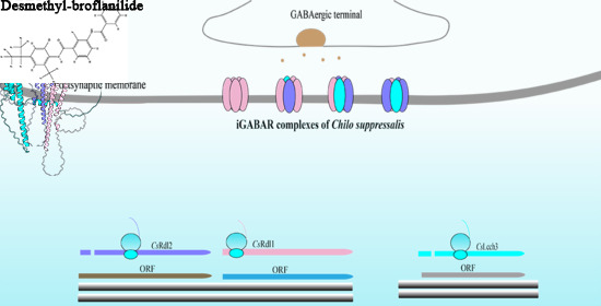
Graphical Abstract

1. Introduction

γ-aminobutyric acid (GABA) is the major inhibitory neurotransmitter in the nervous systems of both vertebrates and invertebrates, and mediates fast inhibitory synaptic transmission by acting on ionotropic GABA receptors (iGABARs) [1]. As a member of the cys-loop ligand-gated ion channel (LGIC) superfamily, iGABARs have been identified as one of the major molecular targets for drugs and insecticides. Notably, the pharmacological properties of insect iGABARs exhibit significant differences to those of vertebrate iGABARs, which provide a foundation for developing insect-specific insecticides [2]. The structural and functional diversity of iGABARs is attributed to the varied composition of subunit complexes within the LGIC superfamily [3]. In vertebrates, the pore-forming subunits of native iGABARs have been isolated and characterized [4], and are assembled by five subunits in vivo [5,6]. For instance, iGABARs containing 2α1/2β/γ2 subunits localize at synapses in the cerebellar granule cells [7]. Furthermore, 12 native subunit assemblies and their corresponding three-dimensional (3D) structures of iGABARs have been precisely defined [6]. Additionally, alternative splicing of pre-mRNA has been recognized as a key mechanism for generating iGABAR diversity [8]. Compared with its N-terminal-truncated transcript, the completed γ2 subunit reduces the cell surface expression of α1β3γ2 receptors by 50% [9]. Moreover, alternative splicing of the mouse Mus musculus Linnaeus α4 subunit results in an N-terminal-truncated α4 (∆N-α4), which selectively attenuates GABA-induced currents [10].
In insects, the predicted subunits of iGABARs include RDL (resistant to dieldrin), LCCH3 (ligand-gated chloride channel homolog 3), GRD (GABA and glycine receptor-like subunit of Drosophila), and CG8916 [2,11,12,13,14]. In vitro experimental data have demonstrated that the functional diversity of homomeric or heteromeric iGABARs can be revealed through a heterogeneous expression system. For example, compared with homomeric iGABAR assembled by RDL subunits, the sensitivity of heteromeric iGABARs assembled by RDL and LCCH3 to bicuculline and picrotoxin is enhanced and reduced, respectively [15]. Furthermore, RDL and LCCH3 of human louse Pediculus humanus humanus Linnaeus reconstitute functional heteromeric iGABARs in the Xenopus oocyte heterologous expression system, which respond to GABA in a concentration-dependent manner and are sensitive to picrotoxin and fipronil [16]. Moreover, post-transcriptional modifications of messenger RNA (mRNA), including alternative splicing and RNA editing, can increase RDL heterogeneity [17]. Two transcript variants of the Rdl, designated as Rdlad and Rdlbd, arose from alternative splicing events at exons 3 and 6 and were identified [1]. Truncated transcripts of Rdl1 and Lcch3 exist in the silkworm Bombyx mori Linnaeus [18]. Alternative splicing events in CsRdl of the rice stem borer (RSB) and the splicing factor Nova have also been reported [19]. However, the native truncated isoforms of iGABAR subunits generated by alternative splicing have not yet been identified in the RSB.
Insect iGABARs are important insecticidal molecular targets [20,21]. Currently, insecticides acting on iGABARs, such as fipronil, avermectin and broflanilide, are widely applied in the control of Lepidoptera pests in agriculture [20,21,22]. As a meta-diamide insecticide, broflanilide has been reported to be metabolized to desmethyl-broflanilide (DMBF) in insects and then embedded into the cavity between the transmembrane domain 1 (TM1) and TM3 of RDL [23]. Furthermore, glycine at the third position (G3’) in TM3 of RDL is associated with the sensitivity of insects to DMBF [23,24]. In recent, 3D structures of iGABAR assembled by honeybee Apis mellifera L. RDL have been precisely defined, and the binding pocket and key residue interacting with abamectin, fluralaner, fluxametamide, and isocycloseram, in iGABAR have been reported [25]. As is well-known, the RSB is an important Lepidoptera pest in Asia, especially in China [26]. Therefore, the native iGABARs of RSB in vivo, and DMBF in silico binding to iGABARs assembled by CsRDL1, CsRDL2 and CsLCCH3 were studied in the present study. These results will provide evidence for alternative splicing events and facilitate the development of targeted insecticides.

2. Materials and Methods

2.1. CsRdl1, CsRdl2 and CsLcch3 Transcripts Analyzation

The genome of RSB (InsectBase ID: IBG_00177) [27] and its transcriptome (InsectBase ID: PRJNA522832) were used to explore the transcripts of CsRdl1, CsRdl2 and CsLcch3. The nucleotide sequences of CsRdl1ad (GenBank accession no. KX856969.1), CsRdl1bd (GenBank accession no. KX856966.1), CsRdl2 (GenBank accession no. KX856970.1) and CsLcch3 (GenBank accession no. KX856968.1) were were aligned against the RSB genome using BLASTN v2.17.0+ (National Center for Biotechnology Information (NCBI), Bethesda, MD, USA) with an e-value threshold of 1e−5 to map their genomic locations. Subsequently, based on the annotations of the genomic GFF3 file, GffRead v0.12.7 (Center for Computational Biology, Johns Hopkins University, Baltimore, MD, USA) was used to extract the corresponding transcript sequences from the RSB genome by matching the transcript coordinates in the GFF3 file with the genomic sequence and combining the genomic location information of the target genes. Raw transcriptomic reads were mapped to the reference genome via HISAT2 v2.2.1 (Center for Computational Biology, Johns Hopkins University, Baltimore, MD, USA) [28]. The alignment outputs were processed with Samtools v1.21 (Wellcome Sanger Institute, Cambridge, UK), and the resulting sorted BAM file was then used for transcript assembly and GTF file generation using StringTie v2.2.1 (Johns Hopkins University, Baltimore, MD, USA). The resulting GTF file was applied to retrieve transcript sequences from the genome with GffRead v0.12.7. Transcript sequences were extracted using BEDTools software v2.31.1 (Quinlan Laboratory, University of Utah, Salt Lake City, UT, USA) [29], and transcript profiles were visualized using the IGV browser [30]. The signal peptide was analyzed in the SignalP v5.0 server (https://services.healthtech.dtu.dk/services/SignalP-5.0/) (Center for Biological Sequence Analysis, Technical University of Denmark, Kongens Lyngby, Denmark). The protein domains were analyzed in the SMART server v10 (https://smart.embl.de/) (European Molecular Biology Laboratory (EMBL), Heidelberg, Germany).

2.2. Insect Rearing and Antibodies Synthesis

Larvae of RSB were collected from Huzhou city (Zhejiang province, China) and reared in an artificial climate incubator at 27 ± 1 °C, with a 16: 8 h light/dark photoperiod and 60–80% relative humidity. Larvae and adults were fed respectively with wild rice stem Zizania latifolia (Griseb.) Hance ex F.Muell. and 10% (v/v) honey water as a nutritional supplement [31]. Peptides of CsRDL1 (DPHTLSKMGTIGRC), CsRDL2 (PLPPPRTSTLNRPLC) and CsLCCH3 (AKLKNRDQMSTSTSC), which located at the intracellular cytoplasmic loop between TM3 and TM4, were used as antigens, and GenScript (Nanjing, China) was commissioned to synthesize rabbit polyclonal antibodies against these peptides [32].

2.3. Postsynaptic Membrane Extraction

The postsynaptic membrane was extracted from the head and thorax of the RSB adults as described by Morató et al. (2017) [33]. In detail, 0.35 g head or thorax from the RSB adults were homogenized with 1 mL isolation buffer (0.32 M sucrose, 0.1 mM CaCl2 and 0.1 mM MgCl2, pH 7.4) and solubilized with 6 mL 2 M sucrose and 2.5 mL 0.1 mM CaCl2 at 4 °C. The solution was transferred into 38.5 mL centrifuge tubes, and 2.5 mL of 1 M sucrose/0.1 mM CaCl2 solution was slowly layered on top of each tube to form a sucrose gradient sequentially. Subsequently, centrifugation was performed at 100,000× g for 3 h at 4 °C using a swinging-bucket rotor centrifuge. The white ring at the interphase between 1.25 M and 1 M sucrose was carefully collected, diluted with nine volumes of isolation buffer, and then centrifuged at 15,000× g for 30 min at 4 °C. The resulting pellet was recovered and resuspended in 1 mL isolation buffer. The suspended solution was diluted in 5 mL of 0.1 mM CaCl2, followed by incubation with 5 mL of ice-cold 2 × Solubilization Buffer (40 mM Tris with 2% Triton X-100, pH 6.0) for 50 min on ice under high agitation. After incubation, centrifugation was conducted at 40,000× g for 30 min at 4 °C, and the pellet was recovered, washed with 2 mL of 1 × Solubilization Buffer I (20 mM Tris with 1% Triton X-100, pH 6.0) and subsequently incubated in 10 mL of ice-cold 1 × Solubilization Buffer II (20 mM Tris with 1% Triton X-100, pH 8.0) for 50 min on ice under high agitation. After incubation, centrifugation was performed at 40,000× g for 30 min at 4 °C, and the pellet corresponds to the postsynaptic fraction was harvested.

2.4. 1-DE Gel Electrophoresis: Blue Native-PAGE (BN-PAGE)

BN-PAGE was performed as described previously [34]. In brief, the postsynaptic fraction was solubilized with 1% Lauryl Maltose-neopentyl glycol (MNG) (809897, MedChemExpress, Shanghai, China) in 40 mM Tris-Cl buffer (pH 8.0) with proteinase inhibitor (Roche, Basel, Switzerland) [35]. The postsynaptic membrane proteins (50 μg) were seperated by BN-PAGE gel (3.5% stacking and 4–12% separating gel). Electrophoresis was performed using the anode buffer (25 mM imidazole, pH 7.0) and cathode buffer B (pH 7.0) containing 50 mM tricine, 7.5 mM imidazole, and 0.02% w/v Coomassie blue G-250. The voltage was set to 100 V for 30 min until the samples entered the separating gel, and then set to 140 V until the blue running front had moved ~1/3 desired total running distance. Subsequently, the cathode buffer B was replaced with cathode buffer B/10 (pH 7.0) containing 50 mM tricine, 7.5 mM imidazole, and 0.002% w/v Coomassie blue G-250, and electrophoresis continued until the dye front reached the bottom of the gel. Molecular weights (Mws) were determined using the high-molecular-weight calibration kit for native electrophoresis (GE Healthcare, Marlborough, MA, USA). The gel was stained with Coomassie Brilliant Blue R-250 (CBB R-250). BN-PAGE gels were used for electroblotting of native proteins [34], and cut into gel pieces for nanoLC-MS/MS [35] (Text S1) and BN/SDS-PAGE. Immobilon® Western Chemiluminescent HRP Substrate (MilliporeSigma, Burlington, MA, USA) was used for visualization. The protein marker was stained with Ponceau S staining solution (0.1% w/v Ponceau S and 5% v/v acetic acid).

2.5. 2-DE Gel Electrophoresis: BN/SDS-PAGE and Western Blots

It has been reported that not all antibodies can work perfectly in SDS-PAGE and BN-PAGE in the Western blot assay because the target peptide may not be exposed in a complex [36]. Thus, BN/SDS-PAGE was applied to further verify the BN-PAGE bands. The native gel lanes were equilibrated with gentle agitation for 1 h in an equilibration buffer (1% w/v SDS and 1% v/v mercaptoethanol), followed by a brief rinse with water. Each gel lane was gently placed onto an SDS-PAGE gel (5% stacking and 10% separating gel). The SDS-PAGE electrophoresis buffer contained 25 mM Tris, 192 mM glycine and 0.1% w/v SDS. The voltage was set at 100 V for 30 min until the sample had entered the gel, and then increased to 120 V until the dye front reached the bottom of the gel. Mws were determined using PageRuler Prestained Protein Ladder (Thermo Fisher Scientific, Waltham, MA, USA). The gel was stained with CBB R-250. Following electrophoresis, proteins were transferred to a PVDF membrane (MilliporeSigma, Burlington, MA, USA) for Western blot analysis. PVDF membrane was incubated with 1:1000 dilution of a primary antibody against CsRDL1, CsRDL2 or CsLCCH3, respectively, followed by a Mouse Anti-Rabbit IgG Antibody [HRP], mAb (GenScript) (1:20,000). Immobilon® Western Chemiluminescent HRP Substrate was used for visualization.

2.6. 3-DE Gel Glectrophoresis: BN/SDS/SDS-PAGE

The BN/SDS/SDS-PAGE gel electrophoresis was performed according to a previously published protocol [37]. The BN/SDS-PAGE gel were cut into individual gel lanes and equilibrated in an equilibration buffer for 30 min. Then, the gel lanes were rinsed with water, and each gel lane was placed onto an SDS-PAGE gel (5% stacking and 10% separating gel). The SDS-PAGE electrophoresis was performed as abovemented procedures, except the voltage was set at 80 V for 30 min at the initial stage. Meanwhile, the western blot analysis was subsequently performed in accordance with the above mentioned procedures.

2.7. Immunofluorescence

The adult RSB heads were removed carefully and immediately embedded in O.C.T. embedding compound (Sakura Finetek, Tokyo, Japan). Cryosections of head were cut at 10 μm and directly melted onto glass microscope slides. Three successive cryosections were collected for subunit localization, blank control, and negative control, respectively. Sections were firstly fixed in 4% paraformaldehyde for 20 min at room temperature, washed with PBS buffer (pH 7.4), and then incubated with methanol for 5 min at −20 °C to permeabilize the membrane. Cryosections of heads were stained with 1:200 dilutions of a primary antibody against CsRDL1, CsRDL2 or CsLCCH3, respectively, after being blocked with PBS buffer containing 3% w/v BSA and 10% v/v goat serum (Solarbio, Beijing, China), and then stained with a 1:1000 dilution of Goat Anti-Rabbit IgG H&L (Alexa Fluor® 488) preadsorbed (ab150081, Abcam, Cambridge, UK). The PBS buffer (3% w/v BSA and 10% v/v goat serum) without primary antibody was used as the blank control. Rabbit pre-immune serum was used as a primary antibody in the negative control. A Zeiss inverted fluorescence microscope was used for observation. The different brain regions were named according to Drosophila melanogaster Meigen adult brains [38].

2.8. Molecular Docking Analysis

Molecular docking was performed to evaluate the binding affinity between iGABAR complex and DMBF. First, the 3D structures of iGABAR models were predicted using AlphaFold 3 based on the protein sequences of CsRDL1bd (GenBank accession no. ASY91958.1), ∆N-CsRDL2, and ∆N-CsLCCH3 (Table S2), and the structural quality assessment studies were performed using ERRAT and PROCHECK [39,40]. The structure of DMBF were retrieved from the PubChem database (https://pubchem.ncbi.nlm.nih.gov/) (NCBI) and ChemDraw 20.0 [41]. The binding pockets of iGABAR models were predicted by the CASTp v3.0 server (http://sts.bioe.uic.edu/castp/) (University of Illinois at Chicago, Chicago, IL, USA) [42]. AutoDock Vina Tools v1.5.7 (Center for Computational Structural Biology (CCSB), The Scripps Research, La Jolla, CA, USA) [43] was utilized to preprocess the iGABAR models and DMBF. This process included the removal of water molecules, the addition of hydrogen atoms, and the calculation of charges to generate the required input files. Subsequently, a docking grid was set around G3’ in TM3 of RDL in complexes, and the grid center was designated at dimension (x, y, and z) (Table S4). Molecular docking simulations were conducted using AutoDock Vina Tools v1.5.7. Finally, docking poses exhibiting optimal binding affinity and reasonable conformations were selected and visualized using PyMOL v3.1.6.1 (Schrödinger, LLC, New York, NY, USA) and the specific interaction modes between the ligands and protein residues were analyzed using BIOVIA Discovery Studio 2021 (Dassault Systèmes, San Diego, CA, USA).

2.9. Statistics

Statistical analyses were performed using GraphPad Prism 6 (GraphPad Software Inc., San Diego, CA, USA). The grayscale intensity of the band was measured with Image J v1.46r software (National Institutes of Health (NIH), Bethesda, MD, USA).

3. Results

3.1. Transcripts of CsRdl1, CsRdl2 and CsLcch3 in the Genome and Transcriptome of RSB

The transcripts of CsRdl1, CsRdl2 and CsLcch3 were obtained from the RSB genome and transcriptome. The transcripts (Csup004269.1 and STRG.8675.1) corresponded to CsRdl1bd were identified respectively from genome and transcriptome (Figure S1 and Table 1) and its encoding amino acid sequences were same as CsRDL1bd (GenBank accession no. ASY91958.1) (Figure S2). The transcripts (Csup004319.1 and STRG.8682.1) were encoded ∆N-CsRDL2 (Figure S3 and Table 1), which lacks 72-amino acid at the N-terminus, including signal peptide, and shared 85.48% similarity with CsRDL2 (GenBank accession no. ASY91962.1) (Figure S4). Three transcripts of CsLcch3 (Csup009529.1, STRG.622.1 and STRG.622.2) were obtained (Figure S5 and Table 1) and their encoded amino acids shared 100%, 66.67% and 67.07% similarity with CsLCCH3 (GenBank accession no. ASY91960.1), respectively. Thereinto, the transcripts (STRG.622.1 and STRG.622.2) were encoded ∆N-CsLCCH3, which lacks 162-amino acid at the N-terminus, including signal peptide and three loops (A, D and E) (Figure S6).

3.2. Identified Native iGABAR Complexes by Antibodies

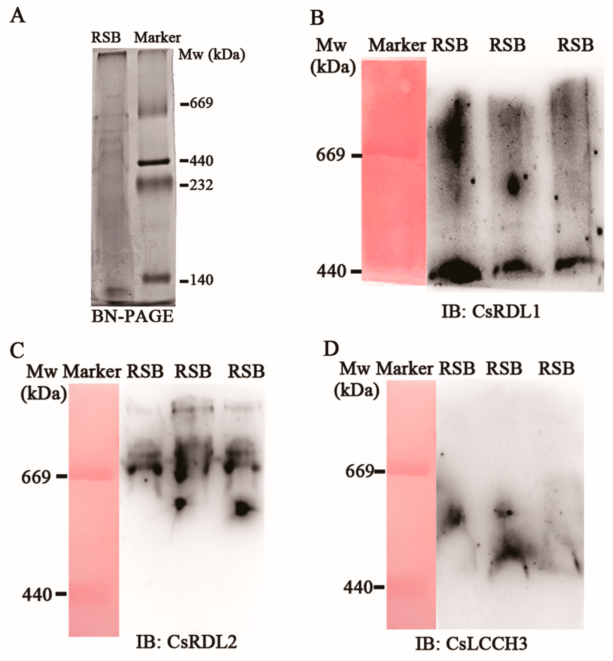
BN-PAGE was used to separate the native iGABARs complexes from the postsynaptic membrane proteins of RSB. Multiple bands were observed at Mws ≥ 440 kDa (Figure 1A). Antibodies against CsRDL1, CsRDL2 and CsLCCH3 were respectively used to identify iGABARs at Mws ≥ 440 kDa. Two bands (440 kDa and Mws > 440 kDa) (Figure 1B), four bands (one bands at 440 kDa < Mws < 669 kDa, one bands at 669 kDa and two bands at Mws > 669 kDa) (Figure 1C) and one band (at 440 kDa < Mws < 669 kDa) (Figure 1D) were identified by the antibodies against CsRDL1, CsRDL2 and CsLCCH3, respectively. Subsequently, the gel pieces with the bands at Mws ≥ 440 kDa were excised and analyzed by nanoLC-MS/MS. Unexpectedly, only the peptides and corresponding abundance of CsRDL2 was identified (Table S1).

3.3. Identified CsRDL1, ∆N-CsRDL2 and ∆N-CsLCCH3 from Native iGABARs

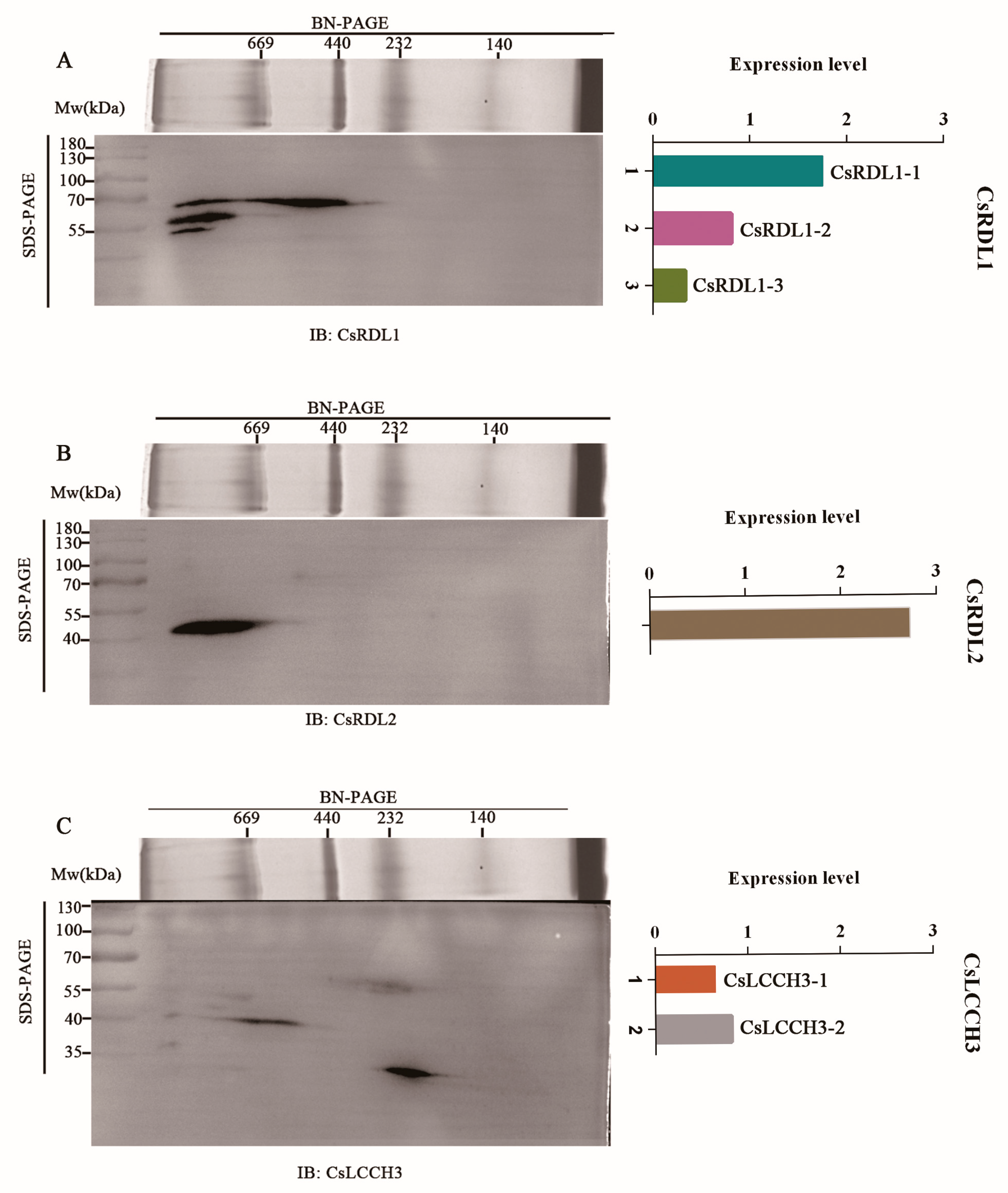
Multiple bands were observed in BN/SDS-PAGE (Figure S7). Three bands with distinct Mws (~54 kDa, ~55 kDa and ~70 kDa) were detected by the antibody against CsRDL1, and defined as CsRDL1-3 isoform, CsRDL1-2 isoform and CsRDL1-1 isoform, respectively (Figure 2A). Thereinto, the expression level of three isoforms in descending order in CsRDL1-1, CsRDL1-2, and CsRDL1-3, respectively (Figure 2A). CsRDL2 exhibited a single, highly intense band at ~48 kDa (Figure 2B). The expression level of CsRDL2 was higher than that of CsRDL1-1 (Figure 2B). Two scattered bands with distinct Mws (~56 kDa and ~37 kDa) were detected by the antibody against CsLCCH3 and defined as CsLCCH3-1 isoform and CsLCCH3-2 isoform, respectively (Figure 2C). Thereinto, the expression level of two isoforms in ascending order in CsLCCH3-1 and CsLCCH3-2, respectively (Figure 2C).
The Mws of iGABARs were divided into three ranges: Mws ≥ 669 kDa, 440 kDa ≤ Mws < 669 kDa and Mws < 440 kDa, and the composition and ratio of each subunit of native iGABARs was speculated based on the expression levels of subunits (CsRDL1-1, CsRDL1-2, CsRDL1-3, CsRDL2, CsLCCH3-1, and CsLCCH3-2) in each range (Figure S8). Furthermore, the BN/SDS-PAGE gel was divided into four gel lanes according to CsRDL2 immunoblot for 3-DE gel electrophoresis (Figure S9A). The spots of CsRDL2 and CsLCCH3-2 in 3-DE gel electrophoresis were observed at 48 and 37 kDa, respectively, which have equal Mws with 2-DE gel electrophoresis (Figure S9B).

3.4. Distribution of CsRDL1, CsRDL2 and CsLCCH3 in the Adult RSB Head

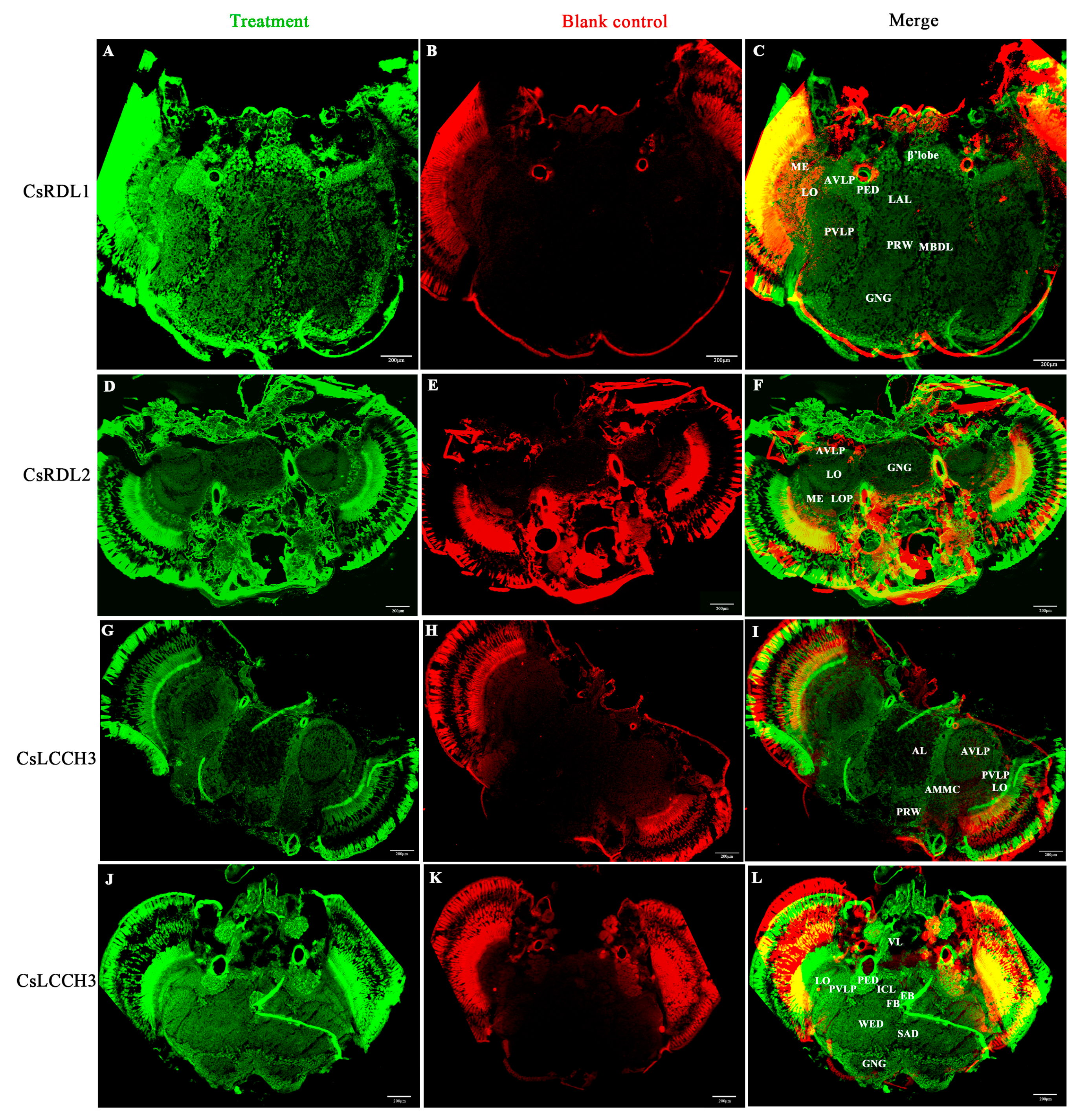
The distribution of CsRDL1, CsRDL2 and CsLCCH3 in the adult RSB head was respectively explored in tissue-cryosections. Compared with the blank control, CsRDL1 was detected in the β’lobe, median bundle (MBDL), anterior ventrolateral protocerebrum (AVLP), lobula (LO) and the periphery of the posterior ventrolateral protocerebrum (PVLP) and gnathal ganglia (GNG) (Figure 3A–C). CsRDL2 was distributed in the AVLP, LO, lobula plate (LOP) and the periphery of the GNG (Figure 3D–F). CsLCCH3 was distributed in the AVLP, ellipsoid body (EB), PVLP, pedunculus (PED), LO, vertical lobe (VL) and the periphery of the GNG, antennal lobe (AL), prow (PRW) and antennal mechanosensory and motor center (AMMC) (Figure 3G–L and Figure S10).

3.5. Binding Affinities of DMBF to Different Assembled iGABAR Models

iGABAR models assembled by CsRDL1bd, ∆N-CsRDL2 and ∆N-CsLCCH3 at different subunit ratios (5:0:0, 3:1:1, 2:2:1 and 1:3:1) were predicted using sequence-based models via AlphaFold 3. The results accessed from ERRAT (Table S3) and PROCHECK (Figure S11) confirmed that the predicted models of iGABAR have good quality.
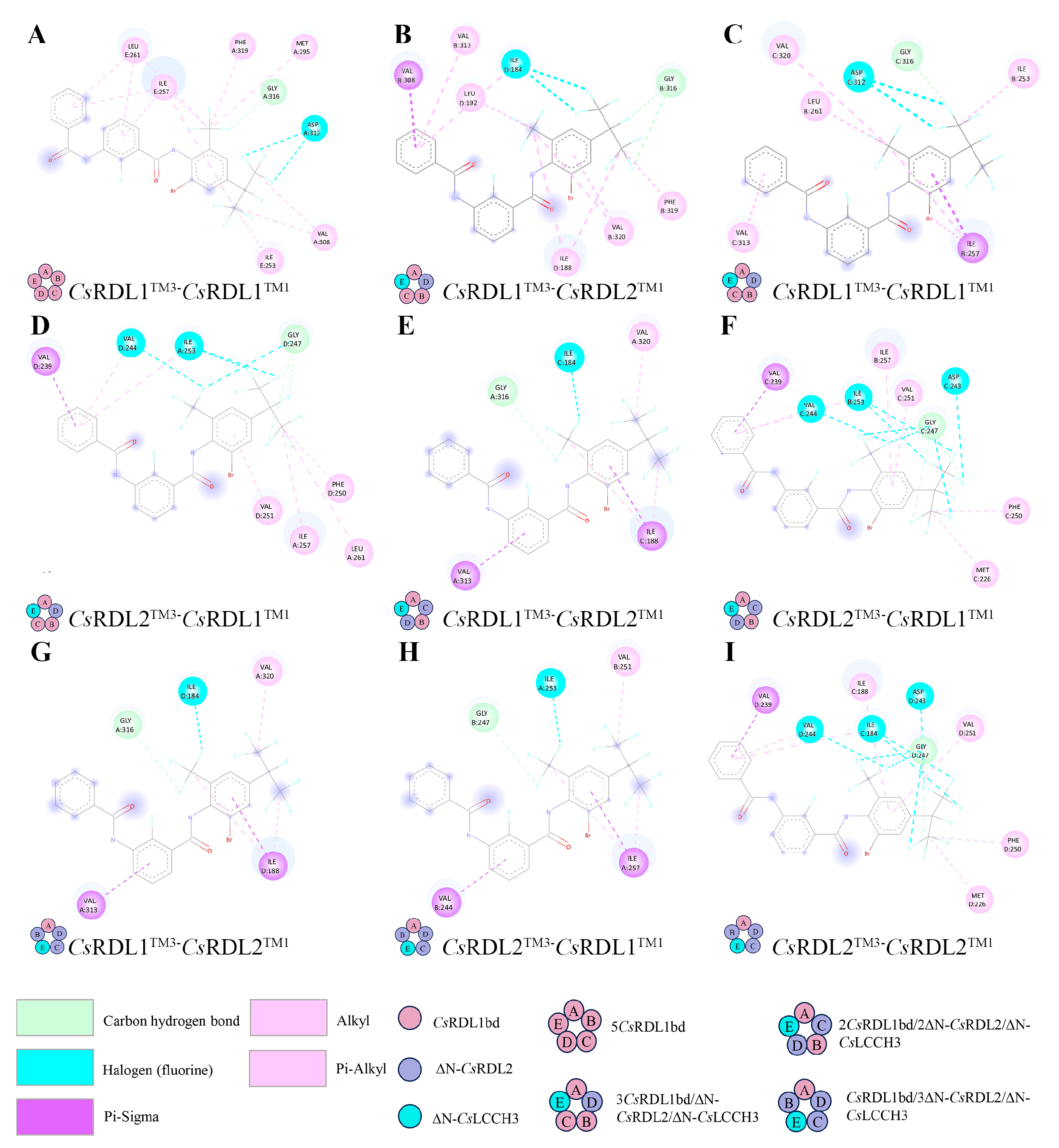
DMBF is bound to the pocket between the TM3 of CsRDL1 or ∆N-CsRDL2, and the TM1 of CsRDL1 or ∆N-CsRDL2 within iGABAR models (Figure S12). The binding affinities of DMBF to the iGABAR model assembled by CsRDL1 and the models assembled by CsRDL1, ∆N-CsRDL2 and ∆N-CsLCCH3 were −7.4 kcal/mol and −7.0 to −6.5 kcal/mol, respectively (Table 2). Furthermore, in iGABAR models assembled by CsRDL1, ∆N-CsRDL2 and ∆N-CsLCCH3, DMBF exhibited lower binding affinities for the TM3 of ∆N-CsRDL2 than for the TM3 of CsRDL1 in the iGABAR models assembled by CsRDL1, ∆N-CsRDL2 and ∆N-CsLCCH3 at subunit ratios of 3:1:1 and 2:2:1 (Table 2). As shown in Figure 4A–I, carbon–hydrogen bonds were observed between G3’ in the TM3 of CsRDL1 (G316) or ∆N-CsRDL2 (G247) and DMBF. Halogen bonds were present between isoleucine (ILE), valine (VAL) or aspartic acid (ASP) residues of CsRDL1 or ∆N-CsRDL2 and the fluorine atoms of DMBF, while pi-alkyl and alkyl interactions were observed between CsRDL1 or ∆N-CsRDL2 and DMBF in homomeric and heteromeric iGABAR models. In addition, pi-sigma interactions were observed between ILE or VAL residues of CsRDL1 or ∆N-CsRDL2 and DMBF in heteromeric iGABAR models (Figure 4B–I).

4. Discussion

Alternative splicing in the Rdl subunit, e.g., Rdlbd and Rdlad, greatly increases the potential diversityof invertebrate iGABARs [1]. In this study, the CsRdl1bd transcript was obtained from the genome and transcriptome of RSB. However, the expression abundance of Rdlad is lower than that of Rdlbd [1], which explains why the transcript of CsRdl1ad cannot be obtained. Furthermore, the truncated transcripts of CsRdl2 and CsLcch3 encode proteins lacking the N-terminal extracellular region. Similarly, the ∆N-α4 subunit sequence has been identified in mouse and human [44,45]. The N-terminal domain of iGABAR subunits as a hitherto functionally unassigned region, could affect agonist potency and aligns closely with known determinants of potency in nicotinic acetylcholine receptors (nAChRs) [46]. Meanwhile, the ∆N-α4 of nAChR in Colorado potato beetle Leptinotarsa decemlineata is possibly a response of diverse populations to exposure of neonicotinoid insecticides [47]. It is valuable to analyze the N-terminal extracellular region role in the structure and function of nAChR [47]. Therefore, ∆N-CsRDL2 and ∆N-CsLCCH3 subunits is not only relevant to the structure and function of iGABARs, but also may be an adaptive response to insecticides exposure.
The Mws of various iGABARs composed of CsRDL1, CsRDL2 or CsLCCH3 subunits were ≥440 kDa in BN-PAGE. Similarly, bands of iGABARs composed of β3 in mouse hippocampus migrated between 450 and 1236 kDa in BN-PAGE [37]. However, native iGABARs of mouse cerebellum have been reported to form two distinct complexes (~700 kDa and ~500 kDa, respectively) in BN-PAGE [35]. In the present study, the bands of iGABARs of RSB were similarly migrated to ~669 kDa and ~440 kDa, but iGABARs composed by CsRDL2 formed more than two different complexes in BN-PAGE. In contrast to vertebrates, diverse native iGABARs are present in the postsynaptic membrane of RSB.
iGABARs of RSB at Mws ≥ 669 kDa are composed of post-translationally modified CsRDL1, CsRDL1bd, CsRDL1ad, ∆N-CsRDL2 and ∆N-CsLCCH3. The predicted Mws of CsRDL1bd, CsRDL1ad (GenBank accession no. ASY91961.1), ∆N-CsRDL2 and ∆N-CsLCCH3 were 55 kDa, 54 kDa, 48 kDa and 37 kDa, respectively. Therefore, the two CsRDL1 bands migrating at ~54 kDa and ~55 kDa correspond to CsRDL1ad and CsRDL1bd, respectively; the CsRDL2 band migrating at ~48 kDa corresponds to ∆N-CsRDL2; and the CsLCCH3 band migrating at ~37 kDa corresponds to ∆N-CsLCCH3. Regarding the one CsRDL1 band migrating at ~70 kDa, evidence for the N-glycosylation of the α1, β1 and β2 iGABAR subunits has been confirmed in vertebrates [48]. Meanwhile, the phosphorylation of the β3 subunit of iGABAR is also in cultured cortical neurons [49]. Furthermore, the phosphorylation process of insect RDL is involved in the sensitivity of iGABAR to fipronil [50]. These results indicated that post-translational modifications, such as N-glycosylation and phosphorylation, possibly explain the increase in the Mw of native CsRDL1.
CsRDL1, CsRDL2 and CsLCCH3 composed native iGABARs in the postsynaptic membrane of RSB. CsRDL1 and ∆N-CsRDL2 were confirmed to be components of the native iGABARs at Mws ≥ 440 kDa. This is consistent with prior in vitro reconstitution studies on the RSB iGABARs. Co-injections of CsRdl1A and CsRdl2S into Xenopus oocytes were shown to form heteromeric iGABARs [51]. Furthermore, while CsLCCH3 forms a cation-selective channel with Cs8916 in the RSB [52], heteromeric LCCH3/GRD channels exhibit low GABA sensitivity and insecticide pharmacology similar to homomeric RDL channels in the honeybee [53]. Meanwhile, the homomeric RDL iGABAR and potentially heteromeric RDL/LCCH3 iGABARs mediate synaptic inhibition in the antennal lobes of honeybee [54]. Likewise, ∆N-CsLCCH3 was identified as a component of iGABARs at Mws ≥ 440 kDa, and full-length CsLCCH3 and the post-translationally modified CsRDL1 were detected in the iGABARs at Mws < 440 kDa in the present study.
CsRDL1, CsRDL2 and CsLCCH3 were distributed in the same regions of the adult RSB head. In sections of Drosophila brain, anti-LCCH3 staining was confined to the cell bodies surrounding the optic neuropil and other cortical regions surrounding the central brain [55]. In contrast, CsLCCH3 was distributed in the PVLP, AVLP, PED, VL, EB, LO and the periphery of the AMMC, GNG, AL and PRW. Furthermore, CsRDL1 immunoreactivity was detected in the β’lobe, AVLP, LO and the periphery of the PVLP. CsRDL2 was distributed in the AVLP, LO, LOP. Similarly, RDL has been observed in the β’lobe of the cricket Acheta domesticus L. brain [56]. Specific RDL immunostaining was evident in the LOP and LO of Drosophila brain [57], and intense RDL staining was observed in the LO, LOP and ventrolateral neuropils (VLP) of Drosophila brain sections [58]. Notably, RDL/LCCH3 do not co-localize in the same tissues of the Drosophila nervous system, as the ionic conductance of RDL/LCCH3 heteromeric complexes showed no significant difference compared with RDL homomers in vitro [55]. However, the present study demonstrated that CsLCCH3, CsRDL1 and CsRDL2 were distributed in the AVLP, LO and the periphery of GNG, providing evidence to verify CsLCCH3 probably assembling with CsRDL1 and/or CsRDL2 in the adult RSB head.
The assembly and intracellular transport of iGABARs in vertebrate cells have been investigated [10,59,60,61]. The intracellular assembly and proper targeting of complex heteromeric membrane proteins (e.g., iGABARs) to specialized regions including synapses, are governed by incompletely elucidated processes [10]. iGABAR’s assembly occurred within the endoplasmic reticulum (ER) and involved the interaction between chaperone molecules, immunoglobulin heavy chain binding protein and calnexin [59]. Furthermore, the N-terminal extracellular region of iGABAR subunits participate in some aspects of receptor assembly [60]. The N-terminus possibly act as a post-translational regulatory role in intracellular folding, glycosylation and assembly of the iGABAR subunits [10]. However, provided that the shorter N-terminal fragment contains an assembly-critical sequence, it will assemble with other subunits independently regardless of an additional sequence exists or is incorporated in longer N-terminal fragments [61]. Finally, assembled receptor complexes are specially targeted to appropriate sites on the membrane and stably anchored at these locations [10]. Hence, iGABARs composed of CsRDL1, ∆N-CsRDL2 and ∆N-CsLCCH3 are likely transported to the postsynaptic membrane after intracellular assembly. Unlike intracellular subunit assembly in vivo, ∆N-CsRDL2 and ∆N-CsLCCH3 failed to express and form functional iGABAR complexes with RDL1 on the plasma membrane of Xenopus oocytes due to the deletion of their N-terminal signal peptides, which target newly synthesized proteins to the membrane of ER and mediate their translocation into the ER lumen [62,63]. Consequently, in silico analysis was implemented to explore the ligand binding characteristics of the iGABAR models assembled by CsRDL1, ∆N-CsRDL2 and ∆N-CsLCCH3.
DMBF was observed to bind to the pocket between the TM3 of the CsRDL1 or ∆N-CsRDL2 subunit and TM1 of the complementary CsRDL1 or ∆N-CsRDL2 subunit in iGABAR models. Similarly, in the honeybee RDL iGABAR, the pocket between the TM3 of the principal subunit and the TM1 of the complementary subunit bind abamectin, and the G3’ in the TM3 of RDL is conserved across invertebrates and represents one of the most common field-identified resistance mutations to abamectin [25]. Furthermore, knockdown of CsRdl1 and CsRdl2 in vivo significantly decreased the susceptibility of the RSB larvae to abamectin and fluralaner [64,65]. Fluralaner, fluxametamide and isocycloseram have been indicated to bind to the same pocket as abamectin [25]. Meanwhile, the G3’M mutation in RDL has been reported to confer resistance to broflanilide and fluralaner [24]. A hydrogen bond formed between DMBF and G277 of the D. melanogaster GABAR model was suggested to be the key interaction mediating DMBF antagonism by in silico simulations [23]. Consistent with this observation, a carbon–hydrogen bond formed between G316 of CsRDL1 or G247 of ∆N-CsRDL2 and DMBF was identified as the key interaction mediating the antagonism of DMBF in different iGABAR models. Additionally, DMBF exhibited higher binding affinities for heteromeric iGABAR models assembled by CsRDL1, ∆N-CsRDL2 and ∆N-CsLCCH3 than for homomeric CsRDL1 iGABAR model. Nevertheless, these in silico predictions are yet to be confirmed experimentally.

5. Conclusions

The truncated transcripts of CsRdl1 and CsLcch3 encode ∆N-CsRDL2 and ∆N-CsLCCH3 with Mws of 48 kDa and 37 kDa, respectively. The Mws of various native iGABARs composed of CsRDL1, CsRDL2 or CsLCCH3 were ≥440 kDa in the postsynaptic membrane of RSB. CsRDL1ad, CsRDL1bd, post-translationally modified CsRDL1, ∆N-CsRDL2 and ∆N-CsLCCH3 composed iGABARs at Mws ≥ 669 kDa. Post-translationally modified CsRDL1, ∆N-CsRDL2 and ∆N-CsLCCH3 composed iGABARs at 440 kDa ≤ Mws < 669 kDa. Post-translationally modified CsRDL1 and CsLCCH3 composed iGABARs at Mws < 440 kDa. Furthermore, CsLCCH3, CsRDL1 and CsRDL2 were distributed in the AVLP, LO and the periphery of the GNG in the adult RSB head. DMBF displayed higher binding affinities for heteromeric iGABARs assembled by CsRDL1bd, ∆N-CsRDL2 and ∆N-CsLCCH3 than for homomeric CsRDL1bd iGABAR models in silico. This study demonstrates that the presence of ∆N-CsRDL2 and ∆N-CsLCCH3 in native iGABARs of RSB provides new insights into the molecular constituents of postsynaptic ion channels and supports the rational design of novel insecticides.

Supplementary Materials

The following supporting information can be downloaded at: https://www.mdpi.com/article/10.3390/insects17050477/s1, Text S1:Materials and methods of peptide analysis by nanoLC-MS/MS. Figure S1: Transcript analysis of CsRdl1. Figure S2: Alignment of the amino acid sequences of CsRDL1. Figure S3: Transcript analysis of CsRdl2. Figure S4: Alignment of the amino acid sequences of CsRDL2. Figure S5: Transcript analysis of CsLcch3. Figure S6: Alignment of the amino acid sequences of CsLCCH3. Figure S7: Separation of the postsynaptic membrane protein via BN/SDS-PAGE. Figure S8: Expression levels of CsRDL1, CsRDL2 and CsLCCH3 in native iGABARs of different molecular weight ranges. Figure S9: Identification of CsRDL2 and CsLCCH3 via BN/SDS/SDS-PAGE. Figure S10: Negative control for CsRDL1 (A), CsRDL2 (B) and CsLCCH3 (C,D). Figure S11: Ramachandran plot statistics of iGABARs assembled by various subunits. Figure S12: Binding of DMBF to the predicted structures of iGABARs assembled by various subunits. Table S1: Peptide and abundance of ∆N-CsRDL2 identified by nanoLC-MS/MS. Table S2: Amino acid sequences of ∆N-CsRDL2 and ∆N-CsLCCH3 used in AlphaFold 3. Table S3: The overall quality factor of iGABAR models evaluated by ERRAT. Table S4: Grid center of the pocket in iGABAR models. Reference [66] was cited in the supplementary materials.

Author Contributions

Conceptualization, E.Z. and C.Z.; methodology, E.Z. and C.Z.; formal analysis, E.Z.; investigation, E.Z., J.L., Y.Z. and J.W.; Resources, E.Z., J.L., Y.Z. and S.N.; Data curation, E.Z. and J.L.; Writing—Original draft preparation, E.Z.; Writing- Reviewing and Editing, C.Z.; visualization, E.Z.; Supervision, C.Z.; Funding acquisition, C.Z. All authors have read and agreed to the published version of the manuscript.

Funding

This work was supported by the Jiangsu Agricultural Science and Technology Independent Innovation Fund Project [grant number CX (24)3010] and National Natural Science Foundation of China [grant number 31501672].

Data Availability Statement

The original contributions presented in this study are included in the article/Supplementary Materials. Further inquiries can be directed to the corresponding author.

Acknowledgments

We thank the anonymous reviewers for their valuable comments to improve the quality of the manuscript.

Conflicts of Interest

The authors declare no conflicts of interest.

Abbreviations

AL, antennal lobe; AMMC, antennal mechanosensory and motor center; AVLP, anterior ventrolateral neuropils; BN-PAGE, blue native PAGE; CBB R-250, Coomassie Brilliant Blue R-250; Cs, Chilo suppressalis; DMBF, desmethyl-broflanilide; EB, ellipsoid body; ER, endoplasmic reticulum; FB, fan-shaped body; GABA, γ-aminobutyric acid; GRD, GABA and glycine receptor-like subunit of Drosophila; GNG, gnathal ganglia; G3’, glycine at the third position; IB, Immunoblot; ICL, interior clamp; iGABARs, ionotropic γ-aminobutyric acid receptors; LAL, lateral accessory lobe; LCCH3, ligand-gated chloride channel homolog 3; LGIC, ligand-gated ion channel; LO, lobula; LOP, lobula plate; MBDL, median bundle; ME, medulla; MNG, lauryl maltose-neopentyl glycol; Mw, molecular weight; mRNA, messenger RNA; PED, pedunculus; PRW, prow; PVLP, posterior ventrolateral protocerebrum; RDL, resistant to dieldrin; RSB, rice stem borer; SAD, saddle; TM, transmembrane domain; VL, vertical lobe; WED, wedge; ∆N-CsRDL2, N-terminal-truncated CsRDL2; ∆N-CsLCCH3, N-terminal-truncated CsLCCH3; 2D, two-dimensional; 3D, three-dimensional.

References

  1. Ffrench-Constant, R.H.; Rocheleau, T.A. Drosophila γ-aminobutyric acid receptor gene Rdl shows extensive alternative splicing. J. Neurochem. 1993, 6, 2323–2326. [Google Scholar] [CrossRef] [Scilit]
  2. Ffrench-Constant, R.H.; Mortlock, D.P.; Shaffer, C.D.; Maclntyre, R.J.; Roush, R.T. Molecular cloning and transformation of cyclodiene resistance in Drosophila: An invertebrate gamma-aminobutyric acid subtype A receptor locus. Proc. Natl. Acad. Sci. USA 1991, 16, 7209–7213. [Google Scholar] [CrossRef] [Scilit]
  3. Martenson, J.S.; Yamasaki, T.; Chaudhury, N.H.; Albrecht, D.; Tomita, S. Assembly rules for GABAA receptor complexes in the brain. eLife 2017, 6, e27443. [Google Scholar] [CrossRef] [Scilit] [PubMed]
  4. Tomita, S. Molecular constituents and localization of the ionotropic GABA receptor complex in vivo. Curr. Opin. Neurobiol. 2019, 57, 81–86. [Google Scholar] [CrossRef] [Scilit] [PubMed]
  5. Jones, A.K. Genomics, cys-loop ligand-gated ion channels and new targets for the control of insect pests and vectors. Curr. Opin. Insect Sci. 2018, 30, 1–7. [Google Scholar] [CrossRef] [Scilit] [PubMed]
  6. Zhou, J.; Noviello, C.M.; Teng, J.; Moore, H.; Lega, B.; Hibbs, R.E. Resolving native GABAA receptor structures from the human brain. Nature 2025, 8050, 562–568. [Google Scholar] [CrossRef] [Scilit]
  7. Nusser, Z.; Sieghart, W.; Somogyi, P. Segregation of different GABAA receptors to synaptic and extrasynaptic membranes of cerebellar granule cells. J. Neurosci. 1998, 5, 1693–1703. [Google Scholar] [CrossRef] [Scilit]
  8. Whiting, P.; McKernan, R.M.; Iversen, L.L. Another mechanism for creating diversity in γ-aminobutyrate type A receptors: RNA splicing directs expression of two forms of γ2 subunit, one of which contains a protein kinase C phosphorylation site. Proc. Natl. Acad. Sci. USA 1990, 24, 9966–9970. [Google Scholar] [CrossRef] [Scilit]
  9. Klausberger, T.; Fuchs, K.; Mayer, B.; Ehya, N.; Sieghart, W. GABAA receptor assembly: Identification and structure of γ2 sequences forming the intersubunit contacts with α1 and β3 subunits. J. Biol. Chem. 2000, 12, 8921–8928. [Google Scholar] [CrossRef] [Scilit]
  10. Mu, W.; Cheng, Q.; Yang, J.; Burt, D.R. Alternative splicing of the GABAA receptor α4 subunit creates a severely truncated mRNA. Brain Res. Bull. 2002, 5, 447–454. [Google Scholar] [CrossRef] [Scilit]
  11. Henderson, J.E.; Soderlund, D.M.; Knipple, D.C. Characterization of a putative γ-aminobutyric-acid (GABA) receptor β-subunit gene from Drosophila melanogaster. Biochem. Biophys. Res. Commun. 1993, 2, 474–482. [Google Scholar] [CrossRef] [Scilit]
  12. Harvey, R.J.; Schmitt, B.; Hermans-Borgmeyer, I.; Gundelfinger, E.D.; Betz, H.; Darlison, M.G. Sequence of a Drosophila ligand-gated ion-channel polypeptide with an unusual amino-terminal extracellular domain. J. Neurochem. 1994, 62, 2480–2483. [Google Scholar] [CrossRef] [Scilit]
  13. Jones, A.K.; Sattelle, D.B. The cys-loop ligand-gated ion channel gene superfamily of the red flour beetle, Tribolium castaneum. BMC Genom. 2007, 8, 327. [Google Scholar] [CrossRef] [Scilit]
  14. Knipple, D.C.; Soderlund, D.M. The ligand-gated chloride channel gene family of Drosophila melanogaster. Pestic. Biochem. Physiol. 2010, 2, 140–148. [Google Scholar] [CrossRef] [Scilit]
  15. Zhang, H.G.; Lee, H.J.; Rocheleau, T.; ffrench-Constant, R.H.; Jackson, M.B. Subunit composition determines picrotoxin and bicuculline sensitivity of Drosophila gamma-aminobutyric acid receptors. Mol. Pharmacol. 1995, 5, 835–840. [Google Scholar] [CrossRef] [Scilit]
  16. Hashim, O.; Charvet, C.L.; Toubaté, B.; Ahmed, A.A.E.; Lamassiaude, N.; Neveu, C.; Dimier-Poisson, I.; Debierre-Grockiego, F.; Dupuy, C. Molecular and functional characterization of GABA receptor subunits GRD and LCCH3 from human louse Pediculus humanus humanus. Mol. Pharmacol. 2022, 2, 116–127. [Google Scholar] [CrossRef] [Scilit] [PubMed]
  17. Jones, A.K.; Buckingham, S.D.; Papadaki, M.; Yokota, M.; Sattelle, B.M.; Matsuda, K.; Sattelle, D.B. Splice-variant- and stage-specific RNA editing of the Drosophila GABA receptor modulates agonist potency. J. Neurosci. 2009, 13, 4287–4292. [Google Scholar] [CrossRef] [Scilit] [PubMed]
  18. Yu, L.L.; Cui, Y.J.; Lang, G.J.; Zhang, M.Y.; Zhang, C.X. The ionotropic γ-aminobutyric acid receptor gene family of the silkworm, Bombyx mori. Genome 2010, 9, 688–697. [Google Scholar] [CrossRef] [Scilit] [PubMed]
  19. Song, Z.; Jiang, J.; Tang, T.; Luo, G.; Wang, J.; Lin, K.; Mu, L.; Zhao, C. Splicing factor Nova regulates the splice variants in exons 3 and 6 of GABA receptor subunit RDL from Chilo suppressalis Walker (Lepidoptera: Crambidae). Pestic. Biochem. Physiol. 2025, 214, 106556. [Google Scholar] [CrossRef] [Scilit]
  20. Casida, J.E.; Durkin, K.A. Novel GABA receptor pesticide targets. Pestic. Biochem. Physiol. 2015, 121, 22–30. [Google Scholar] [CrossRef] [Scilit]
  21. Tang, T.; Hu, F.; Wang, P.; Fu, W.; Liu, X. Broflanilide effectively controls Helicoverpa armigera and Spodoptera exigua exhibiting diverse susceptibilities to chlorantraniliprole and emamectin benzoate. Pest Manag. Sci. 2021, 3, 1262–1272. [Google Scholar] [CrossRef] [Scilit]
  22. Casida, J.E. Golden age of RyR and GABA-R diamide and isoxazoline insecticides: Common genesis, serendipity, surprises, selectivity, and safety. Chem. Res. Toxicol. 2015, 4, 560–566. [Google Scholar] [CrossRef] [Scilit] [PubMed]
  23. Gao, Y.; Zhang, Y.; Wu, F.; Pei, J.; Luo, X.; Ju, X.; Zhao, C.; Liu, G. Exploring the interaction mechanism of desmethyl-broflanilide in insect GABA receptors and screening potential antagonists by in silico simulations. J. Agric. Food Chem. 2020, 50, 14768–14780. [Google Scholar] [CrossRef] [Scilit]
  24. Zhang, Y.; Huang, Q.; Sheng, C.; Liu, G.; Zhang, K.; Jia, Z.; Tang, T.; Mao, X.; Jones, A.K.; Han, Z.; et al. G3′MTMD3 in the insect GABA receptor subunit, RDL, confers resistance to broflanilide and fluralaner. PLoS Genet. 2023, 6, e1010814. [Google Scholar] [CrossRef] [Scilit]
  25. Labouré, T.; Pandey, M.P.; Zarkadas, E.; Juillan-Binard, C.; Baud, D.; Neyton, J.; Cens, T.; Rousset, M.; Dehez, F.; Charnet, P.; et al. Structures of the honeybee GABAA RDL receptor illuminate allosteric modulation. Neuron 2026, 114, 1234–1245. [Google Scholar] [CrossRef] [Scilit] [PubMed]
  26. Sheng, C.F.; Wang, H.T.; Sheng, S.Y.; Gao, L.D.; Xuan, W.J. Pest status and loss assessment of crop damage caused by the rice borers, Chilo suppressalis and Tryporyza incertulas in China. Chin. Bull. Entomol. 2003, 40, 289–294. [Google Scholar]
  27. Ma, W.; Xianxin, Z.; Yin, C.; Jiang, F.; Du, X.; Chen, T.; Zhang, Q.; Qiu, L.; Xu, H.; Hull, J.; et al. A chromosome-level genome assembly reveals the genetic basis of cold tolerance in a notorious rice insect pest, Chilo suppressalis. Mol. Ecol. Resour. 2019, 1, 268–282. [Google Scholar] [CrossRef] [Scilit]
  28. An, Y.; Wu, J.; Chen, Y.; Li, S. Comprehensive analysis of alternative splicing in Rosa roxburghii Tratt. reveals its role in flavonoid synthesis. Front. Plant Sci. 2025, 16, 1627126. [Google Scholar] [CrossRef] [Scilit] [PubMed]
  29. Quinlan, A.R.; Hall, I.M. BEDTools: A flexible suite of utilities for comparing genomic features. Bioinformatics 2010, 6, 841–842. [Google Scholar] [CrossRef] [Scilit]
  30. Thorvaldsdóttir, H.; Robinson, J.T.; Mesirov, J.P. Integrative Genomics Viewer (IGV): High-performance genomics data visualization and exploration. Brief. Bioinform. 2012, 2, 178–192. [Google Scholar] [CrossRef] [Scilit]
  31. Zhang, K.; Zhan, E.; Chang, X.; Fouad, E.A.; Zhao, C. Resistance realized heritability and fitness cost of cyproflanilide in rice stem borer, Chilo suppressalis (Lepidoptera: Pyralidae). Agronomy 2024, 10, 2249. [Google Scholar] [CrossRef] [Scilit]
  32. Chen, Y. Expoloration of the Components of γ-Aminobutyric Acid Receptor of the Rice Stem Borer, Chilo suppressalis, by Co-Immunoprecipitation. Master’s Thesis, Nanjing Agricultural University, Nanjing, China, June 2017. [Google Scholar]
  33. Morató Arús, X.; López-Cano, M.; Canas, P.; Ciruela, F. Brain membrane fractionation: An ex vivo approach to assess subsynaptic protein localization. J. Vis. Exp. 2017, 123, 55661. [Google Scholar] [CrossRef] [Scilit]
  34. Wittig, I.; Braun, H.P.; Schägger, H. Blue native PAGE. Nat. Protoc. 2006, 1, 418–428. [Google Scholar] [CrossRef] [Scilit]
  35. Yamasaki, T.; Hoyos-Ramirez, E.; Martenson, J.S.; Morimoto-Tomita, M.; Tomita, S. GARLH family proteins stabilize GABAA receptors at synapses. Neuron 2017, 5, 1138–1152. [Google Scholar] [CrossRef] [Scilit]
  36. Swamy, M.; Siegers, G.M.; Minguet, S.; Wollscheid, B.; Schamel, W.W. Blue native polyacrylamide gel electrophoresis (BN-PAGE) for the identification and analysis of multiprotein complexes. Sci. STKE 2006, 345, pl4. [Google Scholar] [CrossRef] [Scilit]
  37. Kang, S.U.; Heo, S.; Lubec, G. Mass spectrometric analysis of GABAA receptor subtypes and phosphorylations from mouse hippocampus. Proteomics 2011, 11, 2171–2181. [Google Scholar] [CrossRef] [Scilit] [PubMed]
  38. Ito, K.; Shinomiya, K.; Ito, M.; Armstrong, J.D.; Boyan, G.; Hartenstein, V.; Harzsch, S.; Heisenberg, M.; Homberg, U.; Jenett, A.; et al. A systematic nomenclature for the insect brain. Neuron 2014, 4, 755–765. [Google Scholar] [CrossRef] [Scilit]
  39. Colovos, C.; Yeates, T.O. Verification of protein structures: Patterns of nonbonded atomic interactions. Protein Sci. 1993, 9, 1511–1519. [Google Scholar] [CrossRef] [Scilit] [PubMed]
  40. Laskowski, R.A.; MacArthur, M.W.; Moss, D.S.; Thornton, J.M. PROCHECK: A program to check the stereochemical quality of protein structures. Appl. Crystallogr. 1993, 2, 283–291. [Google Scholar] [CrossRef] [Scilit]
  41. Nakao, T.; Banba, S. Broflanilide: A meta-diamide insecticide with a novel mode of action. Bioorg. Med. Chem. 2016, 3, 373–377. [Google Scholar] [CrossRef] [Scilit]
  42. Tian, W.; Chen, C.; Lei, X.; Zhao, J.; Liang, J. CASTp 3.0: Computed atlas of surface topography of proteins. Nucleic Acids Res. 2018, 46, W363–W367. [Google Scholar] [CrossRef] [Scilit]
  43. Trott, O.; Olson, A.J. AutoDock Vina: Improving the speed and accuracy of docking with a new scoring function, efficient optimization, and multithreading. J. Comput. Chem. 2010, 2, 455–461. [Google Scholar] [CrossRef] [Scilit] [PubMed]
  44. Wisden, W.; Herb, A.; Wieland, H.; Keinänen, K.; Lüddens, H.; Seeburg, P.H. Cloning, pharmacological characteristics and expression pattern of the rat GABAA receptor α4 subunit. FEBS Lett. 1991, 2, 227–230. [Google Scholar] [CrossRef] [Scilit] [PubMed]
  45. Yang, W.; Drewe, J.A.; Lan, N.C. Cloning and characterization of the human GABAA receptor α4 subunit: Identification of a unique diazepam-insensitive binding site. Eur. J. Pharmacol. Mol. Pharmacol. Sect. 1995, 3, 319–325. [Google Scholar] [CrossRef] [Scilit] [PubMed]
  46. Hosie, A.M.; Buckingham, S.D.; Presnail, J.K.; Sattelle, D.B. Alternative splicing of a Drosophila GABA receptor subunit gene identifies determinants of agonist potency. Neuroscience 2001, 3, 709–714. [Google Scholar] [CrossRef] [Scilit]
  47. Chen, D.; Hawthorne, D.J. The cys-loop ligand-gated ion channel gene superfamily of the Colorado potato beetle, Leptinotarsa decemlineata. BMC Genom. 2025, 1, 702. [Google Scholar] [CrossRef] [Scilit]
  48. Mueller, T.M.; Haroutunian, V.; Meador-Woodruff, J.H. N-glycosylation of GABAA receptor subunits is altered in schizophrenia. Neuropsychopharmacology 2014, 3, 528–537. [Google Scholar] [CrossRef] [Scilit]
  49. Brandon, N.J.; Delmas, P.; Kittler, J.T.; McDonald, B.J.; Sieghart, W.; Brown, D.A.; Smart, T.G.; Moss, S.J. GABAA receptor phosphorylation and functional modulation in cortical neurons by a protein kinase C-dependent pathway. J. Biol. Chem. 2000, 49, 38856–38862. [Google Scholar] [CrossRef] [Scilit]
  50. Murillo, L.; Hamon, A.; Es-Salah-Lamoureux, Z.; Itier, V.; Quinchard, S.; Lapied, B. Inhibition of protein kinase C decreases sensitivity of GABA receptor subtype to fipronil insecticide in insect neurosecretory cells. Neurotoxicology 2011, 6, 828–835. [Google Scholar] [CrossRef] [Scilit]
  51. Sheng, C.W.; Jia, Z.Q.; Ozoe, Y.; Huang, Q.T.; Han, Z.J.; Zhao, C.Q. Molecular cloning, spatiotemporal and functional expression of GABA receptor subunits RDL1 and RDL2 of the rice stem borer Chilo suppressalis. Insect Biochem. Mol. Biol. 2018, 94, 18–27. [Google Scholar] [CrossRef] [Scilit]
  52. Huang, Q.T.; Sheng, C.W.; Jones, A.K.; Jiang, J.; Tang, T.; Han, Z.J.; Zhao, C.Q. Functional characteristics of the lepidopteran ionotropic GABA receptor 8916 subunit interacting with the LCCH3 or the RDL subunit. J. Agric. Food Chem. 2021, 39, 11582–11591. [Google Scholar] [CrossRef] [Scilit]
  53. Henry, C.; Cens, T.; Charnet, P.; Cohen-Solal, C.; Collet, C.; van-Dijk, J.; Rousset, M. Heterogeneous expression of GABA receptor-like subunits LCCH3 and GRD reveals functional diversity of GABA receptors in the honeybee Apis mellifera. Br. J. Pharmacol. 2020, 20, 3924–3940. [Google Scholar] [CrossRef] [Scilit]
  54. Dupuis, J.P.; Bazelot, M.; Barbara, G.S.; Paute, S.; Gauthier, M.; Raymond-Delpech, V. Homomeric RDL and heteromeric RDL/LCCH3 GABA receptors in the honeybee antennal lobes: Two candidates for inhibitory transmission in olfactory processing. J. Neurophysiol. 2010, 1, 458–468. [Google Scholar] [CrossRef] [Scilit]
  55. Aronstein, K.; Auld, V.; ffrench-Constant, R.H. Distribution of two GABA receptor-like subunits in the Drosophila CNS. Invertebr. Neurosci. 1996, 2, 115–120. [Google Scholar] [CrossRef] [Scilit]
  56. Strambi, C.; Cayre, M.; Sattelle, D.B.; Augier, R.; Strambi, A. Immunocytochemical mapping of an RDL-like GABA receptor subunit and of GABA in brain structures related to learning and memory in the cricket Acheta domesticus. Learn. Mem. 1998, 5, 78–89. [Google Scholar] [CrossRef] [Scilit] [PubMed]
  57. Harrison, J.B.; Chen, H.H.; Sattelle, E.; Barker, P.J.; Huskisson, N.S.; Rauh, J.J.; Bai, D.; Sattelle, D.B. Immunocytochemical mapping of a C-terminus anti-peptide antibody to the GABA receptor subunit, RDL in the nervous system of Drosophila melanogaster. Cell Tissue Res. 1996, 2, 269–278. [Google Scholar] [CrossRef] [Scilit] [PubMed]
  58. Aronstein, K.; ffrench-Constant, R.H. Immunocytochemistry of a novel GABA receptor subunit Rdl in Drosophila melanogaster. Invertebr. Neurosci. 1995, 1, 25–31. [Google Scholar] [CrossRef] [Scilit]
  59. Connolly, C.N.; Krishek, B.J.; McDonald, B.J.; Smart, T.G.; Moss, S.J. Assembly and cell surface expression of heteromeric and homomeric γ-aminobutyric acid type A receptors. J. Biol. Chem. 1996, 1, 89–96. [Google Scholar] [CrossRef] [Scilit]
  60. Srinivasan, S.; Nichols, C.J.; Lawless, G.M.; Olsen, R.W.; Tobin, A.J. Two invariant tryptophans on the α1 subunit define domains necessary for GABAA receptor assembly. J. Biol. Chem. 1999, 38, 26633–26638. [Google Scholar] [CrossRef] [Scilit]
  61. Sarto-Jackson, I.; Sieghart, W. Assembly of GABAA receptors. Mol. Membr. Biol. 2008, 4, 302–310. [Google Scholar] [CrossRef] [Scilit]
  62. Altschuler, Y.; Rosenberg, N.; Harel, R.; Galili, G. The N- and C-terminal regions regulate the transport of wheat γ-gliadin through the endoplasmic reticulum in Xenopus oocytes. Plant Cell 1993, 4, 443–450. [Google Scholar] [CrossRef] [Scilit]
  63. Hiss, J.A.; Schneider, G. Architecture, function and prediction of long signal peptides. Brief. Bioinform. 2009, 5, 569–578. [Google Scholar] [CrossRef] [Scilit]
  64. Meng, X.; Yang, X.; Zhang, N.; Jiang, H.; Ge, H.; Chen, M.; Qian, K.; Wang, J. Knockdown of the GABA receptor RDL genes decreases abamectin susceptibility in the rice stem borer, Chilo suppressalis. Pestic. Biochem. Physiol. 2019, 153, 171–175. [Google Scholar] [CrossRef] [Scilit]
  65. Jia, Z.Q.; Sheng, C.W.; Tang, T.; Liu, D.; Leviticus, K.; Zhao, C.Q.; Chang, X.L. Identification of the ionotropic GABA receptor-like subunits from the striped stem borer, Chilo suppressalis Walker (Lepidoptera: Pyralidae). Pestic. Biochem. Physiol. 2019, 155, 36–44. [Google Scholar] [CrossRef] [Scilit]
  66. Rappsilber, J.; Mann, M.; Ishihama, Y. Protocol for micro-purification, enrichment, pre-fractionation and storage of peptides for proteomics using StageTips. Nat. Protoc. 2007, 8, 1896–1906. [Google Scholar] [CrossRef] [Scilit]
Figure 1. Separation of the native iGABAR complexes via BN-PAGE. (A) Proteins were stained with CBB R-250. iGABAR complexes identified with antibodies against CsRDL1 (B), CsRDL2 (C) or CsLCCH3 (D), respectively. Marker was stained with Ponceau S staining solution in red/purple bands. IB, Immunoblot.
Insects 17 00477 g001
Figure 2. Separation of subunits in the native iGABARs via BN/SDS-PAGE. (A) CsRDL1; (B) CsRDL2; (C) CsLCCH3.
Insects 17 00477 g002
Figure 3. Distribution of CsRDL1, CsRDL2 and CsLCCH3 in the adult RSB head. (A) CsRDL1; (D) CsRDL2; (G,J) CsLCCH3; (B,E,H,K) blank controls; (C,F,I,L) merged images. AL, antennal lobe; AMMC, antennal mechanosensory and motor center; AVLP, anterior ventrolateral protocerebrum; EB, ellipsoid body; FB, fan-shaped body; GNG, gnathal ganglia; ICL, interior clamp; LAL, lateral accessory lobe; LO, lobula; LOP, lobula plate; MBDL, median bundle; ME, medulla; PED, pedunculus; PRW, prow; PVLP, posterior ventrolateral protocerebrum; SAD, saddle; VL, vertical lobe; and WED, wedge; β’lobe, medial lobe division in mushroom body. Scale bar, 200 μm. The green channel corresponds to signals from the treatment group, while the red channel represents signals from the blank control group.
Insects 17 00477 g003
Figure 4. 2D structures of DMBF and its interacting amino acid residues in iGABAR models. (A) 5CsRDL1; (BD) 3CsRDL1/∆N-CsRDL2/∆N-CsLCCH3; (E,F) 2CsRDL1/2∆N-CsRDL2/∆N-CsLCCH3; (GI) CsRDL1/3∆N-CsRDL2/∆N-CsLCCH3.
Insects 17 00477 g004
Table 1. Character of proteins encoded by CsRdl1, CsRdl2 and CsLcch3 transcripts.
GeneTranscriptProteinNumber of Amino Acid (aa)Molecular Weight (kDa)
CsRdl1bdCsup004269.1 *CsRDL1-248955
STRG.8675.1 *48955
CsRdl2Csup004319.1CsRDL242448
STRG.8682.142448
CsLcch3Csup009529.1 *CsLCCH3-149256
STRG.622.1CsLCCH3-232937
STRG.622.2CsLCCH3-233037
* the full-length transcript.
Table 2. Binding affinity of DMBF to iGABAR models.
iGABAR ComplexesBinding DomainAffinity (kcal/mol)
5CsRDL1CsRDL1TM3-CsRDL1TM1−7.4
3CsRDL1/∆N-CsRDL2/∆N-CsLCCH3CsRDL1TM3-CsRDL1TM1−6.5
CsRDL1TM3-CsRDL2TM1−6.7
CsRDL2TM3-CsRDL1TM1−6.8
2CsRDL1/2∆N-CsRDL2/∆N-CsLCCH3CsRDL1TM3-CsRDL2TM1−6.5
CsRDL2TM3-CsRDL1TM1−7.0
CsRDL1/3∆N-CsRDL2/∆N-CsLCCH3CsRDL1TM3-CsRDL2TM1−6.6
CsRDL2TM3-CsRDL1TM1−6.5
CsRDL2TM3-CsRDL2TM1−7.0
Disclaimer/Publisher’s Note: The statements, opinions and data contained in all publications are solely those of the individual author(s) and contributor(s) and not of MDPI and/or the editor(s). MDPI and/or the editor(s) disclaim responsibility for any injury to people or property resulting from any ideas, methods, instructions or products referred to in the content.