Article

Ultrastructural Morphology and Descriptive Analysis of Cuticular Sensilla in Adult Tomicus pilifer (Coleoptera: Curculionidae)

1
Key Laboratory of Sustainable Forest Ecosystem Management, Ministry of Education, Northeast Forestry University, Harbin 150040, China
2
Forest Protection Research Institute of Heilongjiang Province, Heilongjiang Academy of Forestry, Harbin 150040, China
*
Author to whom correspondence should be addressed.
These authors contributed equally to this work.
Insects 2025, 16(9), 890; https://doi.org/10.3390/insects16090890
Submission received: 22 July 2025 / Revised: 14 August 2025 / Accepted: 23 August 2025 / Published: 26 August 2025
(This article belongs to the Collection Insect Senses: From Perception to Cognition)

Simple Summary

Insects heavily rely on chemical signals for host selection and mate finding, and sensilla, as the basic units of the sensory system, are distributed across various parts of the insect body, playing a crucial role in the reception and transmission of chemical signals. In this study, we utilized scanning electron microscopy (SEM) to investigate the types, numbers, sizes, and distributions of sensilla on the antennae, mouthparts, and legs of both male and female Tomicus pilifer adults. Additionally, by comparing previous studies on the Tomicus genus and other coleopteran insects, we discussed the potential functions of each sensillum type. These findings will provide a foundation for further research into insect–insect and plant–insect pheromone interactions.

Abstract

Tomicus pilifer is a major pest that threatens Pinus koraiensis forests in the northeast region of China. In this study, we employed scanning electron microscopy (SEM) to observe the sensilla on the antennae, mouthparts, and legs of both male and female T. pilifer adults, analyzing the differences in sensillum types, external morphology, and quantities between sexes. The results revealed six types of sensilla on the antennae of both males and females, including sensilla trichoidea, sensilla zigzag, sensilla coeloconica, sensilla chaetica, Böhm bristles, and sensilla basiconica. Among these, sensilla basiconica was the most abundant, with four subtypes identified, and sensilla basiconica III was observed only on the male antennae, indicating sexual dimorphism in sensillum types. The mouthparts exhibited the greatest diversity in sensilla, with seven types identified: sensilla basiconica, sensilla twig basiconica, sensilla coeloconica, sensilla trichoidea, sensilla chaetica, sensilla zigzag, and sensilla digitiformia. Of these, sensilla basiconica and sensilla twig basiconica, responsible for olfactory or gustatory functions, were concentrated at the distal ends of the maxillary palps and labial palps. On the legs, sensilla trichoidea, sensilla zigzag, and sensilla chaetica were distributed on all segments. The findings of this study provide a scientific basis for further research into the olfactory and feeding behaviors, as well as electrophysiological studies, of T. pilifer.

1. Introduction

Tomicus pilifer is an important pest that threatens Pinus koraiensis, exhibiting a habit of transferring damage throughout its life cycle, which allows it to harm different parts of the tree. Adults lay eggs in the phloem, arranged neatly along the sides of the main galleries. After hatching, the larvae bore perpendicular to the main gallery, while the pupal stage remains concealed within the phloem of Pinus koraiensis until the adult emerges through an exit hole. The adult then migrates to the base of the new shoots of Pinus koraiensis for further nourishment [1,2,3]. This feeding behavior causes significant shoot drop within just one growing season, weakening the tree significantly. Without timely intervention, it can also attract other pest species, exacerbating its damage. In addition to harming Pinus koraiensis, T. pilifer also exhibits feeding behavior on Pinus armandii, Pinus tabulaeformis, and Pinus yunnanensis [1,2]. This species is recorded to be distributed only in Asia (Russia and China). Currently, T. pilifer causes damage to Pinus koraiensis forests in various regions of northeast China, severely affecting local forests’ health and economic development [3]. However, control methods for T. pilifer still largely rely on chemical pesticides, with limited research and promotion of green, low-pollution chemicals. As a result, pesticide choices for T. pilifer are still limited to 40% oxydemeton–methyl emulsion, 80% dichlorvos emulsion, and 80% phosalone emulsion, which not only cause environmental pollution but also have adverse effects on human health [4]. Therefore, there is an urgent need for effective and safe control measures to manage T. pilifer. However, there have been no reports on the sensillum morphology and distribution of T. pilifer, and research on its olfactory recognition mechanisms is lacking. This greatly limits the development and application of pest control technologies based on insect sensory behavior.
Research on the morphology and structure of insect sensilla is fundamental to understanding their olfactory recognition mechanisms and behavior [5,6,7]. Insects use highly specialized sensilla to detect physical and chemical stimuli from the external environment [8,9], enabling them to identify hosts. These sensilla are distributed across the insect’s body, primarily concentrated on the antennae, mouthparts, and legs, acting as the “windows” through which insects interact with their surroundings [5,10,11]. The antennae serve as the main sensory organs in insects, with various types of sensilla present on their surface [12,13,14,15]. Based on their physiological functions, sensilla can be classified into mechanoreceptors, chemoreceptors, and thermohygroreceptors [16,17,18]. Some sensilla have micropores on their surface, particularly chemoreceptors [19,20,21], through which odor molecules pass into the sensillar lymph and are transported by odorant-binding proteins (OBPs) to the vicinity of the odor receptors on the dendritic membranes of sensory neurons. After a series of reactions, chemical signals are converted into electrophysiological signals and transmitted to the central nervous system [22,23,24], thus regulating behaviors such as feeding, host location, oviposition, predator avoidance, and mate seeking [25,26,27,28]. The mouthparts are the feeding organs of insects and also contain various sensillar structures, with sensilla on the maxillary palps and labial palps playing a crucial role in host and food source recognition [29,30,31]. Compared to the antennae and mouthparts, research on the sensilla on the legs of insects is relatively limited. However, some studies suggest that various sensilla on the legs contribute to taste, olfaction, and thermohygroreception [31,32].
This study used scanning electron microscopy (SEM) to examine the morphology, types, and distribution of sensilla on the antennae, mouthparts, and legs of both male and female Tomicus pilifer adults. The abundance and distribution of sensilla were compared and analyzed between the sexes. The findings provide a foundation for further exploration of the olfactory mechanisms underlying host selection, feeding, and mate-seeking behaviors in T. pilifer. Additionally, this study offers theoretical guidance for the development of novel control strategies for T. pilifer.

2. Materials and Methods

2.1. Insect

In early June 2024, infested Pinus koraiensis logs were collected from the Jiulong Village Forestry Farm, Boli County, Harbin City, Heilongjiang Province (130°26′50″ E, 45°43′35″ N). The logs were sealed with wax at both ends and incubated indoors. Freshly emerged adults were collected at regular intervals. Healthy male and female adults that had eclosed were placed separately into small rearing bottles and stored at 4 °C for further use.

2.2. Molecular Identification

Molecular identification was performed using the DP304-2 kit from TianGen Biotech (Beijing) Co., Ltd. (Beijing, China), following the manufacturer’s instructions. The D2 region of the 28S subunit of the rDNA from the beetle samples was amplified using the following primers: 28Sscol1: 5′-AACGAAAGGTCGAAGGAAG-3′; and D3R: 5′-TAGTTCACCATCTTTCGGGTC-3′. The PCR conditions were as follows: initial denaturation at 94 °C for 5 min, followed by 35 cycles of 94 °C for 30 s (denaturation), 50 °C for 40 s (annealing), and 72 °C for 1 min (extension), with a final extension at 72 °C for 10 min. The PCR products were directly sequenced, and sequencing was performed by Beijing Ruibo Xingke Biotechnology Co., Ltd. (Beijing, China). The obtained sequences were assembled using DNAMAN v7.0 (Lynnon Biosoft, Canada) Homologous sequence searches were performed using blast from GenBank. The selected species for alignment and their corresponding GenBank accession numbers are listed in Table S1. Phylogenetic analysis was conducted using the Maximum Likelihood (ML) method in MEGA v11.0 (Figure S1), confirming the identification of the insect as Tomicus pilifer.

2.3. Scanning Electron Microscopy

The antennae, mouthparts, forelegs, midlegs, and hindlegs of both male and female Tomicus pilifer adults were dissected using fine forceps and an anatomical needle under a stereomicroscope (Olympus, SZX16, Tokyo, Japan). Each body part was placed into an ultrasonic cleaner and washed with physiological saline for 30 s, repeating the process three times. Subsequently, the samples were subjected to a gradient dehydration process using different concentrations of ethanol (30%, 50%, 60%, 70%, 80%, 90%, and 100%), with each stage lasting 15 min. After natural drying at room temperature, the samples were mounted on specimen holders using double-sided tape and sputter-coated with gold. Scanning observations were conducted using a scanning electron microscope (Apreo C, Thermo Fisher Scientific, Waltham, MA, USA) within a voltage range of 3–10 kV.

2.4. Terminology and Statistical Analysis

The sensillum classification and identification for Tomicus pilifer were based on the nomenclature standards established by Schneider [16], Brooks [33], and Keil [34], and were compared and analyzed with previous studies on sensilla in other Tomicus species. Image contrast and brightness were adjusted using Adobe Photoshop 2022 (Adobe Systems, San Jose, CA, USA), and extraneous background impurities were removed. The length of each sensillum type was measured using ImageJ v1.53 (NIH, Bethesda, MD, USA), with at least six specimens of the same sensillum type selected for each measurement and analysis. All statistical analyses were performed using SPSS Statistics 27.0 (IBM, Armonk, NY, USA).

3. Results

3.1. Antennal Morphology and Sensilla Types of T. pilifer

The antennae of both male and female T. pilifer adults are club-shaped, with no evident sexual dimorphism in their morphological structure (Figure 1). The male antennae measure 612.7 ± 34.7 μm in length, while the female antennae are 649 ± 39.5 μm long. Both sexes’ antennae are divided into three segments: the scape, funicle, and capitate (Figure 1A). The scape is a single, thickened segment with a club-like shape, measuring approximately 214.7 ± 16.3 μm in length and about 73.4 ± 9.6 μm in width at its widest point. The funicle consists of six segments, labeled F1–F6 (Figure 1B), with F1 being bead-like and F2–F6 forming a tubular structure. From F2 onwards, each segment of the funicle gradually increases in thickness (Figure 1B). The clubbed region is rod-like, divided into four segments (C1–C4), with the segments being straight between each node. The club measures approximately 194.3 ± 13.8 μm in length and about 156.4 ± 19.8 μm in width at its widest point (Figure 1B).
Each segment of the antennae bears different types and quantities of sensilla. Specifically, the scape and funicle have fewer and more sparsely distributed sensilla, with the surface exhibiting distinct polygonal or corrugated patterns. In contrast, the clubbed region has a higher density of sensilla, arranged in a regular, circumferential pattern between the segments.
A total of six types of sensilla, including eleven subtypes, were identified on the antennae of Tomicus pilifer. These included sensilla trichoidea I and II (ST I and II), sensilla zigzag I and II (SZ I and II), sensilla coeloconica (Sco), sensilla chaetica (Sch), Böhm bristles (Bb), and sensilla basiconica I, II, III, and IV (Sb I, II, III, and IV). Notably, sensilla basiconica III were found exclusively on male antennae. Additionally, on segment F2 of the funicle, the male antennae were observed to be smooth, with no sensilla present; in contrast, sensilla chaetica were observed at the corresponding location on the female antennae (Table 1).

3.1.1. Sensilla Trichoidea (ST)

Sensilla trichoidea are exclusively distributed in the band-like depressions of the clubbed region of both male and female antennae (Figure 2A). These sensilla are classified into two types, s. trichoidea I and s. trichoidea II, based on differences in size, curvature, and exine. S. trichoidea I is primarily found on segments C1–C3 of the club, arranged in a relatively neat, whorled pattern, and becomes more densely scattered from segment C4 onwards, with a few scattered across the intersegmental areas of the capitate (Figure 2). S. trichoidea I measures 15.6 ± 1.7 μm in length, with a basal diameter of 1.4 ± 0.3 μm. It is oriented perpendicular to the base, with a tapered distal end, and the surface exhibits a bundled texture (Figure 2B). S. trichoidea II is the longest of all sensilla types, distributed both along the sensillar band and at the apex of the club. It measures 34.3 ± 3.2 μm in length and has a basal diameter of 2.1 ± 0.4 μm. The sensillum is slightly curved, and its abundance is significantly lower than that of s. trichoidea I. No significant difference in the abundance of s. trichoidea was observed between male and female antennae (Figure 2C; Table 2).

3.1.2. Sensilla Zigzag (SZ)

Sensilla zigzag are the most widely distributed sensilla, found at the base of the scape, funicle, and capitate (Table 1). These sensilla are hair-like, sparsely distributed, and may be upright or bent. The surface of these sensilla is characterized by saw-like teeth, and they are situated in relatively deep basal pits. Based on their length, shape, and degree of curvature, they are classified into three subtypes. S. zigzag I are distributed at the base of the scape and capitate (Figure 2A). Typically, they have two to nine teeth on one side, while the other side is smooth. The length of s. zigzag I is 23.1 ± 12.7 μm, with a basal diameter of 2.7 ± 0.4 μm, and the contact angle is 67.4 ± 8.9° (Figure 3A). S. zigzag II is found only on the funicle, and compared to s. zigzag I, it has shorter teeth, with two pairs of teeth opposite each other starting from the second tooth at the distal end. S. zigzag II measures 15.3 ± 1.9 μm in length, with a basal width of 1.3 ± 0.4 μm, and a contact angle of 37.2 ± 11.4° (Figure 3B). Although the abundance of s. zigzag is much lower than that of s. trichoidea, their distribution range on the antennae is broader (Table 1). The abundance of s. zigzag I exceeds that of s. zigzag II (Table 2).

3.1.3. Sensilla Coeloconica (Sco)

Sensilla coeloconica are flower bud-shaped, measuring 8.6 ± 0.7 μm in length, with a basal width of 2.7 ± 0.2 μm. They are exclusively distributed on the capitate of the antennae and are often surrounded by sensilla trichoidea and sensilla basiconica. The number of these sensilla is relatively low. Each sensillum exhibits a distinct morphological difference between its upper and lower parts: the base is conical and smooth, while the upper part resembles a flower bud, encased by filamentous sensillar walls. The distal end gradually narrows and converges, resembling an unopened onion bud, with clear longitudinal striations visible on its surface (Figure 4A; Table 2).

3.1.4. Sensilla Chaetica (Sch)

Sensilla chaetica arise from a relatively thick basal groove, with a noticeable gap between each sensillum and its basal groove. The entire surface of the sensillum is covered with spiral longitudinal grooves that taper sharply toward the distal end, which is pointed. S. chaetica I measures 7.6 ± 0.7 μm in length, with a basal width of 2.9 ± 0.2 μm. This type of sensillum is primarily located on the clubbed region of the antennae, interspersed with sensilla trichoidea, but its abundance is significantly lower than that of sensilla trichoidea (Figure 4B; Table 2).

3.1.5. Böhm Bristles (Bb)

Böhm bristles are upright, conical in shape, with a thicker base and a rounded, blunt distal end. They measure approximately 2.4 ± 0.3 μm in length, with a basal diameter of about 1.1 ± 0.1 μm. This sensillum is the shortest among all sensilla, and it is predominantly concentrated in the depressions at the base of the scape (Figure 4C; Table 1 and Table 2).

3.1.6. Sensilla Basiconica (Sb)

Sensilla basiconica are cone-shaped and represent the most abundant type of sensilla on the antennae, found exclusively in the sensillar band of the intersegmental regions of the capitate. The surface of these sensilla is rough, and based on their shape and surface sculpture, they are classified into four subtypes: s. basiconica I, s. basiconica II, s. basiconica III, and s. basiconica IV. S. basiconica I is the most abundant sensillum within this type, with a surface covered in numerous dot-like micropores. The apex is sharp, measuring approximately 11.2 ± 0.9 μm in length, and the basal width is 2.3 ± 0.2 μm (Figure 5A; Table 2). S. basiconica II is similar in size to s. basiconica I, but it exhibits a noticeable constriction near the distal end and features a bundled, crossed surface texture (Figure 5B). S. basiconica I and S. basiconica II are interspersed and distributed along the C1-C3 sensillar bands of the capitate, where they are enveloped by sensilla trichoidea. S. basiconica III and s. basiconica IV are both located in the sensillar band surrounding the apex of the capitate. S. basiconica III is found exclusively on the male antennae, with no occurrence in females. This sensillum is bent inward, with a rough surface and numerous micropores, and it forms a ring-like arrangement at the apex of the capitate (Figure 5C). S. basiconica IV is thumb-shaped, with a cap-like distal end, and is sparsely distributed in the sensillar band at the end of the capitate. It measures approximately 5.7 ± 0.6 μm in length, with a basal width of 1.6 ± 0.2 μm, and is less numerous than the other subtypes (Figure 5D).

3.2. The Mouthpart Structure and Sensilla Types of T. pilifer

The mouthparts of Tomicus pilifer adults are typical chewing-type mouthparts, consisting of a labrum, two mandibles, two maxillae, and one labium (Figure 6A). The labrum is highly reduced and has a simple, plate-like structure. The mandibles are positioned beneath the labrum and possess a highly sclerotized, hard conical shape. The inner surface of the conical tip is serrated, serving the functions of grinding and cutting food (Figure 6B). The maxillae consist of five parts: the basal segment, the stem, the outer maxillary lobe, the inner maxillary lobe, and the maxillary palps. The maxillary palps are divided into three segments, with various types of sensilla distributed on each segment (Figure 6C,D). The labium is situated between the two mandibles, with two robust labial palps at the distal end. The labial palps are divided into three segments, and the types of sensilla on their surface are similar to those found on the maxillary palps (Figure 6E,F).
A total of seven sensillum types, comprising thirteen subtypes, were observed on the mouthparts of both male and female adults. These included sensilla basiconica I and II (Sb I, II), sensilla twig basiconica I, II, and III (Stb I, II, and III), sensilla coeloconica (Sco), sensilla trichoidea I and II (ST I, II), sensilla chaetica (Sch), sensilla zigzag I, II, and III (SZ I, II, and III), and sensilla digitiformia (Sdi).

3.2.1. Sensilla Basiconica (SB)

Sensilla basiconica are conical in shape and upright, with two subtypes: s. basiconica I and s. basiconica II. S. basiconica I has a smooth surface with a blunt distal end, featuring micropores. It measures approximately 5.2 ± 1.2 μm in length and 0.8 ± 0.2 μm in basal width. These sensilla are located exclusively at the distal ends of the maxillary and labial palps, where they are the largest sensilla in the distal sensillar field (Figure 7A,B,D). S. basiconica II is significantly more abundant and longer than s. basiconica I (Table 3). It is cone-shaped, with a length of approximately 28.7 ± 5.3 μm and a basal width of 5.1 ± 1.2 μm. These sensilla are clustered and concentrated at the ligula of the inside of the labium (Figure 6F and Figure 7C).

3.2.2. Sensilla Twig Basiconica (Stb)

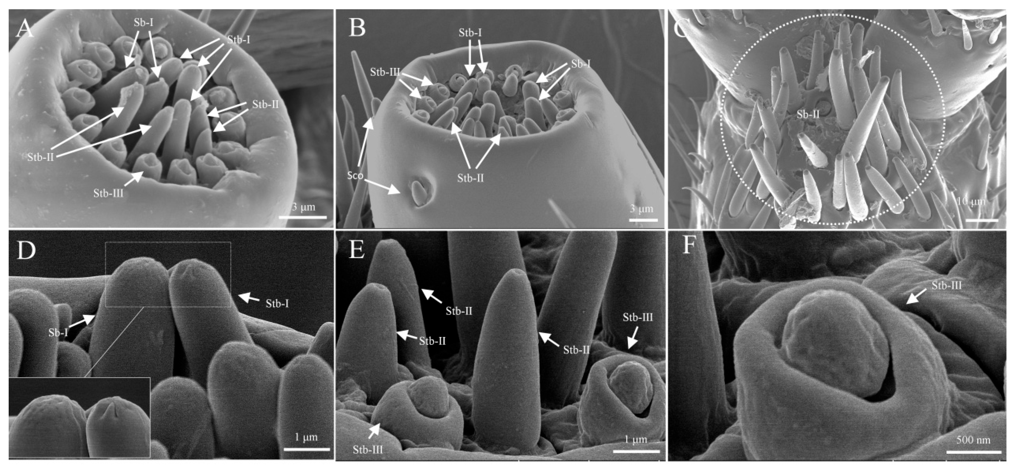
Sensilla twig basiconica are the second-largest sensilla in the distal sensillar field, densely distributed at the tips of the mandibular and labial palps. These sensilla are cone-shaped, with considerable variation in the shape of the conical tip, and can be divided into three subtypes: s. twig basiconica I, s. twig basiconica II, and s. twig basiconica III (Figure 7A,B). The apex of s. twig basiconica I features orderly arranged digitiform projections in the cuticular layer, which converge towards the center. The length of s. twig basiconica I is approximately 2.4 ± 0.2 μm, with a basal width of about 1.2 ± 0.1 μm (Figure 7D). S. twig basiconica II is similar in appearance to s. twig basiconica I, but the distal end has a concave pore resembling a volcanic crater. The length of s. twig basiconica II is approximately 3.1 ± 0.2 μm, with a basal width of about 0.9 ± 0.1 μm (Figure 7E). S. twig basiconica III is the smallest sensillum in the distal sensillar field, featuring a double-layered structure with an outer ring-like arrangement. S. twig basiconica III is located at the edge of the apical tip of the palp and arranged in a ring. The length of s. twig basiconica III is approximately 1.4 ± 0.2 μm, with a basal width of about 1.2 ± 0.1 μm (Figure 7F).

3.2.3. Sensilla Coeloconica (Sco)

Sensilla coeloconica (Sco) are conical protrusions, with multiple sensilla located in a wide, circular groove. The conical structure is small, measuring approximately 1.7 ± 0.2 μm in length and 0.8 ± 0.1 μm in width. These sensilla are found only on the lateral sides at the distal end of the maxillary palps, with a typical count of two to three sensilla (Figure 7B).

3.2.4. Sensilla Trichoidea (ST)

Sensilla trichoidea are hair-like structures, and are classified into two subtypes based on their length: s. trichoidea I and s. trichoidea II. S. trichoidea I has a smooth surface and is upright or slightly curved at the tip, with a length of approximately 87.1 ± 21.2 μm and a base width of around 5.1 ± 1.2 μm. These sensilla are distributed on the outer side of the maxilla and outer maxillary lobe (Figure 6C and Figure 8A). S. trichoidea II is similar in shape to s. trichoidea I but differs significantly in length. S. trichoidea II is much shorter than s. trichoidea I, with a length of approximately 14.2 ± 2.6 μm and a base width of about 2.3 ± 0.7 μm. These sensilla are located on the ventral surface of the inner maxillary lobe and the side of the stem segment (Figure 6C and Figure 8B).

3.2.5. Sensilla Chaetica (Sch)

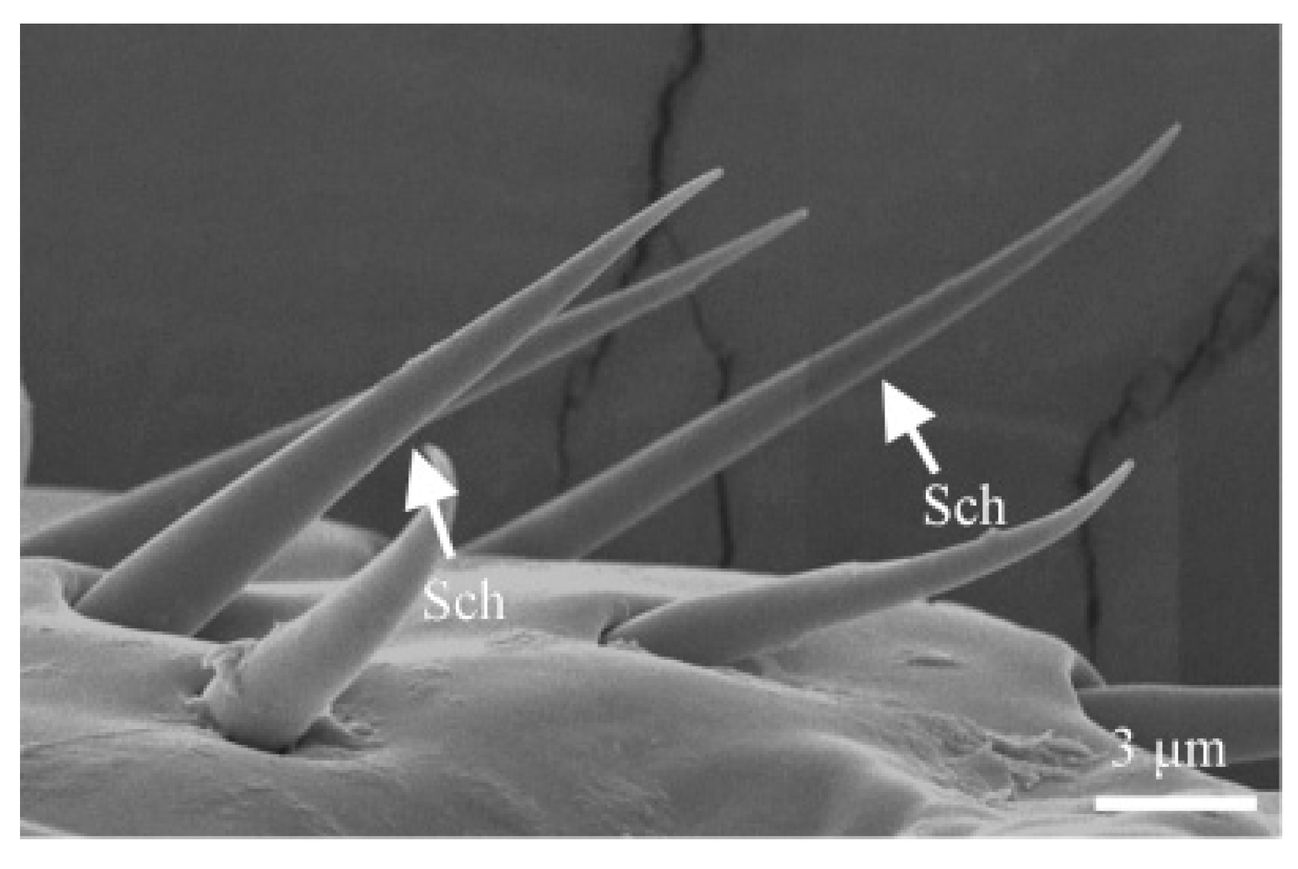
Sensilla chaetica are rigid and spine-like in appearance, with a smooth surface. They are located in a relatively wide, circular groove, measuring approximately 14.2 ± 2.7 μm in length and 1.7 ± 0.4 μm in basal width. These sensilla are distributed on the lateral sides of the maxilla, outer maxillary lobes, and labrum (Figure 6C,F and Figure 9).

3.2.6. Sensilla Zigzag (SZ)

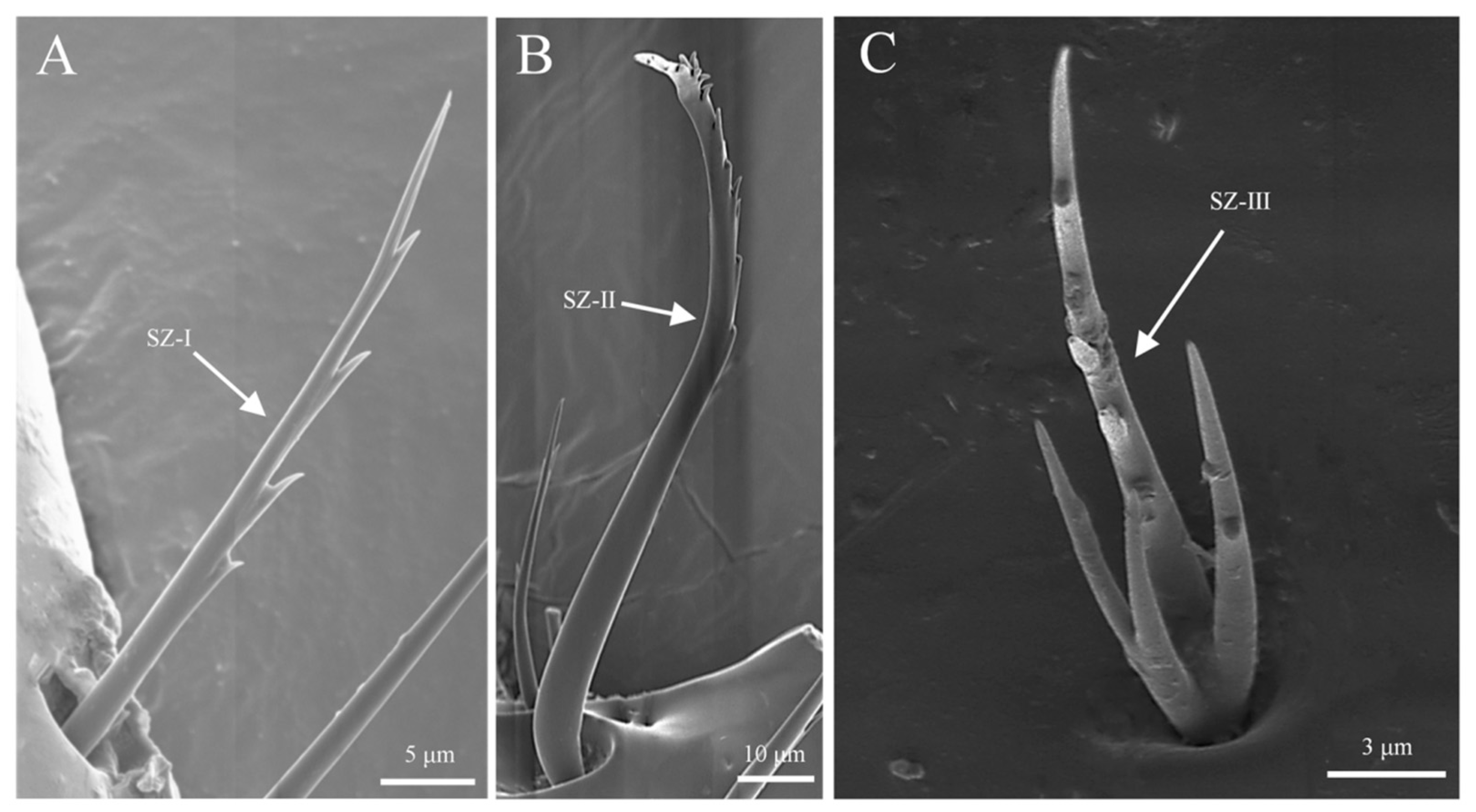
Sensilla zigzag resemble hair and are adorned with serrations. Based on the shape and number of serrations, they are classified into three subtypes: s. zigzag I, s. zigzag II, and s. zigzag III. S. zigzag I is located in a deep, circular groove and begins to develop serrations approximately one-third from the base. Each serration is sharp at the tip, with one serration per segment. The length of this subtype is approximately 34.8 ± 2.1 μm, with a basal width of about 1.9 ± 0.3 μm, and it is distributed on the stem segment, outer maxillary lobes, and labium (Figure 6E and Figure 10A). S. zigzag II starts to form serrations approximately two-thirds from the base and curves into an “S” shape. It is found in a cupped groove, with serrations clustered and rounded at the tips. The length of s. zigzag II is approximately 107.3 ± 19.6 μm, with a basal width of about 5.9 ± 0.7 μm, and it is exclusively distributed on the outer maxillary lobes (Figure 6C and Figure 10B). S. zigzag III differs morphologically from both s. zigzag I and s. zigzag II. It develops three branches from the base of the sensor, with the main shaft standing upright and serrated. This subtype is less abundant in number than s. zigzag I and s. zigzag II, and it is found only on the lateral sides of the labium (Figure 6E and Figure 10C).

3.2.7. Sensilla Digitiformia (Sdi)

Sensilla digitiformia are located exclusively on the lateral surface of the third subsegment of the maxillary palp (Figure 11A), opposite to the distribution of sensilla coeloconica (Figure 11B). Sensilla digitiformia are plate-like or finger-shaped, lying flat within a long elliptical epidermal groove, typically arranged in five parallel structures. Their length is approximately 13.7 ± 0.8 μm, with a width of about 1.3 ± 0.2 μm (Figure 11C).

3.3. Sensilla Types and Morphology on Legs

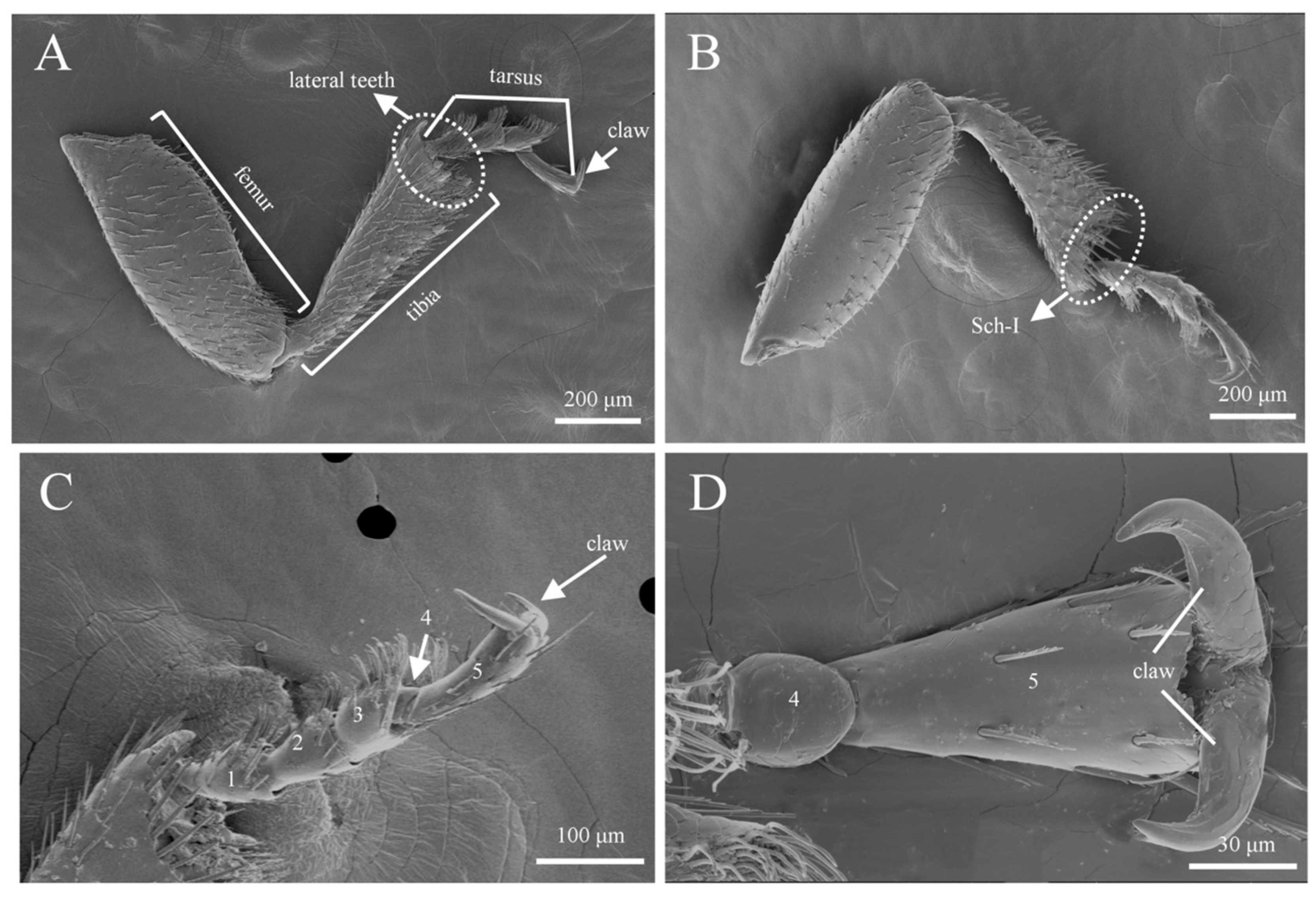
In both male and female adults of Tomicus pilifer, three types of sensilla with six subtypes were observed on the forelegs, midlegs, and hindlegs, including sensilla zigzag I and II (SZ I, II), sensilla trichoidea (ST), and sensilla chaetica I and II (Sch I, II). No significant differences in the types of sensilla were found across the forelegs, midlegs, and hindlegs. Therefore, the forelegs of Tomicus pilifer are used as an example to illustrate the structural features and types of sensilla (Figure 12). The number of each type of sensilla on the forelegs, midlegs, and hindlegs of male and female T. pilifer adults is shown in Table 4.
Each leg of T. pilifer is composed of three segments: the femur, tibia, and tarsus (Figure 12A). The distribution of sensilla on the inner and outer surfaces of the femur shows marked differences, with the outer surface densely covered with sensilla (Figure 12A,B). Additionally, at the junction between the tibia and tarsus, the outer surface exhibits lateral teeth, while the inner surface contains sensilla chaetica (Figure 12B). The tarsus consists of five segments, with segments 1–3 densely populated with sensilla. The fourth segment is small, embedded within the third segment, and lacks any sensilla. The fifth segment is longer, gradually widening from the base to the tip, with a few sensilla present, and ends in a large claw (Figure 12C,D).

3.3.1. Sensilla Zigzag (SZ)

Sensilla zigzag are the most abundant type of sensilla on the legs of Tomicus pilifer adults (Table 4). They are distributed across the femur, tibia, tarsus, and fore-tarsus, with two subtypes: s. zigzag I and s. zigzag II. S. zigzag I is primarily found on the tibia and tarsus, where it is located in a cup-shaped groove. The sensillum is upright and serrated in appearance, with one to eight teeth on one side, while the other side is smooth. The length of s. zigzag I is approximately 42.3 ± 5.6 μm, with a base width of about 2.9 ± 0.3 μm (Figure 13A). S. zigzag II is mainly distributed on the outer surface of the femur, embedded in a scale-like groove. One side near the femur is smooth, while the opposite side bears irregular, interlaced serrations. The number of s. zigzag II is significantly lower than that of s. zigzag I, with a length of approximately 38.7 ± 4.3 μm and a base width of about 3.4 ± 0.7 μm (Figure 13B).

3.3.2. Sensilla Trichoidea (ST)

Sensilla trichoidea are most abundant on the tarsus, concentrated at positions aligned with the direction of the fore-tarsal claw (Figure 12C), with sensilla zigzag sparsely distributed on their dorsal surface. Based on external morphology, they are classified into two types: s. trichoidea I and s. trichoidea II. S. trichoidea I is found on the first and second segments of the tarsus, is hair-like, and has a smooth surface. It gradually tapers from the base to a sharp tip, with a length of approximately 37.6 ± 3.9 μm and a basal width of about 2.1 ± 0.5 μm (Figure 14A,B). S. trichoidea II flattens and widens along its length, with a bundled spiral texture on the surface, resembling a bean sprout. It measures approximately 46.8 ± 5.6 μm in length and 18.2 ± 0.3 μm in basal width, and it is exclusively distributed on the third segment of the tarsus, being less abundant than s. trichoidea I (Figure 14; Table 4).

3.3.3. Sensilla Chaetica (Sch)

Sensilla chaetica are the second-largest type of sensilla on the legs, classified into two subtypes: s. chaetica I and s. chaetica II. S. chaetica I is primarily distributed on the tibia and tarsus, with a smooth surface, growing upright towards the tarsus. It gradually tapers from the base to a sharp tip, measuring approximately 35.4 ± 4.5 μm in length and 3.7 ± 0.6 μm in basal width (Figure 15A). S. chaetica II is the most robust type of sensillum, exclusively located at the junction between the tibia and tarsus. A row of these sensilla encircles the edge of the tibia, with a surface featuring longitudinal striations and a rounded tip. The length of s. chaetica II is approximately 68.5 ± 3.7 μm, with a basal width of about 8.6 ± 1.2 μm (Figure 15B,C).

4. Discussion

The antennae of Tomicus pilifer are club-shaped, consisting of three parts: the scape, funicle, and capitate. No obvious sexual dimorphism is observed in their morphological structure. Additionally, a total of six types of sensilla, including twelve subtypes, were identified on the antennae of T. pilifer. These include sensilla trichoidea I and II, sensilla zigzag I and II, sensilla coeloconica, sensilla chaetica, Böhm bristles, and sensilla basiconica I, II, III, and IV. Compared to previously reported sensilla types on the antennae of Tomicus species [35], we identified three new subtypes of sensilla basiconica (II, III, and IV) on T. pilifer antennae. Notably, sensilla basiconica III were observed only on the male antennae. At segment F2 of the funicle, we found the male antennae to be smooth, without any sensilla, while the same position on the female antennae exhibited sensilla chaetica. These results indicate the presence of sexual dimorphism in the types and distribution of sensilla on T. pilifer antennae. Furthermore, no significant differences in the size and number of each sensillum type were observed between the sexes, suggesting a high degree of functional similarity between the sensilla of males and females. Similar results have been reported in studies of other coleopteran insects, which may be related to specific environmental conditions [11,16,36,37].
Sensilla trichoidea (ST) are primarily concentrated in the sensillar band of the capitate region of the antennae, with few found on the funicle and scape. This distribution pattern is similar to those reported by Wang [35] and Chen [38]. ST I is located at the outermost edge of the sensillar band in the capitate, with a surface featuring vertical striations and a robust structure. This is highly similar to the ST I observed on the antennae of Tomicus yunnanensis and Tomicus piniperda, where they are commonly considered to be a mechanoreceptor and chemoreceptor [39]. Additionally, they may also play a protective role for the sensillar field, including sensilla basiconica and sensilla coeloconica [35,40]. ST II is the longest sensillum on the antennae, mainly distributed at the distal end of the capitate. It has a surface with interlaced vertical striations, resembling the ST observed in Megabruchidius dorsalis [41], Ips typographus [42], and Bruchidius coreanus [43]. This suggests that ST II may play a role in collecting and detecting sex pheromones or aggregation pheromones [37,44]. Sensilla zigzag are the most widely distributed type of sensilla, found on the scape, funicle, and capitate of the antennae. Similar findings have been reported in many Curculionidae species [42,45,46]. It is worth mentioning that due to the significant morphological differences and variety of subtypes observed in sensilla zigzag, we did not classify them as a subtype of sensilla chaetica, as was suggested by Shi [42] and Shewale [45], but instead treated them as a separate sensilla type, as suggested by Chen [46] and Lu [47]. Zacharuk [48] proposed that these unique serrated sensilla often function as both contact chemoreceptors and mechanoreceptors. Sensilla coeloconica have a distinctive flower bud-like shape, with longitudinal grooves on the walls, and have been reported in many Curculionidae species [38,45,49]. Previous studies have shown that Sco in moths play an olfactory role [50], and Hallberg [51] discovered that the Sco on the antennae of Ips typographus have a dual-wall structure, which is similar to that of certain chemoreceptors [52]. Therefore, it is inferred that Sco may function in olfaction as well as thermohygroreception. Sensilla chaetica (Sch) and sensilla zigzag are introduced separately. Therefore, in this study, only one subtype of Sch was observed. Sch was found exclusively at the distal end of the funicle and the capitate, characterized by deep longitudinal striations. Some studies suggest that Sch is the most likely sensillum to function as both a mechanoreceptor and a chemoreceptor [6,53]. Böhm bristles (Bb), located on the scape, are the shortest type of sensilla on the antennae. These sensilla have a smooth surface and are arranged like small spines in clusters around the intersegmental regions of the scape and pedicel. In this study, the distribution and morphological characteristics of Bb were similar to those observed in Curculionidae insects. Wang [35] observed, through transmission electron microscopy, that these sensilla lack wall pores, suggesting they do not possess olfactory functions. Krishnan [54] hypothesized that Bb are mechanoreceptors that sense gravity, primarily functioning to detect mechanical impacts and buffer the effects of gravity. Sensilla basiconica represent the most abundant type of sensillum on the antennae, with the highest number of subtypes. These sensilla are densely distributed in the sensillar band of the intersegmental region of the club. The presence of numerous micropores and branched dendrites suggests that Sb may function as olfactory receptors, likely involved in odor recognition and host location [36,55,56]. Interestingly, we observed the unique Sb III only on male antennae. Our field observations revealed that during the breeding and mating period after overwintering, females are the first to bore entry holes into the host galleries for nutritional supplementation, followed by males, who enter through the female’s entry hole to feed and complete mating (Figure S2). We hypothesize that Sb III may play a role in collecting and detecting the sex pheromones released by females. Further studies are needed to elucidate its specific function.
We also observed two types of sensilla on the mouthparts of Tomicus pilifer that were strikingly similar to those found on the antennae: ST and Sch. These sensilla are widely distributed on the mouthparts and serve a mechanoreceptive function [41,57]. Additionally, we observed sensilla basiconica I and II; sensilla twig basiconica I, II, and III; sensilla coeloconica; and sensilla digitiformia at the distal ends of the maxillary and labial palps. The types, numbers, and distribution patterns of these sensilla are consistent with those previously reported in Tomicus species [58]. This suggests that the types, numbers, and distributions of mouthpart sensilla are highly similar within different species of Tomicus, and similar findings have also been reported in studies of Ips species’ mouthpart sensilla [42,43,51]. In terms of sensillum function, Cui [58] found that the cross-section of Sb exhibits a single-wall structure, with 2–3 dendritic nerves entering the lymph cavity. Therefore, it is inferred that an Sb located on the mouthparts is more likely a gustatory receptor, sensing the chemicals present in food during ingestion. Sensilla twig basiconica are the most abundant sensilla at the distal ends of the maxillary and labial palps. These sensilla may simultaneously function in mechanoreception and gustation [58,59]. Sensilla coeloconica and sensilla digitiformia are both located on the third segment of the maxillary palps, where they are arranged oppositely. Their distribution on the third segment of the labial palps is less frequent. Cui [58] observed branched tubular crystals in the cross-section of Sco and inferred that it may function as a mechanoreceptor [60]. Some studies suggest that Sdi is a poreless sensillum [51,61]. Based on the observation of numerous dendrites innervating the lymph cavity in the cross-section of Sdi in the mouthparts of Tomicus, it is inferred that Sdi may play a role in detecting vibrational signals for intraspecific communication [58,62].
Compared to the antennae and mouthparts of beetles, studies on the ultrastructure of sensilla on the legs are scarce. In Tomicus pilifer, we identified three types of sensilla with five subtypes on the legs, including sensilla zigzag I and II, sensilla trichoidea I and II, and sensilla chaetica. Among these, sensilla zigzag and sensilla chaetica are almost entirely distributed on the femur and tibia. These types of sensilla are also common on the antennae and mouthparts and are often considered to primarily function as mechanoreceptors [32,63]. Sensilla trichoidea (ST) are densely distributed on one side of the tarsus, the side that is in direct contact with the ground when the beetle is crawling. As they come into direct contact with surfaces, sensilla located on the tarsus are commonly believed to be involved in sensing tactile, gustatory, and mechanical stimuli, as well as in gravity [63,64,65].

5. Conclusions

In this study, we investigated the types, numbers, sizes, and distribution patterns of sensilla on the antennae, mouthparts, and legs of Tomicus pilifer. Notably, we identified three new subtypes of sensilla basiconica, sensilla basiconica II, III, and IV, on the antennae of T. pilifer, with sensilla basiconica III observed exclusively on male antennae. At segment F2 of the funicle, we found the male antennae to be smooth, with no sensilla present, while the same location on the female antennae exhibited sensilla chaetica. These findings indicate sexual dimorphism in the types and distribution of sensilla in T. pilifer. Our study further expands the research on the ultrastructure of sensilla in Tomicus beetles and aims to establish a standardized nomenclature for sensilla in this genus in the future. The findings of this study also lay the foundation for future research on the olfactory, feeding, and mating behaviors, as well as the electrophysiological aspects, of this destructive forest pest. Additionally, while speculating on the functions of sensilla in Tomicus pilifer, it was observed that the same type of sensillum may have different functions depending on its location. For example, in this study, the sensilla trichoidea on the antennae and mouthparts primarily function in mechanoreception, whereas those located on the pedicel may have a dual function, serving both mechanoreception and gustation. Therefore, further investigations using techniques such as antenna potential recordings, single-cell recordings, transmission electron microscopy, and molecular biology are necessary to deepen our understanding of the roles and functions of these sensilla in insect behavior.

Supplementary Materials

The following supporting information can be downloaded at https://www.mdpi.com/article/10.3390/insects16090890/s1, Figure S1: Phylogenetic tree of the 28S rDNA gene constructed using the Maximum Likelihood method of Tomicus pilifer. Figure S2: Physiological development of Tomicus pilifer. Table S1. Species and GenBank accession numbers used for DNA sequence analysis in Figure S1.

Author Contributions

Conceptualization, S.Y.; methodology, Q.W., L.W., and Y.L.; data curation, L.W. and Y.L.; writing—original draft, L.W., Q.W., and Y.L.; writing—review and editing, Q.W., L.W., and S.Y.; visualization, Q.W. and L.W.; supervision, S.Y.; funding acquisition, Q.W. All authors have read and agreed to the published version of the manuscript.

Funding

This research was funded by research business expenses of provincial research institutes in Heilongjiang Province (LKSB2022-1), and Key Research and Development projects in Heilongjiang (2023ZX02B05).

Data Availability Statement

We agree to share existing datasets or raw data that have been analyzed in the manuscript, and they will be made available to other researchers following publication. Data availability status: All data are contained within the article and Supplementary Materials.

Conflicts of Interest

The authors declare no conflicts of interest.

References

  1. Lieutier, F.; Langstrom, B.; Faccoli, M. “The Genus Tomicus”, in Bark Beetles: Biology and Ecology of Native and Invasive Species; Vega, F.E., Hofstetter, R.W., Eds.; Elsevier: Amsterdam, The Netherlands, 2015; pp. 371–426. [Google Scholar]
  2. Wang, H.; Liu, C.; Yue, F.; Yan, D.H.; Lu, Q. Identification of ophiostomatalean fungi associated with Tomicus pilifer infesting Pinus koraiensis in Northeastern China. Front. Microbiol. 2022, 2, 919302. [Google Scholar] [CrossRef]
  3. Wang, Y.C.; Zhao, B.; Wang, T.K.; Li, C.; Ma, L.; Li, G. Study on occurring regulation and control method of Tomicus pilifer. Jilin For. Sci. Technol. 2000, 29, 10–11. (In Chinese) [Google Scholar]
  4. Zhang, X.M.; Liu, Y.H.; Zhou, J.Q. Bionomics and control of Tomicus pilifer. Forest Pest. Dis. 2000, 5, 30–31. (In Chinese) [Google Scholar]
  5. Schneider, D. Insect Olfaction: Deciphering System for Chemical Messages. Science 1969, 163, 1031–1037. [Google Scholar] [CrossRef]
  6. Daly, P.J.; Ryan, M.F. Ultrastructure of antennal sensilla of Nebria brevicollis (Fab.) (Coleoptera: Carabidae). Int. J. Insect Morphol. Embryol. 1979, 8, 169–181. [Google Scholar] [CrossRef]
  7. Rebora, M.; Piersanti, S.; Gaino, E. The antennal sensilla of the adult of Libellula depressa (Odonata: Libellulidae). Arthropod Struct. Dev. 2008, 37, 504–510. [Google Scholar] [CrossRef]
  8. McIver, S.B. Structure of cuticle mechanoreceptors of arthropods. Annu. Rev. Entomol. 1975, 20, 381–397. [Google Scholar] [CrossRef]
  9. Chapman, R.F. Mechanoreception. Chemoreception. In The Insects, Structure and Function, 4th ed.; Cambridge University Press: Cambridge, UK, 1998; pp. 610–652. [Google Scholar]
  10. Brożek, J.; Poprawa, I.; Wegierek, P.; Stroiński, A. Functional Morphology and Ultrastructure of the Peripheral Antennal Sensillar System of Graphosoma italicum (Müller, 1766) (Insecta: Hemiptera: Pentatomidae). Insects 2024, 15, 528. [Google Scholar] [CrossRef]
  11. Li, W.B.; Liu, N.Y.; Xu, Q.; Sun, M.; Fang, J.; Wang, S.Y. Ultrastructure structure of antennal sensilla of carabid beetle Carabus elysii Thomson, 1856 (Coleoptera: Carabidae). Entomol. Res. 2019, 49, 81–86. [Google Scholar] [CrossRef]
  12. Gao, Y.; Luo, L.Z.; Hammond, A. Antennal morphology, structure and sensilla distribution in Miroplitis pallidipes (Hymenoptera: Braconidae). Micron 2007, 38, 684–693. [Google Scholar] [CrossRef]
  13. Ren, L.L.; Wu, Y.; Shi, J.; Zhang, L.; Luo, Y.Q. Antenna morphology and sensilla ultrastructure of Tetrigus lewisi Candèze (Coleoptera: Elateridae). Micron 2014, 60, 29–38. [Google Scholar] [CrossRef]
  14. Guo, J.; Du, Z.; Cui, G.; Wang, Z.; Wang, J.; Zhou, X. Ultrastructure Characteristics and Sexual Dimorphism of Antennal Sensilla in Tirathaba rufivena (Lepidoptera: Pyralidae). Insects 2022, 13, 797. [Google Scholar] [CrossRef]
  15. Wang, Q.; Wang, L.; Wang, Q.; Yan, S. Comparative Morphology and Ultrastructure of Antennal Sensilla in Dendrolimus superans (Lepidoptera: Lasiocampidae) and Lymantria dispar (Lepidoptera: Lymantriidae). Insects 2024, 15, 655. [Google Scholar] [CrossRef]
  16. Schneider, D. Insect antennae. Annu. Rev. Entomol. 1964, 9, 103–122. [Google Scholar] [CrossRef]
  17. Keil, T.A.; Steinbrecht, R.A. Mechanosensitive and Olfactory Sensilla of Insects. In Insect Ultrastructure; King, R.C., Akai, H., Eds.; Springer: Berlin/Heidelberg, Germany, 1984; pp. 477–516. [Google Scholar]
  18. Huang, X.; Xu, X.L.; Li, R.X.; Wang, S.; Tian, L.X. Ultrastructure and distribution of antennal sensilla of Bombus terrestris (Hymenoptera: Apidae). Zool. Anz. 2023, 302, 239–247. [Google Scholar] [CrossRef]
  19. Altner, H.; Routil, C.; Loftus, R. The structure of bimodal chemo-, thermo-, and hygroreceptive sensilla on the antenna of Locusta migratoria. Cell Tissue Res. 1981, 215, 289–308. [Google Scholar] [CrossRef]
  20. Zacharuk, R.Y.; Shields, V.D. Sensilla of Immature Insects. Annu. Rev. Entomol. 1991, 36, 331–354. [Google Scholar] [CrossRef]
  21. Keil, T.A. Sensilla on the maxillary palps of Helicoverpa armigera caterpillars: In search of the CO2-receptor. Tissue Cell 1996, 28, 703–717. [Google Scholar] [CrossRef]
  22. Zacharuk, R.Y. Ultrastructure and Function of Insect Chemosensilla. Annu. Rev. Entomol. 1980, 25, 27–47. [Google Scholar] [CrossRef]
  23. Wilson, R.I.; Mainen, Z.F. Early events in olfactory processing. Annu. Rev. Neurosci. 2006, 29, 163–201. [Google Scholar] [CrossRef]
  24. Fleischer, J.; Krieger, J. Insect Pheromone Receptors-Key Elements in Sensing Intraspecific Chemical Signals. Front. Cell Neurosci. 2018, 12, 425. [Google Scholar] [CrossRef]
  25. Visser, J.H. Host odor perception in phytophagous insects. Annu. Rev. Entomol. 1986, 31, 121–144. [Google Scholar] [CrossRef]
  26. Vosshall, L.B. Olfaction in Drosophila. Curr. Opin. Neurobiol. 2000, 10, 498–503. [Google Scholar] [CrossRef]
  27. Guo, M.; Chen, Q.; Liu, Y.; Wang, G.; Han, Z. Chemoreception of mouthparts: Sensilla morphology and discovery of chemosensory genes in proboscis and labial palps of adult Helicoverpa armigera (Lepidoptera: Noctuidae). Front. Physiol. 2018, 9, 970. [Google Scholar] [CrossRef]
  28. Li, Y.; Xiao, D.; Liu, F.F.; Yin, L.; Liu, T.X. Ultrastructure of the sensilla on antennae and mouthparts of larval and adult Plutella xylostella (Lepidoptera: Plutellidae). J. Integr. Agric. 2018, 17, 1409–1420. [Google Scholar] [CrossRef]
  29. Vega, F.E.; Bauchan, G.; Infante, F.; Davis, S. Mouthpart structure and elemental composition of the mandibles in the coffee berry borer (Coleoptera: Curculionidae: Scolytinae). Ann. Entomol. Soc. Am. 2017, 110, 381–389. [Google Scholar] [CrossRef]
  30. Wang, H.; Zheng, H.; Zhang, Y.; Zhang, X. Morphology and distribution of antennal, maxillary palp and labial palp sensilla of the adult bruchid beetles, Callosobruchus chinensis (L.) (Coleoptera: Bruchidae). Entomol. Res. 2018, 48, 466–479. [Google Scholar] [CrossRef]
  31. Lei, G.; Fu, Y.; Wu, W. Fine structure of mouthparts and forelegs of Euplatypus parallelus (Coleoptera: Curculionidae) with emphasis on the behavior of gallery excavation. Micron 2020, 130, 102815. [Google Scholar] [CrossRef]
  32. Nowińska, A.; Franielczyk-Pietyra, B.; Polhemus, D.A. The Leg Sensilla of Insects from Different Habitats—Comparison of Strictly Aquatic and Riparian Bugs (Corixidae, Ochteridae, Gelastocoridae: Nepomorpha: Insecta: Heteroptera). Insects 2023, 14, 441. [Google Scholar] [CrossRef]
  33. Brooks, G.T. Comprehensive insect physiology, biochemistry and pharmacology: Edited by G. A. Kerkut and L. I. Gilbert. Pergamon Press, Oxford. 1985. 13 Volumes. 8200 pp approx. £1700.00/$2750.00. ISBN 0 08 026850 1. Insect Biochem. 1985, 15, i–xiv. [Google Scholar] [CrossRef]
  34. Keil, T.A. Morphology and development of the peripheral olfactory organs. In Insect Olfaction; Hansson, B.S., Ed.; Springer: Berlin/Heidelberg, Germany, 1999; pp. 5–47. [Google Scholar]
  35. Wang, P.Y.; Zhang, Z.; Kong, X.B.; Wang, H.B.; Zhang, S.F.; Gao, X.R.; Yuan, S.R. Antennal morphology and sensilla ultrastructure of three Tomicus species (Coleoptera: Curculionidae, Scolytinae). Microsc. Res. Tech. 2012, 75, 1672–1681. [Google Scholar] [CrossRef]
  36. Merivee, E.; Ploomi, A.; Rahi, M.; Luik, A.; Sammelselg, V. Antennal sensilla of the ground beetle Bembidion lampros Hbst (Coleoptera: Carabidae). Acta Zool. 2000, 81, 339–350. [Google Scholar] [CrossRef]
  37. Ploomi, A.; Merivee, E.; Rahi, M.; Bresciani, J. Antennal sensilla in ground beetles (Coleoptera: Carabidae). Agron. Res. 2003, 1, 221–228. [Google Scholar]
  38. Chen, H.B.; Zhang, Z.; Wang, H.B.; Kong, X.B. Antennal morphology and sensilla ultrastructure of Dendroctonus valens LeConte (Coleoptera: Curculionidae, Seolytinae), an invasive forest pest in China. Micron 2010, 41, 735–741. [Google Scholar] [CrossRef]
  39. Merivee, E.; Rahi, M.; Luik, A. Antennal sensilla of the click beetle, Melanotus villosus (Geoffroy) (Coleoptera: Elateridae). Int. J. Insect Morphol. Embryol. 1998, 28, 41–51. [Google Scholar] [CrossRef]
  40. Dickens, J.C.; Payne, T.L. Structure and function of the sensilla on the antennal club of the southern pine beetle, Dendroctonus frontalis (Zimmerman) (Coleoptera: Scolytidae). Int. J. Insect Morphol. Embryol. 1978, 7, 251–265. [Google Scholar] [CrossRef]
  41. Chen, S.; Li, Y.; Xu, F.; Yang, M.; Wang, X.; Wu, C. Ultrastructure of the Sensilla on the Antennae and Mouthparts of Bean Weevils, Megabruchidius dorsalis (Coleoptera: Bruchinae). Insects 2021, 12, 1112. [Google Scholar] [CrossRef]
  42. Shi, X.; Zhang, S.F.; Liu, F.; Zhang, Z.; Xu, F.Y.; Yin, S.Y.; Kong, X.B. Sensilla on antennae and mouthparts of adult spruce bark beetle Ips typographus (Coleoptera: Curculionidae). Microsc. Res. Tech. 2021, 84, 1484–1497. [Google Scholar] [CrossRef]
  43. Peng, Q.Y.; Xie, M.H.; Pan, X.K.; Li, Y.; Gao, L.; Xu, F.; Wu, C.X.; Yang, M.F. Morphology and distribution of sensilla on antennae and mouthparts of the adult bruchid beetles, Bruchidius coreanus (Coleoptera: Bruchidae). Microsc. Res. Tech. 2024, 87, 922–932. [Google Scholar] [CrossRef]
  44. John, H.B. Antennal Morphology of Ips confuses (Coleoptera: Scolytidae). Ann. Entomol. Soc. Am. 1968, 61, 10–13. [Google Scholar]
  45. Shewale, M.K.; Nebesářová, J.; Grosse-Wilde, E.; Kalinová, B. Microscopic morphology and distribution of the antennal sensilla in the double-spined bark beetle, Ips duplicatus (Coleoptera: Curculionidae). Microsc. Res. Tech. 2023, 86, 1610–1625. [Google Scholar] [CrossRef]
  46. Chen, H.W.; Xie, D.; Jia, N.Y.; Yu, X.H.; Xu, J.J.; Chi, D.F.; Yu, J. Ultrastructure of the antennal sensilla of the red-haired pine bark beetle, Hylurgus ligniperda (Fabricius) (Coleoptera: Curculionidae). Plant Protection. 2023, 49, 236–246. (In Chinese) [Google Scholar]
  47. Lu, S.D.; Zhang, X.M.; Yang, B.; Zhao, N. Antennal Structure and Sensilla of Adult Tomicus yunnanensis Observed by Scanning Electron Microscope. J. Southwest. For. Univ. 2019, 39, 86–91. [Google Scholar]
  48. Zacharuk, R.Y. Antennae and Sensilla. Comparative Insect Physiology, Biochemistry and Pharmacology; Pergamon Press: Oxford, UK, 1985; pp. 1–69. [Google Scholar]
  49. Fernanda, L.; Francisco, A.T.; Arnulfo, A.; Gerardo, Z. Morphology of antennae of Dendroctonus vitei (Coleoptera: Curculionidae: Scolytinae), with special reference to sensilla clustered into pit craters. Can. Entomol. 2018, 150, 471–480. [Google Scholar]
  50. De, B.M.; Baker, T.C. Odor detection in insects: Volatile codes. J. Chem. Ecol. 2008, 34, 882–897. [Google Scholar] [CrossRef]
  51. Hallberg, E. Sensory Organs in Ips typographus (Insecta: Coleoptera) Fine structure of the sensilla of the maxillary and labial palps. Acta Zool. 1982, 63, 191–198. [Google Scholar] [CrossRef]
  52. Altner, H. Insect sensillum specificity and structure: An approach to a new typology. Olfaction Tast. 1977, 6, 295–303. [Google Scholar]
  53. Dai, H.G.; Honda, H. Sensilla on the antennal flagellum of the yellow spotted longicorn beetle, Psacothea hilaris (Pascoe) (Coleoptera: Cerambycidae). Appl. Entomol. Zool. 1990, 25, 273–282. [Google Scholar] [CrossRef]
  54. Krishnan, A.; Prabhakar, S.; Sudarsan, S.; Sane, S.P. The neural mechanisms of antennal positioning in flying moths. J. Exp. Biol. 2012, 215, 3096–3105. [Google Scholar] [CrossRef]
  55. Orden, J.H.; Wood, D.L. The antennal receptors and olfactory response of Ips confusus (Coleoptera: Scolytidae) to male sex attractant in the Laboratory1. Ann. Entomol. Soc. Am. 1966, 59, 253–261. [Google Scholar] [CrossRef]
  56. Liu, G.Q.; Tian, M.Y. Scanning electron microscopic observation of Carabus prodigus antennae and their electroantennographic responses. J. South. China Agric. Univ. 2008, 29, 50–55. [Google Scholar]
  57. Urbanek, A.; Luszczek, D.; Kapusta, M. Antennal and mouthpart sensilla of Acanthoscelides obtectus say (Coleoptera: Bruchidae). Microsc. Res. Technol. 2016, 79, 1230–1235. [Google Scholar] [CrossRef]
  58. Cui, Y.; Zhang, M.; Zhu, H.; Yang, P.; Yang, B.; Li, Z. Fine Structure of the Mouthparts of Three Tomicus Beetles Co-Infecting Pinus yunnanensis in Southwestern China with Some Functional Comments. Insects 2023, 14, 933. [Google Scholar] [CrossRef]
  59. Altner, H.; Prillinger, L. Ultrastructure of invertebrate chemo-, thermo-, and hygroreceptors and its functional significance. Int. Rev. Cytol. 1980, 67, 69–139. [Google Scholar]
  60. Whitehead, A.T. Ultrastructure of sensilla of the female mountain pine beetle, Dendroctonus ponderosae Hopkins (Coleoptera: Scolytidae). Int. J. Insect Morphol. Embryol. 1981, 10, 19–28. [Google Scholar] [CrossRef]
  61. Shi, X.; Shen, J.C.; Zhang, S.F.; Liu, F.; Xu, F.Y.; Wang, G.L.; Zhang, Z.; Kong, X.B. Comparative analysis of the type and number of larval sensilla on the antennae and mouthparts of Ips typographus and Ips subelongatus using SEM. Zool. Anz. 2020, 289, 18–25. [Google Scholar] [CrossRef]
  62. Zacharuk, R.Y.; Albert, P.J.; Bellamy, F.W. Ultrastructure and function of digitiform sensilla on the labial palp of a larval elaterid (Coleoptera). Can. J. Zool. 1977, 55, 569–578. [Google Scholar]
  63. Li, X.M. Phylogenetic Relationship Analysis of Coleoptera Based on Antenna Character. Master Dissertation, Hebei University, Tianjin, China, 2010; pp. 1–75. (In Chinese). [Google Scholar]
  64. Zhao, H.Y.; Gao, Y.; Ma, X.Q.; Yu, W.J.; Sun, Y. Ultrastructure of Tarsal Sensilla of the Asian Longhorned Beetle Anoplophora glabripennis (Motschulsky) (Coleoptera: Cerambycidae). Chin. Agric. Sci. Bull. 2016, 32, 40–43. [Google Scholar]
  65. Zhao, W.W.; Xie, G.L.; Wang, W.K. Ultrastructure of sensilla on the antennae, mouthparts and tarsi of Aromia bungii (Cerambycidae: Cerambycinae). J. Environ. Entomol. 2024, 46, 281–295. [Google Scholar]
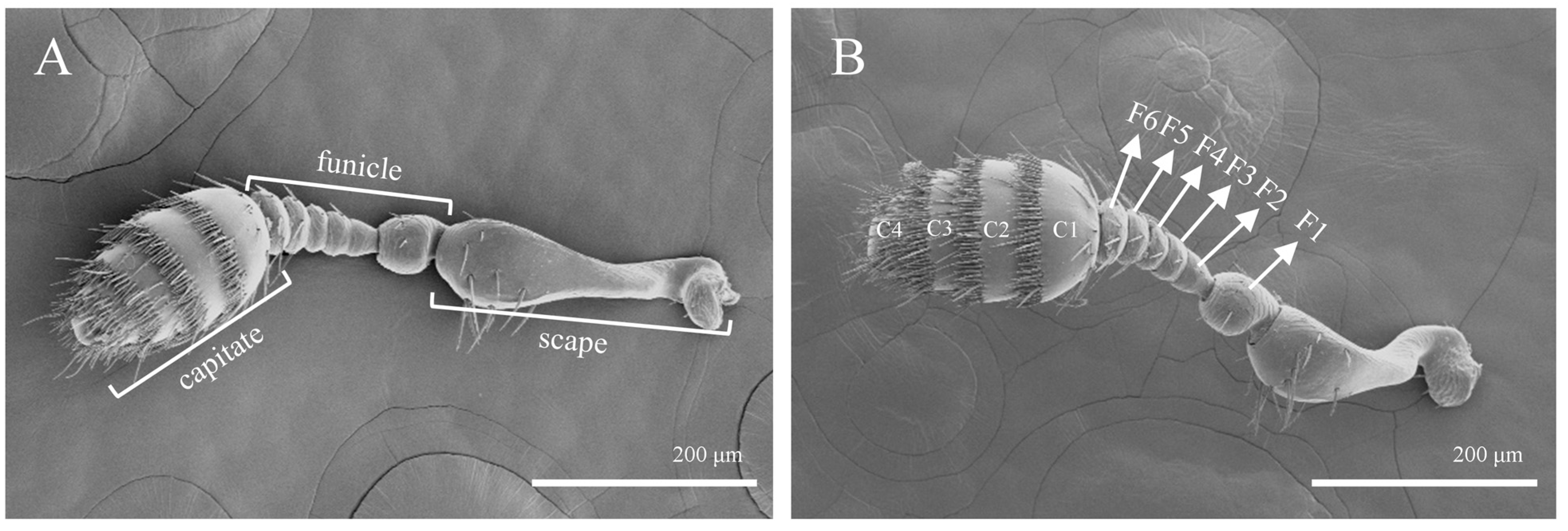
Figure 1. Morphology of T. pilifer antennae. (A) Male; (B) female; F1–F6: the 1st to 6th subsections of the funicle; C1–C4: the 1st to 4th subsections of the capitate.
Insects 16 00890 g001
Figure 2. Morphology of sensilla trichoidea. (A) Sensillum distribution status of the capitate; (B) s. trichoidea I on the antennae; (C) s. trichoidea II on the antennae.
Insects 16 00890 g002
Figure 3. Morphology of sensilla zigzag on the antennae. (A) Distribution of sensilla zigzag on the funicle; (B) s. zigzag I on the antennae; (C) s. zigzag II on the antennae.
Insects 16 00890 g003
Figure 4. The ultrastructure of several sensilla. (A) S. coeloconica on the antennae; (B) s. chaetica on the antennae; (C) Böhm bristles on the antennae.
Insects 16 00890 g004
Figure 5. The ultrastructure of Sensilla basiconica. (A) S. basiconica I on the antennae; (B) s. basiconica II on the antennae; (C) s. basiconica III on the antennae; (D) s. basiconica IV on the antennae.
Insects 16 00890 g005
Figure 6. General morphology and structure of the mouthparts of T. pilifer. (A) The mouthparts of male insects; (B) the mouthparts of female insects; (C) the maxillae in ventral view; (D) the maxillae in dorsal view, 1–3: sections 1 to 3 of the maxillary palp; (E) the labium from external view, 1–3: sections 1 to 3 of the labial palp; (F) the labium from internal view.
Insects 16 00890 g006
Figure 7. Ultrastructure of sensilla types on the distal regions of the maxillary palps, labial palps, and the ventral surface of the labium. (A) Sensory field at the tip of the labial palp; (B) sensory field at the tip of the maxillary palp; (C) s. basiconica II on the ventral surface of the labium; (D) s. basiconica I and s. twig basiconica I at the tips of the maxillary and labial palps; (E) s. twig basiconica II at the tips of the maxillary and labial palps; (F) s. twig basiconica III at the tips of the labial palps.
Insects 16 00890 g007
Figure 8. Morphology of sensilla trichoidea on mouthparts. (A) S. trichoidea I on mouthparts; (B) s. trichoidea II on mouthparts.
Insects 16 00890 g008
Figure 9. S. chaetica on mouthparts.
Insects 16 00890 g009
Figure 10. Morphology of sensilla zigzag on mouthparts. (A) S. zigzag I on mouthparts; (B) s. zigzag II on mouthparts; (C) s. zigzag III on mouthparts.
Insects 16 00890 g010
Figure 11. Morphology of the digitiform sensilla on the mouthparts. (A) Position of sensilla digitiformia on the maxillary palp. (B) Distribution of sensilla digitiformia and sensilla coeloconica on the mouthparts. (C) Sensilla digitiformia on the mouthparts.
Insects 16 00890 g011
Figure 12. General morphology and structure of the leg of T. pilifer. (A) The lateral surface of the forelegs; (B) the inner surface of the forelegs; (C) the structure of the tarsus, 1–5: the 1st to 5th subsegments of the tarsus; (D) the shape of the claws.
Insects 16 00890 g012
Figure 13. Morphology of sensilla zigzag on the leg. (A) S. zigzag I on the leg; (B) s. zigzag II on the leg.
Insects 16 00890 g013
Figure 14. Morphology of sensilla trichoidea on the leg, 1-5: segments 1–5 of the podomere. (A) The distribution of s. trichoidea on the tarsus; (B) s. trichoidea I on the leg. (C) The distribution of s. trichoidea II on the tarsus; (D) s. trichoidea II on the leg; (E) the surface of s. trichoidea II.
Insects 16 00890 g014
Figure 15. Morphology of sensilla chaetica on the foot. (A) S. chaetica I on the leg; (B) s. chaetica II on the leg; (C) the surface of s. chaetica II.
Insects 16 00890 g015
Table 1. Distribution of sensilla on the antennae of male and female Tomicus pilifer adults.
ST IST ⅡSZ I SZ Ⅱ ScoSch BbSb ISb ⅡSb ⅢSb Ⅳ
Scape ----17.3 ± 2.818.1 ± 2.3------14.7 ± 0.814.3 ± 1.1--------
FunicleF1----4.7 ± 0.64.3 ± 0.21.3 ± 0.21.1 ± 0.1--2.3 ± 0.22.7 ± 0.5----------
F2-----------2.3 ± 0.2----------
F3----2.1 ± 0.22.3 ± 0.21.3 ± 0.11.7 ± 0.4--------------
F4----3.7 ± 0.63.9 ± 0.5----------------
F5----1.9 ± 0.31.7 ± 0.4----------------
F6 ---7.8 ± 1.68.1 ± 1.5----------------
CapitateC162.9 ± 11.359.7 ± 9.7--38.7 ± 6.241.2 ± 5.7--8.7 ± 2.19.6 ± 2.4----127.3 ± 13.9121.2 ± 11.633.7 ± 5.936.8 ± 7.4----
C251.5 ± 9.247.8 ± 8.7--18.5 ± 3.116.4 ± 2.7--13.2 ± 2.513.7 ± 2.7----117.6 ± 17.3114.8 ± 15.327.1 ± 3.131.8 ± 3.5----
C347.1 ± 6.846.7 ± 7.23.6 ± 0.73.7 ± 0.4----9.1 ± 1.58.6 ± 1.718.2 ± 2.317.5 ± 2.7--41.2 ± 3.742.7 ± 3.521.2 ± 3.722.7 ± 3.5----
C412.9 ± 2.713.7 ± 3.26.9 ± 1.56.7 ± 1.2------13.5 ± 1.213.8 ± 1.79.5 ± 0.59.2 ± 0.714.7 ± 2.913.1 ± 2.614.7 ± 2.913.1 ± 2.6-28.6 ± 6.18.7 ± 1.29.1 ± 1.4
Note: Data are the mean ± S.E.
Table 2. Length, width, morphological characteristics, and quantity of various types of sensilla in female and male adults of T. pilifer.
Sensillum TypeSexLength (μm)Basal Width (μm)ExineTipShapeTotal
ST Ⅰ15.6 ± 1.71.4 ± 0.3stripesharperect174.4 ± 29.7
15.7 ± 1.51.4 ± 0.2stripesharperect167.9 ± 28.8
ST Ⅱ34.3 ± 3.22.1 ± 0.4stripesharpbend10.5 ± 2.2
34.1 ± 3.12.1 ± 0.4stripesharpbend10.4 ± 1.6
SZ Ⅰ23.1 ± 12.72.7 ± 0.4sawtoothsharperect94.7 ± 15.4
23.2 ± 12.72.6 ± 0.4sawtoothsharperect95.9 ± 13.5
SZ Ⅱ15.3 ± 1.91.3 ± 0.4sawtoothblunterect3.1 ± 0.4
15.3 ± 1.81.5 ± 0.4sawtoothblunterect3.3 ± 0.4
Sco8.6 ± 0.73.7 ± 0.2stripesharperect30.8 ± 6.1
8.8 ± 0.73.5 ± 0.3stripesharperect31.9 ± 6.8
Sch 7.6 ± 0.72.9 ± 0.2stripesharperect36.9 ± 8.7
7.7 ± 0.72.8 ± 0.3stripesharperect36.4 ± 8.2
Bb2.4 ± 0.31.1 ± 0.1smoothsharperect14.7 ± 0.8
2.4 ± 0.21.3 ± 0.1smoothsharperect14.3 ± 0.1
Sb Ⅰ11.2 ± 0.92.1 ± 0.2porebluntbend295.3 ± 17.1
11.7 ± 1.12.1 ± 0.3porebluntbend293.8 ± 16.1
Sb Ⅱ11.6 ± 1.31.8 ± 0.2porebluntbend96.4 ± 19.3
11.3 ± 1.51.9 ± 0.2porebluntbend93.8 ± 16.1
Sb Ⅲ7.7 ± 0.63.1 ± 0.2porebluntbend28.6 ± 6.1
------
Sb Ⅳ5.7 ± 0.61.6 ± 0.2porebluntbend8.7 ± 0.6
5.8 ± 0.81.6 ± 0.3porebluntbend9.1 ± 1.4
Note: Data are the mean ± S.E.
Table 3. The number, size, and distribution of various types of sensilla in the mouthparts of female and male adults of T. pilifer.
SensillaTotalLength (μm)Basal Width (μm)Location
Sb Ⅰ13.2 ± 0.613.7 ± 0.65.2 ± 1.25.1 ± 0.90.8 ± 0.20.8 ± 0.3Mpt, Lpt
Sb Ⅱ42.7 ± 5.943.1 ± 5.728.7 ± 5.328.4 ± 5.25.1 ± 1.25.3 ± 1.3Lig
Stb Ⅰ14.9 ± 1.715.3 ± 1.72.4 ± 0.22.3 ± 0.21.2 ± 0.11.1 ± 0.1Mpt, Lpt
Stb Ⅱ27.4 ± 3.727.1 ± 3.23.1 ± 0.23.2 ± 0.20.9 ± 0.10.9 ± 0.1Mpt, Lpt
Stb Ⅲ58.7 ± 6.758.3 ± 6.31.4 ± 0.21.4 ± 0.31.2 ± 0.1 1.2 ± 0.1Mpt, Lpt
Sco5.4 ± 0.35.2 ± 0.31.7 ± 0.21.7 ± 0.30.8 ± 0.10.8 ± 0.1Mpt, Lpt
ST Ⅰ96.7 ± 11.5101.4 ± 11.787.1 ± 21.286.6 ± 21.35.1 ± 1.25.3 ± 1.3Mx, Lr, Li
ST Ⅱ137.5 ± 12.9135.1 ± 12.214.2 ± 2.6 13.9 ± 3.12.3 ± 0.72.3 ± 0.5Glc
Sch119.7 ± 8.4119.2 ± 8.714.2 ± 2.714.5 ± 2.51.7 ± 0.4 1.7 ± 0.4Md, Mx, Li
SZ Ⅰ51.1 ± 7.248.6 ± 7.834.8 ± 2.134.2 ± 2.61.9 ± 0.31.8 ± 0.4Mx, Li
SZ Ⅱ18.7 ± 1.718.2 ± 1.6107.3 ± 19.6105.9 ± 16.25.9 ± 0.75.7 ± 0.6Mx
SZ Ⅲ13.2 ± 2.311.6 ± 2.77.5 ± 0.77.2 ± 0.72.8 ± 0.42.6 ± 0.5Li
Sdi12.7 ± 0.613.1 ± 0.713.7 ± 0.813.2 ± 0.71.3 ± 0.21.3 ± 0.2Mpl
Note: Data are the mean ± S.E.
Table 4. The number of sensilla of different types on the front, middle, and hind legs of female and male adults of T. pilifer.
SensillaForelegsMidlegsHindlegs
SZ Ⅰ687.9 ± 57.4677.5 ± 53.2685.7 ± 49.6681.2 ± 51.3691.2 ± 53.2682.3 ± 51.7
SZ Ⅱ43.2 ± 7.645.3 ± 7.243.9 ± 6.843.1 ± 6.944.9 ± 7.343.7 ± 7.0
ST Ⅰ132.2 ± 19.6131.7 ± 19.2129.7 ± 21.3131.4 ± 20.7131.9 ± 19.7131.5 ± 19.4
ST Ⅱ87.2 ± 9.788.5 ± 9.287.6 ± 9.287.5 ± 9.586.5 ± 10.187.3 ± 9.8
Sch Ⅰ76.5 ± 11.679.7 ± 10.277.4 ± 10.877.6 ± 11.178.2 ± 10.977.9 ± 10.7
Sch Ⅱ31.4 ± 6.730.8 ± 6.331.7 ± 6.532.4 ± 6.932.7 ± 6.231.8 ± 6.7
Note: Data are the mean ± S.E.
Disclaimer/Publisher’s Note: The statements, opinions and data contained in all publications are solely those of the individual author(s) and contributor(s) and not of MDPI and/or the editor(s). MDPI and/or the editor(s) disclaim responsibility for any injury to people or property resulting from any ideas, methods, instructions or products referred to in the content.