Article

Functional Divergence of Two General Odorant-Binding Proteins to Sex Pheromones and Host Plant Volatiles in Adoxophyes orana (Lepidoptera: Tortricidae)

1
Key Laboratory for Applied Ecology of Loess Plateau (Shaanxi Province), College of Life Sciences, Yan’an University, Yan’an 716000, China
2
Shaanxi Key Laboratory of Research and Utilization of Resource Plants on the Loess Plateau, Yan’an University, Yan’an 716000, China
*
Author to whom correspondence should be addressed.
Insects 2025, 16(9), 880; https://doi.org/10.3390/insects16090880
Submission received: 5 August 2025 / Revised: 16 August 2025 / Accepted: 22 August 2025 / Published: 24 August 2025
(This article belongs to the Section Insect Molecular Biology and Genomics)

Simple Summary

Adoxophyes orana (Lepidoptera: Tortricidae) is a significant leafroller that damages fruit trees in Rosaceae and other plant families. Insects rely on their olfactory system to locate host plants, oviposition sites, and mates. In this study, we assessed tissue and temporal expression profiles of AoraGOBP1 and AoraGOBP2. The binding affinities of recombinant AoraGOBP1 and AoraGOBP2 were determined by fluorescence competition binding assays. Then, key amino acid residues interacting with ligand were identified by molecular docking, molecular dynamics simulations, and per-residue binding free energy decompositions. In general, AoraGOBP2 showed higher binding affinities for sex pheromones, whereas AoraGOBP1 exhibited a wider binding spectrum for host plant volatiles.

Abstract

Adoxophyes orana (Lepidoptera: Tortricidae) is a significant polyphagous leafroller that damages trees and shrubs in Rosaceae and other families. However, the molecular mechanisms by which this pest recognizes sex pheromones and host plant volatiles remain largely unknown. Tissue expression profiles indicated that two general odorant-binding proteins (AoraGOBP1 and AoraGOBP2) were more abundant in the antennae and wings of both sexes, with AoraGOBP1 being rich in the female head and abdomen. Temporal expression profiles showed that AoraGOBP1 was expressed at the highest level in 5 day-nmated adults, while AoraGOBP2 exhibited high expression in 5 day-unmated, 7 day-unmated, and mated female adults. Fluorescence competitive binding assays of heterologous expressed AoraGOBPs demonstrated that AoraGOBP2 strongly bound to the primary sex pheromone Z9-14:Ac, and two minor sex pheromones Z9-14:OH and Z11-14:OH, whereas AoraGOBP1 only showed a high binding affinity to Z9-14:Ac. What is more, AoraGOBP1 exhibited a broader binding spectrum for host plant volatiles than AoraGOBP2. Molecular dockings, molecular dynamic simulations, and per-residue binding free decompositions indicated that the van der Waals interaction was the predominant contributor to the binding free energy. Electrostatic interactions between aldehydes, or alcohols and AoraGOBPs stabilized the conformational structures. Phe12 from AoraGOBP1, and Phe13 from AoraGOBP2 were identified as the most important residues that contributed to bind free energy. Our findings provide a comprehensive insight into the molecular mechanisms of olfactory recognition in A. orana, facilitating the development of chemical ecology-based approaches for the control.

1. Introduction

Insects rely on their sensitive olfactory system to receive semiochemicals from external environment, to locate host plants, identify mates, and keep away from dangers [1,2]. Numerous studies have demonstrated that chemical odorants, including host plant volatiles (HPVs) and pheromones, served as signals in the behavioral processes [3,4,5]. For example, a mixture of (Z)-3-hexenyl acetate, (Z)-3-hexenol, (E)-2-hexenal, benzaldehyde, and benzonitrile has been reported to attract female Grapholita molesta [6,7], while nonanal, a peach-specific aldehyde, also elicits attraction of females of this species [8]. Three isothiocyanates and five green leaf volatiles were found as key olfactory attractants for host location of P. xylostella [9]. Therefore, exploring the olfactory system of insects is indispensable for understanding their behavioral patterns.
A set of proteins constitutes the complex olfactory system, involving odorant-binding proteins (OBPs), chemosensory proteins (CSPs), olfactory receptors (ORs), gustatory receptors (GRs), ionotropic receptors (IRs), and sensory neuron membrane proteins (SNMPs) [10]. When hydrophobic olfactory molecules enter the antenna lymph, they were wrapped and transported by OBPs or CSPs, which subsequently deliver them to ORs and GRs in sensory neurons [1,10]. OBPs in Lepidoptera have been generally classified into pheromone binding proteins (PBPs), general odorant-binding proteins (GOBPs), and antennal-binding proteins (ABPs). PBPs participate in sex pheromone recognition, while GOBPs are traditionally associated with host plant volatile detection [11]. However, in addition to binding host plant volatiles, an increasing number of studies have revealed that GOBPs are also involved in recognizing sex pheromones and insecticides [12,13,14,15,16]. For example, GOBP1 played a key role in semiochemical recognition of Conogethes punctiferalis [12]. AlepGOBP2 in Athetis lepigone showed high binding affinities to two pheromone components, two maize plant volatiles and two insecticides, while AlepGOBP1 bound ocimene and two insecticides [13]. All three GfunGOBPs in G. funebrana strongly bound sex pheromones, and GfunGOBP1 and GfunGOBP3 also exhibited affinity for insecticides [14].
The summer fruit tortrix moth, Adoxophyes orana (Lepidotera: Tortricidae), distributed in Asia and Europe, is a polyphagous leafroller species that damages trees and shrubs in Rosaceae and other families [17,18]. Although trees and shrubs can tolerate limited larvae feeding and leaf rolling, high larvae densities lead to loss of fruit production by larvae chewing. It is imperative to develop eco-friendly approaches to control A. orana, including traps with sex pheromone causing mate-disruption in orchards [19,20,21]. Previous researchers have determined the sex pheromone of A. arona, consisting of (Z)-9-tetradecenyl acetate (Z9-14:Ac), (Z)-11-tetradecenyl acetate (Z11-14:Ac), (Z)-9-tetradecen-1-ol (Z9-14:OH), and (Z)-9-tetradecen-1-ol (Z11-14:OH), with the former two serving as primary components and the latter two as secondary components [22,23,24]. However, limited understanding of olfactory mechanism of A. orana and other leafroller species restricts the development of control Tortricinae species. As far as we know, it seems that there are no reports on the molecular mechanisms of olfactory recognition in Tortricinae species based on the binding affinity of recombinant odorant-binding proteins. Relatively few studies involved in the molecular dynamic simulation compare the homologous modeling and molecular docking when exploring the interactions between proteins and ligands [16]. Herein, we comprehensively determined the expression of GOBPs across different developmental stages and in various adult tissues. Then, we characterized the binding affinities of AoraGOBP1 and AoraGOBP2 to sex pheromones and HPVs. Molecular docking, combined with molecular dynamics simulations, further revealed critical amino acid residues involved in GOBP–ligand interaction, providing new insights into the molecular basis of olfaction in Tortricinae pest species.

2. Materials and Methods

2.1. Insect Rearing

The larvae of Adoxophyes orana tested were originally collected from apple orchards in Ganquan, Shaanxi, China (109°22′10″ E, 36°22′34″ N) in July in 2018 [19]. Larvae were fed on fresh mulberry leaves in an 800 mL polyethylene cuboid box (16 cm × 10 cm × 5 cm). Pupae were collected into polyethylene cups. The emerged adults were fed on 5% sugar solution. The insects were reared in an artificial climate chamber with the following conditions: (24 ± 1) °C, (60 ± 5)%, and L:D = 15 h:9 h. Approximately two days after emergence, the adults could mate.

2.2. RNA Extraction, cDNA Synthesis, and qRT-PCR

Total RNA for GOBPs amplification were extracted from antennae from about approximately 100 pairs of adults 3 days after eclosion following the protocols of AG RNAex reagent (Accurate Biology, Changsha, China). For tissue expression profile, total RNA was extracted from about 100 pairs of antennae, heads or thorax of 10, abdomen from 5, legs from 25, and wings from 20 adults. For temporal expression profile, total RNA was expected from eighty eggs, sixty 1st-instar larvae, thirty 2nd-instar larvae, fifteen 3rd-instar larvae, ten 4th-instar larvae, five 5th-instar larvae, or four pupae on the 2nd and 6th day after pupation. First-strand cDNA was generated following the instructions of HiScript III 1st Strand cDNA Synthesis Kit (+gDNA wiper) (Vazyme, Nanjing, China). The RT-qPCR analyses were conducted on StepOnePlus™ Real-Time PCR System (ABI, Carlsbad, CA, USA) using 2 × Q3 SYBR qPCR Master Mix (TOLOBIO, Shanghai, China) based on the manufacturer’s protocols. Each Rt-qPCR reaction was conducted in a total volume of 20 µL mixture containing 10 µL 2 × Q3 SYBR qPCR Master Mix, 0.4 µL of each primer (10 µM), 1 µL of diluted cDNA, and 8.2 µL of sterile water. The amplification procedure was as follows: 95 °C for 30 s, followed by 40 cycles at 95 °C for 10 s, and 60 °C for 30 s. All developmental stages and tissues were performed with three biological replications and three technical duplicates. β-actin and EF1-α were used as reference genes. The relative expressions of GOBPs were calculated using the 2−ΔΔCt method [25], and normalized using the geometric means of the two reference genes.

2.3. Signal Peptides Prediction, Sequence Alignment and Phylogenetic Analyses

The signal peptides of AoraGOBPs and GOBPs from other moths were predicted using SignalP 6.0 [26]. These sequences after the signal peptides have been removed were aligned using MUSCLE (v. 3.8.1551) [27] and further edited with the WebLogo Online program (https://weblogo.berkeley.edu/logo.cgi) (accessed on 15 February 2025) [28]. Phylogenetic analyses were performed using iqtree2 (v. 2.0.7) [29] and were then visualized with iTOL v6 [30].

2.4. Prokaryotic Expression and Purification of rAoraGOBPs

Specific primers incorporating restriction enzyme sites were designed to amplify the DNA fragments encoding AoraGOBPs without signal peptides. PCR-amplified AoraGOBPs were verified by 1.0% agarose gel and extracted using a Gel Extraction Kit (BioFlux, Hangzhou, China). The purified products were ligated into the 5×TA/Blunt-Zero Cloning Kit (Vazyme, Nanjing, China), and transformed into DH5α competent cells (TOLOBIO, Beijing, China). Positive clones were then screened by PCR. Plasmids containing the AoraGOBPs (pMD™19-T-AoraGOBPs) were extracted using the FastPure Plasmid Mini Kit (Vazyme, Nanjing, China). After verification, pMD™19-T-AoraGOBPs plasmids were subjected to double-digestion with restriction endonucleases HandIII and BamHI for 4 h at 37 °C. Recombinant AoraGOBPs fragments were ligated into the expression vector pET28a (+) using T4 DNA ligase (ThermoFisher Scientific, Vilnius, Lithuania) at 4 °C overnight.
The pET28a(+)-GmolGOBPs plasmids were extracted and transferred into BL21 competent cells (TransGen Biotech., Beijing, China). The cells were cultured in LB liquid medium with kanamycins (50 μg/mL) for 15 h under oscillatory conditions. When the culture OD600 reached 0.6 to 0.8, the cells were induced to expression by adding isopropyl-β-D-thiogalactoside (IPTG) overnight at 28 °C and 220 rpm to a final concentration of 0.5 mM. The rAoraGOBPs were validated using SDS-PAGE and identified as inclusion bodies. The bacterial cells were harvested via centrifugation at 4 °C (8000 rpm, 5 min). After proteins were purified by a standard Ni-NTA agarose magnetic beads (7sea biotech, Shanghai, China) to obtain a soluble protein of about 17 kDa in size, the proteins were recovered in 20 mM Tris-HCl (pH 7.4) by dialysis and used for in vitro fluorescence competitive binding assays. The purity and concentration of rAoraGOBPs was verified by SDS-PAGE and BCA protein assay kit (Vazyme, Nanjing, China), respectively.

2.5. Fluorescence Competitive Binding Assay

The fluorescent competitive binding assays were conducted using an F-2700 spectrophotofluorometer (Hitachi, Tokyo, Japan) to evaluate the binding affinities of AoraGOBP1 and AoraGOBP2 to sex pheromones, and HPVs. At the beginning, the fluorescence probe 1 − NPN and the tested ligands were dissolved in chromatographic methanol to create 100 mM stock solution, which were subsequently diluted to 1 mM for assays. rAoraGOBP were diluted to 2 μM in 20 mM Tris-HCl buffer (pH 7.4), and 2 mL of this solution was titrated with 1 − NPN to a final concentration of 18 μM, and the binding affinity was determined by measuring fluorescence intensity (excitation at 337 nm, emission scanned from 370 to 550 nm). With the increase of 1 − NPN concentration, the change in fluorescence intensity decreased and eventually stabilized, and the data were recorded. Binding dissociation constant K1−NPN (Kd) of 1 − NPN to rAoraGOBPs were determined. Then, 2 mL protein solution containing 2 μM rAoraGOBPs was titrated with 1 μM of each ligand to a final concentration of 18 µM. The fluorescence intensity was measured and recorded after a 2 min reaction. The Ki for competitive binding of each ligand to rAoraGOBPs using the formula Ki = [IC50]/(1 + [1 − NPN]/K1−NPN), where IC50 is the ligand concentration corresponding to 50% displacement of 1 − NPN. Each assay was replicated three times biologically. The Kd and Ki are presented as mean ± S.E.M.

2.6. Homology Modeling, Molecular Docking, and Molecular Dynamic Simulation

The 3D structures of AoraGOBP1 and AoraGOBP2 were constructed on ColabFold server (MIT, Cambridge, USA) [31,32]. The 3D structures of the ligands were downloaded from PubChem [33]. Molecular docking was applied on the CB-Dock 2 server with the default settings [34]. The interactions of AoraGOBP–ligand complexes were visualized by PyMOL-open-source v2.5.0 [35] and Ligplot+ (v. 2.2.8) [36].
The MD simulations of AoraGOBP–ligand complexes were carried out for 200 nanoseconds (ns) using GROMACS (v. 2024.4) [37] with the AMBER99SB protein forcefield [38]. The simulation started with solvating the complex to neutralize the system. Then, energy minimization was achieved using the steepest descent algorithm with a step size of 0.01 nm. System equilibration was performed under NVT and NPT ensembles. After that, a 200 ns production molecular dynamic was performed. Analyses of root-mean-square deviation (RMSD) and root-mean-square fluctuation (RMSF) were conducted using GROMACS (v. 2024.4) built-in tools [37], and visualized using ggplot2 package (v. 3.4.3) [39] in R (v. 4.3.2). Finally, the binding energies for protein and ligands were calculated using the gmx-MMPBSA tool (v. 1.5.7) [40].

2.7. Statistic Analysis

Statistical analyses for the results of RT-qPCR and fluorescence competitive binding assays were conducted using Graphpad Prism 6.0. Every data point is presented as the mean ± S.E.M with three replicates. Normality in each group and homogeneity of variance were tested before perimetric analyses. One-way ANOVA with Tukey’s HSD comparison was employed to assess differences among multiple samples, with significance set at p < 0.05. Student’s t-test was used to compare differences between two sexes (**: p < 0.01; *: p < 0.05; ns: p ≥ 0.05).

3. Results

3.1. Sequence Analysis of AoraGOBP1 and AoraGOBP2

The ORF of AoraGOBP1 and AoraGOBP2 cover 492 and 486 bp, encoding 163 and 161 amino acid residues, respectively. The signal peptides of AoraGOBP1 and AoraGOBP2 were composed of 19 and 20 amino acids, respectively (Figure S1). The mature AoraGOBP1 has a predicted molecular weight (MW) of 16.82 kDa and an isoelectric point (pI) of 5.10. The mature AoraGOBP2 has a MW of 16.17 kDa and a pI of 5.10. Both AoraGOBPs have six conserved cysteines (Figure 1). The 3D structures of AoraGOBP1 and AoraGOBP2 contain seven α-helices: Val2-Ser23 (α1), Glu27-His35 (α2), Arg46-Phe59 (α3), His70-Ser79 (α4), Gly83-His101 (α5), His107-Arg125 (α6), and Met101-Glu143 (α7) for AoraGOBP1 (Figure 2A), and Ala2-Ser23 (α1), Pro27-His35 (α2), Arg46-Phe59 (α3), His70-Ser79 (α4), Gly83-Tyr100 (α5), Asp107-Ala125 (α6), and Val131-Lys140 (α7) for AoraGOBP2 (Figure 2B). Phylogenic analysis indicated GOBP1 and GOBP2 were distributed in different branches (Figure S2).

3.2. Spatial–Temporal Patterns of OBPs in A. orona

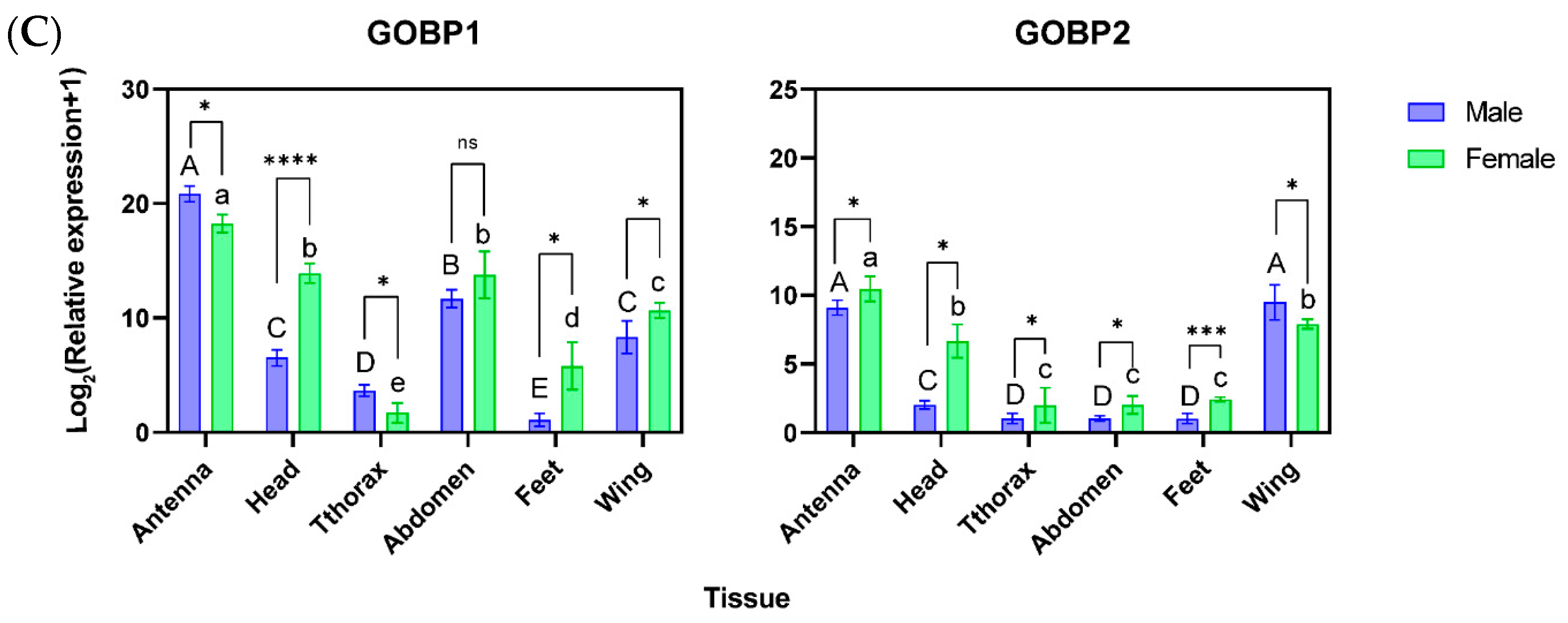
For immature stages, expression of AoraGOBP1 was significantly higher in first-instar larvae than in the other developmental stages, while AoraGOBP2 was abundantly expressed in eggs, and poorly expressed in first-instar larvae and 2-day-old pupae (Figure 2A). The expression of AoraGOBP1 in adults was significantly elevated in 5 day- and 7 day-unmated males after eclosion compared to 1 day-unmated, 3 day-unmated, and mated males, while its expression of females kept relatively stable. The expression of AoraGOBP2 was higher on 5 day-unmated, 7 day-unmated, and mated adults for both sexes than in 1 day-unmated and 3 day-unmated adults (Figure 2B). Regarding tissue expression patterns in adults, both AoraGOBPs exhibited maximal expression in antennae, although AoraGOBP2 was also abundantly expressed in wings. What is more, AoraGOBP1 showed rich expression in female heads and abdomens in both sexes (Figure 2C).

3.3. Expression and Purification of rAoraGOBPs

rAoraGOBP1 and rAoraGOBP2 were successfully expressed in BL21 after IPTG induction. These two proteins were mainly expressed in inclusion bodies (Figure 3). SDS-PAGE results showed that each of the purified rAoraGOBPs migrated as a single band with an MW of approximately 17 kDa (Figure 3).

3.4. Ligand Binding of rAoraGOBPs

Fluorescent binding assays showed that Kd for rAoraGOBP1 of 18.36 μM (95% CI: 15.59~21.86 μM), and 1.65 μM (95% CI: 1.49~1.88 μM) for rAoraGOBP2, suggesting 1 − NPN as a suitable fluorescent probe. Then, we accessed the binding affinities of both rAoraGOBPs towards 55 chemical ligands, including five sex pheromone components and 51 HPVs. rAoraGOBP2 showed strong binding affinity for Z9-14:OH (Ki = 1.49 ± 0.01 μM) and Z11-14:OH (Ki = 1.21 ± 0.02 μM), along with moderate binding affinity to Z9-14:Ac, whereas rAoraGOBP1 showed no binding affinity to sex pheromone (Table 1, Figure S3A). Regarding host plant volatiles, rAoraGOBP1 displayed strong binding affinities to decanal (Ki = 0.78 ± 0.15 μM) and methyl salicylate (Ki = 4.41 ± 0.20 μM), moderate binding affinities to pear ester (Ki = 5.08 ± 0.13 μM) and Z-3-hexen-1-yl 3-methylbutanoate (Ki = 6.21 ± 0.18 μM), and weak binding affinities to five other volatile host plants. rAoraGOBP2 strongly bound farnesol (Ki = 1.13 ± 0.02 μM) and 12:OH (Ki = 1.74 ± 0.03 μM), with moderate affinity to 1,2-Benzenedicarboxylic acid dibutyl ester (Ki = 5.33 ± 0.04 μM) and farnesene (Ki = 6.96 ± 0.32 μM) (Table 1, Figure S3B).

3.5. Protein Structure Modeling and Molecular Docking

The homologous 3D model of AoraGOBP1 and AoraGOBP2 showed that both AoraGOBPs shared seven α-helicals (α1–α7) (Figure 4). Ramachandran plots verified the stereochemical validity of both protein models (Figure S4). Three disulfide bridges in each AoraGOBP stabilized the 3D structures (Figure 4).
Based on the results of fluorescence competitive binding assays, we used molecular docking to investigate the interactions of GOBP–ligand complexes. The docking analyses indicated that several nonpolar amino acid residues formed a hydrophobic cavity, ensuring the binding of GOBP to hydrophobic ligand molecules (Figure 5 and Figure S5). In addition, specific residues (such as Thr, Ser, and Val) in the complexes formed hydrogen bonds with ligands; Thr9 in AoraGOBP1 interacted with decanal, methyl salicylate, and dibutyl phthalate. In AoraGOBP2, Val112 formed hydrogen bonds with Z9-14:OH and Z11-14:OH, respectively, and Ser 57 with farnesol and dibutyl phthalate.

3.6. Per-Residue Free Energy Decomposition

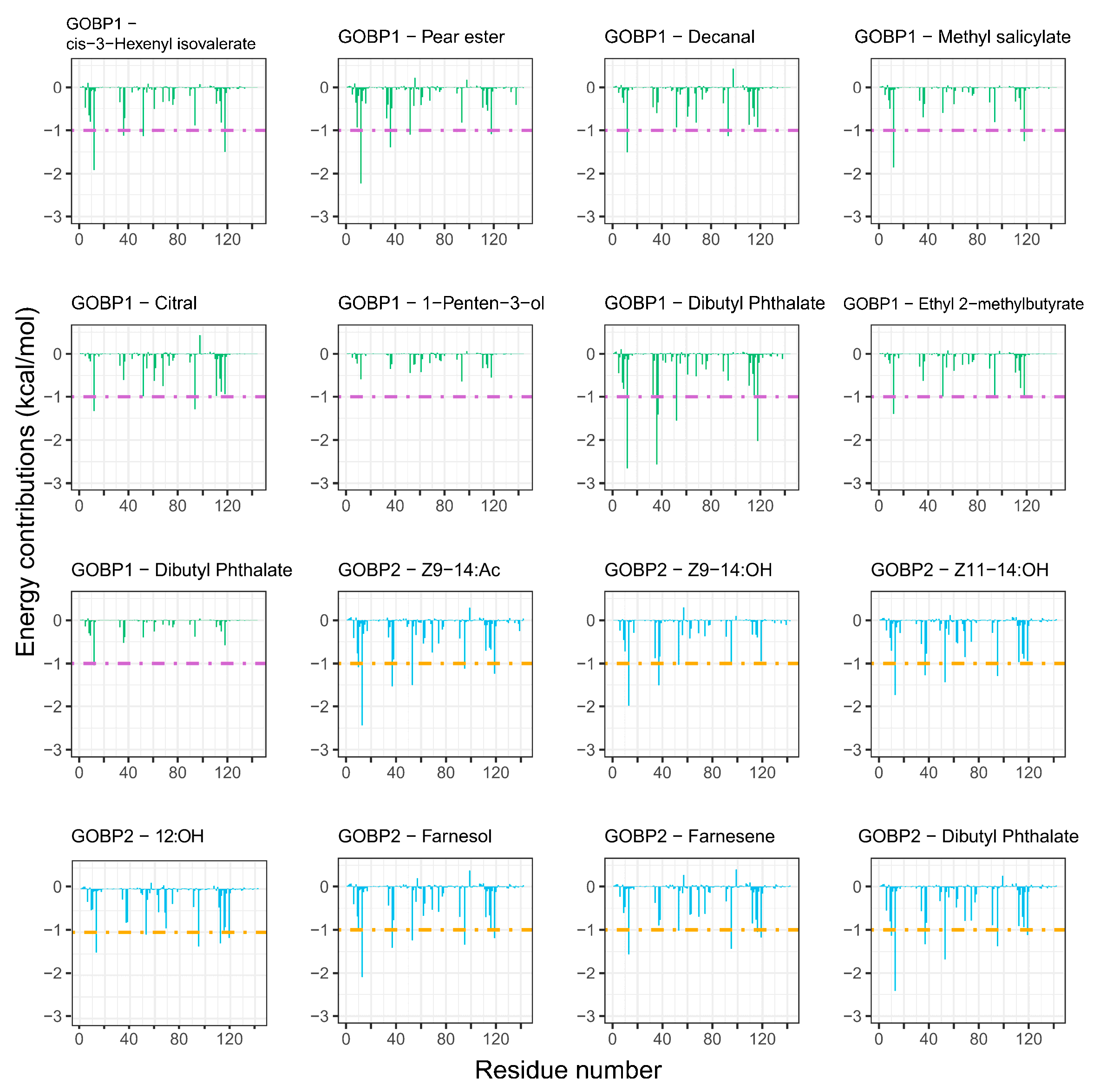
Per-residue free energy decomposition analyses (Table 2) showed that Phe12, Phe36, Ile52, Ile94, and Phe118 contributed the most to the binding free energy in the bindings of AoraGOBP1 to different ligands (Figure 6. Similarly, Phe13, Phe37, Ile53, Val112, and Phe119 played a critical role in the binding of AoraGOBP2 to sex pheromones and HPV ligands (Figure 6. These hydrophobic amino acid residues exhibited relatively low RMSF values (Figure S7), confirming the stability of GOBP–ligand interactions.

4. Discussion

The tissue expression patterns AoraGOBPs suggested that AoraGOBP1 and AoraGOBP2 were richly expressed in antennae and moderately expressed in wings. In addition, AoraGOBP1 was also expressed in the head and abdomen. GOBP genes were specifically expressed in several lepidopteran species, such as Spodoptera frugiperda [15], Orthaga achatina [41], and Glyphodes pyloalis [42]. EoblGOBP2 was highly expressed in the female abdomen, legs, and wings of Ectropis obliqua, except the antennae [43]. Temporal expression profiles showed that AoraGOBP1 was expressed in first-instar larvae, and AoraGOBP2 in the eggs of second- to fifth-instar larvae. Expression of SfruGOBPs also showed expression difference in S. frugiperda, where SfruGOBP1 was detected after pupation, and SfruGOBP2 was detected after egg hatching [44]. SlitGOBP2 was highly expressed in first-instar larvae and adults of S. litura [45].
OBPs were traditionally classified into three subfamilies: PBPs, GOBPs, and ABPs. PBPs were well-known for their abilities to bind sex pheromones, and GOBPs were usually considered to bind host plant volatiles. However, with the wide application of genomic sequences, it has been observed that GOBPs and PBPs in Lepidoptera were closely located in the same chromosome [15,46], suggesting the role of GOBPs in recognizing sex pheromones. Fluorescent competing binding assays showed that AoraGOBP1 and AoraGOBP2 exhibited functional differentiation. AoraGOBP2 strongly bound two secondary sex pheromone components, Z9-14:OH and Z11-14:OH, and weakly bound Z9-14:Ac, while AoraGOBP1 did not. AoraGOBP1 exhibited a broader binding spectrum for host plant volatiles than AoraGOBP2, including one alcohol, three aldehyde, and five esters (Table 1). The functional divergence of GOBPs has been observed in other lepidopteran species. For instance, AlepGOBP2 in A. lepigone showed high binding affinity to Z7-12:Ac and Z9-14:Ac, while AlepGOBP1 did not bind to the sex pheromone [13]. Similarly, SfruGOBP2 in S. frugiperda exhibited strong binding affinities to Z7-12:Ac, Z9-12:Ac, Z9-14:Ac and Z9,E12-14:Ac, while SfruGOBP1 did not bind to the sex pheromone [15]. AipsGOBP2 in A. ipsilon had strong binding affinities to Z7-12:Ac and Z9-14:Ac, whereas AipsGOBP1 showed moderate binding to five sex pheromones [47]. SexiGOBP2 showed strong binding to five sex pheromone components, and SexiGOBP1 only showed weak binding to alcohol sex pheromone compounds [48]. GmolGOBP1 in G. molesta exhibited broad binding affinities for both HPVs and primary sex pheromone, while GmolGOBP2 demonstrated specific binding affinity for 12:OH, the minor sex pheromone, and a narrow spectrum of HPVs [49]. It is interesting that AoraGOBP2 had a high affinity to 12:OH. We speculated that maybe A. orana relies on AoraGOBP2 to recognize signals from G. molesta in the same orchard to avoid niche competition. This speculation needs to be tested using behavior experiments both in the laboratory and in the field. Taken together, the primary function of AoraGOBP1 was the binding of host plant volatiles, whereas AoraGOBP2 was more involved in recognizing sex pheromones.
MD simulations combined with per-residue energy decompositions revealed that the binding energy of Z9-14:Ac interacting with the following residues surpassed −1.00 kcal/mol: Phe12, Phe36, Trp37, Ile52, and Phe118 in AoraGOBP1, and Thr10, Phe13, Phe37, Leu54, Ile95, and Phe119 in AoraGOBP2 (Figure 6). In addition, Z9-14:Ac interacted with the benzene ring of Phe36 and Phe118 in AoraGOBP1, and of Phe13 from AoraGOBP2 by π-σ interactions (Figure 5A,I). Site-directed mutagenesis of SfruGOBP2 from S. frugiperda demonstrated that mutations of Thr9, Phe12, Phe33, Phe36, or Phe118 reduced or even abolished their ability to bind Z9-12:Ac, Z7-14:Ac, Z9-14:Ac, and Z9,E12-14:Ac [15]. AoraGOBP2 showed strong binding affinities to Z9-14:OH and Z11-14:OH. Although the total binding energies of AoraGOBP2–Z9-14:OH and AoraGOBP2–Z11-14:OH were higher than those of AoraGOBP2–Z9-14:Ac, hydrogen bonds were formed between the oxygen atom in the sidechain of Val112 and each of the hydroxyl group of Z9–14:OH and Z11–14:OH (Figure 5K,L). The binding energies of Phe13, Phe37, and Ile53 in these two complexes were below −1.00 kcal/mol. In M. separata, the sex pheromone Z11-16:Ald was stabilized by seven hydrophobic residues, each contributing a free energy below −0.95 kcal/mol, while a hydrogen bond was formed between Ser56 and Z11-16:Ald [16]. Therefore, similar patterns were observed in lepidopteran GOBPs that highly hydrophobic residues, especially Phe, created a binding pocket to stabilize the ligands. When sex pheromone molecules contain hydroxyl or aldehyde carbonyl groups, hydrogen bonds enhance the stability of the protein–ligand complex.
Interestingly, AoraGOBP2 strongly bound Z9-14:OH and Z11-14:OH, while AoraGOBP1 did not. This could be contributed to the differences in the 3D structures. Distance between each of the 9 amino acid residues from AoraGOBP1 and Z9-14:OH (Met5, Thr9, Phe12, Phe36, Trp37, Ile52, Phe76, Ala115, and Phe118) were within 4 Å, whereas distance between the 11 amino acid residues from AoraGOBP2 and Z9-14:OH (Thr10, Phe13, Phe34, Phe36, Trp37, Ile53, Ile95, Val112, Val115, Ala116, and Phe119) were within 4 Å. As for residues within 4 Å away from Z11-14:OH, 10 residues (Val8, Thr9, Phe12, Gly13, Phe33, Phe36, Trp37, Ile52, Ala115, Phe118) were found in AoraGOBP1 and 11 in AoraGOBP2 (Thr10, Phe13, Phe34, Phe36, Trp37, Ile53, Met 91, Val112, Val115, Ala118, Phe119). In addition, α7 of AoraGOBP1 seems far away from the other six helices, while this phenomenon does not exist in AoraGOBP2 (Figure 4). Most of the residues above belong to hydrophobic amino acids, which formed hydrophobic pockets. Therefore, hydrophobic pocket in AoraGOBP2 could tightly wrap the minor sex pheromone components more than the pocket in AoraGOBP1.
AoraGOBP2 showed strong binding affinities to farnesene and farnesol (Table 1), two terpenoid biosynthesized from mevalonate. Previous studies have demonstrated that farnesene stimulated oviposition of Ostrinia nubilalis, while farnesol preceded the oviposition [50]. Larvae of Plutella xylostella were strongly attracted by farnesol [51]. α-farnesene, a compound from apple skin, has been shown to be an attractant of larvae of Cydia pomonella. Female adults of C. pomonella were attracted to low concentrations of α-farnesene and repelled by high concentrations [52], whereas males showed no significant behavioral response over a wide range of doses [53]. Molecular dockings, MD simulations, and per-residue free energy decompositions revealed that Phe13, Ile53, Ile95, Ala116, and Phe119 in AoraGOBP2 strongly interacted with farnesene via van der Waals forces (Figure 5 and Figure 6). A similar interaction pattern was observed for farnesol, with the highest energy contributions coming from Phe13, Phe37, Ile53, Ile95, and Phe119 in AoraGOBP2 (Figure 6). Ser57 from AoraGOBP2 formed strong hydrogen bonds with farnesol, with a bond distance of 2.8 Å, respectively (Figure 5). Therefore, hydrophobic interactions primarily stabilize the AoraGOBP2–farnesol complex, while hydrogen bonds enhance the conformational stabilities.

5. Conclusions

AoraGOBP1 displayed high binding affinities for multiple host plant volatiles, including Decanal, Methyl salicylate, Pear ester, and (Z)-3-hexen-1-yl 3-methylbutanoate. In contrast, AoraGOBP2 strongly bound to sex pheromone components, Z9-14:OH, Z11-14OH, and Z9-14:Ac, as well as some host plant components, including farnesol, farnesene, and Dibutyl phthalate. van der Waals forces between hydrophobic residues and ligands contributed the majority of binding free energy in these protein–ligand complexes. Although for the limitations of this study, as a lack of behavior assays for insects whose AoraGOBP genes were interfered with dsRNA, these findings provide a comprehensive understanding of the molecular mechanisms underlying olfactory recognition in A. arona.

Supplementary Materials

The following supporting information can be downloaded at: https://www.mdpi.com/article/10.3390/insects16090880/s1, Figure S1: Amino acid sequences of AoraGOBP1 and AoraGOBP2. Signal peptides are signed with underlines; Figure S2: Phylogenetic relationship analysis of GOBPs of A. arona and other lepidopteron species; Figure S3: Binding of ligands to AoraGOBPs; Figure S4: Ramachandran plots of AoraGOBP1 and AoraGOBP2 models; Figure S5: 2D plots of molecular docking for the binding of AoraGOBP1 and AoraGOBP2 to plant volatiles and sex pheromones; Figure S6: root-mean-square deviation (RMSD) curves of AoraGOBP–ligand complexes; Figure S7: The root-mean-square fluctuation (RMSF) curves of AoraGOBP–ligand complexes; Table S1: Primers used for gene cloning, RT-qPCR and prokaryotic expression; Table S2: NCBI accession numbers of amino acid sequences for sequence alignment and phylogenetic analyses.

Author Contributions

Conceptualization, G.L. and B.L.; methodology, S.R. and Y.L.; software, Y.L. and X.C.; validation, G.L. and B.L.; formal analysis, S.R. and J.Z.; investigation, G.L.; data curation, K.L. and B.L.; writing—original draft preparation, S.R. and Y.L.; writing—review and editing, K.L., G.L. and B.L.; visualization, X.C. and K.L.; supervision, B.L.; project administration, G.L. and B.L.; funding acquisition, J.Z., G.L., and B.L. All authors have read and agreed to the published version of the manuscript.

Funding

This research was funded by The Youth Innovation Team of Shaanxi Universities (grant number 24JP204), Shaanxi Province “two chains” integration key project (grant number 2023LLRH-01), Research Fund for the Doctoral Start-up Foundation of Yan’an University (grant number YDBK2019-47), and Research Fund of Yan’an Universe (grant number 2023CGZH-016).

Data Availability Statement

Data inquiries can be directed to the corresponding author.

Acknowledgments

We thank Yamei Meng for insect rearing.

Conflicts of Interest

The authors declare no conflicts of interest.

Abbreviations

The following abbreviations are used in this manuscript:
OBPOdorant-binding protein
CSPChemosensory protein
OROdorant receptor
IRIonotropic receptor
GRGustory receptor
SNMPSensory neuron membrane proteins
RT-qPCRReal-time quantitative polymeric chain reaction
One-way ANOVAOne-way analysis of variance
SDS-PAGESodium dodecyl sulfate polyacrylamide gel electrophoresis
MD simulationMolecular dynamics simulation

References

  1. Pelosi, P.; Iovinella, I.; Zhu, J.; Wang, G.; Dani, F.R. Beyond Chemoreception: Diverse Tasks of Soluble Olfactory Proteins in Insects. Biol. Rev. 2018, 93, 184–200. [Google Scholar] [CrossRef] [PubMed]
  2. Xu, H.; Turlings, T.C.J. Plant Volatiles as Mate-Finding Cues for Insects. Trends Plant Sci. 2018, 23, 100–111. [Google Scholar] [CrossRef]
  3. Gadenne, C.; Barrozo, R.B.; Anton, S. Plasticity in Insect Olfaction: To Smell or Not to Smell? Annu. Rev. Entomol. 2016, 61, 317–333. [Google Scholar] [CrossRef]
  4. Anton, S.; Cortesero, A.-M. Plasticity in Chemical Host Plant Recognition in Herbivorous Insects and Its Implication for Pest Control. Biology 2022, 11, 1842. [Google Scholar] [CrossRef]
  5. Zhou, S.; Jander, G. Molecular Ecology of Plant Volatiles in Interactions with Insect Herbivores. J. Exp. Bot. 2022, 73, 449–462. [Google Scholar] [CrossRef]
  6. Piñero, J.C.; Dorn, S. Synergism between Aromatic Compounds and Green Leaf Volatiles Derived from the Host Plant Underlies Female Attraction in the Oriental Fruit Moth. Entomol. Exp. Appl. 2007, 125, 185–194. [Google Scholar] [CrossRef]
  7. Varela, N.; Avilla, J.; Anton, S.; Gemeno, C. Synergism of Pheromone and Host-plant Volatile Blends in the Attraction of Grapholita Molesta Males. Entomol. Exp. Appl. 2011, 141, 114–122. [Google Scholar] [CrossRef]
  8. Piñero, J.C.; Dorn, S. Response of Female Oriental Fruit Moth to Volatiles from Apple and Peach Trees at Three Phenological Stages. Entomol. Exp. Appl. 2009, 131, 67–74. [Google Scholar] [CrossRef]
  9. Yan, X.; Ma, L.; Li, X.; Chang, L.; Liu, Q.; Song, C.; Zhao, J.; Qie, X.; Deng, C.; Wang, C.; et al. Identification and Evaluation of Cruciferous Plant Volatiles Attractive to Plutella xylostella L. (Lepidoptera: Plutellidae). Pest. Manag. Sci. 2023, 79, 5270–5282. [Google Scholar] [CrossRef] [PubMed]
  10. Leal, W.S. Odorant Reception in Insects: Roles of Receptors, Binding Proteins, and Degrading Enzymes. Annu. Rev. Entomol. 2013, 58, 373–391. [Google Scholar] [CrossRef]
  11. Krieger, J.; Breer, H. Olfactory Reception in Invertebrates. Science 1999, 286, 720–723. [Google Scholar] [CrossRef]
  12. Jing, D.; Zhang, T.; Bai, S.; Prabu, S.; He, K.; Dewer, Y.; Wang, Z. GOBP1 Plays a Key Role in Sex Pheromones and Plant Volatiles Recognition in Yellow Peach Moth, Conogethes punctiferalis (Lepidoptera: Crambidae). Insects 2019, 10, 302. [Google Scholar] [CrossRef] [PubMed]
  13. Zhang, X.Q.; Yan, Q.; Li, L.L.; Xu, J.W.; Mang, D.; Wang, X.L.; Hoh, H.H.; Ye, J.; Ju, Q.; Ma, Y.; et al. Different Binding Properties of Two General-Odorant Binding Proteins in Athetis lepigone with Sex Pheromones, Host Plant Volatiles and Insecticides. Pestic. Biochem. Phys. 2020, 164, 173–182. [Google Scholar] [CrossRef]
  14. Li, L.L.; Xu, B.Q.; Li, C.Q.; Li, B.L.; Chen, X.L.; Li, G.W. Different Binding Affinities of Three General Odorant-Binding Proteins in Grapholita funebrana (Treitscheke) (Lepidoptera: Tortricidae) to Sex Pheromones, Host Plant Volatiles, and Insecticides. J. Econ. Entomol. 2022, 115, 1129–1145. [Google Scholar] [CrossRef]
  15. Yang, H.H.; Li, S.-P.; Yin, M.Z.; Zhu, X.Y.; Li, J.B.; Zhang, Y.N.; Li, X.M. Functional Differentiation of Two General Odorant-Binding Proteins to Sex Pheromones in Spodoptera frugiperda. Pestic. Biochem. Phys. 2023, 191, 105348. [Google Scholar] [CrossRef] [PubMed]
  16. Tian, Z.; Li, R.; Cheng, S.; Zhou, T.; Liu, J. The Mythimna separata General Odorant Binding Protein 2 (MsepGOBP2) Is Involved in the Larval Detection of the Sex Pheromone (Z)-11-Hexadecenal. Pest. Manag. Sci. 2023, 79, 2005–2026. [Google Scholar] [CrossRef] [PubMed]
  17. Milonas, P.G.; Savopoulou-Soultani, M. Seasonal Abundance and Population Dynamics of Adoxophyes orana (Lepidoptera: Tortricidae) in Northern Greece. Int. J. Pest. Manag. 2006, 52, 45–51. [Google Scholar] [CrossRef]
  18. Park, H.; Park, I.J.; Lee, S.Y.; Han, K.S.; Yang, C.Y.; Boo, K.S.; Park, K.T.; Lee, J.; Cho, S. Molecular Identification of Adoxophyes orana Complex (Lepidoptera: Tortricidae) in Korea and Japan. J. Asia-Pac. Entomol. 2008, 11, 49–52. [Google Scholar] [CrossRef]
  19. Li, G.; Wang, H.; Yang, W.; Chen, X.; Li, B.; Chen, Y. Influence of Host Plants on the Development, Survivorship, and Fecundity of the Summer Fruit Tortrix Moth, Adoxophyes orana (Lepidoptera: Tortricidae). Entomol. Res. 2021, 51, 499–508. [Google Scholar] [CrossRef]
  20. Sun, L.; Liu, Y.; Zhang, H.; Yan, W.; Yue, Q.; Qiu, G. Molecular Characterization of the Ryanodine Receptor from Adoxophyes orana and Its Response to Lethal and Sublethal Doses of Chlorantraniliprole. J. Integr. Agric. 2021, 20, 1585–1595. [Google Scholar] [CrossRef]
  21. Tsai, C.L.; Shih, L.C.; Yeh, W.B.; Byun, B.K.; Jinbo, U.; Ning, F.Y.; Sung, I.H. Genetic Differentiation and Species Diversification of the Adoxophyes orana Complex (Lepidoptera: Tortricidae) in East Asia. J. Econ. Entomol. 2023, 116, 1885–1893. [Google Scholar] [CrossRef]
  22. Minks, A.K.; Voerman, S. Sex Pheromones of the Summerfruit Tortrix Moth, Adoxophyes orana: Trapping Performance in the Field. Entomol. Exp. Appl. 1973, 16, 541–549. [Google Scholar] [CrossRef]
  23. Den Otter, C.J.; Schuil, H.A.; Oosten, A.S. Reception of Host-Plant Ddors and Female Sex Pheromone in Adoxophyes orana (Lepidoptera: Tortricidae): Electrophysiology and Morphology. Entomol. Exp. Appl. 1978, 24, 570–578. [Google Scholar] [CrossRef]
  24. Yang, C.Y.; Han, K.S.; Boo, K.S. Sex Pheromones and Reproductive Isolation of Three Species in Genus Adoxophyes. J. Chem. Ecol. 2009, 35, 342–348. [Google Scholar] [CrossRef] [PubMed]
  25. Schmittgen, T.D.; Livak, K.J. Analyzing Real-Time PCR Data by the Comparative CT Method. Nat. Protoc. 2008, 3, 1101–1108. [Google Scholar] [CrossRef] [PubMed]
  26. Teufel, F.; Almagro-Armenteros, J.J.; Johansen, A.R.; Gíslason, M.H.; Pihl, S.I.; Tsirigos, K.D.; Winther, O.; Brunak, S.; von Heijne, G.; Nielsen, H. SignalP 6.0 Predicts All Five Types of Signal Peptides Using Protein Language Models. Nat. Biotech. 2022, 40, 1023–1025. [Google Scholar] [CrossRef]
  27. Edgar, R.C. MUSCLE: Multiple Sequence Alignment with High Accuracy and High Throughput. Nucleic Acids Res. 2004, 32, 1792–1797. [Google Scholar] [CrossRef]
  28. Crooks, G.E.; Hon, G.; Chandonia, J.M.; Brenner, S.E. WebLogo: A Sequence Logo Generator. Genome Res. 2004, 14, 1188–1190. [Google Scholar] [CrossRef]
  29. Minh, B.Q.; Schmidt, H.A.; Chernomor, O.; Schrempf, D.; Woodhams, M.D.; Von Haeseler, A.; Lanfear, R. IQ-TREE 2: New Models and Efficient Methods for Phylogenetic Inference in the Genomic Era. Mol. Biol. Evol. 2020, 37, 1530–1534. [Google Scholar] [CrossRef] [PubMed]
  30. Letunic, I.; Bork, P. Interactive Tree of Life (iTOL) v6: Recent Updates to the Phylogenetic Tree Display and Annotation Tool. Nucleic Acids Res. 2024, 52, W78–W82. [Google Scholar] [CrossRef]
  31. Mirdita, M.; Schütze, K.; Moriwaki, Y.; Heo, L.; Ovchinnikov, S.; Steinegger, M. ColabFold: Making Protein Folding Accessible to All. Nat. Methods 2022, 19, 679–682. [Google Scholar] [CrossRef]
  32. Kim, G.; Lee, S.; Levy Karin, E.; Kim, H.; Moriwaki, Y.; Ovchinnikov, S.; Steinegger, M.; Mirdita, M. Easy and Accurate Protein Structure Prediction Using ColabFold. Nat. Protoc. 2025, 20, 620–642. [Google Scholar] [CrossRef]
  33. Kim, S.; Chen, J.; Cheng, T.; Gindulyte, A.; He, J.; He, S.; Li, Q.; Shoemaker, B.A.; Thiessen, P.A.; Yu, B.; et al. PubChem 2023 Update. Nucleic Acids Res. 2023, 51, D1373–D1380. [Google Scholar] [CrossRef] [PubMed]
  34. Liu, Y.; Yang, X.; Gan, J.; Chen, S.; Xiao, Z.X.; Cao, Y. CB-Dock2: Improved Protein–Ligand Blind Docking by Integrating Cavity Detection, Docking and Homologous Template Fitting. Nucleic Acids Res. 2022, 50, W159–W164. [Google Scholar] [CrossRef] [PubMed]
  35. The PyMOL Molecular Graphics System, Version 2.5.0, Schrödinger, LLC. Available online: https://github.com/schrodinger/pymol-open-source (accessed on 21 June 2025).
  36. Laskowski, R.A.; Swindells, M.B. LigPlot+: Multiple Ligand–Protein Interaction Diagrams for Drug Discovery. J. Chem. Inf. Model. 2011, 51, 2778–2786. [Google Scholar] [CrossRef]
  37. Abraham, M.J.; Murtola, T.; Schulz, R.; Páll, S.; Smith, J.C.; Hess, B.; Lindahl, E. GROMACS: High Performance Molecular Simulations through Multi-Level Parallelism from Laptops to Supercomputers. SoftwareX 2015, 1–2, 19–25. [Google Scholar] [CrossRef]
  38. Showalter, S.A.; Brüschweiler, R. Validation of Molecular Dynamics Simulations of Biomolecules Using NMR Spin Relaxation as Benchmarks:  Application to the AMBER99SB Force Field. J. Chem. Theory Comput. 2007, 3, 961–975. [Google Scholar] [CrossRef]
  39. Wickham, H. ggplot2: Elegant Graphics for Data Analysis; Springer: New York, NY, USA, 2016. [Google Scholar]
  40. Valdés-Tresanco, M.S.; Valdés-Tresanco, M.E.; Valiente, P.A.; Moreno, E. gmx_MMPBSA: A New Tool to Perform End-State Free Energy Calculations with GROMACS. J. Chem. Theory Comput. 2021, 17, 6281–6291. [Google Scholar] [CrossRef]
  41. Ma, Y.; Li, Y.; Wei, Z.Q.; Hou, J.H.; Si, Y.X.; Zhang, J.; Dong, S.L.; Yan, Q. Identification and Functional Characterization of General Odorant Binding Proteins in Orthaga achatina. Insects 2023, 14, 216. [Google Scholar] [CrossRef]
  42. Li, Y.; Song, W.; Wang, S.; Miao, W.; Liu, Z.; Wu, F.; Wang, J.; Sheng, S. Binding Characteristics and Structural Dynamics of Two General Odorant-Binding Proteins with Plant Volatiles in the Olfactory Recognition of Glyphodes pyloalis. Insect Biochem. Mol. Biol. 2024, 173, 104177. [Google Scholar] [CrossRef]
  43. Zhang, Y.L.; Fu, X.B.; Cui, H.C.; Zhao, L.; Yu, J.Z.; Li, H.L. Functional Characteristics, Electrophysiological and Antennal Immunolocalization of General Odorant-Binding Protein 2 in Tea Geometrid, Ectropis obliqua. Int. J. Mol. Sci. 2018, 19, 875. [Google Scholar] [CrossRef]
  44. Liu, X.L.; Wu, Z.R.; Liao, W.; Zhang, X.Q.; Pei, Y.W.; Liu, M. The Binding Affinity of Two General Odorant Binding Proteins in Spodoptera Frugiperda to General Volatiles and Insecticides. Int. J. Biol. Macromol. 2023, 252, 126338. [Google Scholar] [CrossRef]
  45. Liu, N.Y.; Yang, K.; Liu, Y.; Xu, W.; Anderson, A.; Dong, S.L. Two General-Odorant Binding Proteins in Spodoptera litura Are Differentially Tuned to Sex Pheromones and Plant Odorants. Comp. Biochem. Physiol. A Mol. Integr. Physiol. 2015, 180, 23–31. [Google Scholar] [CrossRef]
  46. Yin, N.N.; Yang, A.J.; Wu, C.; Xiao, H.Y.; Guo, Y.R.; Liu, N.Y. Genome-Wide Analysis of Odorant-Binding Proteins in Papilio xuthus with Focus on the Perception of Two PxutGOBPs to Host Odorants and Insecticides. J. Agric. Food. Chem. 2022, 70, 10747–10761. [Google Scholar] [CrossRef]
  47. Huang, G.Z.; Liu, J.T.; Zhou, J.J.; Wang, Q.; Dong, J.Z.; Zhang, Y.J.; Li, X.C.; Li, J.; Gu, S.H. Expressional and Functional Comparisons of Two General Odorant Binding Proteins in Agrotis ipsilon. Insect Biochem. Mol. Biol. 2018, 98, 34–47. [Google Scholar] [CrossRef] [PubMed]
  48. Liu, N.Y.; Yang, F.; Yang, K.; He, P.; Niu, X.H.; Xu, W.; Anderson, A.; Dong, S.-L. Two Subclasses of Odorant-binding Proteins inSpodoptera exigua Display Structural Conservation and Functional Divergence. Insect Mol. Biol. 2015, 24, 167–182. [Google Scholar] [CrossRef]
  49. Li, G.; Chen, X.; Li, B.; Zhang, G.; Li, Y.; Wu, J. Binding Properties of General Odorant Binding Proteins from the Oriental Fruit Moth, Grapholita Molesta (Busck) (Lepidoptera: Tortricidae). PLoS ONE 2016, 11, e0155096. [Google Scholar] [CrossRef] [PubMed]
  50. Binder, B.F.; Robbins, J.C.; Wilson, R.L. Chemically Mediated Ovipositional Behaviors of the European Corn Borer, Ostrinia nubilalis (Lepidoptera: Pyralidae). J. Chem. Ecol. 1995, 21, 1315–1327. [Google Scholar] [CrossRef] [PubMed]
  51. Wang, P.; Liu, M.; Lv, C.; Tian, Z.; Li, R.; Li, Y.; Zhang, Y.; Liu, J. Identifying the Key Role of Plutella xylostella General Odorant Binding Protein 2 in Perceiving a Larval Attractant, (E,E)-2,6-Farnesol. J. Agric. Food Chem. 2024, 72, 5690–5698. [Google Scholar] [CrossRef]
  52. Bradley, S.J.; Suckling, D.M. Factors Influencing Codling Moth Larval Response to α-Farnesene. Entomolo. Exp. Appl. 1995, 75, 221–227. [Google Scholar] [CrossRef]
  53. Hern, A.; Dorn, S. Sexual Dimorphism in the Olfactory Orientation of Adult Cydia pomonella in Response to α-Farnesene. Entomol. Exp. Appl. 1999, 92, 63–72. [Google Scholar] [CrossRef]
Figure 1. Alignment of AoraGOBPs and GOBPs from the other 19 lepidopteron species. (A) GOBP1; (B) GOBP2. The red arrows at the bottom indicate conserved cysteines. The insect species and GeneBank accession numbers and signal peptide sequences are listed in Table S2.
Insects 16 00880 g001
Figure 2. Relative expression of AoraGOBP1 and AoraGOBP2 from A. orana using qRT-PCR. (A) Various immature developmental stages. (B) Different ages and mating status of female and male adults. (C) Different tissues of female and male adults. Different upper- and lower-case letters indicate significant differences among different samples using Tukey’s multiple comparisons after one-way ANOVA (p < 0.05). ns, *, **, ***, and **** mean there were significant differences (p ≥ 0.05, p < 0.05, p < 0.01, p < 0.001, and p < 0.0001, respectively) between sexes in the same tissue, or between sexes on the same day after eclosion. Error bar represents S.E.M.
Insects 16 00880 g002aInsects 16 00880 g002b
Figure 3. SDS-PAGE analysis of rAoraGOBPs. (A) rAoraGOBP1; (B) rAoraGOBP2. Lane M, protein molecular weight marker; Lane 1 and 2, before and after IPTG-induced pET28a(+)-AoraGOBP1/2; Lane 3, supernatant of IPTG-induced pET28a(+)-AoraGOBP1/2; Lane 4, inclusion bodies of IPTG-induced pET28a(+)-AoraGOBP1/2. Lane 5, purified rAoraGOBP1/2.
Insects 16 00880 g003
Figure 4. The 3D structure of (A) AoraGOBP1, and (B) AoraGOBP2 by alphafold2 modeling.
Insects 16 00880 g004
Figure 5. Interactions of the AoraGOBP–ligand complexes. (A) AoraGOBP1–cis-3-Hexenyl isovalerate. (B) AoraGOBP1–Pear ester. (C) AoraGOBP1–Decanal. (D) AoraGOBP1–Methyl salicylate. (E) AoraGOBP1–Citral. (F) AoraGOBP1–1-penten-3-ol. (G) AoraGOBP1–Dibutyl Phthalate. (H) AoraGOBP1–Ethyl 2-methylbutyrate. (I) AoraGOBP1–Isobutyraldehyde. (J) AoraGOBP2–Z9-14:Ac. (K) AoraGOBP2–Z9-14:OH. (L) AoraGOBP2–Z11-14:OH. (M) AoraGOBP2–12:OH. (N) AoraGOBP2–Farnesol. (O) AoraGOBP2–Farnesene. (P) AoraGOBP2–Dibutyl Phthalate. The green, magenta, and purple dashed lines represent hydrogen bonds, π-π stacks, and π-σ interactions, respectively.
Insects 16 00880 g005
Figure 6. The per-residue free energy decomposition of AoraGOBP1–ligand complexes (green bar) and AoraGOBP2–complexes (blue bar). Dashed lines indicate total binding energy below -1.00 kcal/mol.
Insects 16 00880 g006
Table 1. Binding affinities of AoraGOBPs to sex pheromones and host plant volatiles.
Ligand NameCAS NumberKi (μM)Ligand NameCAS NumberKi (μM)
GOBP1GOBP2GOBP1GOBP2
Z9-14:Ac35153-15-2 6.46 ± 0.181-hexanol111-27-3--
Z11-14:Ac20711-10-8--Benzyl alcohol100-51-6--
Z9-14:OH35153-15-2-1.49 ± 0.01(Z)-3-hexen-1-ol928-96-1--
Z11-14:OH34010-15-6-1.24 ± 0.02Farnesol4602-84-0 1.13 ± 0.02
E9-14:Ac --Nerolidol3790-78-1--
12:OH88170-32-5-1.74 ± 0.031-Penten-3-ol616-25-110.19 ± 0.14
Butyl butanoate109-21-7--Linalool78-70-6--
Methyl salicylate119-36-84.41 ± 0.20-Heptan-1-ol111-70-6--
ethyl acetate141-78-6--Decan-1-ol112-30-1--
Ethyl valerate539-82-2--Cineole470-82-6--
Dibutyl phthalate84-74-210.27 ± 0.075.33 ± 0.04(Z,E)-phytol7541-49-3--
Ethyl isovalerate108-64-5--Isooctyl alcohol26952-21-6--
cis-3-Hexenyl butyrate16491-36-4--Farnesene502-61-4 6.96 ± 0.32
Methyl oleate112-62-9--Camphene5794-04-7--
Butyl acetate123-86-4--(Z)-β-ocimene13877-91-3--
Ethyl Butyrate105-54-4--α-Pinene7785-70-8--
Pear ester3025-30-75.08 ± 0.13-β-Caryophyllene87-44-5--
Ethyl Tiglate5837-78-5--3-carene4497-92-1--
Isoamyl acetate123-92-2--2-methylprop-1-enylbenzene768-49-0--
Methyl palmitate112-39-0--α-phellandrene99-83-2--
Butyl propanoate590-01-2--3,7-Dimethyl-2,6-octadienenitrile5146-66-7--
(Z)-3-Hexen-1-yl 3-methylbutanoate35154-45-16.21 ± 0.18-Benzonitrile100-47-0--
cis-3-hexenyl acetate1708-82-3--
Ethyl hexanoate123-66-0--
Ethyl 2-methylbutanoate7452-79-111.78 ± 0.08-
Heptanal111-71-7--
Hexanal66-25-1--
Decanal112-31-20.78 ± 0.15-
Benzaldehyde100-52-7--
Trans-2-Hexenal6728-26-3--
Citral5392-40-511.70 ± 0.44-
Nonanal124-19-6--
Isobutyraldehyde78-84-211.88 ± 0.41-
Table 2. Binding free energy for the GOBP–ligand complexes (kcal/mol).
ComplexElectrostatic
Energy (ΔGele)
van der Waals
Energy (ΔGvdw)
Polar Solvation Energy (ΔGPB)Nonpolar Solvation Energy (ΔGSA)Total Binding Energy (ΔGbind)
AoraGOBP1–cis-3-Hexenyl isovalerate−1.89 ± 0.01−31.21 ± 0.018.96 ± 0.01−4.49 ± 0.00−28.62 ± 0.02
AoraGOBP1–Pear ester−1.12 ± 0.01−35.63 ± 0.0110.48 ± 0.01−5.34 ± 0.00−31.61 ± 0.02
AoraGOBP1–Decanal−2.62 ± 0.01−28.95 ± 0.018.40 ± 0.01−4.37 ± 0.00−27.54 ± 0.02
AoraGOBP1–Methyl salicylate−1.71 ± 0.01−21.97 ± 0.019.75 ± 0.01−3.36 ± 0.00−17.29 ± 0.01
AoraGOBP1–Citral−0.70 ± 0.02−26.81 ± 0.017.15 ± 0.02−4.10 ± 0.00−24.46 ± 0.01
AoraGOBP1–1-Penten-3-ol−3.00 ± 0.02−13.32 ± 0.016.33 ± 0.02−3.99 ± 0.00−12.34 ± 0.02
AoraGOBP1–Dibutyl phthalate−6.42 ± 0.02−44.93 ± 0.0218.69 ± 0.02−6.04 ± 0.00−24.46 ± 0.01
AoraGOBP1–
Ethyl 2-methylbutanoate
−1.57 ± 0.01−21.82 ± 0.016.75 ± 0.02−3.34 ± 0.00−19.97 ± 0.01
AoraGOBP1–Isobutyraldehyde−1.13 ± 0.01−12.28 ± 0.014.20 ± 0.01−1.99 ± 0.00−11.20 ± 0.01
AoraGOBP2–Z9-14:Ac−2.31 ± 0.02−46.26 ± 0.0212.07 ± 0.01−6.69 ± 0.00−43.18 ± 0.02
AoraGOBP2–Z9-14:OH−4.96 ± 0.02−38.65 ± 0.0210.02 ± 0.01−5.75 ± 0.00−39.34 ± 0.02
AoraGOBP2–Z11-14:OH−7.86 ± 0.04−38.41 ± 0.0212.94 ± 0.03−5.87 ± 0.00−39.20 ± 0.02
AoraGOBP2–12:OH−11.39 ± 0.03−34.23 ± 0.0214.49 ± 0.02−5.39 ± 0.00−36.53 ± 0.02
AoraGOBP2–Farnesol−0.82 ± 0.01−36.78 ± 0.027.41 ± 0.01−5.30 ± 0.00−35.49 ± 0.02
AoraGOBP2–Farnesene−3.56 ± 0.02−39.35 ± 0.0210.99 ± 0.02−5.57 ± 0.00−37.49 ± 0.02
AoraGOBP2–Dibutyl phthalate−7.13 ± 0.02−44.73 ± 0.0419.34 ± 0.01−6.41 ± 0.01−38.94 ± 0.02
Disclaimer/Publisher’s Note: The statements, opinions and data contained in all publications are solely those of the individual author(s) and contributor(s) and not of MDPI and/or the editor(s). MDPI and/or the editor(s) disclaim responsibility for any injury to people or property resulting from any ideas, methods, instructions or products referred to in the content.