Article

Effects of the Ecdysone Receptor on the Regulation of Reproduction in Coccinella septempunctata

1
Guizhou Institute of Plant Protection, Guiyang 550006, China
2
Guizhou Key Laboratory of Agricultural Biosecurity, Guiyang 550006, China
3
Guizhou Branch of State Key Laboratory for Biology of Plant Diseases and Insect Pests, Guiyang 550006, China
4
Guizhou Provincial Laboratory of Green Technology and Application Engineering of Plant Protection, Guiyang 550006, China
*
Author to whom correspondence should be addressed.
Insects 2025, 16(6), 643; https://doi.org/10.3390/insects16060643
Submission received: 15 April 2025 / Revised: 11 June 2025 / Accepted: 16 June 2025 / Published: 19 June 2025
(This article belongs to the Section Insect Physiology, Reproduction and Development)

Simple Summary

The ladybug Coccinella septempunctata L. (Coleoptera: Coccinellidae), a natural enemy, is widely used in the biological control of insect pests. At present, the primary method for producing C. septempunctata is to rear aphids as their diet. This method requires considerable space and is labor-intensive. To solve these problems and achieve large-scale production of ladybugs, the development of an artificial diet is needed. Previous studies have shown that adding the molting hormone (MH) to artificial diets caused a significant increase in the reproductive capacity of ladybugs. The main function of the MH is to promote physiological processes such as the tanning and hardening of the epidermis and regulate insect behavior, diapause, and reproduction. The MH regulates the transcription of downstream genes by binding to the EcR/USP complex, thereby regulating the biological response of developmental stages or tissues to hormonal signals. The impact of EcR on ovary and testis development and reproductive capacity in C. septempunctata remains unclear. In this study, we use RNAi to confirm the regulatory function of EcR in C. septempunctata reproduction. The findings enrich our understanding of MH regulation during ladybug reproduction. This study contributes to improved methods for cultivating natural enemies and will ultimately benefit pest control.

Abstract

The effects of the gene encoding the ecdysone receptor (EcR) on the reproduction of the ladybug Coccinella septempunctata was evaluated. EcR transcription was measured by quantitative real-time PCR in ladybug adults reared on artificial diets with and without 20-hydroxyecdysone (20E). EcR expression levels in 5 d old male and female ladybugs supplied with the 20E-amended artificial diet were lower than expression levels in ladybugs reared on an artificial diet lacking 20E. However, EcR expression levels in 10 d old ladybugs supplied with the 20E artificial diet were higher than those lacking 20E supplementation. The regulatory effects of EcR were studied in female and male ladybugs by RNA interference. EcR expression in female ladybugs injected with EcR-dsRNA was significantly downregulated after 5 d but remained unaffected in 10 d old females. EcR expression levels in males microinjected with EcR-dsRNA were significantly lower at 5 and 10 d after microinjection than GFP-dsRNA-treated males. The ovary volume in females injected with EcR-dsRNA at 5 d was smaller than females microinjected with GFP-dsRNA, but volumes at 10 d were larger than in GFP-dsRNA-treated females. The testes of males injected with EcR-dsRNA were larger than those injected with GFP-dsRNA at 5 d but the testes at 10 d after injection with EcR-dsRNA were smaller than those injected with GFP-dsRNA. When females were microinjected with EcR-dsRNA and mated with noninjected males, egg production decreased by 34.80% for 20 days. When males were microinjected with EcR-dsRNA and mated with noninjected females, egg production decreased by 30.38% for 20 days. Injection of female and male ladybugs with EcR-dsRNA had no significant effect on egg hatching rates. Our results show that EcR plays an important role in the development of reproductive organs and egg development in C. septempunctata.

1. Introduction

The ladybug Coccinella septempunctata L. (Coleoptera: Coccinellidae) is widely distributed in Asia, Europe, and northern Africa [1]. This natural enemy is widely used in the biological control of insect pests [2,3] and possesses a large appetite, robust reproductive ability, and strong adaptability. At present, the primary method for producing C. septempunctata is to rear aphids on host plants and allow ladybugs to reproduce by feeding on the aphids. This method requires considerable space and is labor-intensive. To solve these problems and achieve large-scale production of ladybugs, the development of an artificial diet is needed [4]. Although efforts to formulate an artificial diet for ladybugs began in the 1950s [5], a suitable artificial diet for large-scale rearing is lacking. Problems associated with artificial diets include prolonged larval stages, low survival rates, reduced egg production, and low egg hatching rates. The emergence of these problems is sometimes related to feeding methods and environmental conditions but is primarily due to the nutritional composition of diets and hormone regulation.
The molting hormone (MH), also known as ecdysterol, is produced by the prothoracic gland. The main function of the MH is to promote physiological processes such as the tanning and hardening of the epidermis and regulate insect behavior, diapause, and reproduction [6,7,8]. In insects, the MH is primarily present as 20-hydroxyecdysone (20E). After insects ingest cholesterol or other steroid hormones from food, ecdysteroid precursors are converted to active 20E by cytochrome monooxygenases in the P450 family [9]. An early target of 20E is the ecdysone receptor (EcR)/superspiracle (USP) complex, which belongs to the nuclear receptor superfamily. The MH regulates the transcription of downstream genes (e.g., E75, E78, FTZ-F1, HR4, HR38, HR39) by binding to the EcR/USP complex, thereby regulating the biological response of developmental stages or tissues to hormonal signals [6,10,11].
In adult insects, the MH is synthesized in gonads, where it promotes their maturation. The MH is then used for reproductive regulation and induces the synthesis of vitellogenin (Vg) in fat bodies [12,13]. In the early stages of female oogenesis, disruption of the MH signaling pathway can cause changes in ovarian stem cells and may interfere with meiosis and the development of primary oocytes [14,15]. In the later stages of oogenesis, the MH can regulate the synthesis of yolk proteins by fat bodies, the absorption of yolk granules by oocytes, egg maturation, and egg deposition [16,17]. The MH can also impact the development of male accessory glands and sperm formation by regulating mitosis and meiosis [18]. When 20E was injected into silkworms or added to mulberry leaves, BmVg transcription was induced in Bombyx mori fat bodies [19]. In Tribolium castaneum, RNA interference (RNAi) demonstrated that EcR and USP were essential for ovary development, oocyte maturation, and the growth and migration of follicle cells [16].
Previous studies have shown that adding the MH or juvenile hormones (JHs) to artificial diets caused a significant increase in the reproductive capacity of ladybugs [20]. RNAi studies showed that genes encoding the methotrexate receptor (Met) and the transcription factor krüppel homolog 1 (Kr-h1) on the JH signaling pathway play important roles in regulating the reproduction of ladybugs by directly affecting Vg formation, ovary and testis development, and fertility [21,22]. Previous studies focused on the role of the MH signaling pathway on larval molting, whereas research on the reproductive function of EcR in adults was limited to a relatively few insect species. Consequently, relevant studies on the reproductive regulation of ladybugs have been inadequate. In this report, we use RNAi to investigate the regulatory function of EcR in C. septempunctata reproduction. The results contribute to our knowledge of reproductive regulation in ladybugs and provide further guidance in formulating artificial diets for ladybugs.

2. Materials and Methods

2.1. Insects

The ladybugs used herein were obtained from a population raised in a growth chamber at the Guizhou Institute of Plant Protection. This population was reared on aphids [Aphis craccivora Koch (Hemiptera: Aphididae)] as a food source and maintained in growth chambers at 70 ± 5% RH, 25 ± 1 °C, and under a 16:8 h light/dark photoperiod [23].

2.2. Ladybug Diets

The compounds used in artificial diets were obtained and formulated as reported previously [20]. The methods for rearing ladybugs on A. craccivora and aphids on horsebean seedlings have been described [23].
The compounds in diet 1 were described in a prior publication [22] and included the following: milk powder, 15 g; pig liver, 105 g; eggs, 10 g; olive oil, 2 g; corn oil, 2 g; casein, 7.5 g; cholesterol, 5 g; sucrose, 45 g; protein powder, 4.5 g; powdered yeast, 0.5 g; vitamin C, 1 g; honey, 7.5 g; vitamin E, 1 g; sterile water, 370 g; and agar, 6.17 g.
The diet 2 included all substances in diet 1 as well as 1.7 mg of 20-hydroxyecdysone (95.0%, Shanghai Acmec Biochemical Co., Shanghai, China).

2.3. Sample Handling and Collection

Selected 1 d old ladybug adults were fed on aphids or diet 1 or diet 2. Five- and ten-day-old ladybugs were reared on diet 1 or 2 or aphids. Each sample contained four adults, and each treatment was repeated three times. The collected samples were transferred to 10 mL centrifuge tubes washed in 1 × PBS buffer, dried, frozen in liquid nitrogen, and stored at −80 °C until needed.

2.4. RNA Extraction and cDNA Synthesis

Ladybug adults were ground to a powder in liquid nitrogen, and RNA was obtained using the Eastep® Super RNA kit (Promega, Beijing, China) as described [22]. The iScript cDNA Synthesis Kit (Bio-Rad, Hercules, CA, USA) was utilized to synthesize cDNA templates using established protocols [22]. Samples were stored at −20 °C until needed.

2.5. RNA Interference

2.5.1. Synthesis of EcR dsRNA

Based on previous transcriptome sequencing of C. septempunctata by our research group [24], the EcR and E78 sequences were obtained, and primers were designed to synthesize double-stranded RNA (EcR-dsRNA) (Table 1). T7 promoter sequences were incorporated into the 5′ end of EcR-dsRNA primers. Ladybug DNA served as the template, and DNA fragments specific for EcR and GFP (encoding green fluorescent protein) were amplified using the primers in Table 1 as described [21]. The established protocols and primers EcR-dsRNA-F/R and GFP-dsRNA-F/R (Table 1) were used to synthesize EcR-dsRNA and GFP-dsRNA, respectively [22].

2.5.2. Injection of dsRNA

Newly emerged one-day-old adults were injected with 1 μL EcR-dsRNA or GFP-dsRNA (sham-injected controls) using the Eppendorf Transferman 4R Micromanipulator (Eppendorf, Germany). The adult ladybugs were placed in 25 mL plastic bottles and exposed to CO2 for 15 s; the CO2-exposed adults were then removed, viewed with a stereomicroscope, and injected at the 3rd and 4th abdominal intermembrane. The needle was withdrawn 5 s after injection to prevent excess liquid from flowing out of the injected ladybugs. Each treatment consisted of 50 adults, and experiments were repeated three times.

2.5.3. Effects of RNAi on Ladybug Adults

Three RNAi experiments were conducted. In the first experiment, adult ladybugs were injected with dsRNA and supplied with an aphid diet. Adults were selected on the 5th and 10th d after injection; RNA was extracted, cDNA was synthesized, and expression was analyzed by quantitative real-time PCR (qPCR). Each sample contained four adults, and treatments contained three biological replicates. In the second experiment, adults were injected with dsRNA as described above, and the ovaries and testes were removed by dissection at 5 and 10 d post-injection. Reproductive organs were examined with a stereomicroscope and measured with Image View software (x64, 4.11.18709.20210403). The following measurements were taken: ovary length, lengths and widths of the left and right egg chambers, accessory gland lengths and widths, and testis lengths. Thirty dissected adults were used from each treatment. The ovary and testes were quantitatively detected using the Bradford method for protein.
In the third experiment, egg production was measured in two groups of mating pairs. Group I consisted of females injected with dsRNA and noninjected males, whereas group II comprised males injected with dsRNA and noninjected females. Each treatment consisted of three replicates, with 10 pairs of ladybugs per replicate. Egg production was recorded for 20 days after injection.

2.6. Quantitative Real-Time PCR (qPCR)

The EcR-F/EcR-R primers (Table 1) were used to measure EcR expression in ladybugs. qPCR was performed in 10 μL containing the cDNA template (1 μL), EcR-F/EcR-R primers (1 μL each), 5 μL SsoAdvanced Universal SYBR Green Supermix (Bio-Rad, Hercules, CA, USA), and 2 μL ddH2O. The reaction conditions for qPCR included 2 min at 95 °C (pre-denaturation), 5 s at 95 °C (denaturation), and 30 s of 60 °C for 39 cycles (annealing and extension). Actin expression was determined with Actin-F/R primers (Table 1) following annealing temperatures described previously [25]. Melting curve analysis was conducted to determine primer specificity after the reaction. All experiments were conducted using three biological and three technical replicates. The 2−∆∆Ct method was used to obtain relative expression levels [26].

2.7. Data Analysis

Data were analyzed with DPS v.19.05 [27]. Two treatments were compared using the t-test, and three treatments were analyzed using one-way analysis of variance (ANOVA). Duncan’s multiple comparison method was used to assess significance at p < 0.05, and the data points shown represent means ± standard error (SE).

3. Results

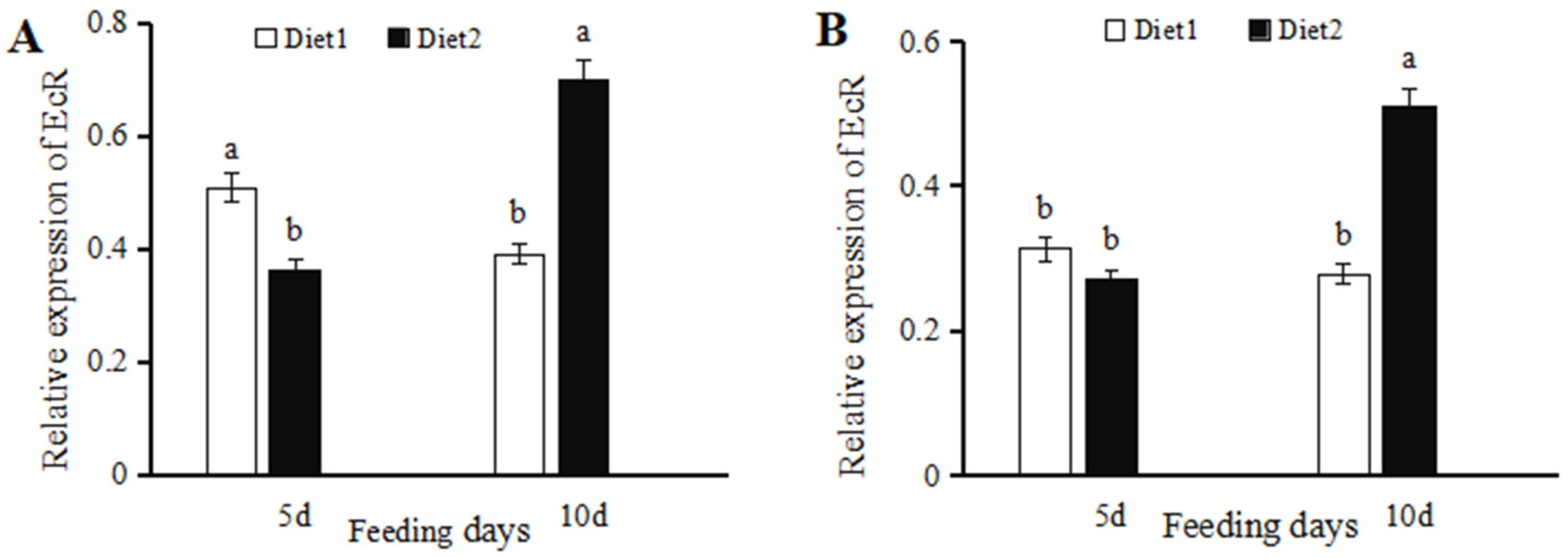
3.1. 20-Hydroxyecdysone-Mediated Changes in EcR Transcription

EcR expression in female adults fed with diet 2 for five days was significantly reduced (28.45% lower) (t = 3.3211, p = 0.0293) as compared to those supplied with diet 1 (Figure 1A). In contrast, EcR expression levels in females fed with diet 2 for ten days were significantly higher (78.41%) (t = 3.6314, p = 0.0221) than females fed with diet 1. EcR expression in male adults supplied with diet 2 for five days was 13.32% lower than those on diet 1, and the difference was not significant (t = 0.6717, p = 0.5386) (Figure 1B). However, EcR expression in males supplied with diet 2 for ten days was significantly higher (83.24%) (t = 7.7019, p = 0.0015) than the expression in those fed on diet 1.

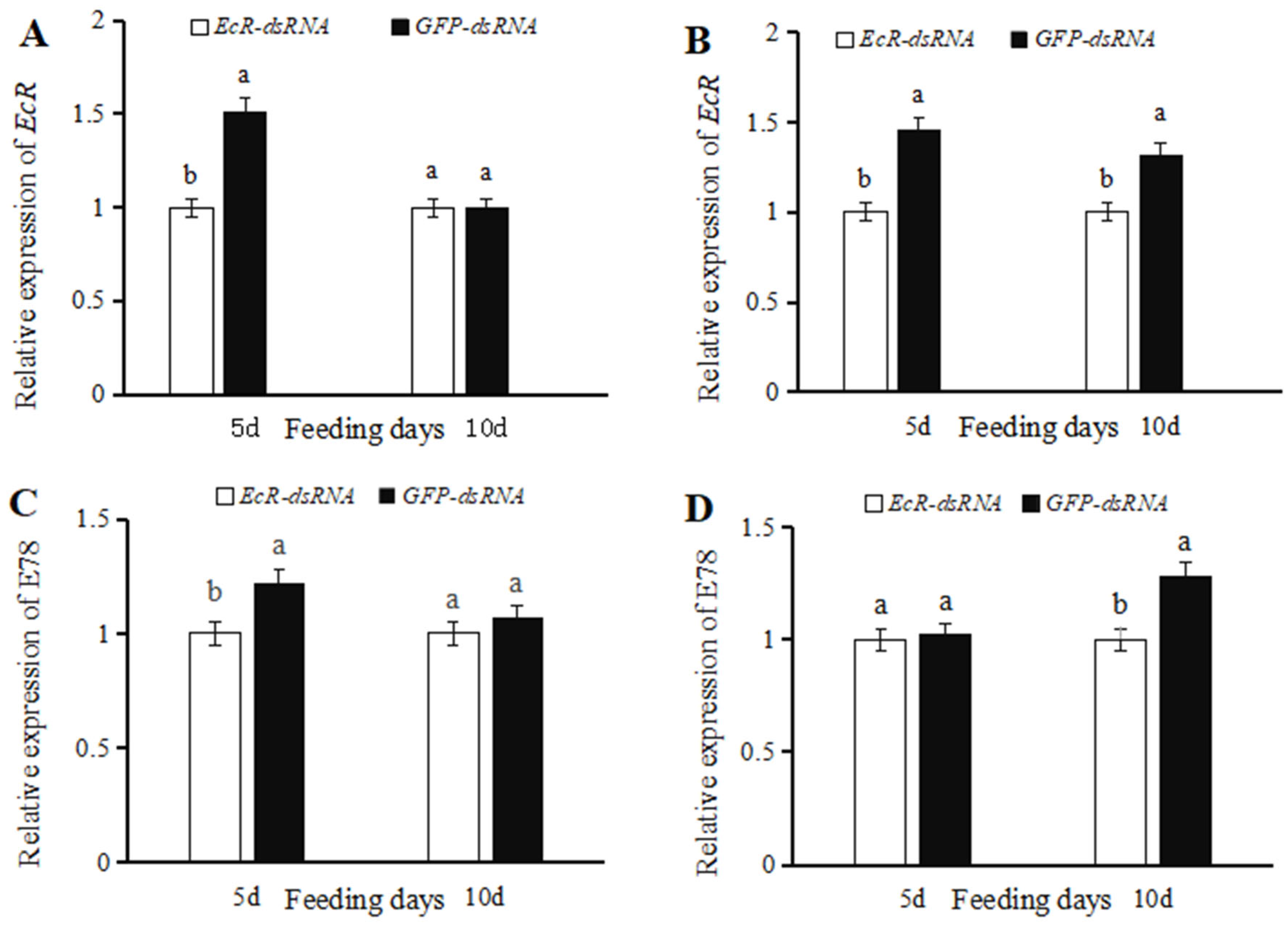
3.2. Effects of dsRNA on EcR Expression

EcR expression levels in female ladybugs injected with EcR-dsRNA were significantly reduced (33.96% lower) (t = 3.8289, p = 0.0186) than insects microinjected with GFP-dsRNA at five days (Figure 2A). No significant differences in EcR expression were observed in females injected with EcR-dsRNA or GFP-dsRNA at ten days post-injection (t = 0.0467, p = 0.9650) (Figure 2A). EcR expression levels in males treated with EcR-dsRNA and analyzed at five days and ten days were significantly reduced (31.22% and 24.19% lower, respectively) (t = 5.0927, p = 0.0070) as compared to males treated with GFP-dsRNA (Figure 2B).
E78 expression levels in female ladybugs injected with EcR-dsRNA were significantly reduced (17.97% lower) (t = 2.8759, p = 0.0452) than insects microinjected with GFP-dsRNA at five days (Figure 2C). No significant differences in E78 expression were observed in females injected with EcR-dsRNA or GFP-dsRNA at ten days post-injection (t = 0.4474, p = 0.6984) (Figure 2C). E78 expression levels in males treated with EcR-dsRNA at five days showed no significant differences (t = 0.1301, p = 0.9028) as compared to males treated with GFP-dsRNA (Figure 2D). E78 expression levels in males injected with EcR-dsRNA were significantly reduced (21.98% lower) (t = 3.0271, p = 0.0389) than insects microinjected with GFP-dsRNA at ten days (Figure 2D).

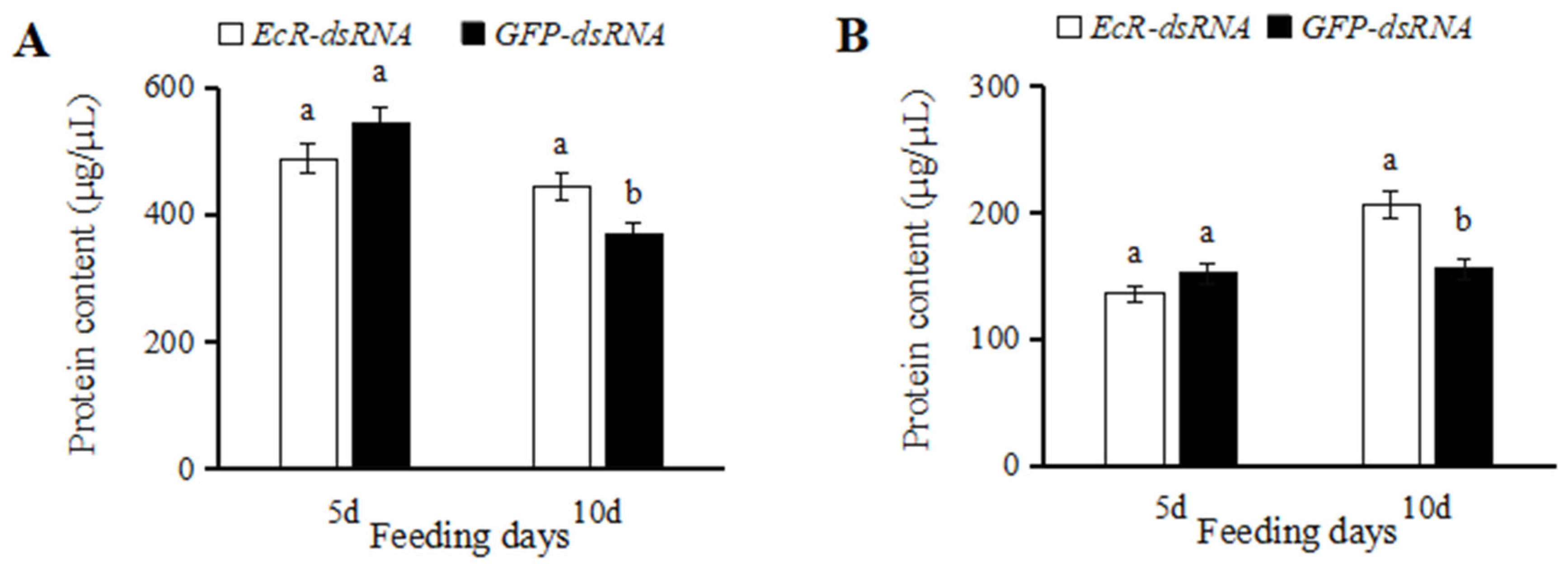
3.3. Effects of dsRNA Injection on Ovaries and Testes

Ovary formation in female adults was evaluated at five days and ten days after microinjection with EcR-dsRNA and GFP-dsRNA. Very few eggs were produced in the ovaries of ladybugs injected with EcR-dsRNA at five days, whereas eggs in GFP-dsRNA-injected females filled approximately half of the ovary at five days (Figure 3A,B). When sampled ten days after injection with EcR-dsRNA, approximately two-thirds of the ovaries were filled with eggs, whereas eggs in females microinjected with GFP-dsRNA filled the entire ovary (Figure 3D,E). No obvious differences were noted in the egg development of females treated with GFP-dsRNA and the noninjected control.
Testis development in male adults was evaluated at five days and ten days post-injection with EcR-dsRNA or GFP-dsRNA. The formation of testes in males injected with EcR-dsRNA was delayed at both time points as compared to males treated with GFP-dsRNA (Figure 4A,D). Males treated with GFP-dsRNA had more white substances (containing semen proteins [28]) in the accessory glands, whereas the group injected with EcR-dsRNA had fewer white substances and contained collapsed testicular tubes. No obvious differences were apparent in the formation of white substances in the accessory glands of GFP-dsRNA-injected and noninjected males.
After injecting EcR-dsRNA into C. septempunctata for five days, the protein content of female and male insects was lower than that of GFP-dsRNA-injected insects, and the difference was not significant (Figure 5A,B). However, at 10 d after injection, the protein content in insects treated with EcR-dsRNA was significantly higher than that of GFP-dsRNA-treated insects (Figure 5A,B).

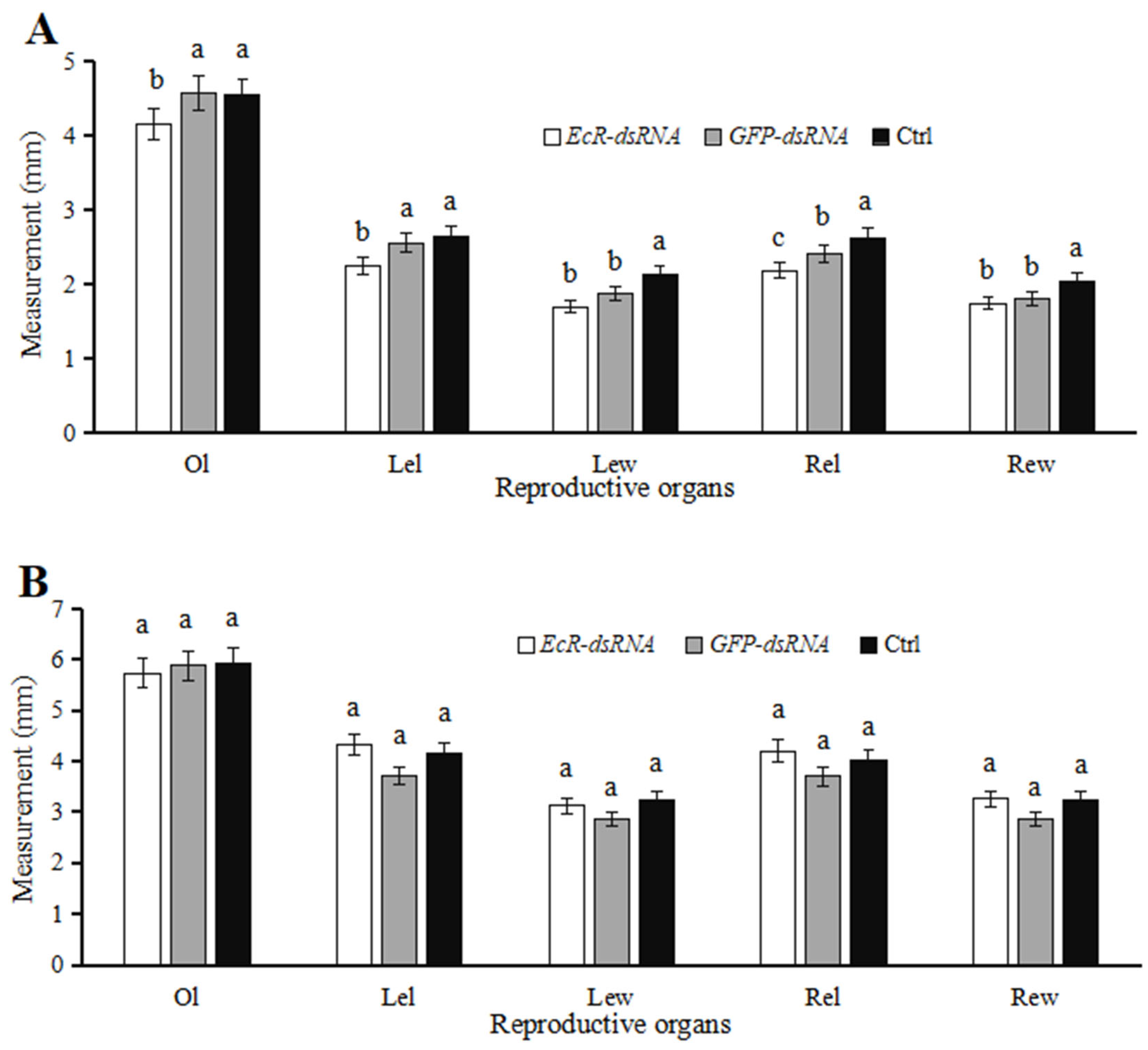
3.4. Effects of dsRNA Injection on Development of Reproductive Systems

The ovary length (Ol), width of the left egg chamber (Lew), length of the left egg chamber (Lel), width of the right egg chamber (Rew), and length of the right egg chamber (Rel) in ladybugs injected with EcR-dsRNA decreased by 9.14%, 9.28% 12.05%, 3.16%, and 8.93% at five days, respectively, as compared to ladybugs microinjected with GFP-dsRNA (Figure 6A). Among these, the lengths of the ovaries and the right and left egg chambers were significantly lower (p < 0.05) for EcR-dsRNA-injected females than in GFP-dsRNA-treated females (Figure 6A). Interestingly, ladybugs injected with EcR-dsRNA, analyzed at ten days, showed increases of 16.48%, 9.43%, 13.48%, and 13.93% in Lel, Lew, Rel, and Rew, respectively, when compared to the GFP-dsRNA group (Figure 6B); however, these differences were not significant.
In ladybugs injected with EcR-dsRNA, testis lengths decreased by 2.13% and accessory gland lengths and widths increased by 5.79% and 3.47%, respectively, as compared to insects microinjected with GFP-dsRNA at five days (Figure 7A); however, these differences were not significant at p > 0.05. At ten days after microinjection with EcR-dsRNA, testis lengths, accessory gland lengths, and accessory gland widths decreased by 1.74%, 2.54%, and 7.03%, respectively, when compared to males microinjected with GFP-dsRNA (Figure 7B). In the GFP-dsRNA-treated group, there were no significant differences in testis lengths and accessory gland lengths at 10 d when compared to the noninjected group (p > 0.05), indicating that the injection wound did not have a significant impact on the male testes.

3.5. Effect of dsRNA Injection on Egg Production and Hatching

When female ladybugs were treated with EcR-dsRNA or GFP-dsRNA, paired with noninjected males and reared for twenty days, the mean egg production was 148 and 227 eggs, respectively (Figure 8A), whereas the mean egg production in the noninjected group was 241 eggs. In females injected with EcR-dsRNA, egg production was significantly lower than that of the GFP-dsRNA group (F = 82.5768, p = 0.0006). The hatching rates of female insects injected with EcR-dsRNA decreased when paired with males, but the difference was not significant when compared to GFP-dsRNA-injected or noninjected females (F = 0.0376, p = 0.9634) (Figure 9A).
When males were treated with EcR-dsRNA or GFP-dsRNA and paired with noninjected females, the mean egg production for twenty days was 220 and 316 eggs, respectively (Figure 8B). Interestingly, when males were microinjected with EcR-dsRNA, egg production by females was significantly reduced (F = 26.5189, p = 0.0049); however, microinjection of males with GFP-dsRNA had no significant impact on fecundity, similar to the noninjected control group. The hatching rates of male insects injected with EcR-dsRNA decreased when paired with females, but the difference was not significant when compared to GFP-dsRNA-injected or noninjected males (F = 2.9089, p = 0.1600) (Figure 9B).

4. Discussion

The role of ecdysteroids in adult insects is mediated by receptors, especially the heterodimeric nuclear receptor EcR/USP. In this study, ladybug adults supplied with an artificial diet containing 20E showed decreased EcR expression at five days; this may be the outcome of increased 20E metabolism and concomitant binding to EcR receptors, resulting in decreased EcR expression. Furthermore, EcR expression was upregulated up to ten days when ladybug adults were supplied with an artificial diet supplemented with 20E. We speculated that the addition of 20E to artificial diets may increase EcR transcription with prolonged feeding, as the amount of EcR consumed in 20E metabolism was less than the synthesis amount. When 20E was supplied to Antheraea pernyi pupae, diapause was induced, and ApEcRB1 and ApUSP1 expression decreased [11]. ApEcRB1 expression remained low for 12 d in the later stages of pupal development; however, expression sharply increased in the early stages of eclosion and reached its highest level at 16 d of pupal development. Similarly, ApUSP1 expression levels began to increase at 8 d after injection and reached peak levels at 16 d [11]. In Apis mellifera, expression levels of EcRA, EcRB1, and USP1 began to decrease 1 h after injection with 20E, and high concentrations of 20E inhibited EcR expression [29]. In a subsequent study, Yu et al. [30] attempted to rescue EcRA and USP expression in A. mellifera EcR-RNAi larvae by injecting 20E, but the expression levels remained low.
The MH receptor EcR is positioned at the onset of cascading reactions that control insect molting, metamorphosis, and reproduction, and EcR plays an essential role in these processes. The 20E signaling mediated by EcR and USP can control the differentiation of germline stem cells during early development of the ovaries [31]. In Drosophila female adults, oogenesis in EcR mutants is defective, and the spectrum of oogenic defects includes the presence of abnormal egg chambers and loss of vitellogenic egg stages [32]. EcR and USP were significantly inhibited at the transcriptional level 6–18 days after injection of dsEcR and dsUFP, and the number of eggs in the ovaries and eggs laid per female significantly decreased compared with the control [33]. In Spodoptera littoralis male adults, injection of 20E inhibited and delayed the release of sperm bundles [34]. The downregulation of molting-hormone-responsive genes (e.g., E75, E78, FTZ-F1, HR4, HR38, HR39) in male T. castanenum can lead to abnormal accessory gland development, reduced sperm count, and a significant decrease in egg production in mating females. However, EcR-RNAi and USP-RNAi had no significant effect on male reproduction in T. castanenum, which may be due to insufficient interference or the presence of other functional ecdysteroid receptors [35]. In our study, injection of EcR-dsRNA into female ladybugs inhibited EcR and E78 transcription, delayed ovary development, and reduced egg production. After EcR-dsRNA was microinjected into male adults, testis volumes decreased and substances in the accessory gland (primarily semen proteins) decreased. Furthermore, egg production decreased when males injected with EcR-dsRNA were paired with noninjected females. These results indicate that the reproductive abilities of both male and female ladybug adults are regulated by EcR.
EcR is widely present in insect tissues, including the central nervous system, fat body, intestines, and reproductive organs of both male and female insects [10]. Fat bodies are responsible for providing yolk protein precursors and the energy required for egg maturation [36,37]. Kamoshida et al. [38] found that EcR-mediated ecdysteroid signaling can reduce lipid accumulation in Drosophila fat bodies. Further research is needed to evaluate whether lipid accumulation in the fat body of C. septempunctata is regulated by EcR.

5. Conclusions

The results clearly demonstrate that EcR is required for normal ovary and testis development and maximal egg production in C. septempunctata, indicating that EcR has a bifunctional role in the reproduction of males and females. This study further contributes to the improvement of methods for cultivating natural enemies and will ultimately benefit pest control.

Author Contributions

Conceptualization, Y.C.; methodology, Y.C., Y.Z. and C.L.; validation, Y.C.; formal analysis, Y.C., Y.Z. and C.L.; investigation, Y.Z. and C.L.; resources, Y.C. and Y.Z.; data curation, Y.C. and C.L.; writing—original draft preparation, Y.C. and C.L.; writing—review and editing, Y.C. and C.L.; supervision, Y.C.; project administration, Y.C. and Y.Z.; funding acquisition, Y.C. and Y.Z. All authors have read and agreed to the published version of the manuscript.

Funding

This work was supported by the Science and Technology Innovation Project of Guizhou Academy of Agricultural Sciences ([2023]08), and Innovative Capabilities Buildup of Green Prevention and Control for Invasive Species in Agriculture ([2023]011), and Guizhou Key Laboratory of Agricultural Biosecurity [Qiankehe ZSYS(2025)024].

Data Availability Statement

The original contributions presented in this study are included in the article. Further inquiries can be directed to the corresponding author.

Conflicts of Interest

The authors declare no conflicts of interest.

References

  1. Cartwright, B.O.; Eikenbary, R.D.; Angalet, G.W.; Ampbell, R.K. Release and establishment of Coccinella septempunctata in Oklahoma. Environ. Entomol. 1979, 8, 819–823. [Google Scholar] [CrossRef]
  2. Hance, T.; Dixon, A.F.G. Insect predator-prey dynamics: Ladybird beetles and biological control. J. Insect Conserv. 2006, 10, 375–376.s. [Google Scholar] [CrossRef]
  3. Hodek, I.; Michaud, J.P. Why is Coccinella septempunctata so successful? (A point-of-view). Eur. J. Entomol. 2008, 105, 1–12. [Google Scholar] [CrossRef]
  4. Duan, Y.J.; He, H.G.; Pu, D.Q.; Yang, H.; Chen, Q.; Lu, Q.C. Basic research status of Coccinella septempunctata. J. Biosaf. 2021, 30, 20–28. [Google Scholar]
  5. Smirnoff, W.A. An artificial diet for rearing Coccinellid beetles. Can. Entomol. 1958, 90, 563–565. [Google Scholar] [CrossRef]
  6. Yang, D.L.; Hao, J.; Wang, Y.N.; Xi, G.S. Research progress of ecdysone in adult insects. Chin. Bull. Life Sci. 2014, 26, 874–881. [Google Scholar]
  7. Niwa, R.; Niwa, Y.S. Enzymes for ecdysteroid biosynthesis: Their biological functions in insects and beyond. Biosci. Biotech. Bioch. 2014, 78, 1283–1292. [Google Scholar] [CrossRef]
  8. Zhu, J.S. Non-genomic action of juvenile hormone modulates the synthesis of 20- hydroxyecdysone in Drosophila. Sci. Bull. 2022, 67, 117–118. [Google Scholar] [CrossRef]
  9. Feyereisen, R. Insect CYP genes and P450 enzymes. Insect Mol. Miol. 2012, 8, 236–316. [Google Scholar]
  10. Wang, J.J.; Hu, Q.B. A review of ecdysone receptor. J. Environ. Entomol. 2017, 39, 721–729. [Google Scholar]
  11. Ru, Y.T.; Wang, Y.; Zhou, J.G.; Wang, D.Y.; Ma, Y.Y.; Jiang, Y.R.; Gao, Q.; Qin, L. The expression patterns of ecdysone receptor and ultraspiracle genes in Antheraea pernyi during development and hormone-induced process. Sci. Seric. 2017, 43, 0594–0602. [Google Scholar]
  12. Fallon, A.M.; Hagedorn, H.H.; Wyatt, G.R.; Laufer, H. Activation of vitellogenin synthesis in the mosquito Aedes aegypti by ecdysone. J. Insect Physiol. 1974, 20, 1815–1823. [Google Scholar] [CrossRef]
  13. Zhou, J.; Li, J.; Wang, Q.; Luo, Y.Q. The regulation of ecdysteroid on insect growth and productive processes. Chinese J. Applied Entomol. 2013, 50, 1413–1418. [Google Scholar]
  14. König, A.; Yatsenko, A.S.; Weiss, M.; Shcherbata, H. Ecdysteroids affect Drosophila ovarian stem cell niche formation and early germline differentiation. EMBO J. 2011, 30, 1549–1562. [Google Scholar]
  15. Morris, L.X.; Spradling, A.C. Steroid signaling within Drosophila ovarian epithelial cells sex-specifically modulates early germ cell development and meiotic entry. PLoS ONE 2012, 7, e46109. [Google Scholar] [CrossRef]
  16. Parthasarathy, R.; Sheng, Z.T.; Sun, Z.Y.; Palli, S.R. Ecdysteroid regulation of ovarian growth and oocyte maturation in the red flour beetle, Tribolium castaneum. Insect Biochem. Molec. 2010, 40, 429–439. [Google Scholar] [CrossRef]
  17. Swevers, L.; Iatrou, K. Ecdysteroids and ecdysteroid signaling pathways during insect oogenesis. In Ecdysone: Structures and Functions; Springer: Dordrecht, The Netherlands, 2009; pp. 127–164. [Google Scholar]
  18. Happ, G.M. Maturation of the male reproductive system and its endocrine regulation. Annu. Rev. Entomol. 1992, 37, 303–320. [Google Scholar] [CrossRef]
  19. Shen, G.W.; Lin, Y.; Lv, Y.H.; Wang, J.Y.; Xing, R.M.; Xia, Q.Y. Regulation of ecdysone on vitellogenin gene expression in silkworm Bombyx mori. Chin. J. Biochem. Mol. Biol. 2014, 30, 1106–1112. [Google Scholar]
  20. Cheng, Y.; Zhou, Y.H.; Ran, H.Y.; Li, F.L. Effects of different hormones as dietary supplements on biological characteristics of Coccinella septempunctata L. J. Appl. Entomol. 2023, 147, 1–7. [Google Scholar] [CrossRef]
  21. Cheng, Y.; Zhou, Y.; Li, C.; Jin, J.X. Cloning and functional analysis of the juvenile hormone receptor gene CsMet in Coccinella septempunctata. J. Insect Sci. 2024, 24, ieae065. [Google Scholar] [CrossRef]
  22. Cheng, Y.; Zhou, Y.; Li, C. Functional analysis of genes encoding juvenile hormone receptor Met and transcription factor Kr-h1 in the reproductive capacity of Coccinella septempunctata males. Insects 2025, 16, 49. [Google Scholar] [CrossRef] [PubMed]
  23. Zhou, Y.H.; Cheng, Y.; Jin, J.X.; Li, W.H.; Li, F.L. Large scale production and release application of Coccinella septempunctata. Southwest China J. Agric. Sci. 2017, 30, 602–605. [Google Scholar]
  24. Cheng, Y.; Zhi, J.R.; Li, F.L.; Wang, H.; Zhou, Y.H.; Jin, J.X. Transcriptome sequencing of Coccinella septempunctata adult (Coleoptera: Coccinellidae) feeding on artificial diet and Aphis craccivora. PLoS ONE 2020, 15, e0236249. [Google Scholar] [CrossRef] [PubMed]
  25. Liu, M.Y.; Wang, J.; Wang, M.Z.; Gao, F.; Zhang, H.Z.; Li, Y.Y.; Zang, L.S.; Zhang, L.S. Cloning and expression analysis of juvenile hormone epoxide hydrolase in Coccinella septempunctata. Plant Prot. 2019, 45, 156–162. [Google Scholar]
  26. Pfaffl, M.W. A new mathematical model for relative quantification in real-time RT-PCR. Nucleic. Acids Res. 2001, 29, e45. [Google Scholar] [CrossRef]
  27. Tang, Q.Y.; Zhang, C.X. Data Processing System (DPS) software with experimental design, statistical analysis and data mining developed for use in entomological research. Insect Sci. 2013, 20, 254–260. [Google Scholar] [CrossRef]
  28. Wang, Z.S.; Zhong, X.C.; Qiu, X.J.; Hu, S.Y.; Guo, F. Observations on the reproduction of Coccinella septempunctata L. Acta Entomol. Sin. 1977, 20, 397–404. [Google Scholar]
  29. Mello, T.R.P.; Aleixo, A.C.; Pinheiro, D.G.; Nunes, F.M.F.; Bitondi, M.M.G.; Hartfelder, K.; Barchuk, A.R.; Simoes, Z.L.P. Developmental regulation of ecdysone receptor (EcR) and EcR-controlled gene expression during pharate-adult development of honeybees (Apis mellifera). Front. Genet. 2014, 5, 445. [Google Scholar] [CrossRef]
  30. Yu, J.; Song, H.Y.; Wang, Y.; Liu, Z.G.; Wang, H.F.; Xu, B.H. 20-hydroxyecdysone upregulates ecdysone receptor (EcR) gene to promote pupation in the honeybee, Apis Mellifera Ligustica. Integr. Comp. Biol. 2023, 63, icad077. [Google Scholar] [CrossRef]
  31. Ables, E.T.; Drummond-Barbosa, D. The steroid hormone ecdysone functions with intrinsic chromatin remodeling factors to control female germ line stem cells in Drosophila. Cell Stem Cell 2010, 7, 581–592. [Google Scholar] [CrossRef]
  32. Carney, G.E.; Bender, M. The Drosophila ecdysone receptor (EcR) gene is required matemally for normal oogenesis. Genetics 2000, 154, 1203–1211. [Google Scholar] [CrossRef] [PubMed]
  33. Huang, X.X.; Zheng, W.Y.; Xue, H.; Jiao, P.Y.; Chen, L.Z. Effects of silencing ecdysone receptor gene on the fecundity of Adelphocris suturalis. Plant Prot. 2023, 49, 164–177. [Google Scholar]
  34. Polanska, M.A.; Maksimiuk-Ramirez, E.; Ciuk, M.A.; Kotwica, J.; Bebas, P. Clock-controlled rhythm of ecdysteroid levels in the haemolymph and testes and its relation to sperm release in the egyptian cotton leafworm, Spodoptera littoralia. J. Insect Physiol. 2009, 55, 426–434. [Google Scholar] [CrossRef]
  35. Xu, J.; Raman, C.; Zhu, F.; Tan, A.; Palli, S.R. Identification of nuclear receptors involved in regulation of male reproduction in the red flour beetle, Tribolium castaneum. J. Insect Physiol. 2012, 58, 710–717. [Google Scholar] [CrossRef] [PubMed]
  36. Attardo, G.M.; Hanseni, A.; Raikhel, A.S. Nutritional regulation of vitellogenesis in mosquitoes: Implications for anautogeny. Insect Biochem. Molec. 2005, 35, 661–675. [Google Scholar] [CrossRef]
  37. Wang, X.L.; Hou, Y.; Saha, T.T.; Pei, G.F.; Raikhel, A.S.; Zou, Z. Hormone and receptor interplay in the regulation of mosquito lipid metabolism. Proc. Nati. Acad. Sci. USA 2017, 114, E2709–E2718. [Google Scholar] [CrossRef]
  38. Kamoshida, Y.; Fujiyama-Nakamur, S.; Kimura, S.; Suzuki, E.; Lim, J.; Shiozaki-Sato, Y.; Kato, S.; Takeyama, K. Ecdysone receptor (EcR) suppresses lipid accumulation in the Drosophila fat body via transcription control. Biochem. Biophys. Res. Commun. 2012, 421, 203–207. [Google Scholar] [CrossRef]
Figure 1. Relative expression of EcR in Coccinella septempunctata at five and ten days after feeding on diet 1 or 2. Diet 2 is diet 1 amended with 20-hydroxyecdysone. Panels show the expression of EcR in females (A) and males (B). Column heights represent means ± SE (n = 3), and columns labeled with different letters indicate a significant difference at p < 0.05 using the t-test.
Insects 16 00643 g001
Figure 2. Relative expression of EcR and E78 in Coccinella septempunctata at five and ten days after injection with EcR-dsRNA or GFP-dsRNA. Panels show the relative expression of EcR in females (A) and males (B). Panels show the relative expression of E78 in females (C) and males (D). Column heights indicate means ± SE (n = 3), and the labeling of columns with different letters indicates significance at p < 0.05 using the t-test.
Insects 16 00643 g002
Figure 3. Ovary development in Coccinella septempunctata females after injection with EcR-dsRNA or GFP-dsRNA. Panels (A,B) show ovary development in females at 5 d after injection with EcR-dsRNA and GFP-dsRNA, respectively. Panels (D,E) show ovary development in females at 10 d after injection with EcR-dsRNA and GFP-dsRNA, respectively. Control (Ctrl) panels (C,F) show ovary development in noninjected females.
Insects 16 00643 g003
Figure 4. Testis development in Coccinella septempunctata males after microinjection with EcR-dsRNA or GFP-dsRNA. Panels (A,B) illustrate testes in males at five days following injection with EcR-dsRNA and GFP-dsRNA, respectively. Panels (D,E) show testis development in males at ten days following injection with EcR-dsRNA and GFP-dsRNA, respectively. Control (Ctrl) panels (C,F) show the development of testes in males that were not injected.
Insects 16 00643 g004
Figure 5. Protein concentration in Coccinella septempunctata after microinjection with EcR-dsRNA or GFP-dsRNA. Panels show protein concentration in females (A) and males (B). Column heights represent means ± SE (n = 3), and columns labeled with different letters indicate a significant difference at p < 0.05 using the t-test.
Insects 16 00643 g005
Figure 6. Ovary measurements in female ladybugs after microinjection with EcR-dsRNA or GFP-dsRNA. Columns labeled as Ctrl represent noninjected controls. Panels show ovary measurements at five (A) and ten (B) days post-injection. Abbreviations: Ol, ovary length; Lel, length of left egg chamber; Lew, width of left egg chamber; Rel, length of right egg chamber; and Rew, width of right egg chamber. Data points represent means ± SE (n = 3). Columns labeled with different letters indicate significance at p < 0.05 using Duncan’s test.
Insects 16 00643 g006
Figure 7. Testis measurements in Coccinella septempunctata after microinjection with EcR-dsRNA or GFP-dsRNA. Columns labeled as Ctrl represent noninjected controls. Panels: testis measurements at (A) five days and (B) ten days after microinjection. Abbreviations: Tl, length of testes; Agl, length of accessory glands; Agw, width of accessory glands. Data points represent means ± SE (n = 3). Columns with different letters represent significance at p < 0.05 using Duncan’s test.
Insects 16 00643 g007
Figure 8. Fecundity of Coccinella septempunctata following microinjection with EcR-dsRNA or GFP-dsRNA. (A) Fecundity of females after injection with EcR-dsRNA or GFP-dsRNA and mating with males. (B) Fecundity of males after injection with EcR-dsRNA or GFP-dsRNA and mating with females. Ctrl, noninjected control. Data points represent mean ± SE (n = 3). Columns with different letters indicate significance at p < 0.05 using Duncan’s test.
Insects 16 00643 g008
Figure 9. Hatching rates of Coccinella septempunctata following microinjection with EcR-dsRNA or GFP-dsRNA. (A) Hatching rate of females after injection with EcR-dsRNA or GFP-dsRNA and mating with males. (B) Hatching rate of males after injection with EcR-dsRNA or GFP-dsRNA and mating with females. Ctrl, noninjected control. Data points represent mean ± SE (n = 3). Columns with different letters indicate significance at p < 0.05 using Duncan’s test.
Insects 16 00643 g009
Table 1. Sequences of primers used in this study.
Primer NameSequenceAnnealing Temperature
EcR-F
EcR-R
AAAGGACCAACACCAAGGCA
TCGTCACACTCCGTCGAAAG
55.5
EcR-dsRNA-F
EcR-dsRNA-R
taatacgactcactatagggCGATGACTTGCTGCTGGTTA
taatacgactcactatagggGTCGTAGCTGCCTGATGACA
GFP-dsRNA-F
GFP-dsRNA-R
E78-F
E78-R
taatacgactcactatagggGCCAACACTTGTCACTACTT
taatacgactcactatagggGGAGTATTTTGTTGATAATGGTCTG
AGCTCCTGTTTCATGATGCGG
ATTCATCCCCGGTCGAATGTG
58.0
Actin-F
Actin-R
GATTCGCCATCCAGGACATCTC
TCCTTGCTCAGCTTGTTGTAGTC
60.0
Letters in lowercase indicate the T7 promoter region.
Disclaimer/Publisher’s Note: The statements, opinions and data contained in all publications are solely those of the individual author(s) and contributor(s) and not of MDPI and/or the editor(s). MDPI and/or the editor(s) disclaim responsibility for any injury to people or property resulting from any ideas, methods, instructions or products referred to in the content.

