Article

Molecular Characterization and Expression of the Ecdysone Receptor and Ultraspiracle Genes in the Wheat Blossom Midge, Sitodiplosis mosellana

1
Shandong Institute of Sericulture, Jiaodong Innovation Center, Shandong Academy of Agricultural Sciences, Yantai 264002, China
2
Key Laboratory of Plant Protection Resources and Pest Management of Ministry of Education, College of Plant Protection, Northwest A&F University, Yangling 712100, China
3
Department of Entomology, Texas A&M University, College Station, TX 77843, USA
*
Authors to whom correspondence should be addressed.
Insects 2025, 16(5), 537; https://doi.org/10.3390/insects16050537
Submission received: 23 April 2025 / Revised: 14 May 2025 / Accepted: 17 May 2025 / Published: 19 May 2025
(This article belongs to the Section Insect Molecular Biology and Genomics)

Simple Summary

20-hydroxyecdysone (20E) has a major role in the regulation of development and diapause in insects. Ecdysone receptor (EcR) and ultraspiracle (USP) proteins are primary regulators involved in 20E signaling. However, the functions of these genes during diapause are poorly understood. This study aimed to characterize the genes encoding EcR and USP (SmEcR, SmUSP-A, and SmUSP-B) in Sitodiplosis mosellana, a major wheat pest that undergoes obligatory diapause during the mature third larval instar, and analyze their expression dynamics across various developmental stages, as well as their responsiveness to 20E treatment. The results showed that the expression of SmEcR, SmUSP-A, and SmSP-B was not only developmentally controlled but also responsive to 20E. The transcription levels of the genes increased as the larvae entered pre-diapause, followed by downregulation during diapause and upregulation during transition to post-diapause quiescence, with higher expression observed in adult females compared to males. These results suggest that 20E-responsive SmEcR and SmUSP genes mediate critical functions during diapause initiation, maintenance, and post-diapause quiescence, and also contribute to adult reproduction, while the larval–pupal transition may be associated with a downregulation of their expression.

Abstract

20-hydroxyecdysone (20E) is essential for insect development and diapause. Ecdysone receptor (EcR) and ultraspiracle (USP) proteins are crucial regulators of 20E signaling. To explore their potential roles in the development of Sitodiplosis mosellana, a major wheat pest that undergoes obligatory diapause as a larva, one SmEcR and two SmUSPs (SmUSP-A and SmUSP-B) from this species were isolated and characterized. The deduced SmEcR and SmUSP-A/B proteins contained a conserved DNA-binding domain with two zinc finger motifs that bind to specific DNA sequences. Expression of SmEcR and the SmUSPs was developmentally controlled, as was 20E induction. Their transcription levels increased as the larvae entered pre-diapause, followed by downregulation during diapause and upregulation during the shift to post-diapause quiescence, which is highly consistent with ecdysteroid titers in this species. Topical application of 20E to diapausing larvae also elicited a dose-dependent expression of the three genes. Expression of SmEcR and SmUSPs decreased markedly during the pre-pupal stage and was higher in adult females compared to males. These findings suggested that 20E-induced expression of SmEcR and SmUSPs has key roles in diapause initiation and maintenance, post-diapause quiescence, and adult reproduction, while the larval–pupal transformation may be associated with a decrease in their expression levels.

Graphical Abstract

1. Introduction

Insects are the most successful animal group in terms of species number. Although multiple factors contribute to this profusion, one of the most important is the characteristic insect developmental plan of metamorphosis and diapause. Diapause is a genetically determined state of developmental arrest, which permits insects to survive in adverse environmental conditions and also to synchronize their life cycle with seasons favorable for development and reproduction [1,2]. Due to the central role of diapause in life-cycle synchronization and adaptation to environmental extremes, an in-depth understanding of the molecular mechanism underlying the process is essential for the development of effective pest management strategies [3].
Steroid hormone 20-hydroxyecdysone (20E) is known to modulate many important aspects of insect life, including not only molting, metamorphosis, and reproduction [4,5,6], but also diapause. It has been demonstrated that in some insects undergoing larval diapause, a drop in or low level of 20E titer is associated with the initiation and maintenance of diapause, while a rise in 20E or its exogenous application can terminate diapause, such as the bamboo borer Omphisa fuscidentalis [7,8], the corn earworm Helicoverpa zea [9], the peach fruit moth Carposina sasakii [10], and the cabbage white butterfly Pieris rapae [11]. The effects of 20E are mediated through two nuclear receptors: the ecdysone receptor (EcR) and its heterodimeric partner ultraspiracle (USP), a homolog of the vertebrate retinoid X receptor (RXR) [12,13,14]. The EcR/USP heterodimer binds to ecdysone response elements (EcREs) within the promoter regions of 20E-responsive genes, triggering transcriptional cascades that drive developmental transitions [15,16,17].
The genes encoding EcR and USP have been identified and characterized in a variety of insect species. Three EcR isoforms, namely, EcRA, EcRB1, and EcRB2, which have different N-terminal regions but the same DNA- and hormone-binding domains, were identified in Drosophila melanogaster [18], while one or two isoforms of EcR have been observed in the majority of other investigated insects, including Bombyx mori, Diaphorina citri, Leptinotarsa decemlineata, and Monochamus alternates [19,20,21,22,23]. In contrast to EcR, only one USP form has been found in Drosophila, while two isoforms with unique N-termini are present in many other species [24,25,26]. Recent studies have investigated the functions of EcR and the USP isoforms in the control of insect metamorphosis and molting [27,28,29]. However, to date, little information is available about their relationship with larval diapause [30,31].
The wheat blossom midge Sitodiplosis mosellana Géhin (Diptera: Cecidomyiidae), a major wheat pest, is widely distributed in the main wheat-producing regions of the northern hemisphere [32,33,34]. This species is univoltine with an obligatory larval diapause. In most areas of northern China, adult emergence and subsequent oviposition on wheat heads typically occur from mid- to late April [35]. The hatched larvae feed on the developing kernels until the larvae mature to the third instar. Matured larvae fall to the ground in mid-to-late May, spinning cocoons in the soil to initiate diapause. While the larvae remain in the cocoons until mid-March of the following year, diapause is concluded at the onset of cold weather in early December, after which the larvae enter post-diapause quiescence [36], which is morphologically identical to diapause. Post-diapause development is initiated by increased ambient temperature in spring, with the larvae leaving their cocoons and pupating on the soil surface. Therefore, the prolonged diapause and quiescence enable this species to combat harsh environmental conditions during summer and winter, and also synchronize its developmental cycle with that of its wheat host.
Previous studies have suggested the involvement of 20E in regulating the S. mosellana diapause [37], although the underlying molecular mechanism remains unclear. To explore the molecular basis of 20E effects on S. mosellana, the present study cloned and characterized cDNAs encoding EcR and USP from this species, namely, SmEcR, SmUSP-A, and SmUSP-B. The dynamic expression of these genes at different stages of diapause and direct development was also investigated, and the effects of topical application of 20E on expression were examined. The results of this study provide a foundation for further investigation into the functions of these genes in regulating the development of S. mosellana, assisting in the development of novel control strategies for this major pest.

2. Materials and Methods

2.1. Experimental Insects

Samples of S. mosellana at all developmental stages except the egg were collected from a natural setting using our previously developed method [38]. Briefly, the first two instars and pre-diapause third instar larvae, which were differentiated by body size, coloration, and the sternal Y-shaped structure [39], were obtained during May 2022 by dissecting wheat ears at the grain filling stage that were infested with S. mosellana. Concurrently, wheat ears containing 3rd instar larvae were collected in bulk and placed on soil in a field insectary established in Yangling, China (34°16′ N, 108°4′ E). The ears were sprayed with water to induce larval egress from the glumes and their subsequent migration into the soil. Successful initiation of diapause was confirmed by the presence of larval cocoons. Cocooned larvae, including those in the diapause (June–November) and post-diapause quiescent stages (December–February) [36] were harvested by sieving the soil in the insectary monthly, from 25 June 2022 to 25 February 2023. Post-diapause larvae (i.e., larvae exiting cocoons), pre-pupae, early-to-late-stage pupae, and adults were subsequently collected from mid-March. Groups of 50 1st and 2nd instar larvae or 20 individuals from each developmental stage were placed in 1.5 mL tubes, with at least three replicates from each sample. The samples were snap-frozen in liquid nitrogen and subsequently maintained at −80 °C for later use.

2.2. Cloning of S. mosellana EcR and USP Genes

Total RNA was extracted from 20 homogenized 3rd instar larvae using RNAiso Plus reagent (TaKaRa, Dalian, China) as per the manufacturer’s instructions. The concentration and purity of the RNA were assessed spectrophotometrically by the absorbance ratio at 260/280 nm (i.e., OD260/OD280), and the integrity was confirmed by 1% agarose gel electrophoresis. First-strand complementary DNA (cDNA) was synthesized from 1 μg of purified RNA using the PrimeScriptTM RT reagent Kit with gDNA Eraser (TaKaRa, Dalian, China), following the provided instructions, and preserved at −20 °C until use.
An EcR sequence and two USP isoforms (i.e., USP-A and USP-B) were identified in an S. mosellana transcriptomic dataset established in our laboratory and a genomic database [40] using similar gene sequences from the copepod Tigriopus japonicus [41]. Specific primers (Table 1) for amplification of the open reading frames (ORFs) were designed and synthesized. The PCR reactions (in 20 μL) consisted of 10 μL of 2×Taq MasterMix (CWbiotech, Beijing, China), 1 μL of cDNA, 0.5 μL of each primer (10μM), and 8 μL of ddH2O. The amplification conditions were an initial denaturation at 94 °C for 3 min, followed by 30 cycles (95 °C, 30 s), annealing (55 °C, 30 s), and extension (72 °C, 60 s for USPs or 90 s for EcR), concluding with a 10 min final extension at 72 °C. The PCR amplicons were purified from gels and then ligated into the pMD19-T vector. Recombined plasmids were transformed into DH5α-competent E. coli cells and sequenced.

2.3. Sequencing and Phylogenetic Analysis

The molecular weights and theoretical isoelectric points of EcR, USP-A, and USP-B proteins from S. mosellana were determined using the Compute pI/Mw tool (http://web.expasy.org/protparam, accessed on 15 May 2023). Functional domains and motifs were identified using a conserved domain search tool (http://www.ncbi.nlm.nih.gov/Structure/cdd/wrpsb.cgi, accessed on 15 May 2023) and the SMART tool (http://smart.embl-heidelberg.de/, accessed on 15 May 2023). Multiple sequence alignments of the SmEcR and SmUSP proteins with those from other insect species were performed using DNAMAN 6.0 software (Lynnon Corporation, Pointe-Claire, QC, Canada). A neighbor-joining phylogenetic tree was constructed [42] using MEGA7 with 1000 bootstrap repeats.

2.4. 20E Treatment

To examine the effects of 20-hydroxyecdysone (20E) on the expression of S. mosellana EcR and USP genes in diapause larvae, 20E was diluted in 50% ethanol to concentrations of 0.1–0.6 pg/nL. These solutions (23 nL) were injected using a Nanoject Ⅱ Auto-Nanoliter Injector (Drummond Scientific Company, Broomall, PA, USA) into the abdomens of cocooned larvae collected in October. The doses of 20E applied were based on the highest reported levels of endogenous 20E in the larvae of this insect [37]. The treated larvae were placed in petri dishes with moist filter paper and incubated for 1, 3, or 6 h at 25 °C in environmental incubators with 70% relative humidity before being frozen in liquid nitrogen and stored at −80 °C for subsequent RNA isolation. Insects treated with the same volume of 50% ethanol acted as the controls. Each treatment was performed using at least three replicates with 20 individuals per replicate.

2.5. Expression Analysis Using Reverse-Transcription Quantitative PCR (RT-qPCR)

To investigate the expression patterns of the S. mosellana EcR and USP genes during diapause and metamorphosis, approximately 50 1st instar larvae, 50 2nd instar larvae, and 20 3rd instar larvae at the different developmental stages related to diapause, as well as 20 pupae at different developmental stages, and adults, were used for RNA extraction. To examine the transcript levels in 20E-treated larvae, 20 individuals from each treatment group were used for RNA extraction. RNA was extracted using the RNAiso Plus reagent (TaKaRa, Dalian, China). The template cDNA was prepared from 1 μg of RNA with the PrimeScriptTM RT reagent Kit with gDNA Eraser (TaKaRa, Dalian, China).
RT-qPCR was conducted on an ABI 7500 real-time PCR system (Applied Biosystems, Foster City, CA, USA) (Thermofisher, Waltham, MA, USA) in a 20 μL reaction volume containing 2 μL of template cDNA, 10 μL of 2×SYBR Green Premix EX TaqTM II (TliRNaseH Plus) (TIANGEN, Beijing, China), 0.8 μL each of forward and reverse 10 μM primers (Table 1), 0.4 μL of 50×ROX Reference Dye, and 6.0 μL of ddH2O. GAPDH (GenBank accession number: KR733066) was used as an endogenous reference. The amplification procedure was performed using the two-step method, with an initial pre-denaturation at 95 °C for 30 s, followed by 40 cycles of denaturation (95 °C, 10 s), and annealing (60 °C, 30 s). The amplification specificity was verified through melting curve analysis with temperature gradients of 95 °C for 5 s, 60 °C for 1 min, and 95 °C for 15 s. The negative controls used RNase-free water instead of the cDNA template. Standard curves were generated using serially diluted pooled cDNA samples for target genes and GAPDH. The amplification efficiencies for primers of all genes were similar and close to 100%. Each treatment was performed in triplicate, with technical triplicates per sample. The cycle threshold value (Ct) of each target gene was normalized to that of GAPDH in the different samples, and relative expression was calculated using the 2−∆∆CT method [43].

2.6. Statistical Analysis

Data are reported as means and standard error (SE) from three independent replicates per sample. Duncan’s multiple range test was used with analysis of variance (ANOVA) to compare differences between samples and treatments, with p < 0.05 considered statistically significant. All statistical computations were conducted in SPSS 22.0 software (SPSS Inc., Chicago, IL, USA).

3. Results

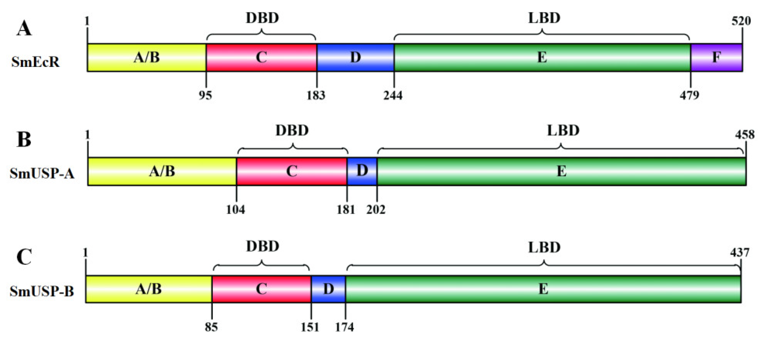
3.1. Characterization of SmEcR and SmUSP cDNAs

The complete open reading frame (ORF) of S. mosellana EcR (SmEcR) cloned by PCR was found to be 1563 bp in length (Figure S1, accession No. MN853890), encoding a protein with 520 amino acids, a molecular weight (MW) of 59.6 kDa, and an isoelectric point (pI) of 5.51. The ORFs of the S. mosellana USP isoforms, SmUSP-A (Figure S2, accession No. MN853891) and SmUSP-B (Figure S2, accession No. MN853892), were 1377 and 1314 bp, respectively. These encoded proteins of 458 and 437 amino acids have estimated MWs of 51.6 and 49.2 kDa, and pI values of 9.00 and 8.78, respectively.
Domain searching showed that SmEcR and two SmUSPs shared several typical structural hallmarks of the nuclear hormone receptor superfamily. These were N-terminal A/B (transactivation domain), C (DNA binding domain, DBD), D (hinge domain), and E (ligand binding domain, LBD) domains; however, SmEcR contained an additional F domain (Figure 1A,C) [44]. Further, two highly conserved C2C2 Zn-finger motifs, a P-box (EGCKG for SmEcR and SmUSPs) and a D-box (KFGHE for SmEcR and REDKN for SmUSPs), which are responsible for the recognition and binding to the hormone response elements (HREs) in target genes [24,45], were identified in the DBD of three putative proteins; while a T-box sequence (REAVQEERQ), also a DNA-recognition motif, was discovered in the D domain of two SmUSPs (Figure 2A,B).

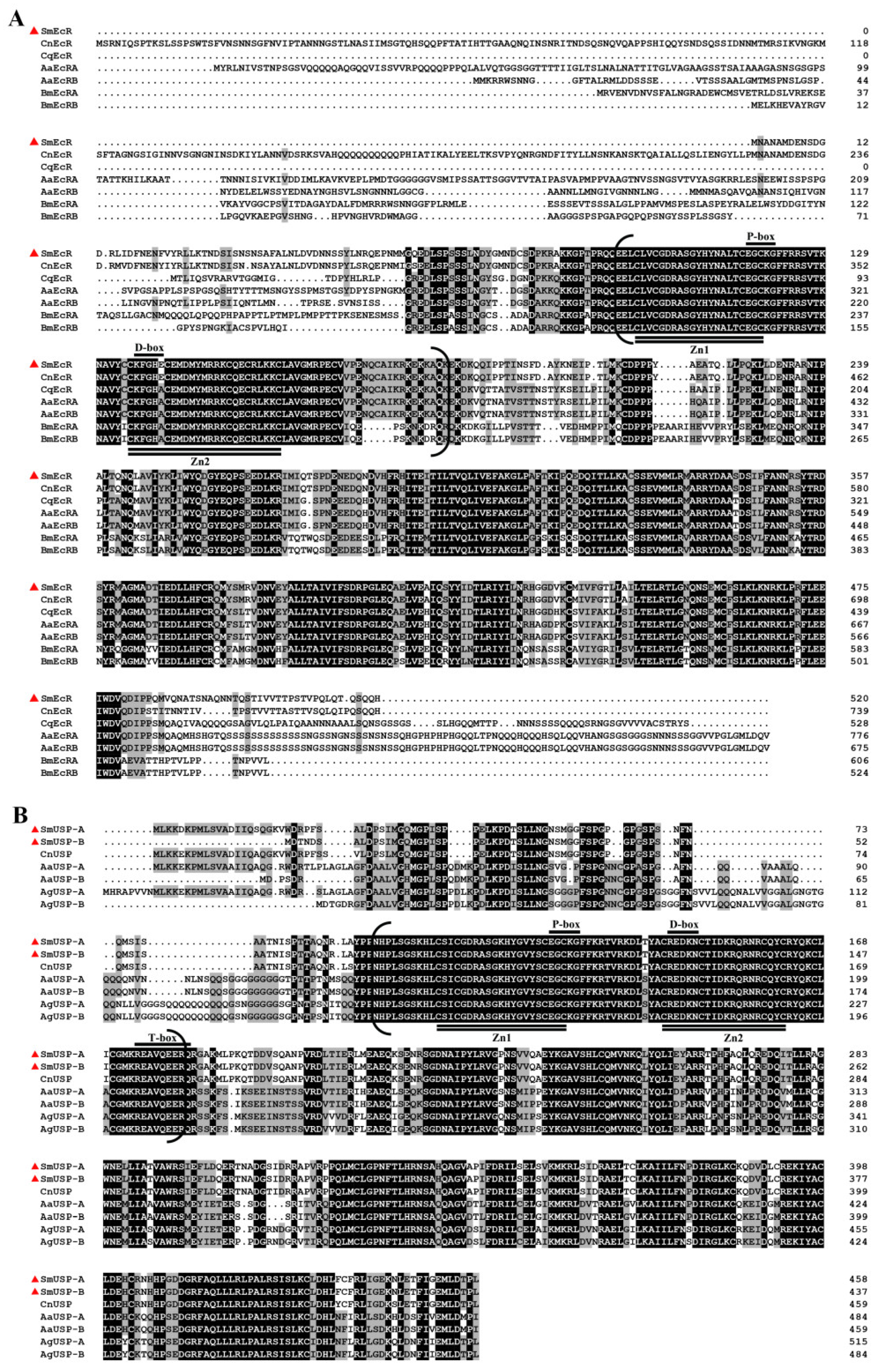
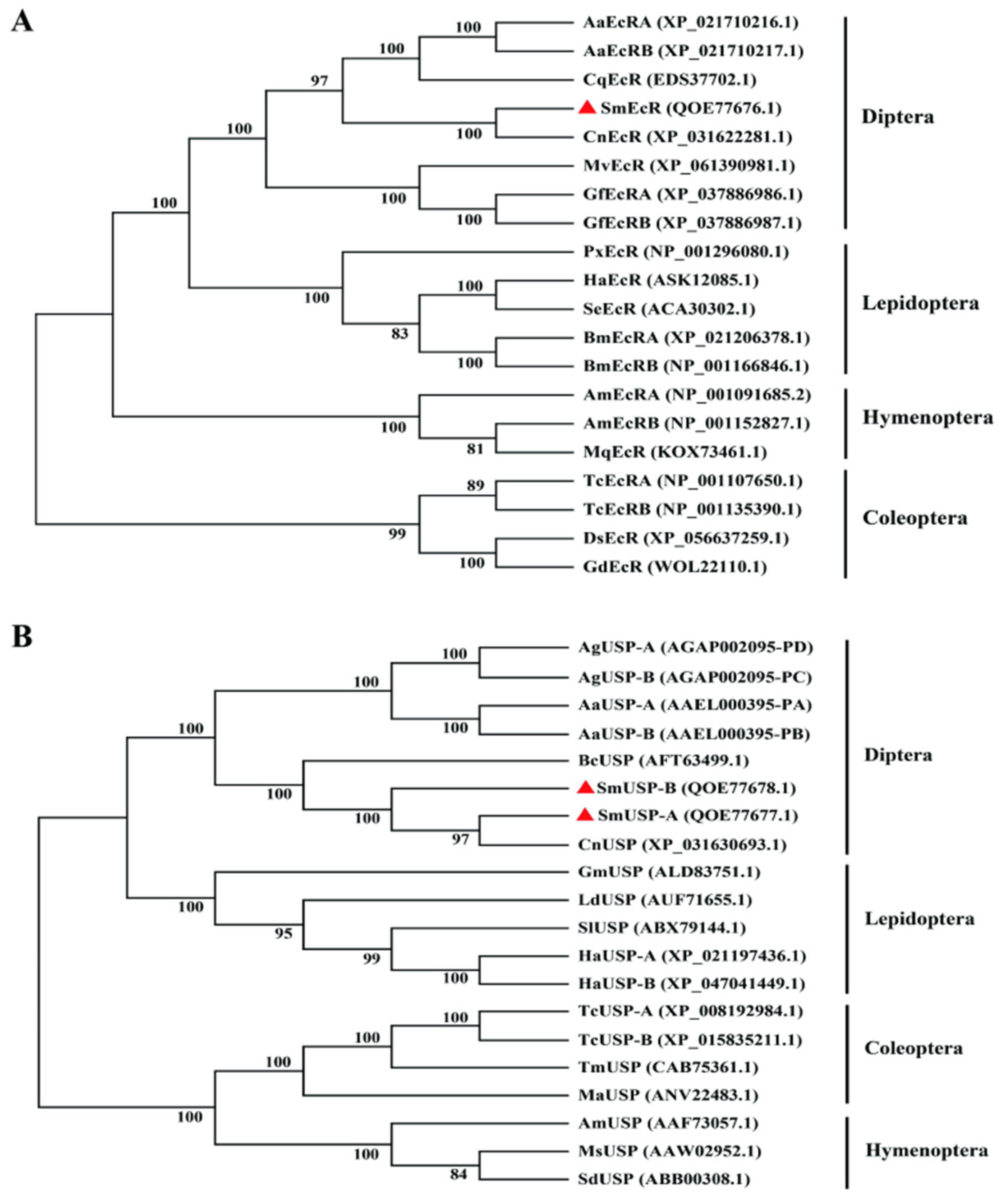
3.2. Sequence Comparison and Phylogenetic Analyses

A comparison of the deduced amino acid sequences showed that SmEcR had the highest sequence identity (92%) with EcR from Contarinia nasturtii (XP_031622281.1), and 86% identity to EcRs from Culex quinquefasciatus (EDS37702.1) and Aedes aegypti (XP_021710216.1, XP_021710217.1). In contrast, SmEcR showed less than 67% identity with Bombyx mori EcR (XP_021206378.1, NP_001166846.1) (Figure 2A). Similarly, SmUSP-A (QOE77677.1) and SmUSP-B (QOE77678.1) exhibited the highest identity with C. nasturtii USP (XP_031630693.1), achieving 98% and 92% identity, respectively, and 62–67% identity with USPs from Aedes aegypti (AAEL000395-PA, AAEL000395-PB) and Anopheles gambiae (AGAP002095-PD, AGAP002095-PC). The two SmUSPs were 94% identical (Figure 2B).
As anticipated, phylogenetic analysis revealed distinct clustering of EcRs and USPs derived from Dipteran, Lepidopteran, Coleopteran, and Hymenopteran insects, corresponding to their established taxonomic divergence. Notably, SmEcR and SmUSP-A/B were more closely related to their counterparts in the dipteran Cecidomyiidae family (C. nasturtii), emphasizing their close phylogenetic ties (Figure 3A,B).

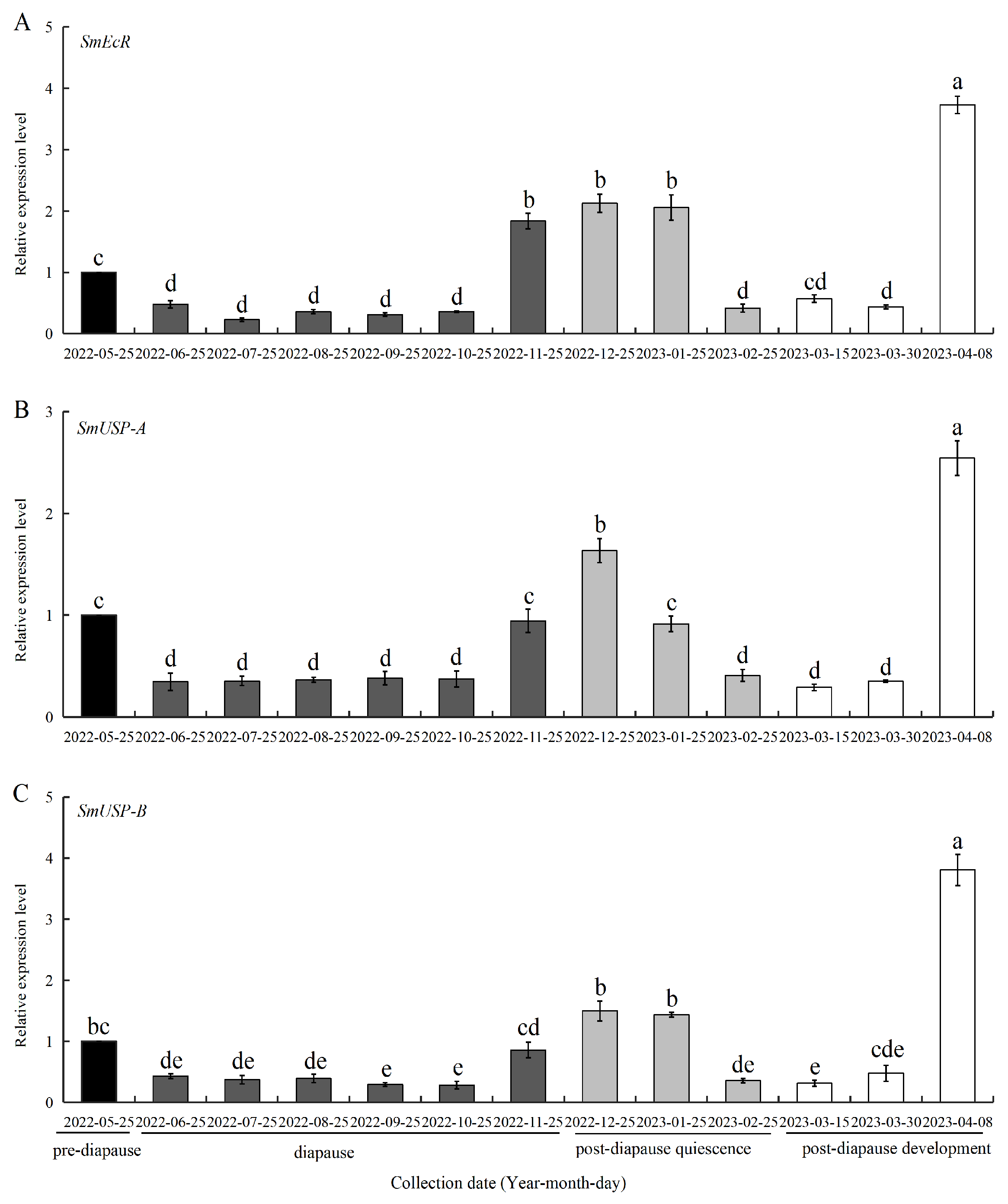
3.3. Expression of SmEcR and SmUSPs During Diapause

The expression profiles of SmEcR and the SmUSPs were determined in four successive diapause processes of S. mosellana; specifically, 3rd instar larvae, including the pre-diapause, diapause, post-diapause quiescence, and post-diapause developmental stages, as shown in Figure 4. SmEcR expression was downregulated upon entry into diapause (June), with low expression throughout diapause (June-October), but was significantly upregulated from late November, when most S. mosellana end diapause and enter post-diapause quiescence [36], after which expression remained constant until the mid-quiescent period (January). Expression then dropped sharply, falling back to the level at the early-to-mid period of post-diapause development (mid-to-late March). Thus, the highest expression was observed during the late developmental stage (early April) (Figure 4A).
Similarly, the expression levels of SmUSP-A and SmUSP-B were low during diapause and the early-to-mid stage of post-diapause development, peaking at the late stage of post-diapause development, which was then the quiescent period in December. In contrast to SmEcR, whose expression during early-to-mid quiescence (November–January) was stable and significantly higher than that at pre-diapause (May), the levels of SmUSP-A and SmUSP-B expression were both higher in December than those in November and January, with the November and January levels similar to those observed again during the mid-to-late period of post-diapause development (Figure 4B,C).

3.4. 20E Regulation of SmEcR and SmUSP Genes During Diapause

Treatment of larvae in diapause with 20E (0.1–0.6 pg/nL) resulted in significant upregulation of SmEcR and SmUSP-A 1 h after treatment with 0.4 pg/nL 20E and 3 h after treatment at concentrations of 0.2–0.6 pg/nL for SmEcR and 0.2–0.4 pg/nL for SmUSP-A. The maximum expression values were observed at 3 h after treatment with 0.4 pg/nL 20E for SmEcR and SmUSP-A, with 2.5-fold and 2.1-fold increases relative to the control, respectively (Figure 5A,B). The expression of SmUSP-B only increased significantly at 3 h after treatment with 0.4 pg/nL (1.5-fold) and 0.6 pg/nL (1.3-fold) (Figure 5C). Notably, the expression of all three genes was downregulated for all concentrations at the 6 h time point after treatment.

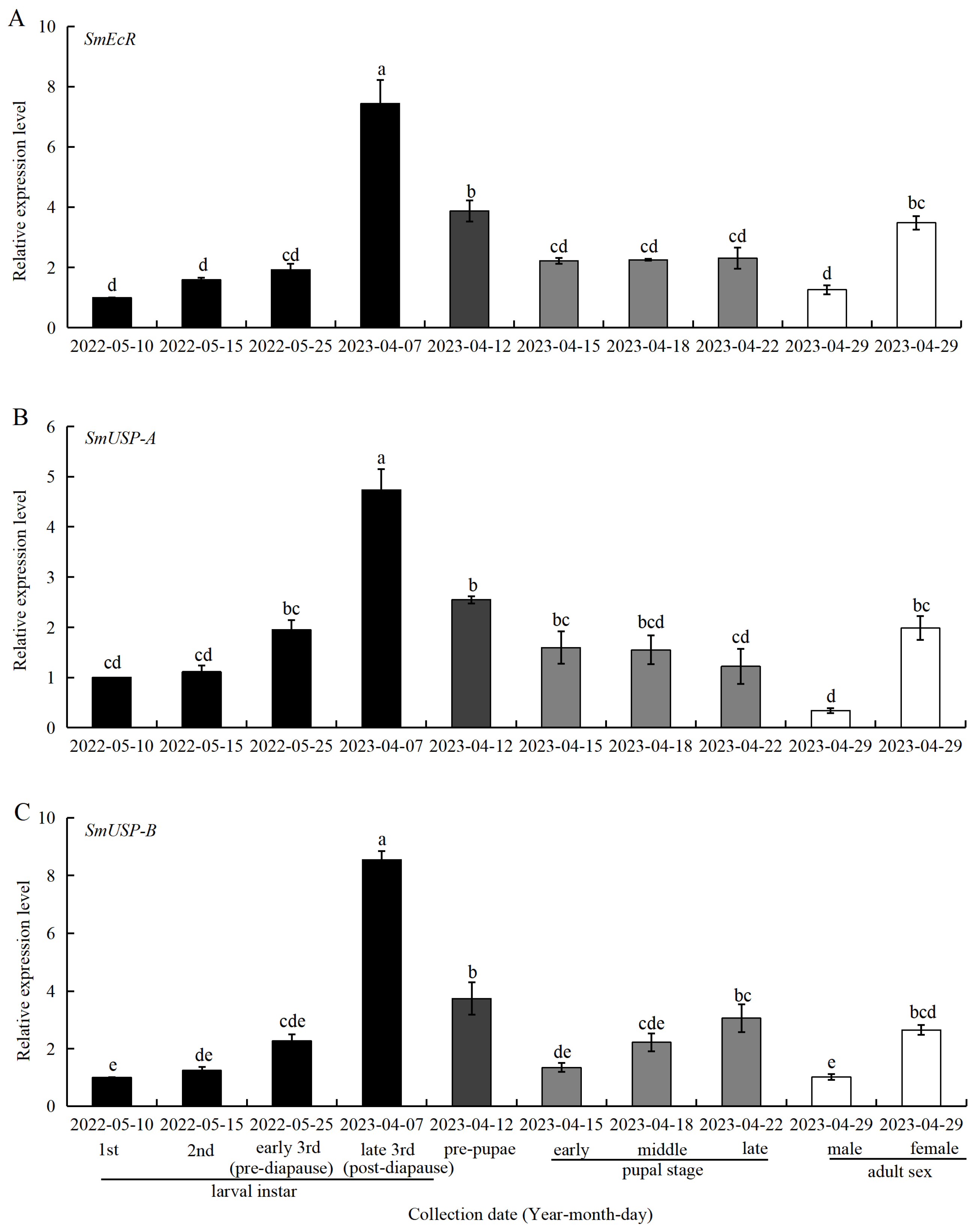
3.5. Expression of SmEcR and SmUSP Genes at Different Developmental Stages

The expression profiles of SmEcR, SmUSP-A, and SmUSP-B were also determined across all developmental stages of S. mosellana, including larval instars (1st, 2nd, early-3rd, and late-3rd instar larvae), pre-pupae, and pupae (early, middle, and late-stage pupae), as well as adults (males and females) (Figure 6).
The expression profiles of the three genes varied significantly among the different developmental stages (p < 0.05), with the highest expression levels seen in the late-3rd instar larvae. Marked reductions in expression were observed in the transition to the pre-pupal stage, with further declines on development into pupae. The expression of SmEcR remained unchanged during the pupal stage, while that of SmUSP-A decreased gradually and that of SmUSP-B increased gradually. Notably, the expression of all three genes was significantly higher in females than in male adults.

4. Discussion

As functional ecdysone receptors, EcR and USP are considered pivotal in 20E-mediated larval diapause. However, the functions of these proteins during larval diapause are less well understood. In this study, we identified and characterized EcR and USP genes (SmEcR, SmUSP-A, and SmUSP-B) from S. mosellana, a significant wheat pest that undergoes typical obligatory larval diapause, and explored their expression profiles throughout the insect life cycle, as well as their responsiveness to 20E treatment. In contrast to many molecular investigations of insect development, all insect samples utilized in this study were obtained directly from field collections, potentially mirroring natural conditions more accurately.
Similar to the findings of previous studies, the identified SmEcR and SmUSPs contained the canonical structural domains of the nuclear receptor superfamily (Figure 1) [46,47]. In particular, the DNA-binding domains (DBDs), characterized by two zinc-finger motifs containing a proximal P-box and D-box sequences, were found to be highly conserved in the three putative proteins (Figure 2A,B). The P-box mediates base-specific recognition of hormone response element half-sites [48], whereas the D-box stabilizes homo-/heterodimer formation via hydrophobic packing [49], a mechanism co-opted in Drosophila to mediate EcR/USP transcriptional activity during metamorphosis [50], suggesting a conserved role for SmEcR/SmUSP in stage-specific gene regulation. Notably, the two SmUSP proteins also contained a T-box within the DBD domain (Figure 2B), which is crucial in mediating response element recognition via monomeric interactions with cognate sequences [44,51].
Deficiencies in 20E are documented to attenuate ecdysis-triggering hormone signaling and suppress the synthesis of juvenile hormone (JH), ultimately triggering diapause in Colaphellus bowringi [6]. Conversely, elevated concentrations of 20E promote diapause termination in Bactrocera minax and Helicoverpa zea by inducing the expression of EcR [9,52]. The potential roles of SmEcR and SmUSPs in the sensing and relaying of 20E signals during larval diapause were supported by the observed correspondence between gene expression patterns (Figure 4) and ecdysteroid titers [37], as well as the responsiveness of these genes to 20E induction (Figure 5). Notably, elevated expression of SmEcR and the SmUSP genes was observed in the early period of post-diapause quiescence (December), characterized by identical morphology to diapause [36]. At this stage, S. mosellana shows increased biosynthesis of both JH and ecdysteroid [37,53]. Presumably, the stage of larval quiescence state arises from the synergistic actions of ecdysteroid and JH, wherein ecdysteroid triggers diapause termination and growth by regulating the expression of SmEcR and the SmUSPs, counteracted by the antagonistic effect of JH mediated via SmMet and SmKr-h1 [54]. These findings suggest that the diapause of Sitodiplosis mosellana is related to the expression of EcR or USPs. Therefore, future pest management strategies that focus on regulating diapause by interfering with the expression of these genes may provide a means to break their diapause characteristics and reduce the accumulation of pest sources, thereby promoting effective pest control of this major pest.
It is generally believed that a conspicuous drop in JH titer coupled with a sharp increase in ecdysteroid levels during the last instar larval stage of holometabolous insects heralds the imminent transformation from larva to pupa [55,56,57]. During this period, 20E actions are mediated by the EcR and USP heterodimer [14]. In the melon fly, Zeugodacus cucurbitae, EcR expression in the penultimate (second) and final (third) instar larvae was found to be substantially higher than that in the pupae, and its suppression halted the larval–pupal transition [58]. Similarly, Grapholita molesta exhibited heightened expression of USP prior to the final larval molt and during the nascent pupal stage [59]. In the current study, the expression of S. mosellana EcR and USPs in the late last instar larval stage was significantly higher than that in the pre-pupae and pupae (Figure 6A,C), echoing the significant elevations in ecdysteroid titers seen before pupation and the concomitant reduction in the expression levels of the JH receptor SmMet and its downstream transcription factor SmKr-h1 [37,54]. The temporal expression patterns of SmEcR and SmUSPs are indicative of their pivotal role in the progression of larval and pupal development. Interestingly, the expression of SmUSP-A gradually decreased from the primary pupa stage to the postpupal stage, but SmUSP-B gradually increased (Figure 6B,C). Previous studies have shown that reduced USP expression in Apis mellifera may trigger the developmental pathways that stabilize cuticle formation by inhibiting the formation of the ecdysone receptor complex [60], whereas USP expression in Drosophila melanogaster was associated with stage-specific transcriptional regulation and its synergistic interaction with EcR [61]. Presumably, the opposite expression patterns of SmUSP-A and SmUSP-B in the pupal stage suggest potential antagonistic or complementary roles in regulating pupal development, though their functional divergence remains unresolved.
The results of recent studies have also implicated EcR and USP in the regulation of insect reproduction. For instance, inhibiting the expression of EcR and USP in female adults of the melon aphid, Aphis gossypii, and the desert locust, Schistocerca gregaria, reduces both insect chorion formation and oviposition, thereby markedly decreasing their reproductive capacity [46,62]. Suppression of EcR and USP expression in the adult female citrus psyllids, Diaphorina citri, was found to significantly impede ovarian maturation and curtail egg output [23]. The observed higher expression of EcR and the USPs in female S. mosellana adults compared to their male counterparts (Figure 6) suggests the potential involvement of these genes in the reproductive process.

5. Conclusions

In summary, the results of the present study shed light on the molecular characterization of S. mosellana EcR and USP proteins, ecdysone-responsive transcription factors involved in 20E-mediated larval diapause and adult reproduction. A deeper understanding of their regulatory mechanisms may offer novel strategies for the effective management of this insect pest.

Supplementary Materials

The following supporting information can be downloaded at https://www.mdpi.com/article/10.3390/insects16050537/s1. Figure S1. Nucleic acid and predicted amino acid sequences of SmEcR in Sitodiplosis mosellana. Start codon (ATG) and stop codon (TAG) were marked with an ellipse. A/B, C, D, E and F domains are indicated by shadows. Two zinc-finger motifs “CLVCGDRASGYHYNALTCEGC” and “CKFGHECEMDMYMRRKCQECRLKKC” are emphasized with underlines. The P-box (EGCKG) and D-box (KFGHE) are boxed; Figure S2. Nucleic acid and predicted amino acid sequences of SmUSP-A and SmUSP-B in Sitodiplosis mosellana. Start codon (ATG) and stop codon (TAA) were marked with an ellipse. A/B, C, D and E domains are indicated by shadows. Two zinc-finger motifs “CSICGDRASGKHYGVYSCEGC” and “CREDKNCTIDKRQRNRCQYC” are emphasized with underlines. The P-box (EGCKG), D-box (REDKN) and T-box (REAVQEERQ) are boxed.

Author Contributions

Conceptualization, W.C. and K.Z.-S.; methodology, Q.H.; investigation, Q.H.; formal analysis, Q.H., L.M., Y.L. and W.C.; writing—original draft preparation, Q.H. and W.C.; writing—review and editing, W.C. and K.Z.-S. All authors have read and agreed to the published version of the manuscript.

Funding

This work was supported by the Natural Science Foundation of Shandong Province (ZR2023QC051), Shandong Academy of Agricultural Sciences, Agricultural Science and Technology Innovation Engineering (CXGC2024F19, CXGC2025F18), the National Natural Science Foundation of China (31371933), and the Key Research and Development Program of Shaanxi Province, China (2020NY-059).

Data Availability Statement

The original contributions presented in this study are included in the article/Supplementary Materials. Further inquiries can be directed to the corresponding authors.

Conflicts of Interest

The authors declare no conflicts of interest.

References

  1. Hahn, D.A.; Denlinger, D.L. Meeting the energetic demands of insect diapause: Nutrient storage and utilization. J. Insect Physiol. 2007, 53, 760–773. [Google Scholar] [CrossRef]
  2. Zonato, V.; Collins, L.; Pegoraro, M.; Tauber, E.; Kyriacou, C.P. Is diapause an ancient adaptation in Drosophila? J. Insect Physiol. 2017, 98, 267–274. [Google Scholar] [CrossRef]
  3. Denlinger, D.L. Why study diapause? Entomol. Res. 2008, 38, 1–9. [Google Scholar] [CrossRef]
  4. Riddiford, L.M.; Hiruma, K.; Zhou, X.; Nelson, C.A. Insights into the molecular basis of the hormonal control of molting and metamorphosis from Manduca sexta and Drosophila melanogaster. Insect Biochem. Mol. Biol. 2003, 33, 1327–1338. [Google Scholar] [CrossRef] [PubMed]
  5. Yamanaka, N.; Rewitz, K.F.; O’Connor, M.B. Ecdysone control of developmental transitions: Lessons from Drosophila research. Annu. Rev. Entomol. 2013, 58, 497–516. [Google Scholar] [CrossRef]
  6. Guo, S.; Tian, Z.; Wu, Q.W.; King-Jones, K.; Liu, W.; Zhu, F.; Wang, X.P. Steroid hormone ecdysone deficiency stimulates preparation for photoperiodic reproductive diapause. PLoS Genet. 2021, 17, e1009352. [Google Scholar] [CrossRef]
  7. Singtripop, T.W.; Wanichacheewa, S.; Tsuzuki, S.; Sakurai, S. Larval growth and diapause in a tropical moth, Omphisa fuscidentalis Hampson. Zool. Sci. 1999, 16, 725–733. [Google Scholar] [CrossRef]
  8. Singtripop, T.; Oda, Y.; Wanichacheewa, S.; Sakurai, S. Sensitivities to juvenile hormone and ecdysteroid in the diapause larvae of Omphisa fuscidentalis based on the hemolymph trehalose dynamics index. J. Insect Physiol. 2002, 48, 817–824. [Google Scholar] [CrossRef] [PubMed]
  9. Reynolds, J.A.; Nachman, R.J.; Denlinger, D.L. Distinct microRNA and mRNA responses elicited by ecdysone, diapause hormone and a diapause hormone analog at diapause termination in pupae of the corn earworm, Helicoverpa zea. Gen. Comp. Endocrinol. 2019, 278, 68–78. [Google Scholar] [CrossRef]
  10. Li, Y.L.; Yan, Z.B.; Yin, X.M.; Lei, Z.S.; Zhou, Z. Dynamics of ecdysone titer and the interference of methoxyfenozide on diapause during the development of Carposina sasakii (Lepidoptera: Carposinidae). Acta Entomol. Sin. 2022, 65, 791–798. [Google Scholar]
  11. Liao, J.; Cai, D.X.; Geng, S.L.; Lyu, Z.P.; Wu, Y.L.; Guo, J.J.; Li, H.Y. Transcriptome-based analysis reveals a crucial role of the 20E/HR3 pathway in the diapause of Pieris rapae. Pestic. Biochem. Physiol. 2024, 199, 105787. [Google Scholar] [CrossRef]
  12. Yao, T.P.; Segraves, W.A.; Oro, A.E.; McKeown, M.; Evans, R.M. Drosophila ultraspiracle modulates ecdysone receptor function via heterodimer formation. Cell 1992, 71, 63–72. [Google Scholar] [CrossRef]
  13. Swevers, L.; Cherbas, L.; Cherbas, P.; Iatrou, K. Bombyx EcR (BmEcR) and Bombyx USP (BmCF1) combine to form a functional ecdysone receptor. Insect Biochem. Mol. Biol. 1996, 26, 217–221. [Google Scholar] [CrossRef] [PubMed]
  14. Mazina, M.Y.; Kocheryzhkina, E.V.; Nikolenko, J.V.; Krasnov, A.N.; Georgieva, S.G.; Vorobyeva, N.E. Nuclear receptors EcR, Usp, E75, DHR3, and ERR regulate transcription of ecdysone cascade genes. Dokl. Biochem. Biophys. 2017, 473, 145–147. [Google Scholar] [CrossRef] [PubMed]
  15. King-Jones, K.; Charles, J.P.; Lam, G.; Thummel, C.S. The ecdysone-induced DHR4 orphan nuclear receptor coordinates growth and maturation in Drosophila. Cell 2005, 121, 773–784. [Google Scholar] [CrossRef] [PubMed]
  16. Li, K.; Li, S.; Cao, Y. Transcriptional regulation by 20-hydroxyecydsone and its nuclear receptor EcR-USP. Acta Entomol. Sin. 2011, 54, 933–937. [Google Scholar]
  17. Lenaerts, C.; Van Wielendaele, P.; Peeters, P.; Vanden Broeck, J.; Marchal, E. Ecdysteroid signalling components in metamorphosis and development of the desert locust, Schistocerca gregaria. Insect Biochem. Mol. Biol. 2016, 75, 10–23. [Google Scholar] [CrossRef]
  18. Talbot, W.S.; Swyryd, E.A.; Hogness, D.S. Drosophila tissues with different metamorphic responses to ecdysone express different ecdysone receptor isoforms. Cell 1993, 73, 1323–1337. [Google Scholar] [CrossRef]
  19. Minakuchi, C.; Nakagawa, Y.; Kiuchi, M.; Tomita, S.; Kamimura, M. Molecular cloning, expression analysis and functional confirmation of two ecdysone receptor isoforms from the rice stem borer Chilo suppressalis. Insect Biochem. Mol. Biol. 2002, 32, 999–1008. [Google Scholar] [CrossRef]
  20. Ogura, T.; Minakuchi, C.; Nakagawa, Y.; Smagghe, G.; Miyagawa, H. Molecular cloning, expression analysis and functional confirmation of ecdysone receptor and ultraspiracle from the Colorado potato beetle Leptinotarsa decemlineata. FEBS J. 2005, 272, 4114–4128. [Google Scholar] [CrossRef]
  21. Zheng, W.W.; Yang, D.T.; Wang, J.X.; Song, Q.S.; Gilbert, L.I.; Zhao, X.F. Hsc70 binds to ultraspiracle resulting in the upregulation of 20-hydroxyecdsone-responsive genes in Helicoverpa armigera. Mol. Cell. Endocrinol. 2010, 315, 282–291. [Google Scholar] [CrossRef]
  22. Weng, H.B.; Shen, W.F.; Liu, Y.; He, L.H.; Niu, B.L.; Meng, Z.Q.; Mu, J.J. Cloning and characterization of two EcR isoforms from Japanese pine sawyer, Monochamus alternates. Arch. Insect Biochem. Physiol. 2013, 84, 27–42. [Google Scholar] [CrossRef] [PubMed]
  23. Zhang, C.; Wan, B.; Jin, M.R.; Wang, X.; Wei, Y.J.; Zhong, L.; Xia, B. Inhibition of ecdysone receptor (DcEcR) and ultraspiracle (DcUSP) expression in Diaphorina citri increased susceptibility to pesticides. Pestic. Biochem. Physiol. 2023, 194, 105518. [Google Scholar] [CrossRef] [PubMed]
  24. Kapitskaya, M.; Wang, S.; Cress, D.E.; Dhadialla, T.S.; Raikhel, A.S. The mosquito ultraspiracle homologue, a partner of ecdysteroid receptor heterodimer: Cloning and characterization of isoforms expressed during vitellogenesis. Mol. Cell. Endocrinol. 1996, 121, 119–132. [Google Scholar] [CrossRef]
  25. Vögtli, M.; Imhof, M.O.; Brown, N.E.; Rauch, P.; Spindler-Barth, M.; Lezzi, M.; Henrich, V.C. Functional characterization of two Ultraspiracle forms (CtUSP-1 and CtUSP-2) from Chironomus tentans. Insect Biochem. Mol. Biol. 1999, 29, 931–942. [Google Scholar] [CrossRef] [PubMed]
  26. Morishita, C.; Minakuchi, C.; Yokoi, T.; Takimoto, S.; Hosoda, A.; Akama, Y. cDNA cloning of ecdysone receptor (EcR) and ultraspiracle (USP) from Harmonia axyridis and Epilachna vigintioctopunctata and the evaluation of the binding affinity of ecdysone agonists to the in vitro translated EcR/USP heterodimers. J. Pestic. Sci. 2014, 39, 76–84. [Google Scholar] [CrossRef]
  27. Xu, J.J.; Tan, A.J.; Palli, S.R. The function of nuclear receptors in regulation of female reproduction and embryogenesis in the red flour beetle, Tribolium castaneum. J. Insect Physiol. 2010, 56, 1471–1480. [Google Scholar] [CrossRef]
  28. Xu, Q.Y.; Deng, P.; Zhang, Q.; Li, A.; Fu, K.Y.; Guo, W.C.; Li, G.Q. Ecdysone receptor isoforms play distinct roles in larval-pupal-adult transition in Leptinotarsa decemlineata. Insect Sci. 2020, 27, 487–499. [Google Scholar] [CrossRef]
  29. Wu, J.J.; Mu, L.L.; Kang, W.N.; Ze, L.J.; Shen, C.H.; Jin, L.; Anjum, A.A.; Li, G.Q. RNA interference targeting ecdysone receptor blocks the larval-pupal transition in Henosepilachna vigintioctopunctata. Insect Sci. 2021, 28, 419–429. [Google Scholar] [CrossRef]
  30. Imhof, M.O.; Rusconi, S.; Lezzi, M. Cloning of a Chironomus tentans cDNA encoding a protein (cEcRH) homologous to the Drosophila melanogaster ecdysteroid receptor (dEcR). Insect Biochem. Mol. Biol. 1993, 23, 115–124. [Google Scholar] [CrossRef]
  31. Perera, S.C.; Palli, S.R.; Ladd, T.R.; Krell, P.J.; Retnakaran, A. The ultraspiracle gene of the spruce budworm, Choristoneura fumiferana: Cloning of cDNA and developmental expression of mRNA. Dev. Genet. 1998, 22, 169–179. [Google Scholar] [CrossRef]
  32. Jacquemin, G.; Chavalle, S.; De Proft, M. Forecasting the emergence of the adult orange wheat blossom midge, Sitodiplosis mosellana (Géhin) (Diptera: Cecidomyiidae) in Belgium. Crop Prot. 2014, 58, 6–13. [Google Scholar] [CrossRef]
  33. Shrestha, G.; Reddy, G.V.P. Field efficacy of insect pathogen, botanical, and jasmonic acid for the management of wheat midge Sitodiplosis mosellana and the impact on adult parasitoid Macroglenes penetrans populations in spring wheat. Insect Sci. 2019, 26, 523–535. [Google Scholar] [CrossRef]
  34. Wang, Q.; Liu, X.B.; Liu, H.; Fu, Y.; Cheng, Y.M.; Zhang, L.J.; Shi, W.P.; Zhang, Y.; Chen, J.L. Transcriptomic and metabolomic analysis of wheat kernels in response to the feeding of orange wheat blossom midges (Sitodiplosis mosellana) in the Field. J. Agric. Food. Chem. 2022, 70, 1477–1493. [Google Scholar] [CrossRef] [PubMed]
  35. Wang, Y.; Long, Z.R.; Feng, A.R.; Cheng, W.N. Effects of initial population number, wheat varieties and precipitation on infestation of Sitodiplosis mosellana (Diptera: Cecidomyiidae). Acta Agri. Boreali-Occident. Sin. 2015, 24, 165–171. [Google Scholar]
  36. Cheng, W.N.; Long, Z.R.; Zhang, Y.D.; Liang, T.T.; Zhu-Salzman, K.Y. Effects of temperature, soil moisture and photoperiod on diapause termination and post-diapause development of the wheat blossom midge, Sitodiplosis mosellana (Géhin) (Diptera: Cecidomyiidae). J. Insect Physiol. 2017, 103, 78–85. [Google Scholar] [CrossRef]
  37. Cheng, W.N.; Li, J.J.; Li, Y.P.; Li, X.L.; Wu, J.X.; Wang, H.L. Quantitative analysis of ecdysteroid in adults and the pre-diapause, diapause and post-diapause larvae of wheat blossom midge, Sitodiplosis mosellana Gehin. Acta Phytophy. Sin. 2009, 36, 163–167. [Google Scholar]
  38. Cheng, W.N.; Li, D.; Wang, Y.; Liu, Y.; Zhu-Salzman, K.Y. Cloning of heat shock protein genes (hsp70, hsc70 and hsp90) and their expression in response to larval diapause and thermal stress in the wheat blossom midge, Sitodiplosis mosellana. J. Insect Physiol. 2016, 95, 66–77. [Google Scholar] [CrossRef] [PubMed]
  39. Gagne, R.J.; Doane, J.E. The larval instars of the wheat midge, Sitodiplosis mosellana (Gehin) (Diptera: Cecidomyiidae). Proc. Entomol. Soc. Wash. 1999, 101, 57–63. [Google Scholar]
  40. Gong, Z.J.; Li, T.; Miao, J.; Duan, Y.; Jiang, Y.L.; Li, H.L.; Guo, P.; Wang, X.Q.; Zhang, J.; Wu, Y.Q. A chromosome-level genome assembly of the orange wheat blossom midge, Sitodiplosis mosellana Géhin (Diptera: Cecidomyiidae) provides insights into the evolution of a detoxification system. G3 2022, 12, jkac161. [Google Scholar] [CrossRef]
  41. Hwang, D.S.; Lee, B.Y.; Kim, H.S.; Lee, M.C.; Kyung, D.H.; Om, A.S.; Rhee, J.S.; Lee, J.S. Genome-wide identification of nuclear receptor (NR) superfamily genes in the copepod Tigriopus japonicus. BMC Genom. 2014, 15, 993. [Google Scholar] [CrossRef] [PubMed]
  42. Tamura, K.; Stecher, G.; Peterson, D.; Filipski, A.; Kumar, S. MEGA6: Molecular evolutionary genetics analysis version 6.0. Mol. Biol. Evol. 2013, 30, 2725–2729. [Google Scholar] [CrossRef]
  43. Livak, K.J.; Schmittgen, T.D. Analysis of relative gene expression data using real-time quantitative PCR and the 2−ΔΔCT method. Methods 2001, 25, 402–408. [Google Scholar] [CrossRef]
  44. Nakagawa, Y.; Henrich, V.C. Arthropod nuclear receptors and their role in molting. FEBS J. 2009, 276, 6128–6157. [Google Scholar] [CrossRef] [PubMed]
  45. Yao, Q.; Xu, S.; Dong, Y.Z.; Quan, L.F.; Chen, B.X. Ecdysone Receptor (EcR) and ultraspiracle protein (USP) genes from Conopomorpha sinensis Bradley eggs: Identification and expression in response to insecticides. Front. Physiol. 2020, 11, 851. [Google Scholar] [CrossRef]
  46. Yan, T.; Chen, H.M.; Sun, Y.W.; Yu, X.D.; Xia, L.Q. RNA interference of the ecdysone receptor genes EcR and USP in Grain Aphid (Sitobion avenae F.) affects its survival and fecundity upon feeding on wheat plants. Int. J. Mol. Sci. 2016, 17, 2098. [Google Scholar] [CrossRef]
  47. Abdullah-Zawawi, M.R.; Afiqah-Aleng, N.; Ikhwanuddin, M.; Sung, Y.Y.; Tola, S.; Fazhan, H.; Waiho, K. Recent development in ecdysone receptor of crustaceans: Current knowledge and future applications in crustacean aquaculture. Rev. Aquacult. 2021, 13, 1938–1957. [Google Scholar] [CrossRef]
  48. Germain, P.; Staels, B.; Dacquet, C.; Spedding, M.; Laudet, V. Overview of nomenclature of nuclear receptors. Pharmacol. Rev. 2006, 58, 685–704. [Google Scholar] [CrossRef]
  49. Bain, D.L.; Heneghan, A.F.; Connaghan-Jones, K.D.; Miura, M.T. Nuclear receptor structure: Implications for function. Annu. Rev. Physiol. 2007, 69, 201–220. [Google Scholar] [CrossRef]
  50. King-Jones, K.; Thummel, C.S. Nuclear receptors-a perspective from Drosophila. Nat. Rev. Genet. 2005, 6, 311–323. [Google Scholar] [CrossRef]
  51. Devarakonda, S.; Harp, J.M.; Kim, Y.; Ozyhar, A.; Rastinejad, F. Structure of the heterodimeric ecdysone receptor DNA-binding complex. EMBO J. 2003, 22, 5827–5840. [Google Scholar] [CrossRef] [PubMed]
  52. Chen, Z.Z.; Dong, Y.C.; Wang, Y.H.; Andongma, A.A.; Rashid, M.A.; Krutmuang, P.; Niu, C.Y. Pupal diapause termination in Bactrocera minax: An insight on 20-hydroxyecdysone induced phenotypic and genotypic expressions. Sci. Rep. 2016, 6, 27440. [Google Scholar] [CrossRef] [PubMed]
  53. Li, Y.M.; Wu, J.X.; Cheng, W.N.; Li, Y.P.; Hou, J.J. Determination of juvenile hormone in wheat blossom midge, Sitodiplosis mosellana (Gehin). Acta Agric. Boreali-Occident. Sin. 2006, 15, 73–75. [Google Scholar]
  54. Cheng, W.N.; Li, X.J.; Zhao, J.J.; Zhu-Salzman, K. Cloning and characterization of Methoprene-tolerant (Met) and Krüppel homolog 1 (Kr-h1) genes in the wheat blossom midge, Sitodiplosis mosellana. Insect Sci. 2020, 27, 292–303. [Google Scholar] [CrossRef]
  55. Ureña, E.; Chafino, S.; Manjón, C.; Franch-Marro, X.; Martín, D. The occurrence of the holometabolous pupal stage requires the interaction between E93, Krüppel-Homolog 1 and Broad-Complex. PLoS Genet. 2016, 12, e1006020. [Google Scholar] [CrossRef] [PubMed]
  56. Kayukawa, T.; Jouraku, A.; Ito, Y.; Shinoda, T. Molecular mechanism underlying juvenile hormone-mediated repression of precocious larval-adult metamorphosis. Proc. Natl. Acad. Sci. USA 2017, 114, 1057–1062. [Google Scholar] [CrossRef]
  57. Pan, X.Y.; Connacher, R.P.; O’Connor, M.B. Control of the insect metamorphic transition by ecdysteroid production and secretion. Curr. Opin. Insect Sci. 2021, 43, 11–20. [Google Scholar] [CrossRef]
  58. Ahmad, S.; Jamil, M.; Jaworski, C.C.; Wu, Y.; Palma-Onetto, V.; Lyu, B.Q.; Luo, Y.P. Knockdown of the ecdysone receptor disrupts development and causes mortality in the melon fly, Zeugodacus cucurbitae. Insect Mol. Biol. 2023, 32, 738–747. [Google Scholar] [CrossRef]
  59. Cao, J.J.; Liu, Y.C.; Yang, Y.H.; Zhang, H.J.; Li, Z.; Yang, Q.B.; Zhang, S.D.; Zhang, Q.W.; Liu, X.X. Molecular characterization and functional analysis of the ultraspiracle (USP) in the oriental fruit moth Grapholita molesta (Lepidoptera: Olethreutidae). Comp. Biochem. Physiol. B Biochem. Mol. Biol. 2015, 190, 54–62. [Google Scholar] [CrossRef]
  60. Barchuk, A.R.; Figueiredo, V.L.; Simões, Z.L. Downregulation of ultraspiracle gene expression delays pupal development in honeybees. J. Insect Physiol. 2008, 54, 1035–1040. [Google Scholar] [CrossRef]
  61. Henrich, V.C.; Szekely, A.A.; Kim, S.J.; Brown, N.E.; Antoniewski, C.; Hayden, M.A.; Lepesant, J.A.; Gilbert, L.I. Expression and function of the ultraspiracle (usp) gene during development of Drosophila melanogaster. Dev. Biol. 1994, 165, 38–52. [Google Scholar] [CrossRef] [PubMed]
  62. Lenaerts, C.; Marchal, E.; Peeters, P.; Vanden Broeck, J. The ecdysone receptor complex is essential for the reproductive success in the female desert locust, Schistocerca gregaria. Sci. Rep. 2019, 9, 15. [Google Scholar] [CrossRef] [PubMed]
Figure 1. Schematic representation of SmEcR (A), SmUSP-A (B), and SmUSP-B (C). Functional domains of the coding region are visually depicted by distinct color-coded boxes: A/B domain (transactivation domain, yellow), C domain (DNA binding domain, DBD, red), D domain (hinge domain, blue), E domain (ligand binding domain, LBD, green), and F domain (purple).
Insects 16 00537 g001
Figure 2. Sequence alignments of Sitodiplosis mosellana EcR (SmEcR) (A) and USP (SmUSP-A and SmUSP-B) (B) proteins in relation to their homologs from other insect species. SmEcR and SmUSPs are indicated with red triangles. Identical and similar amino acids were emphasized through black and grey shading, respectively. DNA-binding domain is denoted by brackets. Two zinc-finger motifs are emphasized with double underlines. The P-box (E117-G121 for SmEcR, E125-G129 for USP-A, and E104-G108 for USP-B), D-box (K136-G140 for SmEcR, R144-N148 for USP-A, and R123-N127 for USP-B), and T-box (R174-Q182 for USP-A and R153-Q161 for USP-B) sequences were underscored with underlines. Species abbreviations: Sm, Sitodiplosis mosellana; Cn, Contarinia nasturtii; Aa, Aedes aegypti; Bm, Bombyx mori; Ag, Anopheles gambiae.
Insects 16 00537 g002
Figure 3. Phylogenetic neighbor-joining tree of Sitodiplosis mosellana EcR (SmEcR) (A) and USP (SmUSP-A and SmUSP-B) (B) proteins in relation to their homologs from other insect species. SmEcR and SmUSPs are indicated with red triangles. Species abbreviations: Sm, Sitodiplosis mosellana; Cn, Contarinia nasturtii; Aa, Aedes aegypti; Bm, Bombyx mori; Cq, Culex quinquefasciatus; Mv, Musca vetustissima; Gf, Glossina fuscipes; Px, Plutella xylostella; Ha, Helicoverpa armigera; Se, Spodoptera exigua; Am, Apis mellifera; Mq, Melipona quadrifasciata; Tc, Tribolium castaneum; Ds, Diorhabda sublineata; Gd, Galeruca daurica; Ag, Anopheles gambiae; Bc, Bradysia coprophila; Gm, Grapholita molesta; Ld, Lymantria dispar; Sl, Spodoptera litura; Tm, Tenebrio molitor; Ma, Monochamus alternatus; Ms, Melipona scutellaris; and Sd, Scaptotrigona depilis.
Insects 16 00537 g003
Figure 4. Relative mRNA levels of Sitodiplosis mosellana EcR (A), USP-A (B), and USP-B (C) in pre-diapause, diapause, and post-diapause larvae. The relative transcript abundance (means ± SE) of all tested stages was normalized against the pre-diapause larvae, which served as a calibrator with an assigned value of 1. Different letters above the bars indicated significant differences using Duncan’s multiple range test after ANOVA (p < 0.05).
Insects 16 00537 g004
Figure 5. Relative mRNA levels of Sitodiplosis mosellana EcR (A), USP-A (B), and USP-B (C) at 1, 3, and 6 h time points after 20E injections at concentrations spanning 0.1 to 0.6 pg/nL into diapausing larvae. The relative transcript abundance (means ± SE) of each treatment was normalized against the ethanol control, which served as a calibrator with an assigned value of 1. Different letters above the bars within each time point indicated significant differences using Duncan’s multiple range test after ANOVA (p < 0.05).
Insects 16 00537 g005
Figure 6. Relative expression levels of Sitodiplosis mosellana EcR (A), USP-A (B), and USP-B (C) in different developmental stages. The relative transcript abundance (means ± SE) of each stage was normalized against 1st instar larvae, which served as a calibrator with an assigned value of 1. Different letters above the bars indicated significant differences using Duncan’s multiple range test after ANOVA (p < 0.05).
Insects 16 00537 g006
Table 1. Primer sequences used in this study.
Primer NameSequence (5′ to 3′)Purpose
EcR senseTCGTGGCTCCGATTTTATORF cloning
EcR antisenseGCAACGAATCTTACGGAT
USP-A senseTGAACGAGCGACACTGAA
USP-A antisenseCACCCCAATCACTGTCAA
USP-B senseCGAGCAGATAAACCTACG
USP-B antisenseCTGGTGTGACTATCTGGC
EcR senseTGCGGCGTAAGTGTCAGGAqPCR
EcR antisenseGCTTTCTTCTCTTTGCGTTTGA
USP-A senseAATGCTTTCCGTAGCCGATATA
USP-A antisenseCTGGTTTGAGTTCAGGCGGT
USP-B senseATGGATACAAATGATTCAGCTTTGG
USP-B antisenseTGGTTGAAATTGCTGGGGC
GAPDH senseCCATCAAAGCAAGCAAGA
GAPDH antisenseCAGCACGGAGCACAAGAC
Disclaimer/Publisher’s Note: The statements, opinions and data contained in all publications are solely those of the individual author(s) and contributor(s) and not of MDPI and/or the editor(s). MDPI and/or the editor(s) disclaim responsibility for any injury to people or property resulting from any ideas, methods, instructions or products referred to in the content.

