Article

Gut Bacteria Mediate Aggregation Pheromone Release in the Borer Beetle Trigonorhinus sp.

1
Department of Forest Conservation, Forestry College, Inner Mongolia Agricultural University, Hohhot 010018, China
2
Inner Mongolia Academy of Forestry Sciences, Hohhot 010013, China
3
Ulanqab Institute of Agricultural and Forestry Sciences, Ulanqab 012001, China
4
Ulanqab Forestry Protection Station, Ulanqab 012000, China
*
Author to whom correspondence should be addressed.
These authors contributed equally to this work.
Insects 2025, 16(10), 999; https://doi.org/10.3390/insects16100999
Submission received: 21 August 2025 / Revised: 12 September 2025 / Accepted: 20 September 2025 / Published: 25 September 2025
(This article belongs to the Section Insect Behavior and Pathology)

Simple Summary

An investigation was conducted on the wood-boring beetle Trigonorhinus sp., a pest of Caragana liouana, to determine the necessity of gut bacteria for male aggregation pheromone release. Using antibiotic depletion, qPCR, GC-MS, and Y-tube olfactometry, it was verified that a marked reduction in gut bacterial load led to more than an 85% decrease in the emission of two key pheromone components (2,6,10,14-tetramethylheptadecane and heptacosane), and females no longer exhibited significant attraction to treated males. Recolonization with a specific gut bacterial isolate, Acinetobacter guillouiae, restored pheromone emission to near-control levels, demonstrating a strain-specific effect. These findings demonstrate a decisive role of specific gut taxa in the beetle’s chemical communication and suggest feasible avenues for symbiont-targeted pest management.

Abstract

Gut microbial symbionts are increasingly recognized as key modulators of host insect physiology and behavior, yet their role in pheromone-mediated chemical communication remains insufficiently understood. In this study, we investigated the wood-boring beetle Trigonorhinus sp., a pest of Caragana liouana, to determine the necessity of gut bacteria for male aggregation pheromone release. A combination of antibiotic-mediated bacterial depletion, quantitative PCR, gas chromatography-mass spectrometry (GC-MS), and Y-tube olfactometry was employed. Antibiotic treatment resulted in a marked reduction in gut bacterial load and a concomitant decrease of more than 85% in the emission of two key pheromone components, 2,6,10,14-tetramethylheptadecane and heptacosane. Behavioral assays demonstrated that females no longer exhibited significant attraction to treated males. Furthermore, defined recolonization with a single cultured gut isolate, Acinetobacter guillouiae, was sufficient to rescue pheromone emission. This indicates that particular gut taxa, rather than microbial biomass alone, are essential for pheromone biosynthesis. These findings demonstrate a decisive role of gut bacteria in the chemical communication of Trigonorhinus sp. and highlight the potential of symbiont-targeted strategies for pest management.

1. Introduction

The gut microbes of insects can strongly influence their chemical communication. This has become a major research area in entomology. These microbes can control behaviors and bodily functions that scientists once thought were controlled by genes alone [1,2,3,4]. One such insect is the wood-boring weevil Trigonorhinus sp. (Anthribidae), a major pest of Caragana liouana shrubs in arid regions [5]. This diminutive beetle (2.7–3.5 mm) exhibits a dark-brown to black integument with greyish-white to brown setae and convergent traits with its woody substrate, including distinctly punctate elytra and elongate, multi-segmented antennae that couple camouflage with chemosensory acuity. Sexual dimorphism is subtle but diagnostic: females possess an elongate sixth abdominal sternite, enabling rapid, reliable identification in the field [6].
The damage from Trigonorhinus sp. is severe. By killing C. liouana, a key plant, it harms the entire dryland ecosystem. Females oviposit on current-year shoots; upon hatching, larvae tunnel into the plant’s veins and create galls. These galls block water flow, which causes the branches to die [7]. These galling processes co-opt host development, redirecting resources toward anomalous tissue growth while provisioning developing larvae. Reproduction peaks from May to August in synchrony with host phenology, suggesting long-term co-adaptation, yet recent range expansions across Ningxia, Inner Mongolia, Shanxi, and Shaanxi indicate that environmental change may be perturbing historical population regulation [8,9,10].
Foundational work integrating morphology, behavior, and chemical ecology has accelerated understanding of this species. Song et al. [7] established taxonomic baselines and damage assessment protocols; Zhang et al. [5] clarified host gall developmental dynamics. Critically, Lei et al. [6] combined Y-tube olfactometry, solid-phase microextraction (SPME), solvent extraction, and gas chromatography–mass spectrometry-electroantennographic detection (GC-MS-EAD) to define an aggregation pheromone system comprised of 2,6,10,14-tetramethylheptadecane and heptacosane. The former is constitutively produced by both mated and unmated males, whereas the latter appears restricted to mated individuals and their frass, implying temporal and physiological regulation of pheromone output.
Studies across insects demonstrate that microbial symbionts can shape pheromone biosynthesis by supplying precursors, enzymatically modifying host compounds, or modulating host gene expression [11,12,13,14,15]. For example, the desert locust Schistocerca gregaria relies on Pantoea agglomerans for guaiacol and phenol production [16,17], and bark beetles leverage microbial partners to generate complex terpene attractants [18,19]. These advances reframe the evolution of chemical communication and highlight microbiome-informed avenues for pest management [20,21,22]. However, whether and how the gut microbiome governs aggregation pheromone biosynthesis in Trigonorhinus sp. has remained untested.
Here, we address this gap by testing the hypothesis that gut bacteria regulate male aggregation pheromone emission in Trigonorhinus sp. To test this, we disrupted the beetle’s gut bacteria and then analyzed its pheromones and behavior. Our goal was to see if the microbes directly control pheromone production. This could lead to new pest control strategies that target these microbes.

2. Materials and Methods

2.1. Insect Collection and Rearing

The study insect was identified as a species of Trigonorhinus (Coleoptera: Anthribidae) by the weevil specialist Boris Korotyaev [5]. Further identification to the species level is ongoing.
The insects were acquired by collecting galls from their host plant, C. liouana, at a single location in Horinger County, Inner Mongolia (111°51′15″ E, 40°30′48″ N). These galls were transported to the laboratory and held in an incubator at 25 °C ± 2 °C, 70 ± 10% RH, and a 16L:8D photoperiod to facilitate adult emergence. Adults from this stock colony were used for all experiments (Figure 1).
All procedures followed institutional and local guidelines, and no permits were required for this non-protected arthropod.

2.2. Gut Dissection and Sample Preparation

Adults were surface-sterilized by immersion in 75% ethanol for 1 min, followed by three rinses in sterile phosphate-buffered saline (PBS; pH 7.2–7.4). Under aseptic conditions on ice, entire guts were dissected using sterile forceps and micro-scissors. A portion of the dissected guts was immediately stored at −80 °C for subsequent DNA extraction and metabolomics analysis, while the remainder was processed fresh for culture-dependent assays. A total of 48 individuals (24 males and 24 females) were used for microbiome analyses.

2.3. Antibiotic Treatment Protocol

To generate aposymbiotic insects, ciprofloxacin was incorporated into the sterile artificial diet at a final concentration of 1 mg/mL. Newly emerged males were fed this antibiotic-containing diet for 7 days (antibiotic group, AT). Control males (CK) were fed the same sterile diet without antibiotics. The efficacy of the treatment was verified by qPCR targeting the bacterial 16S rRNA gene and by plating gut homogenates on LB agar (25 °C for 72 h) to confirm the absence of culturable bacteria. A recovery group was established by transferring antibiotic-treated males back to a normal sterile diet for 14 days to assess the restoration of the microbiome and pheromone production. Sample sizes for these experiments were as follows: CK (n = 30), AT (n = 30), and recovery (n = 20).
For microbial reconstitution, isolated bacterial strains were cultured to log phase, washed, and re-suspended in sterile PBS. The suspensions were then evenly incorporated into the sterile artificial diet to create reconstitution groups designated L1–L6, N3, and N4. Beetles were fed these diets for 7 days before analysis. A vehicle-only control, containing sterile PBS mixed into the diet, was also included. The specific strains and consortia corresponding to each label are detailed in the relevant figures and tables.

2.4. Culture-Dependent Isolation and Identification of Gut Bacteria

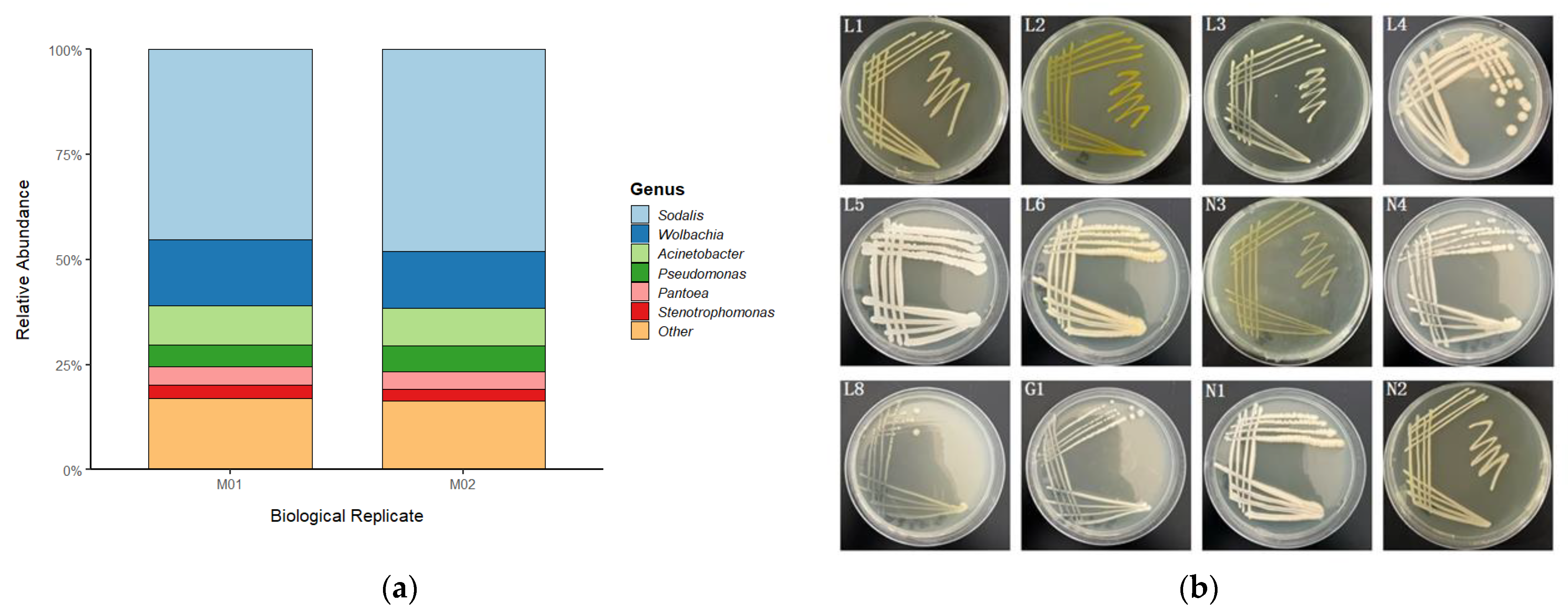
Gut homogenates were plated on Luria-Bertani (LB), Nutrient Agar (NA), and Gao’s No. 1 agar media and incubated at 25 °C. Morphologically distinct colonies were isolated and purified by repeated streaking. Genomic DNA was extracted from each pure isolate, and the 16S rDNA gene was amplified and sequenced. Taxonomic identities were assigned using the BLAST tool (version 2.14.0) against the NCBI database [23]. All isolates were preserved as glycerol stocks at −80 °C. In total, 12 bacterial isolates were obtained (Figure 2b; Table 1), and a neighbor-joining tree was constructed to visualize their phylogenetic relationships (Figure 2b).

2.5. 16S rRNA Gene Amplicon Sequencing for Bacterial Community Profiling

16S rRNA gene amplicon sequencing was employed to obtain a broad profile of the bacterial community across multiple individual insects. Total genomic DNA was extracted from individual guts using the FastDNA SPIN Kit (MP Biomedicals, Solon, OH, USA) for Soil [24]. The V3–V4 hypervariable region of the 16S rRNA gene was amplified using the primers 341F/806R [25]. The resulting amplicon libraries were sequenced on an Illumina MiSeq platform (2 × 250 bp paired-end). Extraction blanks and PCR negative controls were included in each sequencing run. Samples with fewer than 10,000 high-quality reads were excluded from further analysis.

2.6. Shotgun Metagenomic Sequencing for Unbiased Community Characterization

In a complementary approach, shotgun metagenomic sequencing was used for a deeper, unbiased analysis of the total microbial community. This method allowed for the characterization of all microbial members, including fungi and other non-bacterial taxa, and provided higher-resolution taxonomic and functional data. For this purpose, gut DNA from multiple individuals was pooled to construct two biological replicate libraries (M01 and M02) for shotgun metagenomic sequencing on an Illumina NovaSeq 6000 platform (2 × 150 bp paired-end). After quality control and removal of host-derived reads, the data were used to profile the microbial community composition.

2.7. Amplicon and Metagenome Data Processing

Amplicon data were processed using the QIIME 2 pipeline [26]. The DADA2 plugin was used for sequence denoising, merging, and chimera removal to generate amplicon sequence variants (ASVs) [27]. Taxonomy was assigned against the SILVA database [28]. Alpha diversity and beta diversity were calculated. Functional profiles were predicted from the ASV table using PICRUSt2 [29].
Metagenome data were analyzed following quality control and host read removal. Community composition metrics and visualizations, such as Good’s coverage and Principal Component Analysis (PCA), were generated to assess sequencing depth and replicate similarity.

2.8. Pheromone Collection and GC-MS Analysis

Aggregation pheromones from individual males (n = 15 per treatment group) were collected via headspace aeration. Each male was enclosed in a glass chamber, and purified air was passed through at a rate of 200 mL/min for 24 h. Volatiles were trapped on a Porapak Q adsorbent (50 mg). The trapped compounds were then eluted with 500 μL of n-hexane containing n-octadecane (10 ng/μL) as an internal standard.
The samples were analyzed on an Agilent 7890B Gas Chromatograph coupled to a 5977B Mass Spectrometer (GC-MS) (Agilent, Santa Clara, CA, USA), equipped with a 5% phenyl methyl siloxane capillary column (30 m × 250 μm × 0.25 μm). An aliquot of 1 μL of each sample was injected in splitless mode with an inlet temperature of 250 °C. High-purity helium (>99.99%) was used as the carrier gas under a constant pressure mode. The oven temperature was programmed as follows: held at 50 °C for 1 min, then ramped at 10 °C/min to 180 °C and held for 2 min, and finally increased at 20 °C/min to 240 °C with a 5 min hold. The total run time was 32 min. The mass spectrometer was operated in electron ionization (EI) mode at 70 eV, with the ion source and quadrupole temperatures maintained at 230 °C and 150 °C, respectively. Mass spectra were acquired in full scan mode over a mass range of 30–600 amu from 5.5 min to 32 min.
Each sample was analyzed with six technical replicates. The amounts of 2,6,10,14-tetramethylheptadecane and heptacosane were quantified relative to the internal standard. To account for variations in body size, pheromone quantities were normalized by the body weight of each male, which was measured to ±0.1 mg prior to collection.

2.9. Behavioral Bioassays (Y-Tube Olfactometer)

All behavioral bioassays were conducted in a dedicated testing room under controlled environmental conditions of 24–25 °C and 50–60% relative humidity. To ensure uniform lighting and eliminate external visual cues, the windows were covered with thick, opaque curtains, and illumination was provided by incandescent lamps.
A glass Y-tube olfactometer (stem: 15 cm; arms: 15 cm at a 60° angle) was used for all assays. Purified and humidified air was delivered to each arm at a constant flow rate of 400 mL/min using an atmospheric sampler (Model QC-1S; Beijing Ke’an Labor Protection Technology Co., Ltd., Beijing, China), which integrated both the vacuum pump and flowmeters. For each trial, one arm contained a live male from a specific treatment group as the odor source, while the other arm, serving as a blank control, remained empty.
A single virgin female was gently introduced at the base of the olfactometer’s stem and was observed for a total duration of 5 min. A definitive choice was recorded only if the female moved more than 5 cm into one of the arms and remained there for at least 60 consecutive seconds. Any female that entered an arm but stayed for less than 60 s, or failed to make a definitive choice within the 5 min observation period, was recorded as having “no response”. Each insect was tested only once. For each treatment, 25 responding virgin females were tested.
To prevent positional bias and minimize contamination, several procedures were followed. The sequence of testing different treatment groups was randomized. The positions of the odor source and blank control arms were swapped after every five trials. Furthermore, the entire glass apparatus was thoroughly cleaned with 75% ethanol and oven-dried at 60 °C for 30 min between each individual bioassay to eliminate any residual chemical cues.

2.10. Statistical Analysis

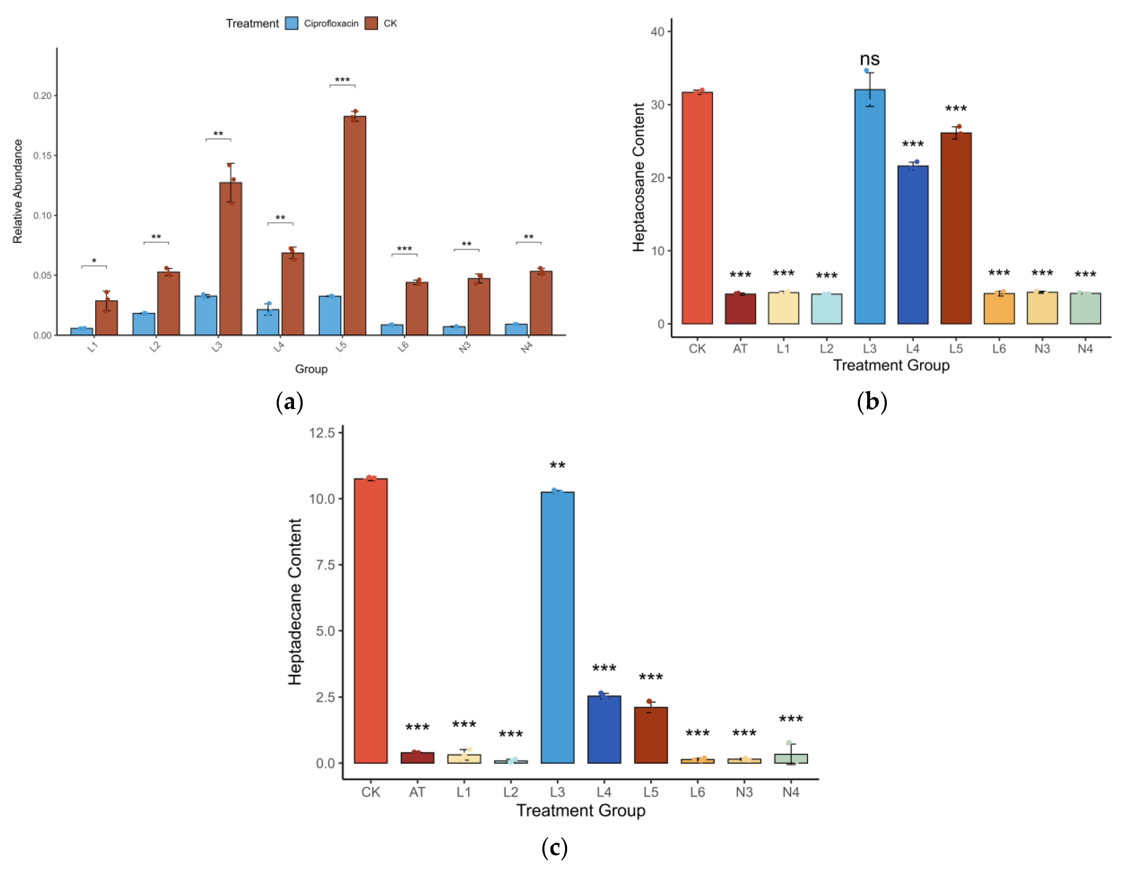
All statistical analyses were performed in R (version 4.5.0) [30]. Data normality was assessed using the Shapiro-Wilk test, and appropriate parametric or non-parametric tests were subsequently applied. Differences in alpha diversity were evaluated with the Wilcoxon rank-sum test. Differences in microbiome composition (beta diversity) were tested using PERMANOVA with the ‘adonis2’ function in the ‘vegan’ package [31]. Correlations between microbial taxa and chemical compounds were assessed using Spearman’s rank correlation with false discovery rate (FDR) correction. Y-tube choice data were analyzed using a chi-square test against an expected 50:50 distribution. A p-value of less than 0.05 was considered statistically significant for all tests. Differences in pheromone content among treatment groups (Figure 3b,c) were analyzed using a one-way Analysis of Variance (ANOVA), followed by Dunnett’s post hoc test to compare each treatment group against the control (CK) group. The effect of ciprofloxacin treatment on the relative abundance of each bacterial group (Figure 3a) was assessed using a paired t-test.

3. Results

3.1. Gut Microbiota Composition of Trigonorhinus sp.

To comprehensively characterize the gut microbiota of Trigonorhinus sp., we employed a combination of metagenomic and traditional culture-based techniques.
The metagenomic analysis revealed a community structure dominated by a few major genera (Figure 2a). Across all samples, Sodalis was the absolute dominant genus, with a relative abundance approaching 50%, followed by Wolbachia with a relative abundance of approximately 15%. Additionally, genera such as Acinetobacter, Pseudomonas, Pantoea, and Stenotrophomonas also constituted a certain proportion of the community. The microbial composition of the two biological replicates demonstrated high consistency, indicating a relatively stable gut community structure.
In a parallel, culture-dependent approach, gut homogenates were plated on various agar media. A total of 12 bacterial strains with distinct colony morphologies were isolated and purified (Figure 2b). Through 16S rDNA gene sequencing and BLAST analysis for definitive identification, these 12 isolates were identified as eight distinct species: Acinetobacter guillouiae (3 strains), Stenotrophomonas lactitubi, Pantoea plantarum, Mammaliicoccus sciuri (3 strains), Pseudomonas allii, Comamonas sediminis, Pantoea endophytica, and Brucella pseudogrignonensis.
Although the two methods differ in resolution, their results show important consistency and complementarity. Several genera obtained through culturing, such as Acinetobacter, Stenotrophomonas, Pantoea, and Pseudomonas, were also detected in the metagenomic results (Figure 2a), confirming their presence as members of this gut ecosystem. The sequencing technology revealed the presence of difficult-to-culture dominant symbionts like Sodalis and Wolbachia, while the culture-based method successfully yielded viable strains for subsequent functional studies.

3.2. Gut Bacteria Are Essential for Pheromone Release

To investigate the relationship between gut microbiota and pheromone production in Trigonorhinus sp., we quantified both bacterial abundance and pheromone levels under different treatments. qPCR analysis showed that ciprofloxacin treatment led to a dramatic reduction in gut bacterial abundance across all tested groups, with decreases ranging from 65% to over 90% compared to the control (Figure 3a). This confirmed the effective elimination of gut microbiota following antibiotic exposure.
Subsequent GC-MS analyses revealed that the elimination of gut bacteria resulted in a marked suppression of aggregation pheromone release (Figure 3b,c). In the antibiotic-treated (AT) group, both heptacosane and heptadecane levels were drastically reduced (averaging 4.07 ng and 0.39 ng, respectively), representing a reduction of over 85% compared to the control (CK) group, which showed the highest pheromone levels (averaging 31.68 ng and 10.75 ng, respectively). Notably, only the L3 group, which was recolonized with a specific microbial community, exhibited a full recovery of pheromone production, with levels comparable to the control. The L4 and L5 groups showed partial recovery, while other groups (L1, L2, L6, N3, N4) failed to restore pheromone synthesis, with levels remaining as low as those in the AT group. These results indicate that gut microbiota are indispensable for pheromone biosynthesis in male beetles, with significant differences observed among the treatment groups for both heptacosane (ANOVA, F(9, 20) = 718.2, p < 0.001) and heptadecane (ANOVA, F(9, 20) = 2047, p < 0.001). Furthermore, they suggest that only certain core bacterial taxa are capable of fully restoring this function.

3.3. Characterization of the Gut Microbiome in Trigonorhinus sp.

We studied the gut bacteria of a beetle called Trigonorhinus sp. We used two separate samples, named M01 and M02. Our sequencing was very thorough, capturing over 99.99% of the bacteria in both samples.
The number of different species in the gut was high. Both samples had similar diversity, with a Shannon index around 4.2. However, the number of unique species was different. We found 2075 species in M01 and 2288 in M02. This shows that even though the samples came from the same group, there was some natural variety.
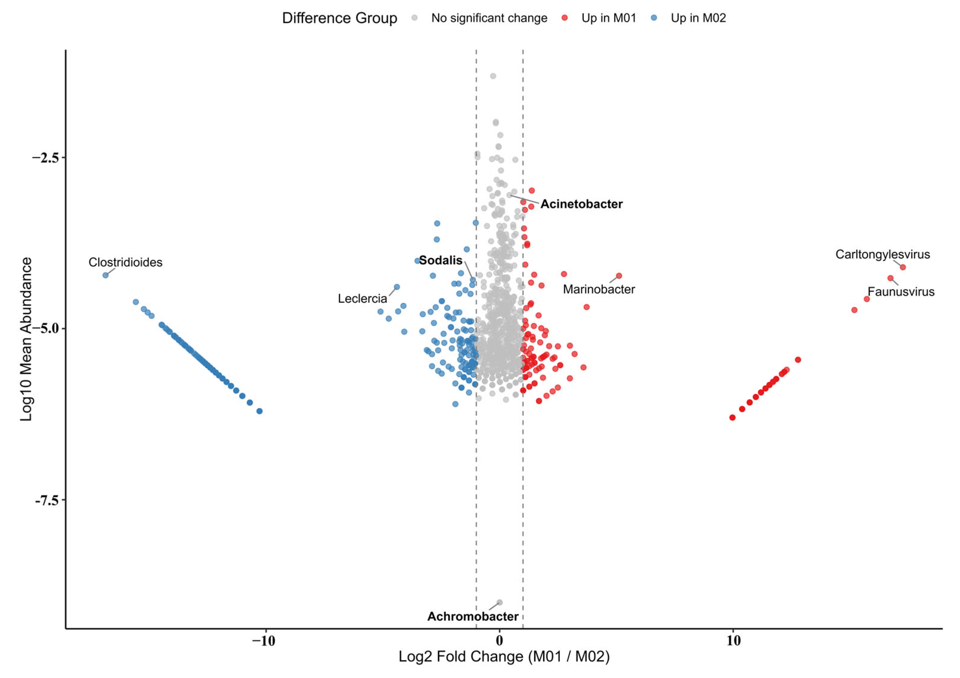
A visual analysis called PCA showed a clear difference between the two samples (Figure 4). M01 was on one side of the plot, and M02 was on the other. This difference was statistically significant. The plot shows that many bacteria were much more common in one sample than the other. For example, some bacteria like Clostridioides were much more abundant in M01. Other bacteria, like Faunusvirus and Carltongylesvirus, were more abundant in M02.
Despite these differences, the main types of bacteria were the same in both samples. The most common bacteria belonged to four main groups: Proteobacteria, Firmicutes, Bacteroidetes, and Actinobacteria. This is a common pattern in insect guts.
At a more detailed level, we found that a few genera dominated the community. Sodalis was the most common genus overall. Other important genera, like Acinetobacter, Leclercia, and Achromobacter, were also present. The consistent presence of these bacteria suggests they are a stable and important part of the beetle’s gut.

3.4. Behavioral Consequences of Microbiome Depletion

In Y-tube olfactometer assays, females showed a strong and significant preference for the odor of control (CG) males over a blank control (χ2 = 31.183, p < 0.001). This attraction was completely abolished when aposymbiotic (AT) males were used as the odor source, with females showing no preference between the odor of the AT male and the blank control (χ2 = 0.065, p = 0.799) (Table 2).

4. Discussion

This study reveals that the gut microbial community is indispensable for the production of aggregation pheromones in the borer beetle Trigonorhinus sp. Our integrated approach, combining microbial depletion and targeted recolonization with chemical and behavioral analyses, establishes a clear causal link between gut bacteria and the host’s chemical communication system. These findings reveal a multi-layered symbiosis in which gut bacteria provide nutritional support and govern host reproduction. This key function of pheromone synthesis is highly specific to Acinetobacter guillouiae, while other symbionts like Mammaliicoccus sciuri and Pseudomonas allii make partial contributions, demonstrating functional redundancy.
Our characterization of the Trigonorhinus sp. gut microbiome revealed a community dominated by the phylum Proteobacteria, which is consistent with findings in other wood-boring beetles that subsist on recalcitrant plant diets [32,33,34,35]. Notably, there was a discrepancy between our culture-dependent and metagenomic sequencing results [36,37,38,39,40,41]. While metagenomic analysis identified Sodalis as the most abundant genus, our culture-based approach successfully isolated twelve distinct strains, including members of Acinetobacter, Pantoea, and Pseudomonas. This highlights a common challenge in microbiology, where the most abundant taxa may not be the most easily culturable or, critically, the most functionally relevant for a specific metabolic task.
The central finding of our study is the definitive link between the gut microbiome and pheromone production. The near-complete cessation of pheromone release and the corresponding loss of behavioral attraction in aposymbiotic beetles provide unequivocal evidence for this dependency. Most significantly, the recolonization experiments demonstrated a high degree of functional specificity. The reintroduction of a single bacterial isolate, Acinetobacter guillouiae (L3), was sufficient to fully restore pheromone emission to control levels. In contrast, other isolates resulted in only partial (L4, L5) or no recovery at all. This strongly indicates that pheromone biosynthesis is not a generic microbial function but is dependent on specific taxa possessing the necessary metabolic capabilities.
The complete rescue by A. guillouiae points to this species as a key player in the metabolic partnership. Members of the genus Acinetobacter are renowned for their versatile metabolic capabilities, particularly in the degradation and synthesis of various hydrocarbons and lipids [42,43,44,45,46,47]. Given that the aggregation pheromones of Trigonorhinus sp. are long-chain alkanes (heptacosane and a tetramethylheptadecane), it is plausible that A. guillouiae contributes essential enzymes or precursors for the biosynthesis or modification of these compounds, a hypothesis that warrants direct investigation in future studies. The partial recovery induced by Mammaliicoccus sciuri (L4) and Pseudomonas allii (L5) suggests these bacteria may also possess some, albeit less efficient, relevant metabolic pathways.
From an applied perspective, the obligate dependence of Trigonorhinus sp. on specific gut symbionts for chemical communication presents a novel and promising target for pest management. Strategies aimed at disrupting this crucial symbiotic relationship, a concept known as “symbiont-based control,” could offer a highly specific and environmentally benign alternative to conventional insecticides [3,48,49,50,51]. This could involve the development of molecules that specifically inhibit key microbial pathways or the use of para-transgenesis to introduce engineered bacteria that interfere with pheromone production, thereby disrupting mating and controlling pest populations [52,53,54].

5. Conclusions

In conclusion, our study reveals that the gut bacterial community plays an indispensable role in the aggregation pheromone biosynthesis of the borer beetle Trigonorhinus sp. We demonstrate a clear functional link between the microbiome and the host’s chemical communication system. Specifically, we identified a single bacterial species, Acinetobacter guillouiae, capable of fully restoring pheromone production in symbiont-depleted beetles, highlighting a sophisticated and highly specific host-microbe co-adaptation. These findings not only fundamentally advance our understanding of the evolution of chemical communication and insect-microbe symbiosis but also open up new avenues for the development of innovative and sustainable symbiont-targeted strategies for the control of this important pest.

Author Contributions

Conceptualization, J.D., X.Y. and Y.Z.; methodology, X.Y. and J.D.; software, J.Y.; validation, X.W., X.L., H.Z., H.J. and J.H.; formal analysis, X.Y. and J.D.; investigation, J.D., X.Y. and J.S.; resources, X.W., X.L. and H.Z.; data curation, J.S. and J.Y.; writing—original draft preparation, J.D. and X.Y.; writing—review and editing, Y.Z.; visualization, J.S.; supervision, Y.Z.; project administration, Y.Z.; funding acquisition, Y.Z. All authors have read and agreed to the published version of the manuscript.

Funding

This research was funded by the National Natural Science Foundation of China, grant number 32160372; the Inner Mongolia Autonomous Region Natural Science Foundation, grant number 2020BS03014; the Key Research and Technology Transformation Program of Inner Mongolia Autonomous Region-Technology supports the ecological protection and high-quality development of the Yellow River Basin, grant number 2025SYFHH0087; and the Inner Mongolia Agricultural University experimental teaching equipment development and specimen making project, grant number YZ2024002. The APC was funded by the National Natural Science Foundation of China (32160372), and the Inner Mongolia Agricultural University, Internally Funded Research Project of the First-Level Discipline of Forestry, grant number LX2024-KYTD001.

Data Availability Statement

The sequencing data presented in this study are openly available in Figshare at https://doi.org/10.6084/m9.figshare.30196861.

Conflicts of Interest

The authors declare no conflicts of interest.

References

  1. Haider, K.; Abbas, D.; Galian, J.; Ghafar, M.A.; Kabir, K.; Ijaz, M.; Hussain, M.; Khan, K.A.; Ghramh, H.A.; Raza, A. The multifaceted roles of gut microbiota in insect physiology, metabolism, and environmental adaptation: Implications for pest management strategies. World J. Microbiol. Biotechnol. 2025, 41, 75. [Google Scholar] [CrossRef]
  2. Jones, R.M.; Neish, A.S. Gut microbiota in intestinal and liver disease. Annu. Rev. Pathol. Mech. Dis. 2021, 16, 251–275. [Google Scholar] [CrossRef]
  3. Mondal, S.; Somani, J.; Roy, S.; Babu, A.; Pandey, A.K. Insect microbial symbionts: Ecology, interactions, and biological significance. Microorganisms 2023, 11, 2665. [Google Scholar] [CrossRef]
  4. Siddiqui, J.A.; Khan, M.M.; Bamisile, B.S.; Hafeez, M.; Qasim, M.; Rasheed, M.T.; Rasheed, M.A.; Ahmad, S.; Shahid, M.I.; Xu, Y. Role of insect gut microbiota in pesticide degradation: A review. Front. Microbiol. 2022, 13, 870462. [Google Scholar] [CrossRef]
  5. Zhang, Y.; Hu, Y.; Jiang, H.; Zhao, S.; Lei, J.; Wang, R.; Chen, Y. Pest characteristics of Trigonorhinus sp. (Coleoptera: Anthribidae) and the developmental process of its galls in Caragana liouana Zhao, Y. Chang and Yakovlev. Can. Entomol. 2023, 155, e14. [Google Scholar] [CrossRef]
  6. Lei, J.W.; Zhao, S.G.; Li, B.L.; Zhao, P.W.; Zhang, L.Y.; Liu, X.Y.; Hu, Y.R.; Zhang, Y.R. Morphological criteria for sexing adult Trigonorhinus sp. Chin. J. Appl. Entomol. 2023, 60. [Google Scholar]
  7. Song, X.; Liu, G.; Mo, Y. Trigonorhinus sp.: A new pest in Caragana korshinskii. J. Northwest For. Univ. 2010, 25, 130–131. [Google Scholar]
  8. Castano-Sanz, V.; Gomez-Mestre, I.; Rodriguez-Exposito, E.; Garcia-Gonzalez, F. Pesticide exposure triggers sex-specific inter-and transgenerational effects conditioned by past sexual selection. Proc. R. Soc. B 2024, 291, 20241037. [Google Scholar] [CrossRef]
  9. Calixto, E.; IasmimPereira, D. Evidence of climate change effects on insect diversity. Eff. Clim. Change Insects Physiol. Evol. Ecol. Responses 2024, 179. [Google Scholar]
  10. Abbott, K.C.; Heggerud, C.M.; Lai, Y.-C.; Morozov, A.; Petrovskii, S.; Cuddington, K.; Hastings, A. When and why ecological systems respond to the rate rather than the magnitude of environmental changes. Biol. Conserv. 2024, 292, 110494. [Google Scholar] [CrossRef]
  11. Engl, T.; Kaltenpoth, M. Influence of microbial symbionts on insect pheromones. Nat. Prod. Rep. 2018, 35, 386–397. [Google Scholar] [CrossRef]
  12. Delclos, P.J.; Bouldin, T.L.; Tomberlin, J.K. Olfactory choice for decomposition stage in the burying beetle Nicrophorus vespilloides: Preference or aversion? Insects 2020, 12, 11. [Google Scholar] [CrossRef]
  13. Wada-Katsumata, A.; Zurek, L.; Nalyanya, G.; Roelofs, W.L.; Zhang, A.; Schal, C. Gut bacteria mediate aggregation in the German cockroach. Proc. Natl. Acad. Sci. USA 2015, 112, 15678–15683. [Google Scholar] [CrossRef]
  14. Wyatt, T.D. Pheromones. Curr. Biol. 2017, 27, R739–R743. [Google Scholar] [CrossRef]
  15. Ali, A.; Zeb, I.; Zahid, H. Climate Change and Plants: Biodiversity, Growth and Interactions; CRC Press: Boca Raton, FL, USA, 2021; pp. 97–111. [Google Scholar]
  16. Lavy, O.; Gophna, U.; Gefen, E.; Ayali, A. Locust bacterial symbionts: An update. Insects 2020, 11, 655. [Google Scholar] [CrossRef]
  17. Dillon, R.; Vennard, C.; Charnley, A. A note: Gut bacteria produce components of a locust cohesion pheromone. J. Appl. Microbiol. 2002, 92, 759–763. [Google Scholar] [CrossRef]
  18. Raffa, K.F. Terpenes tell different tales at different scales: Glimpses into the chemical ecology of conifer-bark beetle-microbial interactions. J. Chem. Ecol. 2014, 40, 1–20. [Google Scholar] [CrossRef]
  19. Zhao, T.; Ganji, S.; Schiebe, C.; Bohman, B.; Weinstein, P.; Krokene, P.; Borg-Karlson, A.-K.; Unelius, C.R. Convergent evolution of semiochemicals across Kingdoms: Bark beetles and their fungal symbionts. ISME J. 2019, 13, 1535–1545. [Google Scholar] [CrossRef]
  20. Douglas, A.E. Multiorganismal insects: Diversity and function of resident microorganisms. Annu. Rev. Entomol. 2015, 60, 17–34. [Google Scholar] [CrossRef]
  21. Frago, E.; Dicke, M.; Godfray, H.C.J. Insect symbionts as hidden players in insect–plant interactions. Trends Ecol. Evol. 2012, 27, 705–711. [Google Scholar] [CrossRef]
  22. Busby, P.E.; Soman, C.; Wagner, M.R.; Friesen, M.L.; Kremer, J.; Bennett, A.; Morsy, M.; Eisen, J.A.; Leach, J.E.; Dangl, J.L. Research priorities for harnessing plant microbiomes in sustainable agriculture. PLoS Biol. 2017, 15, e2001793. [Google Scholar] [CrossRef]
  23. Schoch, C.L.; Ciufo, S.; Domrachev, M.; Hotton, C.L.; Kannan, S.; Khovanskaya, R.; Leipe, D.; Mcveigh, R.; O’Neill, K.; Robbertse, B. NCBI Taxonomy: A comprehensive update on curation, resources and tools. Database 2020, 2020, baaa062. [Google Scholar] [CrossRef]
  24. Biomedicals, M. FastDNA™ SPIN Kit for Soil. Instr. Man. 2021, 1–11. Available online: https://www.mpbio.com/media/document/file/manual/dest/f/a/s/t/d/FastDNA_SPIN_Kit_for_Soil_UM_2021_WEB.pdf (accessed on 13 May 2025).
  25. Imchen, M.; Kumavath, R.; Vaz, A.B.; Góes-Neto, A.; Barh, D.; Ghosh, P.; Kozyrovska, N.; Podolich, O.; Azevedo, V. 16S rRNA gene amplicon based metagenomic signatures of rhizobiome community in rice field during various growth stages. Front. Microbiol. 2019, 10, 2103. [Google Scholar] [CrossRef]
  26. Bolyen, E.; Rideout, J.R.; Dillon, M.R.; Bokulich, N.A.; Abnet, C.C.; Al-Ghalith, G.A.; Alexander, H.; Alm, E.J.; Arumugam, M.; Asnicar, F. Reproducible, interactive, scalable and extensible microbiome data science using QIIME 2. Nat. Biotechnol. 2019, 37, 852–857. [Google Scholar] [CrossRef]
  27. Callahan, B.J.; McMurdie, P.J.; Rosen, M.J.; Han, A.W.; Johnson, A.J.A.; Holmes, S.P. DADA2: High-resolution sample inference from Illumina amplicon data. Nat. Methods 2016, 13, 581–583. [Google Scholar] [CrossRef]
  28. Pruesse, E.; Quast, C.; Knittel, K.; Fuchs, B.M.; Ludwig, W.; Peplies, J.; Glöckner, F.O. SILVA: A comprehensive online resource for quality checked and aligned ribosomal RNA sequence data compatible with ARB. Nucleic Acids Res. 2007, 35, 7188–7196. [Google Scholar] [CrossRef]
  29. Douglas, G.M.; Maffei, V.J.; Zaneveld, J.R.; Yurgel, S.N.; Brown, J.R.; Taylor, C.M.; Huttenhower, C.; Langille, M.G. PICRUSt2 for prediction of metagenome functions. Nat. Biotechnol. 2020, 38, 685–688. [Google Scholar] [CrossRef]
  30. R. Core Team, R. R: A Language and Environment for Statistical Computing. 2020. Available online: https://www.R-project.org/ (accessed on 13 May 2025).
  31. Dixon, P. VEGAN, a package of R functions for community ecology. J. Veg. Sci. 2003, 14, 927–930. [Google Scholar] [CrossRef]
  32. Schloss, P.D.; Delalibera Jr, I.; Handelsman, J.; Raffa, K.F. Bacteria associated with the guts of two wood-boring beetles: Anoplophora glabripennis and Saperda vestita (Cerambycidae). Environ. Entomol. 2006, 35, 625–629. [Google Scholar] [CrossRef]
  33. Montagna, M.; Chouaia, B.; Mazza, G.; Prosdocimi, E.M.; Crotti, E.; Mereghetti, V.; Vacchini, V.; Giorgi, A.; De Biase, A.; Longo, S. Effects of the diet on the microbiota of the red palm weevil (Coleoptera: Dryophthoridae). PLoS ONE 2015, 10, e0117439. [Google Scholar] [CrossRef]
  34. Lenka, J.; González-Tortuero, E.; Kuba, S.; Ferry, N. Bacterial community profiling and identification of bacteria with lignin-degrading potential in different gut segments of African palm weevil larvae (Rhynchophorus phoenicis). Front. Microbiol. 2025, 15, 1401965. [Google Scholar] [CrossRef] [PubMed]
  35. Jing, T.-Z.; Qi, F.-H.; Wang, Z.-Y. Most dominant roles of insect gut bacteria: Digestion, detoxification, or essential nutrient provision? Microbiome 2020, 8, 38. [Google Scholar] [CrossRef] [PubMed]
  36. Edet, U.; Antai, S.; Brooks, A.; Asitok, A.; Enya, O.; Japhet, F. An overview of cultural, molecular and metagenomic techniques in description of microbial diversity. J. Adv. Microbiol. 2017, 7, 1–19. [Google Scholar] [CrossRef]
  37. Wijayawardene, N.N.; Bahram, M.; Sanchez-Castro, I.; Dai, D.-Q.; Ariyawansa, K.G.; Jayalal, U.; Suwannarach, N.; Tedersoo, L. Current insight into culture-dependent and culture-independent methods in discovering Ascomycetous Taxa. J. Fungi 2021, 7, 703. [Google Scholar] [CrossRef]
  38. Forbes, J.D.; Knox, N.C.; Ronholm, J.; Pagotto, F.; Reimer, A. Metagenomics: The next culture-independent game changer. Front. Microbiol. 2017, 8, 1069. [Google Scholar] [CrossRef]
  39. Garza, D.R.; Dutilh, B.E. From cultured to uncultured genome sequences: Metagenomics and modeling microbial ecosystems. Cell. Mol. Life Sci. 2015, 72, 4287–4308. [Google Scholar] [CrossRef]
  40. Liu, C.; Song, X.; Liu, J.; Zong, L.; Xu, T.; Han, X.; Li, F.; Li, B.; Zhu, H.; Shi, D. Consistency between metagenomic next-generation sequencing versus traditional microbiological tests for infective disease: Systemic review and meta-analysis. Crit. Care 2025, 29, 55. [Google Scholar] [CrossRef]
  41. Doughty, E.L.; Sergeant, M.J.; Adetifa, I.; Antonio, M.; Pallen, M.J. Culture-independent detection and characterisation of Mycobacterium tuberculosis and M. africanum in sputum samples using shotgun metagenomics on a benchtop sequencer. PeerJ 2014, 2, e585. [Google Scholar] [CrossRef]
  42. Singha, L.P.; Kumari, R.; Singha, K.M.; Pandey, P.; Shukla, P. Synergistic co-metabolism enhancing the crude oil degradation by Acinetobacter oleivorans DR1 and its metabolic potential. Microbiol. Spectr. 2025, 13, e0302324. [Google Scholar] [CrossRef]
  43. Wang, Y.; Wang, Q.; Liu, L. Crude oil degrading fingerprint and the overexpression of oxidase and invasive genes for n-hexadecane and crude oil degradation in the Acinetobacter pittii H9-3 strain. Int. J. Environ. Res. Public Health 2019, 16, 188. [Google Scholar] [CrossRef]
  44. Throne-Holst, M.; Wentzel, A.; Ellingsen, T.E.; Kotlar, H.-K.; Zotchev, S.B. Identification of novel genes involved in long-chain n-alkane degradation by Acinetobacter sp. strain DSM 17874. Appl. Environ. Microbiol. 2007, 73, 3327–3332. [Google Scholar] [CrossRef]
  45. Salcedo-Vite, K.; Sigala, J.-C.; Segura, D.; Gosset, G.; Martinez, A. Acinetobacter baylyi ADP1 growth performance and lipid accumulation on different carbon sources. Appl. Microbiol. Biotechnol. 2019, 103, 6217–6229. [Google Scholar] [CrossRef]
  46. Zhao, Y.; Wei, H.-M.; Yuan, J.-L.; Xu, L.; Sun, J.-Q. A comprehensive genomic analysis provides insights on the high environmental adaptability of Acinetobacter strains. Front. Microbiol. 2023, 14, 1177951. [Google Scholar] [CrossRef]
  47. Jung, J.; Park, W. Acinetobacter species as model microorganisms in environmental microbiology: Current state and perspectives. Appl. Microbiol. Biotechnol. 2015, 99, 2533–2548. [Google Scholar] [CrossRef]
  48. Lu, M.; Hulcr, J.; Sun, J. The role of symbiotic microbes in insect invasions. Annu. Rev. Ecol. Evol. Syst. 2016, 47, 487–505. [Google Scholar] [CrossRef]
  49. Lv, C.; Huang, Y.-Z.; Luan, J.-B. Insect–microbe symbiosis-based strategies offer a new avenue for the management of insect pests and their transmitted pathogens. Crop Health 2024, 2, 18. [Google Scholar] [CrossRef]
  50. Gonella, E.; Alma, A. The role of symbiont-targeted strategies in the management of Pentatomidae and Tephritidae pests under an integrated vision. Agronomy 2023, 13, 868. [Google Scholar] [CrossRef]
  51. Gonella, E.; Orrù, B.; Marasco, R.; Daffonchio, D.; Alma, A. Disruption of host-symbiont associations for the symbiotic control and management of pentatomid agricultural pests—A review. Frontiers in Microbiology 2020, 11, 547031. [Google Scholar] [CrossRef]
  52. Wilke, A.B.B.; Marrelli, M.T. Paratransgenesis: A promising new strategy for mosquito vector control. Parasites Vectors 2015, 8, 342. [Google Scholar] [CrossRef]
  53. Ratcliffe, N.A.; Furtado Pacheco, J.P.; Dyson, P.; Castro, H.C.; Gonzalez, M.S.; Azambuja, P.; Mello, C.B. Overview of paratransgenesis as a strategy to control pathogen transmission by insect vectors. Parasites Vectors 2022, 15, 112. [Google Scholar] [CrossRef] [PubMed]
  54. Miller, T. Paratransgenesis as a potential tool for pest control: Review of applied arthropod symbiosis. J. Appl. Entomol. 2011, 135, 474–478. [Google Scholar] [CrossRef]
Figure 1. Habitus of adult Trigonorhinus sp. (a) Dorsal view; (b) Lateral view; (c) Ventral view.
Insects 16 00999 g001
Figure 2. Gut microbiota analysis of Trigonorhinus sp. using metagenomic and culture-based methods. (a) Relative abundance of the major bacterial genera in the gut of Trigonorhinus sp. based on 16S rRNA gene sequencing. (b) Colony morphologies of the 12 isolated and purified bacterial strains.
Insects 16 00999 g002
Figure 3. Effects of gut microbiota manipulation on bacterial abundance and aggregation pheromone production in Trigonorhinus sp. (CK: control; AT: antibiotic treatment). (a) Relative abundance of gut bacteria in different groups, comparing control (CK) and ciprofloxacin-treated conditions. Data are shown as mean ± SD (n = 3). Asterisks indicate significant differences determined by a paired t-test (* p < 0.05, ** p < 0.01, *** p < 0.001). (b) Heptacosane content (ng/male/24 h) and (c) 2,6,10,14-tetramethylheptadecane (heptadecane) content (ng/male/24 h) in each treatment group. Data are shown as mean ± SD (n = 3), with individual data points overlaid. Asterisks denote significant differences compared to the control (CK) group, determined by a one-way ANOVA followed by Dunnett’s test (ns: not significant, *** p < 0.001).
Insects 16 00999 g003
Figure 4. Differential Abundance Analysis of Microbial Genera Between Trigonorhinus sp. The X-axis shows the Log2 Fold Change, measuring how much more abundant a genus is in one sample versus the other. Points far to the left are much more abundant in M02, while points far to the right are much more abundant in M01. The Y-axis represents the Log10 Mean Abundance, so genera with a higher overall abundance appear at the top of the plot. The colors on the plot highlight the direction of change: blue points are enriched in M01, red points are enriched in M02, and gray points show no significant change. Additionally, the label text indicates importance: bolded labels are used to highlight the genera with the least significant changes, while unbolded labels point to the genera with the most significant changes.
Insects 16 00999 g004
Table 1. 16S rDNA sequence alignment of intestinal bacteria in Trigonorhinus sp.
Strain No.Most Closely Related StrainSimilarity (%)Accession Number
L1Stenotrophomonas lactitubi99.79NR179509
L2Pantoea plantarum99.79NR104943
L3Acinetobacter guillouiae99.50NR117626
L4Mammaliicoccus sciuri100NR025520
L5Pseudomonas allii100NR179337
L6Comamonas sediminis99.14NR149789
L8Mammaliicoccus sciuri100NR025520
N1Mammaliicoccus sciuri100NR025520
N2Acinetobacter guillouiae99.5NR117626
N3Pantoea endophytica99.71NR178843
N4Brucella pseudogrignonensis100NR042589
G1Acinetobacter guillouiae99.5NR117626
Table 2. Detailed results of Y-tube olfactometer bioassays showing the olfactory responses of Trigonorhinus sp. males and females to odor sources from different treatment groups.
Treatment GroupResponding InsectChose Odor SourceChose Control SourceNo ResponseResponse Rate (%)Chi-Square (χ2)p-Value
CGFemale63141381.8231.183<0.001
Male6615981.4832.111<0.001
ATFemale30322848.390.0650.799
Male32302851.610.0650.799
L1Female26283648.150.0740.785
Male27303347.370.1580.691
L2Female24224452.170.0870.768
Male23224551.110.0220.881
L3Female6416108028.8<0.001
Male6615981.4832.11<0.001
L4Female48202270.5911.5290.001
Male49162575.3816.754<0.001
L5Female58181476.3221.053<0.001
Male6120975.3120.753<0.001
L6Female29263552.730.1640.686
Male30283251.720.0690.793
N3Female31302950.820.0160.898
Male32302851.610.0650.799
N4Female27253851.920.0770.782
Male28253752.830.170.68
Disclaimer/Publisher’s Note: The statements, opinions and data contained in all publications are solely those of the individual author(s) and contributor(s) and not of MDPI and/or the editor(s). MDPI and/or the editor(s) disclaim responsibility for any injury to people or property resulting from any ideas, methods, instructions or products referred to in the content.