


Live Detection of Intracellular Chitin in Butterfly Wing Epithelial Cells In Vivo Using Fluorescent Brightener 28: Implications for the Development of Scales and Color Patterns



Simple Summary
Abstract
1. Introduction
2. Materials and Methods
2.1. Butterfly Rearing
2.2. Experimental Operations
2.3. Fluorescent Probes
2.4. Confocal Imaging
3. Results
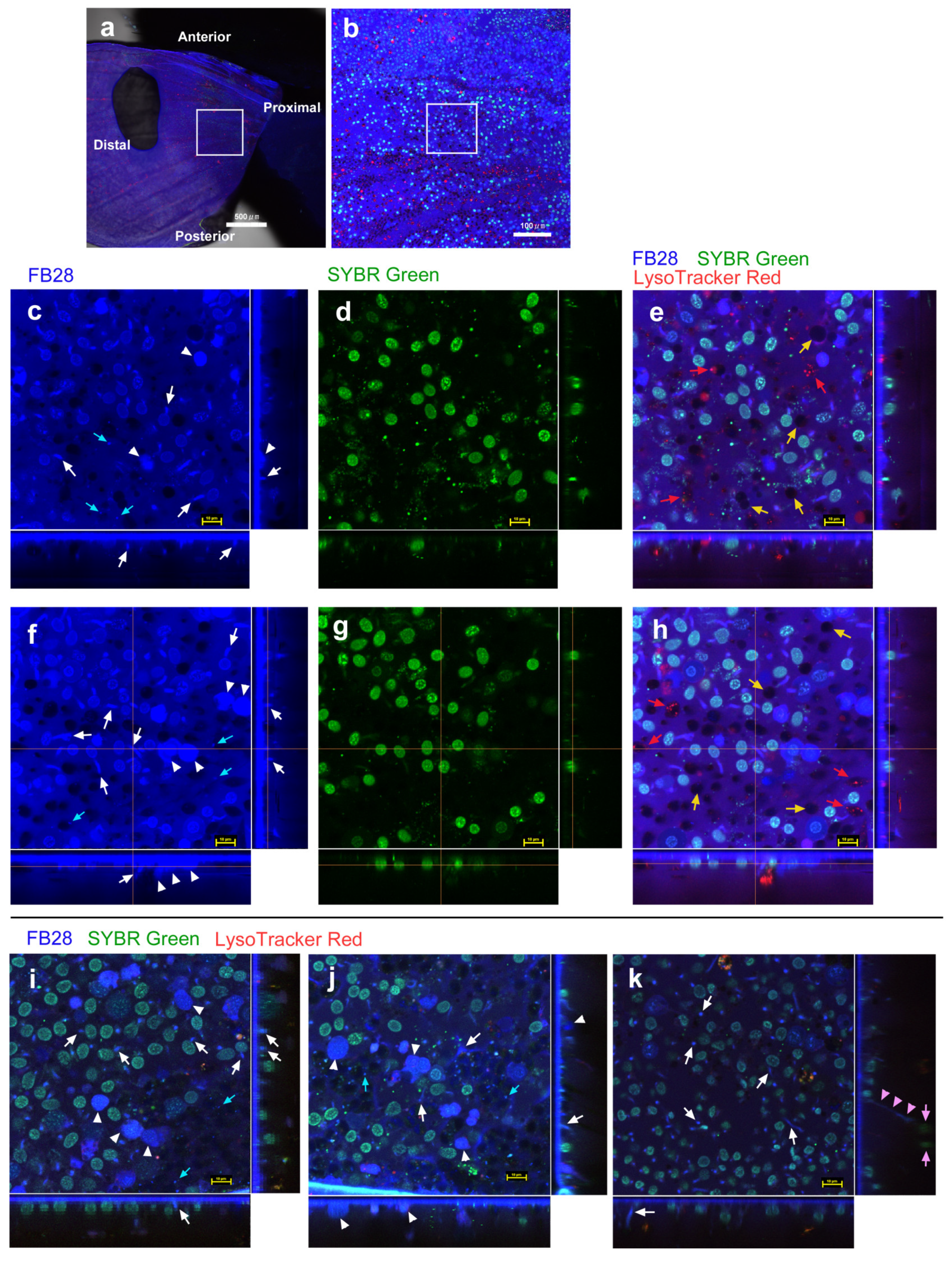
3.1. FB28 and SYBR Green I: Proximal Regions
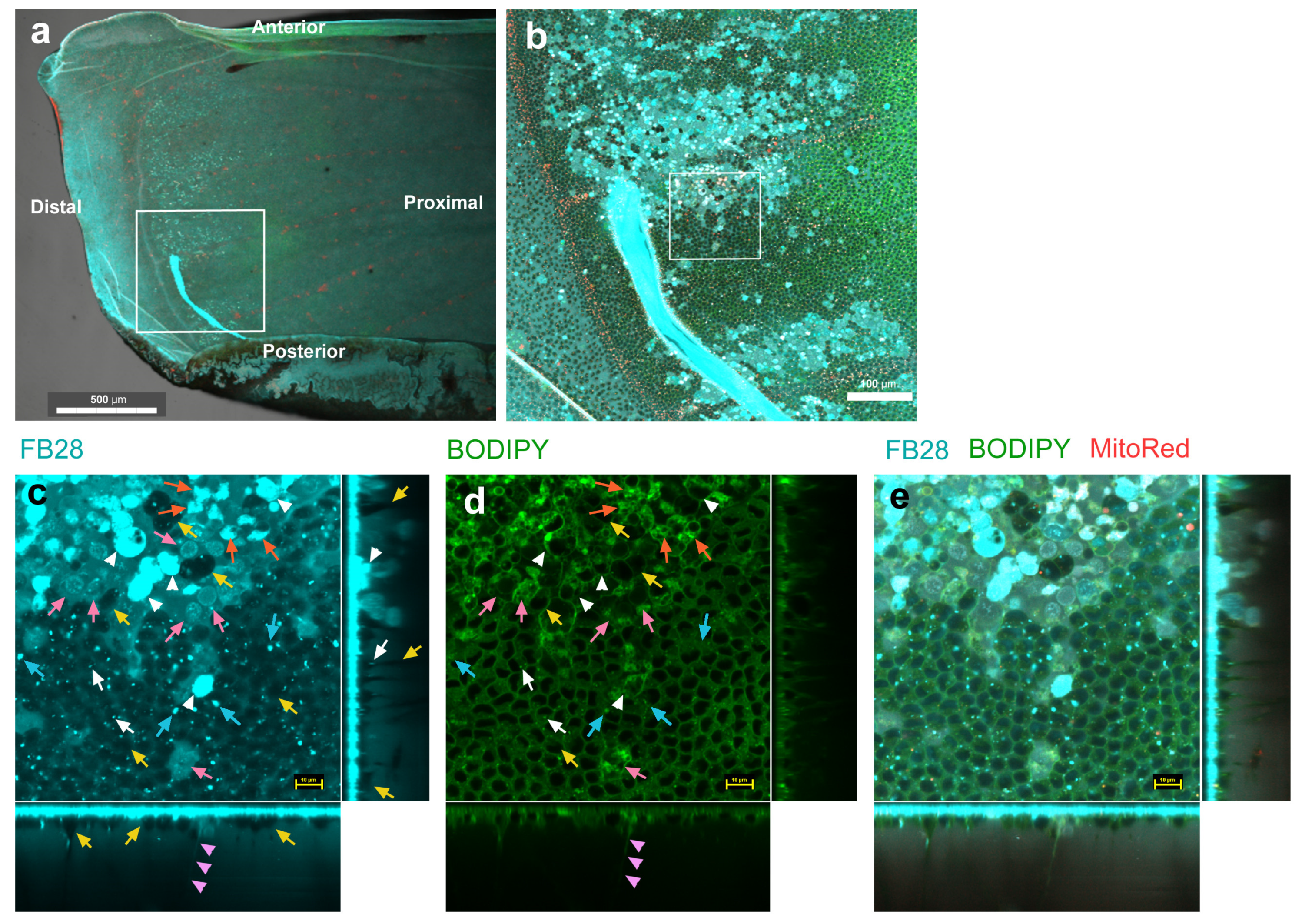
3.2. FB28 and BODIPY: Distal Regions
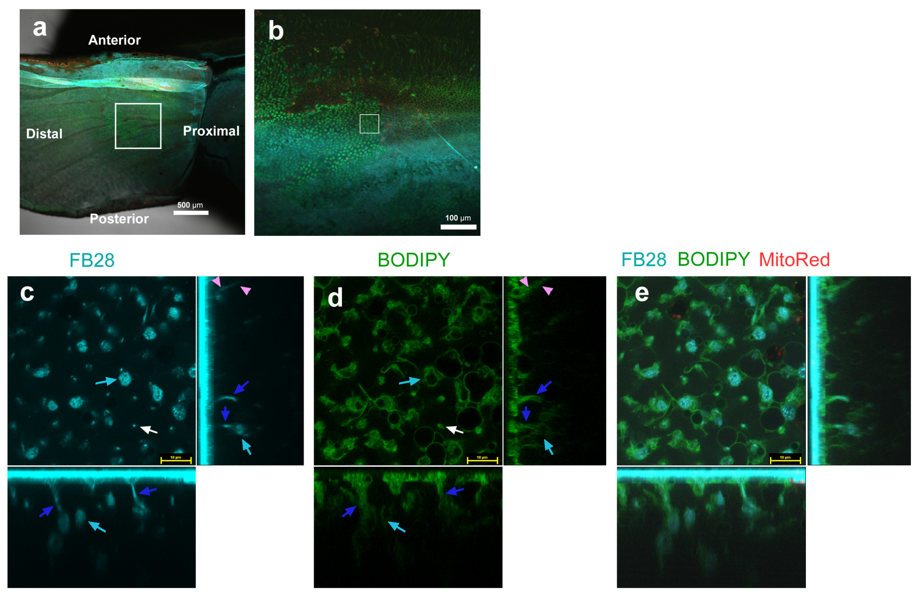
3.3. FB28 and BODIPY: Middle Regions
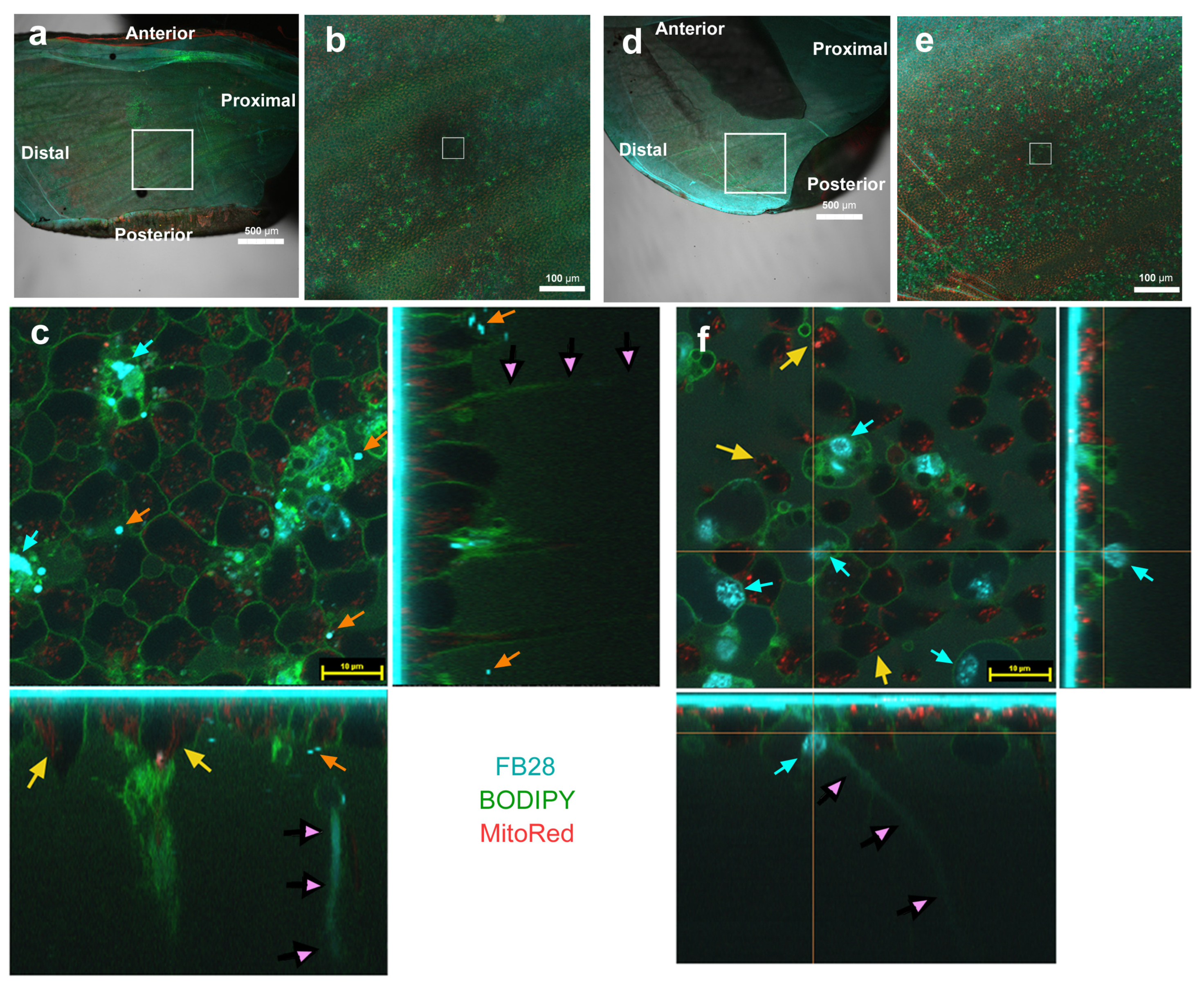
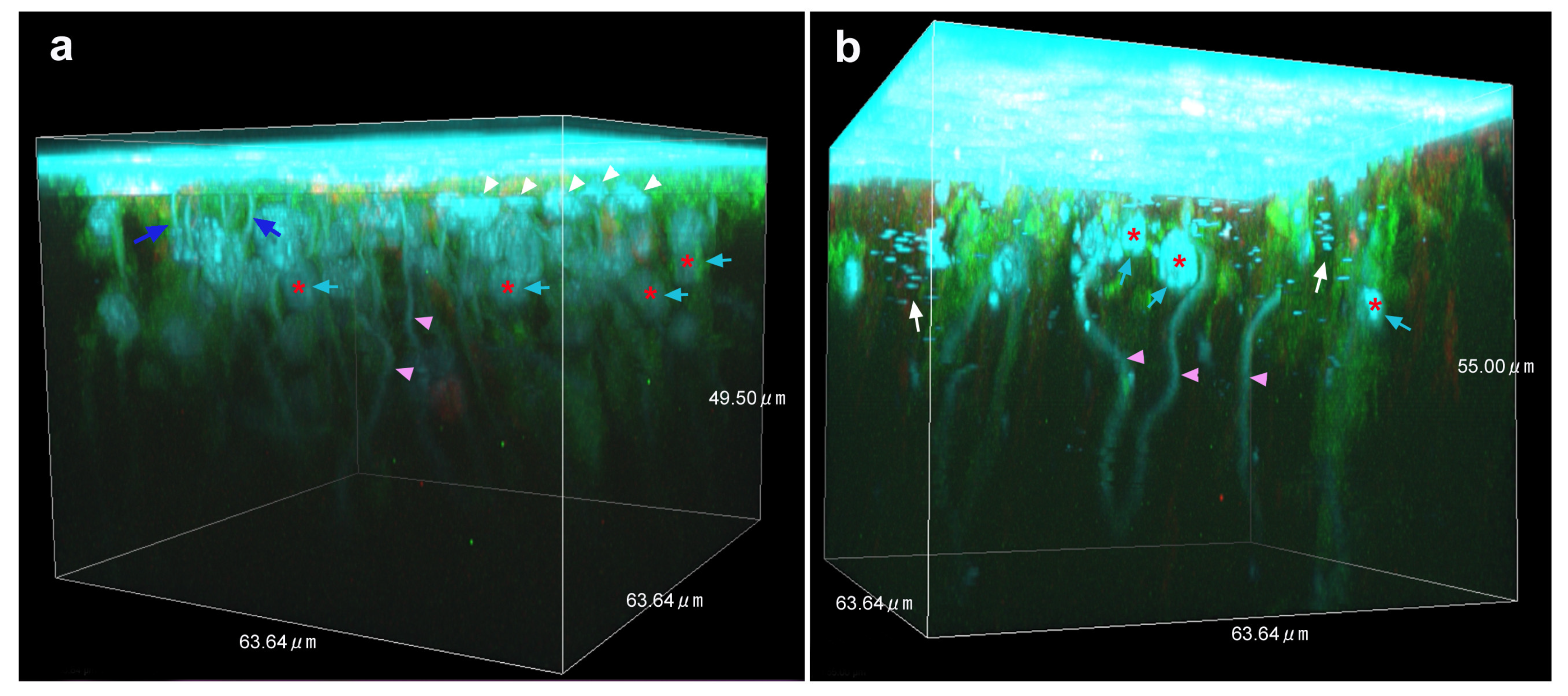
3.4. Three-Dimensional Images
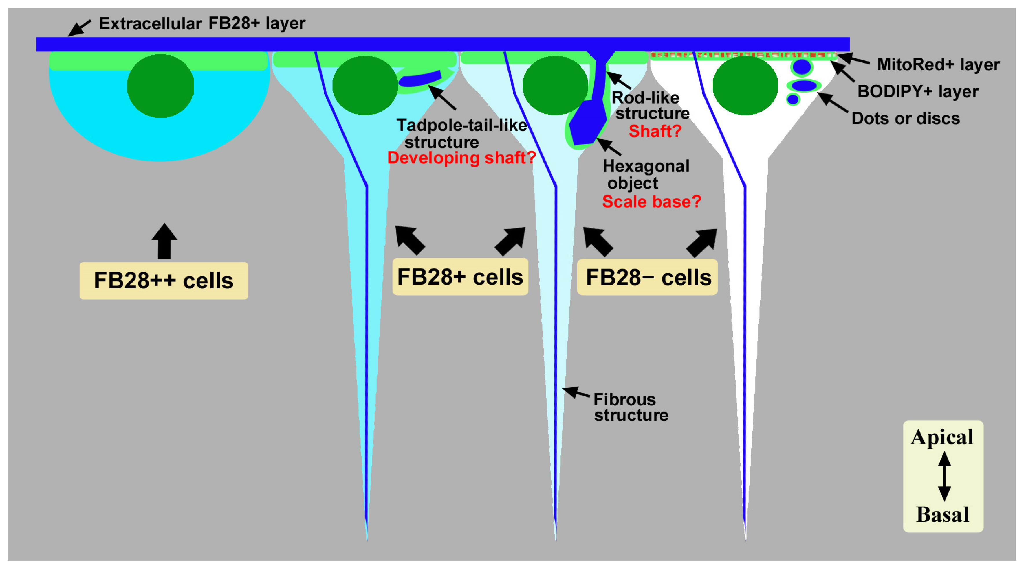
4. Discussion
5. Conclusions
Supplementary Materials
Author Contributions
Funding
Data Availability Statement
Acknowledgments
Conflicts of Interest
References
- Stricker, S.A.; Whitaker, M. Confocal laser scanning microscopy of calcium dynamics in living cells. Microsc. Res. Technol. 1999, 46, 356–369. [Google Scholar] [CrossRef]
- Tovey, S.C.; Brighton, P.J.; Willars, G.B. Confocal microscopy: Theory and applications for cellular signaling. Methods Mol. Biol. 2005, 312, 57–85. [Google Scholar] [CrossRef] [Scilit]
- Hanson, G.T.; Hanson, B.J. Fluorescent probes for cellular assays. Comb. Chem. High Throughput Screen. 2008, 11, 505–513. [Google Scholar] [CrossRef] [Scilit] [PubMed]
- Miyawaki, A.; Nagai, T.; Mizuno, H. Engineering fluorescent proteins. Adv. Biochem. Eng. Biotechnol. 2005, 95, 1–15. [Google Scholar] [CrossRef] [Scilit] [PubMed]
- Terai, T.; Nagano, T. Small-molecule fluorophores and fluorescent probes for bioimaging. Pflug. Arch. Eur. J. Physiol. 2013, 465, 347–359. [Google Scholar] [CrossRef] [Scilit] [PubMed]
- Tsien, R.Y. New calcium indicators and buffers with high selectivity against magnesium and protons: Design, synthesis, and properties of prototype structures. Biochemistry 1980, 19, 2396–2404. [Google Scholar] [CrossRef] [Scilit] [PubMed]
- Tsien, R.Y. A non-disruptive technique for loading calcium buffers and indicators into cells. Nature 1981, 290, 527–528. [Google Scholar] [CrossRef] [Scilit]
- Chalfie, M.; Tu, Y.; Euskirchen, G.; Ward, W.W.; Prasher, D.C. Green fluorescent protein as a marker for gene expression. Science 1994, 263, 802–805. [Google Scholar] [CrossRef] [Scilit]
- Zhu, H.; Fan, J.; Du, J.; Peng, X. Fluorescent probes for sensing and imaging within specific cellular organelles. Acc. Chem. Res. 2016, 49, 2115–2126. [Google Scholar] [CrossRef] [Scilit]
- Choi, N.-E.; Lee, J.-Y.; Park, E.-C.; Lee, J.-H.; Lee, J. Recent advances in organelle-targeted fluorescent probes. Molecules 2021, 26, 217. [Google Scholar] [CrossRef] [Scilit]
- Lin, J.; Yang, K.; New, E.J. Strategies for organelle targeting of fluorescent probes. Org. Biomol. Chem. 2021, 19, 9339–9357. [Google Scholar] [CrossRef] [Scilit]
- Lu, S.; Dai, Z.; Cui, Y.; Kong, D.-M. Recent development of advanced fluorescent molecular probes for organelle-targeted cell imaging. Biosensors 2023, 13, 360. [Google Scholar] [CrossRef] [Scilit] [PubMed]
- Klowden, M.J.; Pallai, S.R. Integumentary system. In Physiological Systems in Insects, 4th ed.; Academic Press: Burlington, NJ, USA, 2022; pp. 87–142. [Google Scholar]
- Andersen, S.O. Cuticle sclerotization and tanning. In Insect Molecular Biology and Biochemistry; Gilbert, L.I., Ed.; Academic Press: New York, NY, USA, 2012; pp. 167–192. [Google Scholar] [CrossRef] [Scilit]
- Muthukrishnan, S.; Merzendorfer, H.; Arakane, Y.; Kramer, K.J. Chitin metabolism in insects. In Insect Molecular Biology and Biochemistry; Gilbert, L.I., Ed.; Academic Press: New York, NY, USA, 2012; pp. 193–235. [Google Scholar] [CrossRef] [Scilit]
- Moussian, B. Recent advances in understanding mechanisms of insect cuticle differentiation. Insect Biochem. Mol. Biol. 2010, 40, 363–375. [Google Scholar] [CrossRef] [Scilit]
- Moussian, B. Chitin: Structure, chemistry and biology. In Targeting Chitin-Containing Organisms—Advances in Experimental Medicine and Biology; Yang, Q., Fukamizo, T., Eds.; Springer: Singapore, 2019; Volume 1142, pp. 5–18. [Google Scholar] [CrossRef] [Scilit]
- Moussian, B. The apical plasma membrane of chitin-synthesizing epithelia. Insect Sci. 2013, 20, 139–146. [Google Scholar] [CrossRef] [Scilit] [PubMed]
- Moussian, B.; Schwarz, H.; Bartoszewski, S.; Nüsslein-Volhard, C. Involvement of chitin in exoskeleton morphogenesis in Drosophila melanogaster. J. Morphol. 2005, 264, 117–130. [Google Scholar] [CrossRef] [Scilit] [PubMed]
- Muthukrishnan, S.; Merzendorfer, H.; Arakane, Y.; Yang, Q. Chitin organizing and modifying enzymes and proteins involved in remodeling of the insect cuticle. In Targeting Chitin-Containing Organisms—Advances in Experimental Medicine and Biology; Yang, Q., Fukamizo, T., Eds.; Springer: Singapore, 2019; Volume 1142, pp. 83–114. [Google Scholar] [CrossRef] [Scilit]
- Sobala, L.F.; Adler, P.N. The gene expression program for the formation of wing cuticle in Drosophila. PLoS Genet. 2016, 12, e1006100. [Google Scholar] [CrossRef] [Scilit]
- Moussian, B.; Tång, E.; Tonning, A.; Helms, S.; Schwarz, H.; Nüsslein-Volhard, C.; Uv, A.E. Drosophila Knickkopf and Retroactive are needed for epithelial tube growth and cuticle differentiation through their specific requirement for chitin filament organization. Development 2006, 133, 163–171. [Google Scholar] [CrossRef] [Scilit]
- Devine, W.P.; Lubarsky, B.; Shaw, K.; Luschnig, S.; Messina, L.; Krasnow, M.A. Requirement for chitin biosynthesis in epithelial tube morphogenesis. Proc. Natl. Acad. Sci. USA 2005, 102, 17014–17019. [Google Scholar] [CrossRef] [Scilit]
- Harrington, B.J.; Raper, K.B. Use of a fluorescent brightener to demonstrate cellulose in the cellular slime molds. Appl. Microbiol. 1968, 16, 106–113. [Google Scholar] [CrossRef]
- Hughes, J.; McCully, M.E. The use of an optical brightener in the study of plant structure. Stain Technol. 1975, 50, 319–329. [Google Scholar] [CrossRef] [Scilit] [PubMed]
- Herth, W.; Schnepf, E. The fluorochrome, Calcofluor white, binds oriented to structural polysaccharide fibrils. Protoplasma 1980, 105, 129–133. [Google Scholar] [CrossRef] [Scilit]
- Fischer, J.M.C.; Peterson, C.A.; Bols, N.C. A new fluorescent test for cell vitality using Calcofluor white M2R. Stain Technol. 1985, 60, 69–79. [Google Scholar] [CrossRef] [Scilit]
- Hoch, H.C.; Galvani, C.D.; Szarowski, D.H.; Turner, J.N. Two new fluorescent dyes applicable for visualization of fungal cell walls. Mycologia 2005, 97, 580–588. [Google Scholar] [CrossRef]
- Sviben, S.; Spaeker, O.; Bennet, M.; Albéric, M.; Dirks, J.-H.; Moussian, B.; Fratzl, P.; Bertinetti, L.; Politi, Y. Epidermal cell surface structure and chitin–protein co-assembly determine fiber architecture in the locust cuticle. ACS Appl. Mater. Interfaces 2020, 12, 25581–25590. [Google Scholar] [CrossRef] [Scilit] [PubMed]
- Sobala, L.F.; Wang, Y.; Adler, P.N. ChtVis-Tomato, a genetic reporter for in vivo visualization of chitin deposition in Drosophila. Development 2015, 142, 3974–3981. [Google Scholar] [CrossRef] [Scilit] [PubMed]
- Dinwiddie, A.; Null, R.; Pizzano, M.; Chuong, L.; Krup, A.L.; Tan, H.E.; Patel, N.H. Dynamics of F-actin prefigure the structure of butterfly wing scales. Dev. Biol. 2014, 392, 404–418. [Google Scholar] [CrossRef] [Scilit]
- Day, C.R.; Hanly, J.J.; Ren, A.; Martin, A. Sub-micrometer insights into the cytoskeletal dynamics and ultrastructural diversity of butterfly wing scales. Dev. Dyn. 2019, 248, 657–670. [Google Scholar] [CrossRef] [Scilit]
- Lloyd, V.J.; Burg, S.L.; Harizanova, J.; Hill, O.; Enciso-Romero, J.; Cooper, R.L.; Flenner, S.; Longo, E.; Greving, I.; Nadeau, N.J.; et al. The actin cytoskeleton plays multiple roles in structural color formation in butterfly wing scales. bioRxiv 2023. bioRxiv:2023.06.01.542791. [Google Scholar] [CrossRef] [Scilit]
- Ghiradella, H. Insect cuticular surface modifications: Scales and other structural formations. Adv. Insect Physiol. 2010, 38, 135–180. [Google Scholar] [CrossRef] [Scilit]
- Kazama, M.; Ichinei, M.; Endo, S.; Iwata, M.; Hino, A.; Otaki, J.M. Species-dependent microarchitectural traits of iridescent scales in the triad taxa of Ornithoptera birdwing butterflies. Entomol. Sci. 2017, 20, 255–269. [Google Scholar] [CrossRef] [Scilit]
- Thayer, R.C.; Allen, F.I.; Patel, N.H. Structural color in Junonia butterflies evolves by tuning scale lamina thickness. eLife 2020, 9, e52187. [Google Scholar] [CrossRef] [Scilit] [PubMed]
- Lloyd, V.J.; Nadeau, N.J. The evolution of structural colour in butterflies. Curr. Opin. Genet. Dev. 2021, 69, 28–34. [Google Scholar] [CrossRef] [Scilit] [PubMed]
- Prakash, A.; Finet, C.; Banerjee, T.D.; Saranathan, V.; Monteiro, A. Antennapedia and optix regulate metallic silver wing scale development and cell shape in Bicyclus anynana butterflies. Cell Rep. 2022, 40, 111052. [Google Scholar] [CrossRef] [Scilit] [PubMed]
- Flaven-Pouchon, J.; Moussian, B. Fluorescent microscopy-based detection of chitin in intact Drosophila melanogaster. Front. Physiol. 2022, 13, 856369. [Google Scholar] [CrossRef] [Scilit] [PubMed]
- Otaki, J.M.; Nakazato, Y. Butterfly wing color pattern modification inducers may act on chitin in the apical extracellular site: Implications in morphogenic signals for color pattern determination. Biology 2022, 11, 1620. [Google Scholar] [CrossRef] [Scilit]
- Nijhout, H.F. The Development and Evolution of Butterfly Wing Patterns; Smithsonian Institution Press: Washington, DC, USA, 1991. [Google Scholar]
- Kusaba, K.; Otaki, J.M. Positional dependence of scale size and shape in butterfly wings: Wing-wide phenotypic coordination of color-pattern elements and background. J. Insect Physiol. 2009, 55, 174–182. [Google Scholar] [CrossRef] [Scilit]
- Iwata, M.; Ohno, Y.; Otaki, J.M. Real-time in vivo imaging of butterfly wing development: Revealing the cellular dynamics of the pupal wing tissue. PLoS ONE 2014, 9, e89500. [Google Scholar] [CrossRef] [Scilit]
- Ohno, Y.; Otaki, J.M. Live cell imaging of butterfly pupal and larval wings in vivo. PLoS ONE 2015, 10, e0128332. [Google Scholar] [CrossRef] [Scilit]
- Ohno, Y.; Otaki, J.M. Spontaneous long-range calcium waves in developing butterfly wings. BMC Dev. Biol. 2015, 15, 17. [Google Scholar] [CrossRef] [Scilit]
- Iwasaki, M.; Ohno, Y.; Otaki, J.M. Butterfly eyespot organiser: In Vivo imaging of the prospective focal cells in pupal wing tissues. Sci. Rep. 2017, 7, 40705. [Google Scholar] [CrossRef] [Scilit]
- Iwata, M.; Tsutsumi, M.; Otaki, J.M. Developmental dynamics of butterfly wings: Real-time in vivo whole-wing imaging of twelve butterfly species. Sci. Rep. 2018, 8, 16848. [Google Scholar] [CrossRef] [Scilit] [PubMed]
- Hirata, K.; Otaki, J.M. Real-time in vivo imaging of the developing pupal wing tissues in the pale grass blue butterfly Zizeeria maha: Establishing the lycaenid system for multiscale bioimaging. J. Imaging 2019, 5, 42. [Google Scholar] [CrossRef] [Scilit] [PubMed]
- Nakazato, Y.; Otaki, J.M. Protein delivery to insect epithelial cells in vivo: Potential application to functional molecular analysis of proteins in butterfly wing development. BioTech 2023, 12, 28. [Google Scholar] [CrossRef] [Scilit] [PubMed]
- Otaki, J.M. Butterfly eyespot color pattern formation requires physical contact of the pupal wing epithelium with extracellular materials for morphogenic signal propagation. BMC Dev. Biol. 2020, 20, 6. [Google Scholar] [CrossRef] [Scilit]
- De Giorgio, E.; Giannios, P.; Espinàs, M.L.; Llimargas, M. A dynamic interplay between chitin synthase and the proteins Expansion/Rebuf reveals that chitin polymerisation and translocation are uncoupled in Drosophila. PLoS Biol. 2023, 21, e3001978. [Google Scholar] [CrossRef] [Scilit]
- Palazzo, R.E.; Vogel, J.M.; Schnackenberg, B.J.; Hull, D.R.; Wu, X. Centrosome maturation. Curr. Top. Dev. Biol. 1999, 49, 449–470. [Google Scholar] [CrossRef] [Scilit]
- Bornens, M. The centrosome in cells and organisms. Science 2012, 335, 422–426. [Google Scholar] [CrossRef] [Scilit] [PubMed]
- Avidor-Reiss, T.; Carr, A.; Fishman, E.L. The sperm centrioles. Mol. Cell. Endocrinol. 2020, 518, 110987. [Google Scholar] [CrossRef] [Scilit]
- Regolini, M. The centrosome as a geometry organizer. In The Golgi Apparatus and Centriole—Results and Problems in Cell Differentiation; Kloc, M., Ed.; Springer: Cham, Switzerland, 2019; Volume 67, pp. 253–276. [Google Scholar] [CrossRef] [Scilit]
- Adler, P.N. Planar signaling and morphogenesis in Drosophila. Dev. Cell 2002, 2, 525–535. [Google Scholar] [CrossRef] [Scilit]
- Eaton, S. Cell biology of planar polarity transmission in the Drosophila wing. Mech. Dev. 2003, 120, 1257–1264. [Google Scholar] [CrossRef] [Scilit]
- McNeill, H. Planar cell polarity: Keeping hairs straight is not so simple. Cold Spring Harb. Perspect. Biol. 2010, 2, a003376. [Google Scholar] [CrossRef] [Scilit]
- Adler, P.N. The frizzled/stan pathway and planar cell polarity in the Drosophila wing. Curr. Top. Dev. Biol. 2012, 101, 1–31. [Google Scholar] [CrossRef] [Scilit] [PubMed]
- Nagaraj, R.; Adler, P.N. Dusky-like functions as a Rab11 effector for the deposition of cuticle during Drosophila bristle development. Development 2012, 139, 906–916. [Google Scholar] [CrossRef] [Scilit]
- Adler, P.N.; Sobala, L.F.; Thom, D.; Nagaraj, R. Dusky-like is required to maintain the integrity and planar cell polarity of hairs during the development of the Drosophila wing. Dev. Biol. 2013, 379, 76–91. [Google Scholar] [CrossRef] [Scilit] [PubMed]
- Zhang, J.; Chen, X.; Lu, Q.; Liu, J.; Ling, X.; Wang, W.; Liu, P.; Chen, H. Foldable units and wing expansion of the oakleaf butterfly during eclosion. J. Bionic Eng. 2022, 19, 724–736. [Google Scholar] [CrossRef] [Scilit]
- Locke, M.; Huie, P. Epidermal feet in insect morphogenesis. Nature 1981, 293, 733–735. [Google Scholar] [CrossRef] [Scilit] [PubMed]
- Nardi, J.B. Tracheole migration in an insect wing. Evidence for guidance by epithelial processes. Wilhelm Roux’s Arch. Dev. Biol. 1984, 194, 1–8. [Google Scholar] [CrossRef] [Scilit]
- Locke, M. The structure of epidermal feet during their development. Tissue Cell 1985, 17, 901–921. [Google Scholar] [CrossRef] [Scilit]
- Nardi, J.; Magee-Adams, S.M. Formation of scale spacing patterns in a moth wing: I. Epithelial feet may mediate cell rearrangement. Dev. Biol. 1986, 116, 278–290. [Google Scholar] [CrossRef] [Scilit]
- Delhanty, P.; Locke, M. The development of epidermal feet in preparation for metamorphosis in an insect. Tissue Cell 1989, 21, 891–909. [Google Scholar] [CrossRef] [Scilit]
- Ramírez-Weber, F.-A.; Kornberg, T.B. Cytonemes: Cellular processes that project to the principal signaling center in Drosophila imaginal discs. Cell 1999, 97, 599–607. [Google Scholar] [CrossRef] [Scilit]
- Rustom, A.; Saffrich, R.; Markovic, I.; Walther, P.; Gerdes, H.H. Nanotubular highways for intercellular organelle transport. Science 2004, 303, 1007–1010. [Google Scholar] [CrossRef] [Scilit]
- Kornberg, T.B. Cytonemes and the dispersion of morphogens. WIREs Dev. Biol. 2014, 3, 445–463. [Google Scholar] [CrossRef] [Scilit]
- Kornberg, T.B.; Roy, S. Cytonemes as specialized signaling filopodia. Development 2014, 141, 729–736. [Google Scholar] [CrossRef] [Scilit] [PubMed]
- Zhang, C.; Scholpp, S. Cytonemes in development. Curr. Opin. Genet. Dev. 2019, 57, 25–30. [Google Scholar] [CrossRef] [Scilit] [PubMed]
- Korenkova, O.; Pepe, A.; Zurzolo, C. Fine intercellular connections in development: TNTs, cytonemes, or intercellular bridges? Cell Stress 2020, 4, 30–43. [Google Scholar] [CrossRef] [Scilit] [PubMed]
- McDougal, A.D.; Kang, S.; Yaqoob, Z.; So, P.T.C.; Kolle, M. In vivo visualization of butterfly scale cell morphogenesis in Vanessa cardui. Proc. Natl. Acad. Sci. USA 2021, 118, e2112009118. [Google Scholar] [CrossRef] [Scilit] [PubMed]
Disclaimer/Publisher’s Note: The statements, opinions and data contained in all publications are solely those of the individual author(s) and contributor(s) and not of MDPI and/or the editor(s). MDPI and/or the editor(s) disclaim responsibility for any injury to people or property resulting from any ideas, methods, instructions or products referred to in the content. |
© 2023 by the authors. Licensee MDPI, Basel, Switzerland. This article is an open access article distributed under the terms and conditions of the Creative Commons Attribution (CC BY) license (https://creativecommons.org/licenses/by/4.0/).
Share and Cite
Nakazato, Y.; Otaki, J.M. Live Detection of Intracellular Chitin in Butterfly Wing Epithelial Cells In Vivo Using Fluorescent Brightener 28: Implications for the Development of Scales and Color Patterns. Insects 2023, 14, 753. https://doi.org/10.3390/insects14090753
Nakazato Y, Otaki JM. Live Detection of Intracellular Chitin in Butterfly Wing Epithelial Cells In Vivo Using Fluorescent Brightener 28: Implications for the Development of Scales and Color Patterns. Insects. 2023; 14(9):753. https://doi.org/10.3390/insects14090753
Chicago/Turabian StyleNakazato, Yugo, and Joji M. Otaki. 2023. "Live Detection of Intracellular Chitin in Butterfly Wing Epithelial Cells In Vivo Using Fluorescent Brightener 28: Implications for the Development of Scales and Color Patterns" Insects 14, no. 9: 753. https://doi.org/10.3390/insects14090753
APA StyleNakazato, Y., & Otaki, J. M. (2023). Live Detection of Intracellular Chitin in Butterfly Wing Epithelial Cells In Vivo Using Fluorescent Brightener 28: Implications for the Development of Scales and Color Patterns. Insects, 14(9), 753. https://doi.org/10.3390/insects14090753

