Article

A New Albomycin-Producing Strain of Streptomyces globisporus subsp. globisporus May Provide Protection for Ants Messor structor

by
Yuliya V. Zakalyukina
1,2,*,
Nikolay A. Pavlov
3,
Dmitrii A. Lukianov
3,4,
Valeria I. Marina
3,4,
Olga A. Belozerova
5,
Vadim N. Tashlitsky
4,
Elena B. Guglya
5,6,
Ilya A. Osterman
1,3,4 and
Mikhail V. Biryukov
1,3,7
1
Center for Translational Medicine, Sirius University of Science and Technology, Olympic Avenue 1, 354340 Sochi, Russia
2
Department of Soil Science, Lomonosov Moscow State University, Leninskie Gory 1, 119991 Moscow, Russia
3
Center of Life Sciences, Skolkovo Institute of Science and Technology, Bolshoy Boulevard 30, Bld. 1, 121205 Moscow, Russia
4
Department of Chemistry, Lomonosov Moscow State University, Leninskie Gory 1, 119991 Moscow, Russia
5
Shemyakin-Ovchinnikov Institute of Bioorganic Chemistry of the Russian Academy of Sciences, Miklukho-Maklaya st. 16/10, 117997 Moscow, Russia
6
Institute of Translational Medicine, Pirogov Russian National Research Medical University, Ostrovityanova st. 1, 117997 Moscow, Russia
7
Department of Biology, Lomonosov Moscow State University, Leninskie Gory 1, 119991 Moscow, Russia
*
Author to whom correspondence should be addressed.
Insects 2022, 13(11), 1042; https://doi.org/10.3390/insects13111042
Submission received: 11 October 2022 / Revised: 3 November 2022 / Accepted: 7 November 2022 / Published: 11 November 2022
(This article belongs to the Special Issue Insect Microbiome and Immunity)

Abstract

:

Simple Summary

Insects are the most numerous and diverse animals on our planet. They have mastered different habitats and are able to resist many external threats. Many of them have long concluded mutually beneficial alliances with microorganisms that are capable of producing biologically active substances. We found that Messor structor ants have a very common actinobacteria that secretes albomycin, an antibiotic capable of suppressing the growth of enthomopathogenic bacteria in the smallest concentrations. Perhaps this is one of the secrets of ants’ resistance to external factors and their successful evolutionary development.

Abstract

There are several well-studied examples of protective symbiosis between insect host and symbiotic actinobacteria, producing antimicrobial metabolites to inhibit host pathogens. These mutualistic relationships are best described for some wasps and leaf-cutting ants, while a huge variety of insect species still remain poorly explored. For the first time, we isolated actinobacteria from the harvester ant Messor structor and evaluated the isolates’ potential as antimicrobial producers. All isolates could be divided into two morphotypes of single and mycelial cells. We found that the most common mycelial morphotype was observed among soldiers and least common among larvae in the studied laboratory colony. The representative of this morphotype was identified as Streptomyces globisporus subsp. globisporus 4-3 by a polyphasic approach. It was established using a E. coli JW5503 pDualRep2 system that crude broths of mycelial isolates inhibited protein synthesis in reporter strains, but it did not disrupt the in vitro synthesis of proteins in cell-free extracts. An active compound was extracted, purified and identified as albomycin δ2. The pronounced ability of albomycin to inhibit the growth of entomopathogens suggests that Streptomyces globisporus subsp. globisporus may be involved in defensive symbiosis with the Messor structor ant against infections.

1. Introduction

The widespread use of antimicrobial compounds in medicine and agriculture has led to the emergence of multidrug-resistant pathogens, recognized now as a significant threat to human health [1,2]. The search for novel compounds possessing antimicrobial properties is still one of the ways we could overcome this global challenge, and microorganisms are the main source in this research [3,4,5]. Recently great attention was attracted by microbes that form symbioses with higher organisms, in particular, plants and animals [6]. The main reason for that is the mutual evolutionary path, wherein microbes have proved their usefulness to the host [7,8].
Some prokaryotes, principally the phylum Actinomycetota [9], are involved in the formation of so-called “defensive (or protective) symbioses” [10,11,12] with many eukaryotic organisms. The Insecta class, with the largest number of species, is remarkable among them for these interactions [13,14]. Through the release of various antibiotic compounds, actinobacteria protect insects, their brood and food substrate from potential pathogens and parasites [7,15,16,17,18]. Well known in this respect are leaf-cutting ants (Atta, Acromyrmex) of the subfamily Myrmicinae. Their existence, and in particular their feeding and development, depends entirely on the symbiotic actinobacteria of the genus Pseudonocardia [19], localized on the insect cuticle.
However, the specificity of “defensive symbioses” in other species of Formicidae remains unclear, including one of the dominant ants of the steppe zone, Messor structor (the steppe harvester ant). In this paper, we report on the isolation of actinobacteria from a laboratory colony of Messor structor, the identification of the produced antimicrobial compound (albomycin δ2), and its high activity against individual entomopathogens.

2. Materials and Methods

2.1. Ant Colony Rearing and Microbial Isolation

Prior to rearing an ant colony, a special incubator was designed; it consisted of a glass tube with sterile water and poppy seeds as the main nutrient substrate. The mated queen was maintained there during a 3-month period, until the adult ant quantity was sufficient to place the colony in a specific formicarium. The formicarium is made from an acrylic plastic with two main chambers—an arena, through which the seeds are supplemented, and a system of chambers, where ants raise their brood. In the center of the formicarium, a specific watering cell is present, which maintains the humidity and water level inside the nest. Humidity is maintained between 70 and 90% and temperature at 24 °C respectively, without direct sunlight on the formicarium.
The queen was collected during the mating season in July 2017 in the Astrakhan region, Russia (46°51′13.5″ N 47°59′06.2″ E). The isolation of actinobacteria strains was performed only after the stabilization of a population number of at least 50 specimens.
Actinobacterial strains were isolated from the bodies of larvae, pupae and imago worker and soldier castes of Messor structor. A total of 14 individuals from each group were examined. Every specimen was washed three times in sterile distilled water and then crushed by a tissue microhomogenizer with sterile saline solution. Aliquots of this mixture and their 10-fold dilutions were spread over mineral agar 1 [20] and organic agar 79 [21] supplemented nystatin and nalidixic acid at final concentrations 250 μg/mL and 10 μg/mL, accordingly, and incubated for 14 days at 28°C [22]. Actinobacteria isolates were purified and maintained on ISP 3 slants [23] and preserved as a suspension of mycelial fragments and spores in 20% glycerol at −20 °C.

2.2. 16S rRNA Phylogeny of Isolated Strains

The extraction of the genomic DNA of isolates and PCR amplification were achieved using procedures described elsewhere [24]. Both the pair of universal primers F27 (5′-AGAGTTTGATCMTGGCTCAG-3′) and R1492 (5′-TACGGYTACCTTGTTACGACTT-3′) and actinobacterial primers 243F (5′-GGATGAGCCCGCGGCCTA-3′) and A3R (5′-CCAGCCCCACCTTCGAC-3′) were used. The amplicons were purified and sequenced using a commercial service (EvroGen). All sequences were identified by searching close relatives with the BLAST service (https://blast.ncbi.nlm.nih.gov/Blast.cgi, accessed on 1 October 2022) and were submitted in GenBank with the assignment of access numbers.

2.3. Genome Features and Phylogenomic Analysis

Genome of strain 4-3 was sequenced de novo by Skoltech Genomics Core Facility, using the Illumina HiSeq 4000 platform (Illumina, San Diego, CA, USA). The quality control and adapter trimming was done by the bbDuk tool from the BBMap suite v38.42 (https://sourceforge.net/projects/bbmap/, accessed on 1 February 2022). Genome assembly was performed by SPAdes v3.13.0 [25]. The genome was annotated using RASTtk pipeline implemented on the PATRIC web service [26]. Assembly is available in the European Nucleotide Archive with project accession PRJEB51905.
Values of average nucleotide identity (ANI), genome completeness and quality were evaluated using a web service MiGA (http://microbial-genomes.org/, accessed on 1 October 2022), and in silico digital DNA:DNA hybridization (DDH) values were calculated by using the GGDC method, with the recommended formula 2, available at the TYGS web service (https://tygs.dsmz.de/, accessed on 1 October 2022) [27].
Phylogenomic analysis was performed using Type (Strain) Genome Server (https://tygs.dsmz.de/, accessed on 1 October 2022). The phylogenomic tree inferred with FastME 2.1.6.1 [28] from GBDP distances calculated from genome sequences. The branch lengths are scaled in terms of GBDP distance formula d5.
The full-length 16S rRNA gene sequences of strain 4-3 was extracted from the whole genome sequence (PRJEB51905) and was compared to sequences of related Streptomyces globisporus subsp. globisporus and some actinobacteria species, firstly isolated from insects. Evolutionary trees based on 16S rRNA gene sequences were inferred with the neighbor-joining [29], maximum-parsimony [30] and maximum-likelihood [31] treemaking algorithms after CLUSTAL W alignment by using MEGA software version X [32].

2.4. Analysis of Bioactive Compound Biosynthetic Gene Clusters

Secondary metabolite biosynthetic gene clusters in complete genome strain 4-3 and its neighbors were identified with the bacterial version of antiSMASH 6.1.0 (https://antismash.secondarymetabolites.org/, accessed on 1 October 2022). Homologous regions on each genome were identified using NCBI Blastn (https://blast.ncbi.nlm.nih.gov/, accessed on 1 October 2022).

2.5. Phenotypic Characterization

Cultural characteristics of strain 4-3 were observed in ISP 2–ISP 7 media [23] after cultivation for up to 14 days at 28 °C. The RAL Classic Standard was used to determine the designations of colony colors. The shape of spore chains and the spore surface of strain 4-3 on ISP 3 after cultivation at 28 °C for 14 days were studied using light microscopy (Fisherbrand AX-502, Fisher Scientific, Merelbeke, Belgium) and scanning electron microscopy (JSM-6380LA, JEOL, Tokyo, Japan).
Carbon source utilization was assessed on basal medium ISP 9 [23] with the addition of 0.04% solution of bromocresol purple at 28 °C for 14 days. Enzyme activities were estimated using a paper indicator system (NPO Microgen, Nizhny Novgorod, Russia) according to the manufacturer’s recommendations at 28 °C for 7 days. The degradation of casein, starch and cellulose was estimated on the clearing of the insoluble compounds around areas of growth [33].

2.6. Biological Activity Testing

2.6.1. Screening of the Antimicrobial Potential

The ability of actinobacteria isolates to inhibit bacterial growth was assessed by the agar diffusion method. The isolates were challenged against different clinically significant microorganisms: Staphylococcus aureus ATCC 25923, Bacillus subtilis ATCC 6633, Candida albicans CBS 8836 and Aspergillus niger INA 00760. Anti-entomopathogenic activity was also investigated against: Bacillus thuringiensis VKM B-6650, Paenibacillus alvei VKM B-502, Beauveria bassiana VKM F-1357 and Entomophthora coronata VKM F-1359.
Test bacteria, yeast and fungi strains were individually inoculated in Luria–Bertani agar, dextrose–peptone-yeast agar and potato dextrose agar, accordingly. Agar plugs of each actinobacterial isolate (2-week-old cultures in ISP6) were placed on the surface of the inoculated media. The plates were incubated at 37 °C, and after 24–48 h, the inhibition zones were checked.

2.6.2. Reporter Assays on Agar Plates

The two E. coli reporter strains: BW25113 wild-type pDualRep2 and JW5503 ΔtolC pDualRep2 [34] were used in this work as previously described [35]. Briefly, 100 µL of cultural broth were placed into wells in agar that had the lawn of a reporter strain. Two control antibiotics, erythromycin (Ery, 2 μg) and levofloxacin (Lev, 0.05 μg), were additionally applied to an agar plate. Plates were incubated at 37 °C overnight and then scanned by ChemiDoc (Bio-Rad) in the modes ‘Cy3-blot’ for RFP and ‘Cy5-blot’ for Katushka2S. The expression of the rfp gene occurred in the case of the activation of the SOS-response system of the cell and katushka2S, in the case of a violation of translation, when the ribosome was stalled on the mRNA template. When scanning, the signal from two black and white images was superimposed on each other, with the assignment of green for the signal from the RFP protein and red for Katushka2S.

2.6.3. Determination of Minimal Inhibitory Concentration (MIC)

Overnight cultures of E. coli ΔtolC, Bacillus thuringiensis and Paenibacillus alvei were diluted 1:1000 in LB medium. A sterile 96-well plate was then loaded with 200 mL of the diluted cultural media, with the initial row having 400 mL prior serial dilution. A stock solution of a HPLC-purified 4-3 sample was variously seeded in the initial row, along with erythromycin (Ery), which was used as a control for the experiment. Other wells were left without antibiotic but with diluted LB-culture media, while the rest were left with LB media only as additional controls. A two-fold serial dilution was then carried out, with gentle mixing in each row. The plates were then incubated overnight at 37 °C with shaking at 200 rpm. Cell growth was measured at 590 nm using a microplate reader (VICTOR X5 Light Plate Reader, PerkinElmer, Waltham, MA, USA). UV absorption of 4-3 was measured with a spectrophotometer (NanoPhotometer™ NP80, Implen, München, Germany), and the concentration was calculated using the known coefficients of extinction at 306 and 425 nm.

2.6.4. Cell-free Translation

Cell-free (in vitro) translation reactions were performed in the presence of HPLC 4-3 fraction (1/10 of final volume) in 5 μL using the PURExpress® In Vitro system (NEB, Ipswich, MA, USA) supplemented with 100 ng Fluc mRNA and 0.05 mM D-luciferin. Chemiluminescence was recorded with VICTOR X5 Light Plate Reader. The Fluc mRNA obtained byMEGAscript™ T7 Transcription Kit (ThermoFisher, Carlsbad, CA, USA) from the circular DNA template.

2.7. Purification and Identification of Albomycin

To obtain a sufficient amount of the active compound for detailed bioactivity studies, strain 4-3 was cultured in four 750 mL Erlenmeyer flasks with 250 mL of liquid ISP 6 at 28 °C for 14 days under static conditions. One liter of fermentation broth 4-3 was subjected to gravity-flow reverse-phase chromatography on the sorbent LPS500H (polyvinylbenzene, pore size 50–1000 Å) (LLC “Technosorbent”, Moscow, Russia). The fermentation broth was applied to chromatographic media, which then was eluted consistently with 10, 20, 30, 40, 50, 75 and 100% solutions of acetonitrile in water. The pDualrep2 double reporter system was used to analyze the activity of the collected fractions. The most active fractions were eluted by 10 and 20% acetonitrile, they induced the expression of reporter protein Katushka2S. The 10% acetonitrile fraction was further purified by high-performance liquid chromatography (HPLC) (Agilent 1260, isocratic elution 4% of MeCN, 10 mM AcONH4, 1 mL/min, 25 °C) using a Phenomenex HPLC column (Luna 5 μm C18 (2) 100 Å, 4.6 × 250 mm), and the collected fractions were analyzed using the reporter pDualrep2.
Fractions with antibacterial activity corresponding to an individual peak on chromatograms were collected, and the active compound was identified using ultra-high-performance liquid chromatography–electrospray ionization–high-resolution mass spectrometry (UPLC–ESI–HRMS). Analysis was carried out on an Ultimate 3000 RSLCnano HPLC system connected to an Orbitrap Fusion Lumos mass spectrometer (ThermoFisher Scientific). A sample of the active compound was separated on Luna Omega C18 100 × 2.1 mm 1.6 μm columns at a 0.2 mL/min flow rate and at RT. Separation was done by a gradient elution in a two-component mixture from the initial 5% to 20% of component B for 10 min. Component A was 0.1% formic acid plus 10 mM formate ammonium in water, and component B was a mixture of 0.1% formic acid in 100% MeCN and 10% 10 mM formate ammonium in water in ratio 9:1. UV data were registered at 290 nm. MS1 and MS2 spectra were collected in positive ion mode and recorded at 30 K and 15 K resolution, respectively, with HCD fragmentation.

3. Results

3.1. Isolation of Actinobacteria Strains, Associated with Messor Structor Ants

All bacterial strains isolated from Messor structor individuals were divided into two morphotypes: one of which formed beige branching mycelium, dark-pigmented and straight spore chains (later labeled as 4-3), and the other, formed rough colorless colonies from Gram-positive cocci (L1). Results demonstrated that strains of 4-3-morphotype had more association with the caste of soldiers with 89% frequency among this group, while workers’ caste, pupae and larvae showed less specific results—50%, 21%, and 7% respectively. On the contrary, L1-bacteria were found in the vast majority of individuals from these groups besides soldiers (Table S1).
A comparison with the GenBank database demonstrated that all strains assigned to the 4-3-morphotype, in addition to their phenotypical similarity, had an identical sequence of the 16S rRNA genes, indicating their belonging to the Streptomyces genus. The closest strains were Streptomyces globisporus subsp. globisporus DSM 40136 (formerly a type strain of Streptomyces albovinaceus), Streptomyces globisporus subsp. globisporus DSM 40199T, Streptomyces rubiginosohelvolus DSM 40176T and Streptomyces pluricolorescens DSM 40019T. The partial 16S rRNA sequences of L1-morphotype strains showed 100% similarity with Staphylococcus gallinarum DSM 20610T. Among the representatives of the mycelial morphotype, strain 4-3 was selected for a more detailed study of the genome features and antagonistic activity.

3.2. Genome Features and Phylogenomic Analysis of Streptomyces sp. Strain 4-3

Phylogenomic analysis based on whole-genome sequences showed that strain 4-3 formed a well-supported monophyletic clade with S. globisporus subsp. globisporus DSM 40199T and S. globisporus subsp. globisporus DSM 40136 with 95% bootstrap value (Figure 1).
The complete genome size of strain 4-3 was 7,941,828 bp with DNA G + C content of 71.6%, which was consistent with the G + C content of the genus Streptomyces [36]. The closest neighbors S. globisporus subp. globisporus DSM 40199T and S. globisporus subp. globisporus DSM 40136 are characterized by similar genome size and G + C content (Table S2).
The ANI and in silico dDDH values between strains 4-3 and S. globisporus subp. globisporus DSM 40136 and S. globisporus subp. globisporus DSM 40199T were above the recommended threshold of 96% and 70% (Table 1) needed for species separation [27,37]. Based on this, strain 4-3 most likely belongs to the species Streptomyces globisporus subp. globisporus.
Neighbor-joining phylogenetic analysis demonstrated that 4-3 was most closely related to type and not type strains of Streptomyces globisporus subsp. globisporus: DSM 40199T, DSM 40139, C-1027, TFH56, as well as to S. rubiginosohelvolus DSM 40176T, S. pluricolorescens DSM 40019T, S. sindenensis DSM 40255T, S. anulatus DSM 40361T and S. griseus subsp. griseus ATCC 13273 and formed a share clade with 100% bootstrap value (Figure S1). However, type strains of actinobacterial species, first isolated from ants and other insects, did not form well-supported clades with 4-3. This relationship was also supported in the phylogenetic trees generated with maximum-parsimony and maximum-likelihood methods (Figures S2 and S3, available in Supplementary Materials).

3.3. Phenotypic Characterization of Streptomyces sp. Strain 4-3

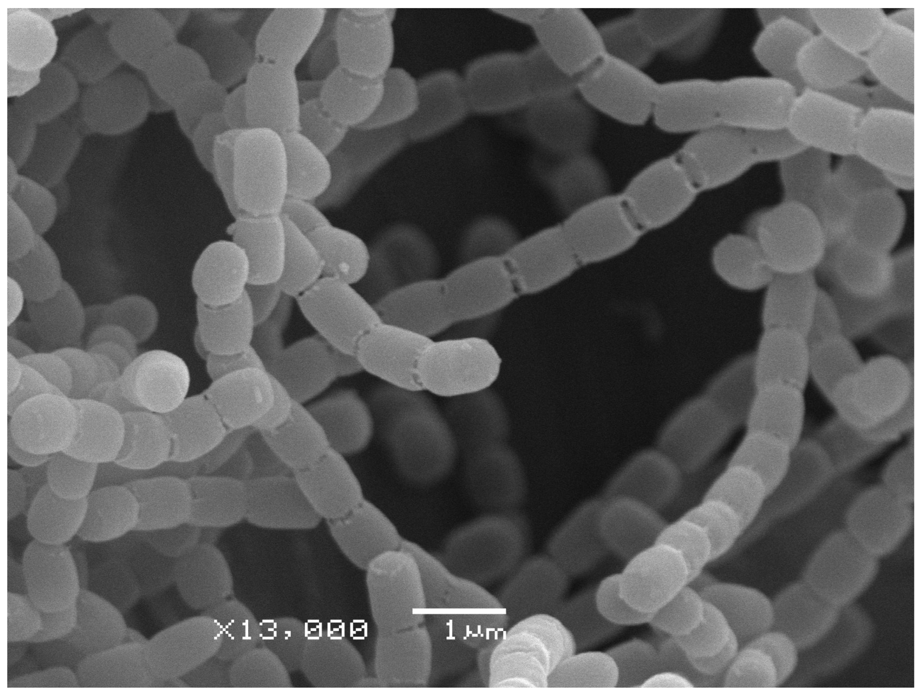
To further evaluate the features of the 4-3 strain using a polyphasic taxonomy approach, the cultural, morphological and physiological properties of 4-3 were compared with ones of the type strains of Streptomyces globisporus subsp. globisporus. Results demonstrated the identity of these organisms in morphology—the shape of sporophores and spore surface (Table S3, Figure 2) and the high similarity of their cultural characteristics on the series of ISP media (Table 2, Figure S4).
However, some differences should be noted in the biochemical and physiological properties of strain 4-3 and the closest type strains of S. globisporus subsp. globisporus (Table S3). For example, the production acid from glucose and xylose was positive in strain 4-3 (Figure S5), whereas the other type strains showed negative results. In the decomposition of polymers, strain 4-3 was unable to use cellulose as a sole carbon source, whereas the other type strains utilized it. Furthermore, the enzyme assay of 4-3 was negative for L-ornithine decarboxylase and L-arginine decarboxylase; in contrast, DSM 40199T and DSM 40136 demonstrated positive responses (Table S3). However, most of the biochemical tests showed similar results.
According to the obtained data, we may conclude that strain 4-3 isolated from Messor structor ants can be classified as Streptomyces globisporus subsp. globisporus.

3.4. Analysis of 4-3 Bioactive Compound Biosynthetic Gene Clusters

The bioinformatics analysis of Streptomyces globisporus subsp. globisporus 4-3 genome revealed a biosynthetic gene cluster of albomycins, consisting of 18 genes from abmA to abmR, completely identical to that of Streptomyces sp. ATCC 700974 (Figure 3), described in detail earlier [38]. The presence of abmK, participating directly in the formation of SB-217452 (the active seryl-tRNA synthetase inhibitor component of albomycin) [39] and also providing self-resistance to albomycins [40], indicates the ability of Streptomyces globisporus subsp. globisporus 4-3 to actively produce albomycin δ2.
Furthermore, the 4-3 strain genome contains a number of second metabolic biosynthesis gene clusters (SMBGCs), coded production of antimicrobial compounds (streptophenazines B/C/E/H/G, mayamycins), odor substances (geosmin), pigments (melanin, isorenieratene), siderophores (streptobactin, coelichelin), cytoprotectants (ectoine) and others (Table S4).

3.5. Screening Antimicrobial Activity

All isolated strains were initially tested for the ability to secrete antimicrobial substances. It was noticed that all phenotypically similar mycelial strains showed the same activity pattern, so we focused on the study of strain 4-3. Analysis of antimicrobial activity demonstrated that the 4-3 strain noticeably inhibited the growth of various pathogenic microorganisms (Table S5): bacteria (Bacillus subtilis, Staphylococcus aureus) and fungi (Aspergillus niger), but it is especially active on entomopathogenic microorganisms (Bacillus thuringiensis, Paenibacillus alvei, Beauveria bassiana, Entomophthora coronata). To evaluate the MIC of the 4-3 compound, we chose strains that were the most susceptible during the screening procedure. The estimated concentration of the HPLC-purified sample was 0.6 μg/mL. In the prepared series of microdilutions, the test strains demonstrated high sensitivity to the 4-3 compound (Table S5), including entomopathogenic bacteria.
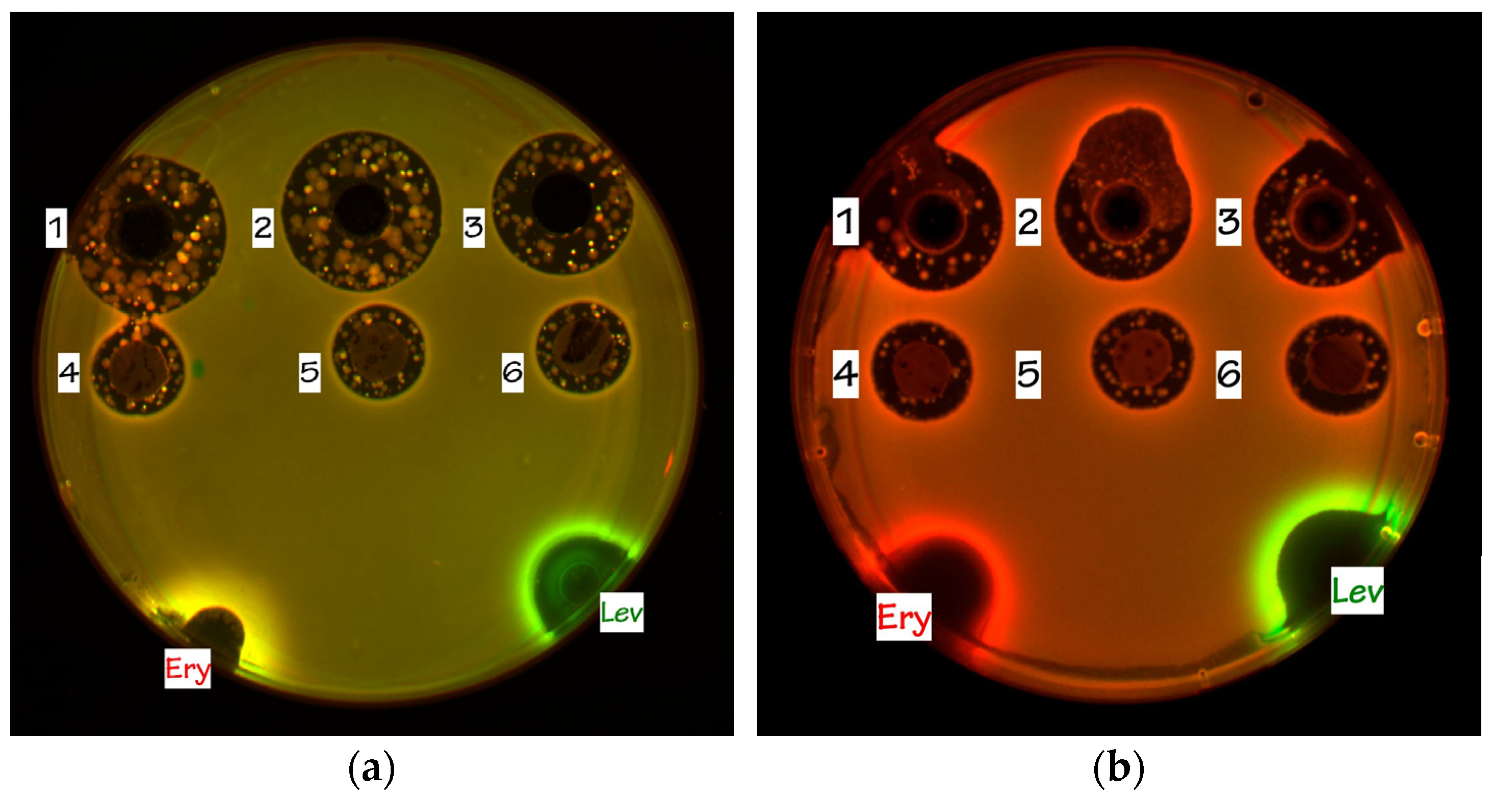
The agar plugs and cultural broth aliquots of 4-3 demonstrated prominent antibiotic activity in tests on the reporter strains (Figure 4) and exhibited strong Katushka2S reporter induction, indicating that the active compound produced by the isolate functions as an inhibitor of protein biosynthesis. To further evaluate the possible mechanism of action, the in vitro translation analysis was performed.

3.6. Analysis of Bioactive Compounds: Cell-free Translation

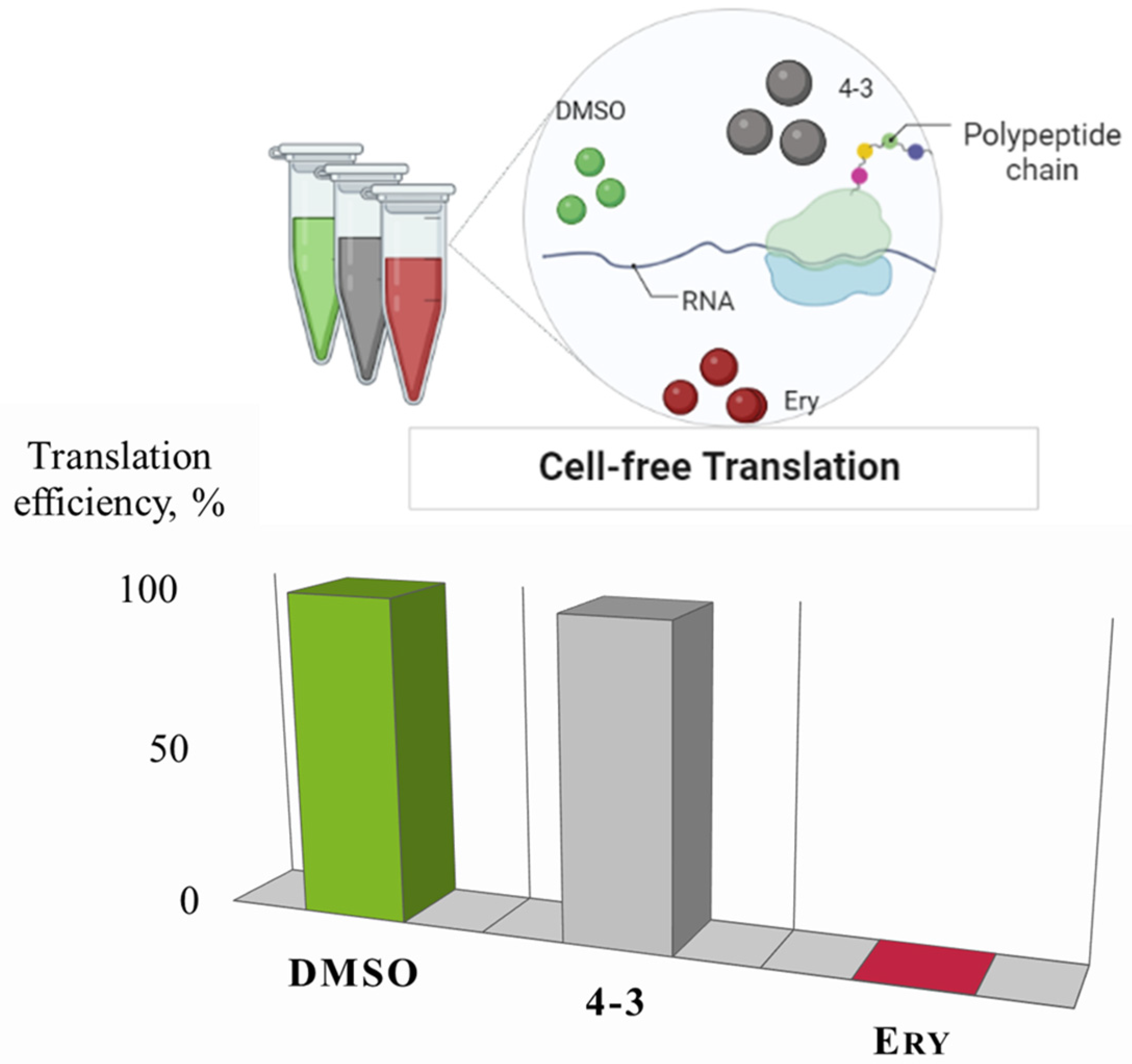
It was decided to test the HPLC-purified sample 4-3 in in vitro translation procedure to completely evaluate its translation inhibitory activity. Despite induction of the Katushka2S, indicating the inhibition of protein synthesis in the cells of the reporter strains (Figure 4), the active compound of the strain 4-3 did not suppress translation in the cell-free system (Figure 5).

3.7. Purification and Identification of Albomycin

The liquid culture of Streptomyces globisporus subsp. globisporus 4-3 was preconcentrated and purified by solid-phase extraction (SPE) on LPS500H resin. It was binded on LPS500H, washed with water and the active compound was eluted with 10% MeCN solution in water. Further purification of active SPE fraction was carried out by RP HPLC on a C18 column in isocratic mode with aqueous solution of ammonium acetate—MeCN as eluent. Thus, a pure compound was isolated with a UV maxima at 283 and 424 nm, and the activity testing confirmed that it corresponded to an active metabolite (Figure S6).
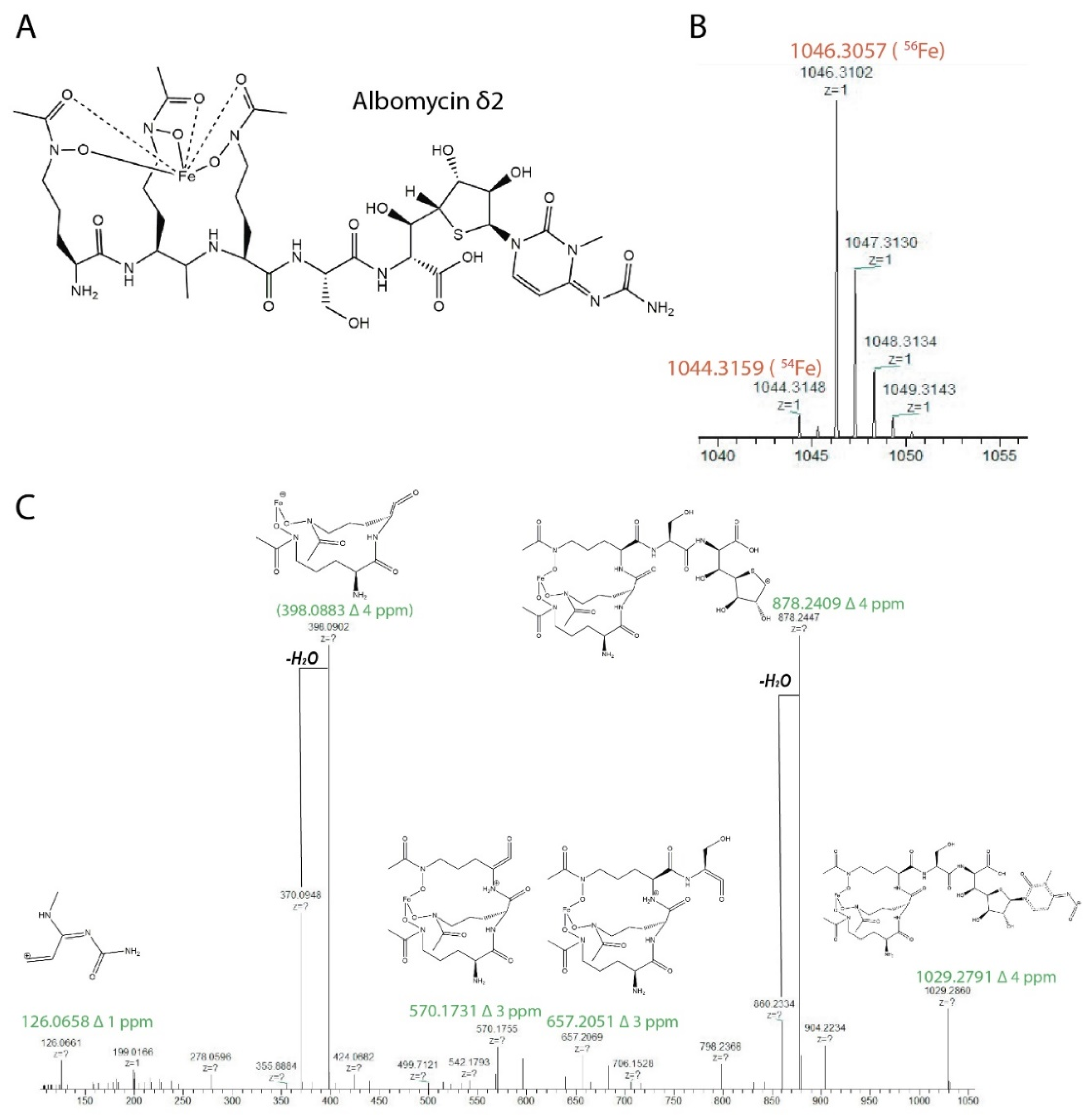
The metabolite identification was carried out using LC-HRMS/MS analysis (Figure S7). The observed exact masses 1046.3102 of molecular ion [M + H]+ of the compound and characteristic isotope distribution corresponded to the composition C37H57FeN12O18S (calculated exact mass 1046.3057). The main fragmentary ion in the MS2 spectrum due to the loss of the cytosine part was observed at m/z 878. MS1–MS2 raw data were analyzed in Compound Discoverer 3.2 software (Thermo Fisher Scientific). Peak annotation was performed with ChemSpider, Natural Product Atlas 2020 and COCONUT databases using the mass spectra information with 5 ppm mass accuracy, isotopic distribution ≥ 50% and match score ≥ 85%. The result of the analysis allowed us to conclude that the active compound is a known inhibitor of bacterial seryl-tRNA synthetase, albomycin δ2 (Figure 6) [41].

4. Discussion

Albomycin δ2 belongs to the group of sideromycins, antibiotics covalently bound to siderophore fragments and penetrating into the cell through siderophore absorption pathways, implementing the so-called “Trojan horse” strategy [42].
Albomycin, originally reported as grisein, were first isolated from soil from the soil-dwelling Streptomyces griseus in 1947 by S. Waksman and colleagues. It was also identified in Streptomyces subtropicus (previously known as Actinomyces subtropicus) by Gause and Brazhnikova in 1951, and their identity was confirmed later [43]. It is noteworthy that albomycin was also known as alveomycin, antibiotics A 1787, LA 5352 and LA 5937, and Ro 5-2667 in the literature [44].
Albomycins have attracted significant attention due to their potent antibacterial activities against both Gram-negative and Gram-positive bacteria, including multi-drug-resistant strains [45]. Moreover, no toxicity was observed during in vivo studies of albomycins, and it was well tolerated and safe up to a maximum dose evaluated in mice [41].
The structure of albomycin and related compounds (δ1, δ2, and ε) was fully established more than 30 years after the initial discovery [46]. Albomycins have a thioribosyl nucleoside moiety linked to an iron-chelating ferrichrome-type siderophore through a serine residue. The L-serine-thioheptose dipeptide partial structure, known as SB-217452, has been found to be the active seryl-tRNA synthetase inhibitor [39].
The iron-chelator portion serves as a vehicle for the active delivery of the albomycin warhead inside both Gram-positive and Gram-negative bacterial cells through the ferrichrome-specific transporter system. The formation of the active inhibitory form of albomycin, SB-217452, occurs intracellularly under the action of PepN peptidase (E. coli), which cleaves off the siderophore part. As a result, the toxic nucleoside part is accumulated in the cytoplasm of E. coli in ~500-fold excess over the concentration of the antibiotic in the medium [42]. This explains the fact that albomycin does not act on translation in a cell-free extract—in such a system, there are no enzymes that cleave off the siderophore (Figure 4). Likewise, added directly to bacterial culture, the nucleoside portion of albomycin does not inhibit cell growth [42] since it cannot get inside the cells without the siderophore part.
In the crops of Messor structor ants contained in laboratory conditions, we constantly observed mycelial bacteria of a certain phenotype, most closely associated with individuals from the soldier caste. A representative of this phenotype, strain 4-3, was identified using a polyphase approach as Streptomyces globisporus subsp. globisporus.
The members of Streptomyces are widely known for their ability to produce various antibiotic compounds and often in association with insects: ants, wasps, beetles [7]. According to the literature, Streptomyces globisporus was most often isolated from soils, plants and so on [47] but rarely from insects. There is an example of the Streptomyces globisporus SP6C4, which plays a significant role in the mutualistic relationship between pineapple strawberries (Fragaria ananassa) and bees (Apidae), protecting both the plant and the insect from pathogenic microorganisms, including the phytopathogenic fungus Botrytis cinerea [48]. In addition, it is reported about Streptomyces globisporus WA5-2-37, isolated from the intestinal tract of the American cockroach (Periplaneta americana), produced actinomycin X2 and collismycin A, which showed great activity against MRSA ATCC 43300 [49]. This is the first reported naturally occurring strain of S. globisporus subsp. globisporus isolated from Formicidae.
The genus Messor (Forel, 1890) is a moderately large genus, with more than 126 species worldwide recognized; they are mainly distributed in the Palearctic, Afrotropical and Oriental regions. Messor species are granivorous and play an important role in ecosystem maintenance and plant-seed dispersal. The harvesting ant, Messor structor (Latreille, 1798), is an ecosystem engineer in many dry biocenoses [50].
The main food resource for harvester ants are grains of cereals and oilseed plants. Such seeds have a solid endosperm and require considerable effort to grind them. Representatives of the soldier caste have a large head—the result of the development of massive occipital muscles responsible for the work of the lower jaw—and powerful mandibles. They initially grind the seeds, and then smaller worker ants process the prepared pieces of seeds, since it requires less effort; turn them into flour; moisten them with saliva and uses them as food for the colony. Their saliva is dominated by amylase enzymes that break down starch [51].
The greatest abundance of actinobacteria, isolated from soldiers and workers, which represent a conveyor for the production of food for the colony, may indicate a possible symbiotic relationship between ants and streptomycetes. Actinobacteria receive food and shelter, and in return produce a substance with a wide spectrum of action that protects the food resource of ants from spoilage. The ability of associated streptomycetes to synthesize albomycin can be extremely useful for hosts—since harvester ants contact with soils and plants and encounter a large number of microorganisms, Gram-positive and Gram-negative, as well as fungi.
As is known, albomycin δ2 is characterized by surprisingly low inhibitory concentrations for many pathogenic microorganisms: minimum inhibitory concentrations (MICs) as low as 5 ng/mL against Escherichia coli and 10 ng/mL against Streptococcus pneumoniae [52]. In our in vitro experiments, Streptomyces globisporus subsp. globisporus 4-3 very actively suppressed the growth of various entomopathogens: Paenibacillus alvei VKM B-502, Bacillus thuringiensis VKM B-6650, Beauveria bassiana VKM F-1357 and Entomophthora coronata VKM F-1359 (Table S5), which can provide one of the forms of protection of the ant family health.
A culture-dependent study of the actinobiome of a Messor structor colony, living under laboratory conditions, revealed the strains dominant in adult individuals from soldier and worker casts. All mycelial isolates demonstrated the same genotypic and phenotypic properties and were identified as Streptomyces globisporus subsp. globisporus. They produced protein synthesis inhibitor albomycin δ2, which was active against entomopathogens. The distribution of these actinobacteria among individuals from different castes suggests their essential role in maintaining the health of the ant family. The confirmation of this assumption, as well as the hypothesis about the methods of the transmission of strains between individuals, needs more detailed studies on a wider range of colonies of harvester ants living in formicaria.

Supplementary Materials

The following supporting information can be downloaded at: https://www.mdpi.com/article/10.3390/insects13111042/s1: Table S1: The isolation of Gram-positive bacteria strains from Messor structor individuals; Table S2: General features of the genome of strain 4-3 and its closely related strains of Streptomyces globisporus; Figure S1: Neighbor-joining phylogenetic tree of strain 4-3; Figure S2: Maximum-parsimony phylogenetic tree of strain 4-3; Figure S3: Maximum-likelihood phylogenetic tree of strain 4-3; Table S3: Morphological, physiological and biochemical characteristics of strain 4-3 and closest Streptomyces globisporus subsp. globisporus strains; Figure S4: Cultural properties of 4-3 on ISP media after 14 days at 28 °C; Figure S5: Utilization of different sugars (1.0%, w/v) as the sole carbon source by strain 4-3; Figure S6. RP HPLC of an active fraction and UV spectrum of an active metabolite peak; Figure S7. LC-MS data for albomycin δ2; Table S4: Some of the secondary metabolite gene clusters in Streptomyces globisporus subsp. globisporus 4-3; Table S5: Antimicrobial activity of Streptomyces globisporus subsp. globisporus 4-3, isolated from Messor structor ants.

Author Contributions

Conceptualization, Y.V.Z.; methodology, Y.V.Z., M.V.B., D.A.L. and I.A.O.; investigation, N.A.P., Y.V.Z., E.B.G., O.A.B., V.N.T., D.A.L. and V.I.M.; resources, N.A.P. and M.V.B.; writing—original draft preparation, Y.V.Z. and N.A.P.; data curation, Y.V.Z.; writing—review and editing, D.A.L., V.I.M., E.B.G. and V.N.T.; visualization, Y.V.Z., E.B.G., O.A.B. and V.N.T.; supervision, Y.V.Z. and M.V.B.; project administration, M.V.B. and I.A.O. All authors have read and agreed to the published version of the manuscript.

Funding

The research was funded by Russian Science Foundation (project No. 22-24-00278).

Data Availability Statement

Genomic data of Streptomyces globisporus subsp. globisporus 4-3 can be found at https://www.ebi.ac.uk/ena/browser/home (accessed on 1 November 2022) with project accession PRJEB51905.

Acknowledgments

SEM studies were carried out at the Shared Research Facility “Electron microscopy in life sciences” at Moscow State University (Unique Equipment “Three-dimensional electron microscopy and spectroscopy”). The authors acknowledge partial support from M.V.Lomonosov Moscow State University Program of Development. This work was partly supported by the Ministry of Health of the Russian Federation project “New antibiotics from marine microorganisms of the Russian Far East: studying structures, mechanisms of action and pharmacological prospectives”. We acknowledge the Skoltech Genomic Core Facility. The authors would like to thank Andrei Osterman, Semen Leyn and Nick Wong for technical help and the critical appraisal of the manuscript.

Conflicts of Interest

The authors declare no conflict of interest.

References

  1. Levy, S.B.; Marshall, B. Antibacterial resistance worldwide: Causes, challenges and responses. Nat. Med. 2004, 10, S122–S129. [Google Scholar] [CrossRef] [PubMed]
  2. Larsson, D.G.J.; Flach, C.-F. Antibiotic resistance in the environment. Nat. Rev. Microbiol. 2022, 20, 257–269. [Google Scholar] [CrossRef] [PubMed]
  3. Abdel-Razek, A.S.; El-Naggar, M.E.; Allam, A.; Morsy, O.M.; Othman, S.I. Microbial natural products in drug discovery. Processes 2020, 8, 470. [Google Scholar] [CrossRef] [Green Version]
  4. Newman, D.J.; Cragg, G.M. Natural products as sources of new drugs over the nearly four decades from 01/1981 to 09/2019. J. Nat. Prod. 2020, 83, 770–803. [Google Scholar] [CrossRef] [Green Version]
  5. Dai, J.; Han, R.; Xu, Y.; Li, N.; Wang, J.; Dan, W. Recent progress of antibacterial natural products: Future antibiotics candidates. Bioorg. Chem. 2020, 101, 103922. [Google Scholar] [CrossRef]
  6. Rosenberg, E.; Zilber-Rosenberg, I. Microbes drive evolution of animals and plants: The hologenome concept. mBio 2016, 7, e01395-15. [Google Scholar] [CrossRef] [Green Version]
  7. Chevrette, M.G.; Carlson, C.M.; Ortega, H.E.; Thomas, C.; Ananiev, G.E.; Barns, K.J.; Book, A.J.; Cagnazzo, J.; Carlos, C.; Flanigan, W.; et al. The antimicrobial potential of streptomyces from insect microbiomes. Nat. Commun. 2019, 10, 516. [Google Scholar] [CrossRef] [Green Version]
  8. Wein, T.; Picazo, D.R.; Blow, F.; Woehle, C.; Jami, E.; Reusch, T.B.H.; Martin, W.F.; Dagan, T. Currency, exchange, and inheritance in the evolution of symbiosis. Trends Microbiol. 2019, 27, 836–849. [Google Scholar] [CrossRef] [Green Version]
  9. Oren, A.; Garrity, G.M.Y. 2021 Valid publication of the names of forty-two phyla of prokaryotes. Int. J. Syst. Evol. Microbiol. 2021, 71, 005056. [Google Scholar] [CrossRef]
  10. Clay, K. Defensive symbiosis: A microbial perspective. Funct. Ecol. 2014, 28, 293–298. [Google Scholar] [CrossRef]
  11. Flórez, L.V.; Biedermann, P.H.W.; Engl, T.; Kaltenpoth, M. Defensive symbioses of animals with prokaryotic and eukaryotic microorganisms. Nat. Prod. Rep. 2015, 32, 904–936. [Google Scholar] [CrossRef] [PubMed] [Green Version]
  12. Hopkins, S.R.; Wojdak, J.M.; Belden, L.K. Defensive symbionts mediate host–parasite interactions at multiple scales. Trends Parasitol. 2017, 33, 53–64. [Google Scholar] [CrossRef] [PubMed]
  13. Arnam, E.B.V.; Currie, C.R.; Clardy, J. Defense contracts: Molecular protection in insect-microbe symbioses. Chem. Soc. Rev. 2018, 47, 1638–1651. [Google Scholar] [CrossRef] [PubMed]
  14. Gil, R.; Latorre, A. Unity makes strength: A review on mutualistic symbiosis in representative insect clades. Life 2019, 9, 21. [Google Scholar] [CrossRef] [PubMed] [Green Version]
  15. Kaltenpoth, M. Actinobacteria as mutualists: General healthcare for insects? Trends Microbiol. 2009, 17, 529–535. [Google Scholar] [CrossRef] [PubMed]
  16. Seipke, R.F.; Kaltenpoth, M.; Hutchings, M.I. Streptomyces as symbionts: An emerging and widespread theme? FEMS Microbiol. Rev. 2012, 36, 862–876. [Google Scholar] [CrossRef] [Green Version]
  17. Kaltenpoth, M.; Engl, T. Defensive microbial symbionts in Hymenoptera. Funct. Ecol. 2014, 28, 315–327. [Google Scholar] [CrossRef]
  18. van der Meij, A.; Worsley, S.F.; Hutchings, M.I.; van Wezel, G.P. Chemical ecology of antibiotic production by Actinomycetes. FEMS Microbiol. Rev. 2017, 41, 392–416. [Google Scholar] [CrossRef] [Green Version]
  19. Currie, C.R.; Wong, B.; Stuart, A.E.; Schultz, T.R.; Rehner, S.A.; Mueller, U.G.; Sung, G.-H.; Spatafora, J.W.; Straus, N.A. Ancient tripartite coevolution in the Attine ant-microbe symbiosis. Science 2003, 299, 386–388. [Google Scholar] [CrossRef] [Green Version]
  20. Gause, G.F.; Preobrazhenskaya, T.P.; Sveshnikova, M.A.; Terekhova, L.P.; Maksimova, T.S. Guide for Determination of Actinomycetes: Genera Streptomyces, Streptoverticillium, and Chainia; Nauka: Moscow, Russia, 1983. [Google Scholar]
  21. Prauser, H.; Falta, R. Phagensensibilität, zellwand-zusammensetzung und taxonomie von actinomyceten. J. Basic Microbiol. 1968, 8, 39–46. [Google Scholar] [CrossRef]
  22. Zakalyukina, Y.V.; Osterman, I.A.; Wolf, J.; Neumann-Schaal, M.; Nouioui, I.; Biryukov, M.V. Amycolatopsis camponoti sp. nov., new tetracenomycin-producing actinomycete isolated from carpenter ant Camponotus vagus. Antonie Van Leeuwenhoek 2022, 115, 533–544. [Google Scholar] [CrossRef] [PubMed]
  23. Shirling, E.B.; Gottlieb, D. Methods for characterization of Streptomyces species. Int. J. Syst. Bacteriol. 1966, 16, 313–340. [Google Scholar] [CrossRef] [Green Version]
  24. Zakalyukina, Y.V.; Birykov, M.V.; Lukianov, D.A.; Shiriaev, D.I.; Komarova, E.S.; Skvortsov, D.A.; Kostyukevich, Y.; Tashlitsky, V.N.; Polshakov, V.I.; Nikolaev, E.; et al. Nybomycin-producing streptomyces isolated from carpenter ant Camponotus vagus. Biochimie 2019, 160, 93–99. [Google Scholar] [CrossRef]
  25. Prjibelski, A.; Antipov, D.; Meleshko, D.; Lapidus, A.; Korobeynikov, A. Using SPAdes de novo assembler. Curr. Protoc. Bioinform. 2020, 70, e102. [Google Scholar] [CrossRef] [PubMed]
  26. Brettin, T.; Davis, J.J.; Disz, T.; Edwards, R.A.; Gerdes, S.; Olsen, G.J.; Olson, R.; Overbeek, R.; Parrello, B.; Pusch, G.D.; et al. RASTtk: A modular and extensible implementation of the RAST algorithm for building custom annotation pipelines and annotating batches of genomes. Sci. Rep. 2015, 5, 8365. [Google Scholar] [CrossRef] [PubMed] [Green Version]
  27. Meier-Kolthoff, J.P.; Göker, M. TYGS is an automated high-throughput platform for state-of-the-art genome-based taxonomy. Nat. Commun. 2019, 10, 2182. [Google Scholar] [CrossRef] [Green Version]
  28. Lefort, V.; Desper, R.; Gascuel, O. FastME 2.0: A comprehensive, accurate, and fast distance-based phylogeny inference program. Mol. Biol. Evol. 2015, 32, 2798–2800. [Google Scholar] [CrossRef] [Green Version]
  29. Saitou, N.; Nei, M. The Neighbor-Joining method: A new method for reconstructing phylogenetic trees. Mol. Biol. Evol. 1987, 4, 406–425. [Google Scholar] [CrossRef]
  30. Nei, M.; Kumar, S. Molecular Evolution and Phylogenetics; Oxford University Press: Oxford, UK, 2000; ISBN 0-19-513584-9. [Google Scholar]
  31. Tamura, K.; Nei, M. Estimation of the number of nucleotide substitutions in the control region of mitochondrial DNA in humans and chimpanzees. Mol. Biol. Evol. 1993, 10, 512–526. [Google Scholar] [CrossRef] [Green Version]
  32. Kumar, S.; Stecher, G.; Li, M.; Knyaz, C.; Tamura, K. MEGA X: Molecular evolutionary genetics analysis across computing platforms. Mol. Biol. Evol. 2018, 35, 1547–1549. [Google Scholar] [CrossRef]
  33. Volynkina, I.A.; Zakalyukina, Y.V.; Alferova, V.A.; Belik, A.R.; Yagoda, D.K.; Nikandrova, A.A.; Buyuklyan, Y.A.; Udalov, A.V.; Golovin, E.V.; Kryakvin, M.A.; et al. Mechanism-based approach to new antibiotic producers screening among actinomycetes in the course of civil science. Antibiotics 2022, 11, 1198. [Google Scholar] [CrossRef] [PubMed]
  34. Osterman, I.A.; Komarova, E.S.; Shiryaev, D.I.; Korniltsev, I.A.; Khven, I.M.; Lukyanov, D.A.; Tashlitsky, V.N.; Serebryakova, M.V.; Efremenkova, O.V.; Ivanenkov, Y.A.; et al. Sorting out antibiotics’ mechanisms of action: A double fluorescent protein reporter for high-throughput screening of ribosome and DNA biosynthesis inhibitors. Antimicrob. Agents Chemother. 2016, 60, 7481–7489. [Google Scholar] [CrossRef] [PubMed] [Green Version]
  35. Osterman, I.A.; Wieland, M.; Maviza, T.P.; Lashkevich, K.A.; Lukianov, D.A.; Komarova, E.S.; Zakalyukina, Y.V.; Buschauer, R.; Shiriaev, D.I.; Leyn, S.A.; et al. Tetracenomycin X inhibits translation by binding within the ribosomal exit tunnel. Nat. Chem. Biol. 2020, 16, 1071–1077. [Google Scholar] [CrossRef] [PubMed]
  36. Nouioui, I.; Carro, L.; García-López, M.; Meier-Kolthoff, J.P.; Woyke, T.; Kyrpides, N.C.; Pukall, R.; Klenk, H.-P.; Goodfellow, M.; Göker, M. Genome-based taxonomic classification of the phylum Actinobacteria. Front. Microbiol. 2018, 9, 2007. [Google Scholar] [CrossRef] [PubMed] [Green Version]
  37. Ciufo, S.; Kannan, S.; Sharma, S.; Badretdin, A.; Clark, K.; Turner, S.; Brover, S.; Schoch, C.L.; Kimchi, A.; DiCuccio, M. Using average nucleotide identity to improve taxonomic assignments in prokaryotic genomes at the NCBI. Int. J. Syst. Evol. Microbiol. 2018, 68, 2386–2392. [Google Scholar] [CrossRef]
  38. Zeng, Y.; Kulkarni, A.; Yang, Z.; Patil, P.B.; Zhou, W.; Chi, X.; Van Lanen, S.; Chen, S. Biosynthesis of albomycin δ2 provides a template for assembling siderophore and aminoacyl-tRNA synthetase inhibitor conjugates. ACS Chem. Biol. 2012, 7, 1565–1575. [Google Scholar] [CrossRef] [Green Version]
  39. Ushimaru, R.; Chen, Z.; Zhao, H.; Fan, P.; Liu, H. Identification of the enzymes mediating the maturation of the seryl-tRNA synthetase inhibitor SB-217452 during the biosynthesis of albomycins. Angew. Chem. Int. Ed. 2020, 59, 3558–3562. [Google Scholar] [CrossRef]
  40. Zeng, Y.; Roy, H.; Patil, P.B.; Ibba, M.; Chen, S. Characterization of two seryl-tRNA synthetases in albomycin-producing Streptomyces sp. strain ATCC 700974. Antimicrob. Agents Chemother. 2009, 53, 4619–4627. [Google Scholar] [CrossRef] [Green Version]
  41. Gause, G.F. Recent studies on albomycin, a new antibiotic. Br. Med. J. 1955, 2, 1177–1179. [Google Scholar] [CrossRef] [Green Version]
  42. Travin, D.Y.; Severinov, K.; Dubiley, S. Natural Trojan horse inhibitors of aminoacyl-tRNA synthetases. RSC Chem. Biol. 2021, 2, 468–485. [Google Scholar] [CrossRef]
  43. Stapley, E.O.; Ormond, R.E. Similarity of albomycin and grisein. Science 1957, 125, 587–589. [Google Scholar] [CrossRef] [PubMed]
  44. Maehr, H. antibiotics and other naturally occurring hydroxamic acids and hydroxamates. Pure Appl. Chem. 1971, 28, 603–636. [Google Scholar] [CrossRef]
  45. Lin, Z.; Xu, X.; Zhao, S.; Yang, X.; Guo, J.; Zhang, Q.; Jing, C.; Chen, S.; He, Y. Total synthesis and antimicrobial evaluation of natural albomycins against clinical pathogens. Nat. Commun. 2018, 9, 3445. [Google Scholar] [CrossRef] [Green Version]
  46. Benz, G.; Schröder, T.D.; Kurz, J.; Wünsche, C.; Karl, W.; Steffens, G.; Pfitzner, J.D.; Schmidt, D. Constitution of the deferriform of the albomycins Δ1, Δ2 and ϵ. Angew. Chem. 1982, 21, 527–528. [Google Scholar] [CrossRef]
  47. Li, X.; Lei, X.; Zhang, C.; Jiang, Z.; Shi, Y.; Wang, S.; Wang, L.; Hong, B. Complete genome sequence of Streptomyces globisporus C-1027, the producer of an enediyne antibiotic lidamycin. J. Biotechnol. 2016, 222, 9–10. [Google Scholar] [CrossRef] [PubMed]
  48. Kim, D.-R.; Cho, G.; Jeon, C.-W.; Weller, D.M.; Thomashow, L.S.; Paulitz, T.C.; Kwak, Y.-S. A Mutualistic interaction between streptomyces bacteria, strawberry plants and pollinating bees. Nat. Commun. 2019, 10, 4802. [Google Scholar] [CrossRef] [PubMed] [Green Version]
  49. Chen, Z.; Ou, P.; Liu, L.; Jin, X. Anti-MRSA Activity of Actinomycin X2 and Collismycin a produced by Streptomyces globisporus WA5-2-37 from the intestinal tract of american cockroach (Periplaneta americana). Front. Microbiol. 2020, 11, 555. [Google Scholar] [CrossRef] [PubMed] [Green Version]
  50. Arnoldi, K.V. survey of harvester ants of the genus Messor (Hymenoptera, Formicidae) of the fauna of the USSR. Zool. Zh. 1977, 56, 1637–1648. [Google Scholar] [CrossRef]
  51. Kipyatkov, V.E. The World of Social Insects, 2nd ed.; Leningrad University Press: Leningrad, Russia, 1991; ISBN 5-288-00376-9. [Google Scholar]
  52. Pramanik, A.; Stroeher, U.H.; Krejci, J.; Standish, A.J.; Bohn, E.; Paton, J.C.; Autenrieth, I.B.; Braun, V. Albomycin is an effective antibiotic, as exemplified with Yersinia enterocolitica and Streptococcus pneumoniae. Int. J. Med. Microbiol. 2007, 297, 459–469. [Google Scholar] [CrossRef]
Figure 1. Phylogenetic tree based on whole-genome sequences from 4-3, related type strains and actinobacteria isolated from ants. The branch lengths are scaled in terms of GBDP distance formula d5. Numbers above branches are GBDP pseudo-bootstrap support values > 60% from 100 replications, with an average branch support of 81.4%. Nocardia camponoti DSM 100526T as outgroup [27].
Insects 13 01042 g001
Figure 2. Scanning electron micrograph of the strain Streptomyces globisporus subsp. globisporus 4-3, showing the spore surface after incubation on ISP 3 medium at 28 °C for 14 days.
Insects 13 01042 g002
Figure 3. Biosynthetic gene clusters of albomycins: genetic organization of the albomycin (abm) gene cluster in Streptomyces sp. ATCC 700974 (top) and gene cluster in Streptomyces globisporus subsp. globisporus 4-3 (bottom). The homologous abm and ctg genes are filled with the same colors.
Insects 13 01042 g003
Figure 4. In Vitro testing of ant-associated strains activity using: (a) E. coli BW25113 wild-type pDualRep2 reporter strain; (b) E. coli JW5503 ΔtolC pDualRep2 reporter strain. 1, 2, 3—crude broth aliquots and 4, 5, 6—agar plugs of X2, X1 and 4-3, accordingly. The agar plates were spotted with erythromycin, 5 μg/mL (Ery) and levofloxacin, 2 μg/mL (Lev).
Insects 13 01042 g004
Figure 5. General scheme of cell-free translation assay (top) and translation effectiveness (bottom) in the presence of sample 4-3, DMSO 1% (negative control) and erythromycin (Ery, 5 μg/mL) as positive control.
Insects 13 01042 g005
Figure 6. Identification of albomycin δ2. (A) Molecular structure of albomycin δ2. (B) Albomycin δ2 molecular ion [M + H]+ observed at 1046.3102 and its isotopic distribution (calculated mass are signed as red). (C) The positive-mode HCD mass spectra of the parent ion at m/z 1046.3102 with the molecular fragment structures (calculated mass for the fragments are signed as green).
Insects 13 01042 g006
Table 1. Genome relatedness of 4-3 and Streptomyces type-strains.
Subject StrainANI,%dDDH (in %)G + C Content Difference (in %)
Streptomyces globisporus subsp. globisporus DSM 4013699.396.30.14
Streptomyces globisporus subsp. globisporus DSM 40199T99.495.90.12
Streptomyces rubiginosohelvolus DSM 40176T96.366.20.18
Streptomyces pluricolorescens DSM 40019T96.266.10.14
Streptomyces parvus NRRL B-1455T94.856.20.01
Streptomyces parvus JCM 4069T94.755.70.05
Streptomyces sindenensis JCM 4164T94.252.90.25
Streptomyces badius JCM 4350T94.252.50.11
Streptomyces anulatus JCM 4721T92.141.80.15
Table 2. Cultural characteristics of strain 4-3 (1) and closely related Streptomyces globisporus subsp. globisporus DSM 40199T (2) and DSM 40136 (3).
Media123
Yeast extract-malt extract (ISP 2)
Growthgoodgoodgood
Aerial spore-mass coloroyster whiteoster whitecream
Substrate mycelial colorbeigeochre yellowbeige
Soluble pigmentnonenonenone
Oatmeal (ISP 3)
Growthgoodgoodgood
Aerial spore-mass coloroyster whiteoster whitecream
Substrate mycelial colorbrown beigeivorybeige
Soluble pigmentbrown beigenonenone
Inorganic salts-starch (ISP 4)
Growthgoodgoodgood
Aerial spore-mass colorwhitelight graysparse
Substrate mycelial colorcolorlessgreen brownbeige
Soluble pigmentnonenonenone
Glycerol-asparagine (ISP 5)
Growthgoodgoodgood
Aerial spore-mass colorlight olivenonenone
Substrate mycelial colorsand yellowivorybeige
Soluble pigmentsand yellownonenone
Peptone-yeast extract iron (ISP 6)
Growthgoodgoodgood
Aerial spore-mass colorwhitenonenone
Substrate mycelial colorbeigesand yellowbeige
Soluble pigmentnonenonenone
Tyrosine (ISP 7)
Growthweakgoodgood
Aerial spore-mass colorivorynonecream
Substrate mycelial coloryellow–redbeigebeige
Soluble pigmentnonenonenone
Data for Streptomyces globisporus subsp. globisporus DSM 40199T and DSM 40136 are from DSMZ catalogue (https://www.dsmz.de/collection/catalogue/microorganisms/catalogue, accessed 1 October 2022).
Publisher’s Note: MDPI stays neutral with regard to jurisdictional claims in published maps and institutional affiliations.