Simple Summary
The innate immune system of insects can recognize various pathogens that invade insects and make rapid immune responses. However, excessive immune activation is detrimental to the survival of insects. Nub gene of the OCT/POU family plays an important role in regulating the intestinal IMD pathway. In this study, an important horticultural pest, Bactrocera dorsalis, was adopted to study its high adaptability in complex habitats. Through NCBI database analysis, we found that the BdNub gene of B. dorsalis produced two transcription isoforms, BdNubX1 and BdNubX2. After Gram-negative bacterium Escherichia coli with system infection, the immunoeffector genes of Imd signaling pathway, antimicrobial peptides Diptcin (Dpt), Cecropin (Cec), AttcinA (AttA), AttcinB (AttB) and AttcinC (AttC) were significantly up-regulated. The expression levels of antimicrobial peptide genes Dpt, Cec, AttA, AttB, and AttC were significantly up-regulated at 6 h and 9 h after intestinal infection with the Gram-negative bacterium Providencia rettgeri. RNAi showed that the silencing of the BdNubX1 and BdNubX2 genes could make the gut more sensitive to Providencia rettgeri infection, reduce the survival rate significantly, and cause changes in the gut microbiota’s structure. These results suggest that the maintenance of immune balance plays an important role in B. dorsalis high invasiveness.
Abstract
Insects face immune challenges posed by invading and indigenous bacteria. They rely on the immune system to clear these microorganisms. However, the immune response can be harmful to the host. Therefore, fine-tuning the immune response to maintain tissue homeostasis is of great importance to the survival of insects. The Nub gene of the OCT/POU family regulates the intestinal IMD pathway. However, the role of the Nub gene in regulating host microbiota remains unstudied. Here, a combination of bioinformatic tools, RNA interference, and qPCR methods were adopted to study BdNub gene function in Bactrocera dorsalis gut immune system. It’s found that BdNubX1, BdNubX2, and antimicrobial peptides (AMPs), including Diptcin (Dpt), Cecropin (Cec), AttcinA (Att A), AttcinB (Att B) and AttcinC (Att C) are significantly up-regulated in Tephritidae fruit fly Bactrocera dorsalis after gut infection. Silencing BdNubX1 leads to down-regulated AMPs expression, while BdNubX2 RNAi leads to increased expression of AMPs. These results indicate that BdNubX1 is a positive regulatory gene of the IMD pathway, while BdNubX2 negatively regulates IMD pathway activity. Further studies also revealed that BdNubX1 and BdNubX2 are associated with gut microbiota composition, possibly through regulation of IMD pathway activity. Our results prove that the Nub gene is evolutionarily conserved and participates in maintaining gut microbiota homeostasis.
1. Introduction
Insects, composed of over 5 million different species, are the most abundant species on earth and can survive in all kinds of complex environments [1]. These highly diverse habitats also exert tremendous survival pressure on insects. Infection by environmental microorganisms and colonization by indigenous bacteria can be detrimental to insects [2]. During their long-term evolution, insects, like all other animals, developed efficient immune defense systems. Although lacking an adaptive immune response, insects can resist bacteria, fungi, viruses, and nematodes using their innate immunity. The IMD pathway plays a vital role in defense against Gram-negative bacteria. This process is marked by antimicrobial peptides (AMPs) production and phagocytosis of bacteria accomplished by blood cells [3,4,5]. Insects also synthesize and accumulate many immune effectors, which are released into the hemolymph and play a role in the immune response, which is called the humoral response [6].
In insects, humoral immune effectors are mainly AMPs. About 20 inducible antimicrobial peptides have been identified in Drosophila melanogaster, showing a broad spectrum of antimicrobial effects [7]. AMPs can act either in a specific or synergistic way [8]. In Drosophila and honeybee, Diptericin, Drosocin, and Attacin play essential roles in the defense against Gram-negative bacteria [7,9]. Defensin mainly resists Gram-positive bacteria, while Drosomycin and Metchnikowin are the main active antifungal substances. Cecropin plays an essential role in both anti-bacterial and anti-fungal processes [10], and the constitutively expressed antimicrobial peptide Andropin is continuously expressed in male reproductive organs for defense [11].
The Nub gene of the POU/OCT family was early discovered in D. melanogaster [12,13]. The POU/OCT family regulates key regulators of metabolism, immunity, and cancer [14]. The Nub gene encodes two distinct proteins with independent functions, Nub-PB and Nub-PD. These two proteins have similar expression patterns but perform different functions. The difference between Nub-PB and Nub-PD proteins’ N-terminal sequence leads to a different regulatory mechanism. Nub-PD is a transcriptional repressor of the antimicrobial peptide gene, while Nub-PB is a transcriptional activator of the antimicrobial peptide gene in Drosophila [14,15]. In Drosophila, overexpressing Nub-PB results in increased AMPs abundance [15]. On the other side, overexpressing Nub-PD results in reduced AMP abundance. Furthermore, co-overexpressing Nub-PB and Nub-PD does not induce changes in AMPs gene expression [14,15].
Microorganisms are found in many plants, animals, and other organisms [16]. Insects host probably the largest group of commensal bacteria [2]. These bacteria are abundant in the gut, body cavity, and specific cells of insects [2]. They have a wide range of functions, including contributions to host growth and development, nutrient acquisition, and resistance to pathogens [17,18,19,20]. On the other hand, dysregulation of gut microbiota can also be harmful to the host [21]. Therefore, the gut microbiota needs to be tightly controlled. In Drosophila, proper gut microbiota composition, density, and localization were altered in IMD-deficient flies, suggesting IMD’s prominent role in gut microbiota control [22]. Maintaining gut microbiota homeostasis and eliminating pathogenic bacteria are essential to the host’s health. In a recent study, compartmentalized IMD pathway receptors PGRP-LC and PGRP-SB, PGRP-LB expression act to eliminate pathogenic bacteria and protect symbiotic bacteria in Bactrocera dorsalis [23].
B. dorsalis is a major horticultural and agricultural pest. It damages more than 250 kinds of fruits and vegetables, causing substantial economic loss. Its larva lives inside rotten fruits and faces threats posed by bacteria. Therefore, its immune system, especially the gut immune system, must be precisely regulated to ensure its survival [21,24,25]. Since Nub gene is essential for AMPs gene expression regulation, we expect it also plays an indispensable role in B. doraslis gut immunity. In this paper, we also aim to decipher BdNub function in B. dorsalis microbiota homeostasis. Gut microbiota has been proven to be necessary for B. dorsalis overall fitness [20,24]. Our findings suggest that BdNub could be a novel target for developing pest control strategies targeting gut homeostasis.
2. Materials and Methods
2.1. Insects Rearing
The experimental insects were collected from Guangdong Province, China using protein bait and maintained in the Institute of Urban and Horticultural Insects, Huazhong Agricultural University, Wuhan, Hubei, China. The photoperiod of the insect-rearing room was 12 h:12 h. The room’s relative humidity was 70–80%, and the temperature was maintained at 28 ± 1 °C. Larvae were raised on larval food (wheat bran 80 g, corn flour 40 g, sucrose 40 g, yeast powder 15 g, water 200 mL). After eclosion, adult flies were moved to 30 cm × 30 cm × 30 cm cages. Adult flies were raised on a sucrose and yeast mix at a ratio of 3:1.
2.2. BdNub Identification
We blasted the Drosophila Nub protein (NCBI REFSEQ: accession NM_001103683.2) sequence against NCBI. BdNub BLAST results showed that BdNub has two different transcripts. The online analysis tool SPLIGN was used to identify BdNub gene introns and exons. The Neighbor-Joining phylogenetic tree was built using MEGA7. DNAMAN was used to perform amino acid homology analysis. We used SnapGene to analyze nucleotide sequence homology. The online tool SMART was used to predict and analyze the conserved protein domains. The protein secondary structure was analyzed using the online tool SOPMA to submit amino acid sequences to the SOPMA working page. The results show an alpha helix (blue), a beta turn (green), a random coil (yellow), and an extended strand (red). SWISS-MODEL was deployed to predict protein structures, input the target amino acid sequence, and build a model. A simple way to evaluate the quality of a model is to look at the GMQE value (Global Model Quality Estimate), which is between 0 and 1. The closer to 1, the better the quality of the model.
2.3. BdNub Spatial and Temporal Expression Profiles
For spatial analysis, adult flies were dissected. Tissue samples were collected from the head, gut, Malpighian tubules, ovary, testis, and fat body. The samples were stored at −80 °C for further use. For temporal analysis, whole eggs or insects were collected from different developmental stages, including eggs, first instar larvae, second instar larvae, third instar larvae, one-day-old pupa, nine-day-old pupa, one-day-old adult flies (male and female apart), and 14-day-old mature adult flies. Samples from different developmental stages were rinsed once in 75% alcohol, followed by two rinses in PBS.
2.4. RNA Extraction and cDNA Synthesis
For spatial expression profiles, total RNA was isolated from 30 different tissue samples per biological replicate; three biological replicates were conducted. For temporal expression profiles, total RNA was isolated from 20 different stage of development samples per biological replicate; three biological replicates were conducted. Samples were placed into a 1.5 mL RNase-free centrifuge tube containing ground beads and homogenized (Jinxin, Shanghai, China). Total RNA was extracted using Trizol (TaKaRa, Otsu, Shiga, Japan) following the manufacturer’s instructions. RNA quality and concentration were determined by electrophoresis (Liuyi Biotechnology, Beijing, China) and a Nanodrop spectrophotometer (Thermo Fisher Scientific Inc., Waltham, MA, USA). RNA was stored at −80 °C for later use. The first-strand complementary DNA (cDNA) of each pool was synthesized from 1 μg of total RNA using the PrimeScriptTM RT reagent kit (Takara, Otsu, Shiga, Japan) with a gDNA eraser to remove residual DNA contamination.
2.5. Real-Time PCR
Real-time PCR was performed using the Hieff UNICON® qPCR SYBR Green Master Mix No Rox kit (Bio-Rad, Hercules, CA, USA)on a real-time Bio-Rad CFX96 (Bio-Rad, Hercules, CA, USA) PCR instrument. Real-time PCR was performed using a Bio-Rad CFX Connect system with the following protocol: initial denaturation at 95 °C for 30 s, followed by 45 cycles of 95 °C for 15 s and 60 °C for 30 s. Melting curve analysis was performed at the end of each amplification run to confirm the presence of a single peak with the following protocol: 55 °C for 60 s, followed by 81 cycles starting at 55 °C for 10 s with a 0.5 °C increase each cycle. Real-time PCR results were relatively quantified using the 2−ΔΔCT method [26]. Relative mRNA abundance was normalized using the RPL32 set as a reference gene. Real-time PCR primers> are listed in Table 1. Each sample was set in a triplicate, and the corresponding blank control was set as required. The total PCR reaction volume was 20 μL, including 10 μL SYBR Green mix, primers 1.6 μL, RNA-free water 6.4 μL, and cDNA 2 μL. The data was analyzed and exported using Graphpad 7.0.

Table 1.
The primers used for Quantitative Real-time PCR.
2.6. DsRNA Synthesis and RNA Interference
BdNubX1 and BdNubX2 specific primers were designed using Prime5 software (Table 2). For BdNubX1 dsRNA synthesis, the T7 polymerase recognition sequence (GGATCCTAATACGACTCACTATAGGN) was added to the 5′ of the primers. BdNubX1 dsRNA was synthesized using the T7 Ribomax express RNAi system (Promega, Madison, WI, USA). Egfp dsRNA was synthesized as a control. Our initial assessment showed that BdNubX2 RNAi could not be achieved by dsRNA injection. Therefore, we choose siRNA for BdNubX2 RNAi. BdNubX2 siRNA was synthesized using specific primers of BdNubX2 (RiboBio, Guangzhou, China).

Table 2.
The primers used for synthesis of dsRNA.
DsRNA integrity and concentration were monitored by 1% agarose gel electrophoresis and a NanoDrop 2000 spectrophotometer (Thermo Fisher Scientific Inc., Waltham, MA, USA). Microinjection was performed using the FemtoJet 5247 micromanipulation system (Microinjector for cell biology, FemtoJet 5247, Hamburg, Germany) with a Pi of 300 hpa and a Ti of 0.3 s. One microliter of 2 μg/μL dsRNA was injected into the adult flies’ abdomen [23]. For ds-egfp, ds-BdNubX1, si-egfp, and si-BdNubX2 treatments, we used 100 flies (age: 2 days after emergence) for injection.
2.7. Bacterial Infection and Survival Assay
Escherichia coli was used for systemic infection. E. coli culture was left to grow in 200 mL LB broth for 14 h at 220 rpm at 37 °C. E. coli was harvested by centrifuging at 3600 g for 5 min. The final concentration of the E. coli solution was adjusted to OD600 = 400(~1011 cfu/mL). For pricking, we used flies 5 days after RNAi treatment. Briefly, a clean insect needle was first surface sterilized by ethanol and then dipped into the bacterial pellet. Flies were then pricked in the abdomen. For oral infections, we used the gram-negative bacteria P. rettgeri. The P. rettgeri strain used in this study was isolated from the B. dorsalis gut. It could induce a strong immune response through oral infection in adult B. dorsalis. The culture method was the same as previously described. The final bacteria concentration for oral infection was OD600 = 50(~1010 cfu/mL). Adult flies were starved and dehydrated for 24 h without food or water supplies. For infection, flies were then fed an artificial diet with 5% sucrose containing the concentrated microbe solution [23]. The control group was fed only 5% sucrose.
For the survival assay, BdNubX1 and BdNubX2 were silenced by injecting corresponding dsRNA and siRNA, and we used 30 flies (age: 2 days after emergence) for injection per biological replicate, and three biological replicates were conducted, respectively. The dsEgfp-injected flies were used as the control group. We fed the flies with P. rettgeri for 24 h. Next, the infected flies were switched to the normal adult diet. Survival was recorded every day.
2.8. Gut Bacterial DNA Extraction
BdNubX1 RNAi, BdNubX2 RNAi, and the control flies (age: 2 days after emergence) were surface sterilized using 75% ethanol and then transferred to PBS (pH = 7.2). DNA was extracted from 30 gut regions of flies (age: BdNubX1 RNAi; BdNubX2 RNAi after 3 d, 5 d, and 7 d) per biological replicate; three biological replicates were conducted. According to the manufacturer’s instructions, total DNA was extracted using an E.Z.N.A. Soil DNA kit (Omega, Norcross, GA, USA).
2.9. Quantification of Bacterial Species or Group by qPCR
For bacteria quantification, qPCR was carried out in a 20 mL reaction volume that included 10 mL of SYBR Green Mix (Bio-Rad, Hercules, CA, USA), 200 nM of each primer, and 5 ng of DNA. Real-time PCR was performed using a Bio-Rad CFX Connect system with the following protocol: (1) preincubation at 50 °C for 2 min and 95 °C for 10 min (2) 45 cycles of denaturation at 95 °C for 15 s and annealing at 60 °C for 1 min; and (3) one cycle at 95 °C for 15 s, 53 °C for 15 s, and 95 °C for 15 s. Real-time quantitative PCR (qPCR) was performed and normalized to the host β-actin gene (Table 2). In this study, bacterial primers are listed in (Table 3).

Table 3.
The primers used for different bacteria genera.
2.10. Statistics and Analysis
Student’s t-test was used for two independent samples to compare the mean values. For multiple comparisons among samples, the Tukey’s test in one-way ANOVA was used, and the significance level was set at p < 0.05. An analysis of variance was completed with SPSS 18 software. GraphPad Prism 7.0 and Excel software were used to plot and analyze the experimental data.
3. Results
3.1. Identification of Nub Gene in B. dorsalis
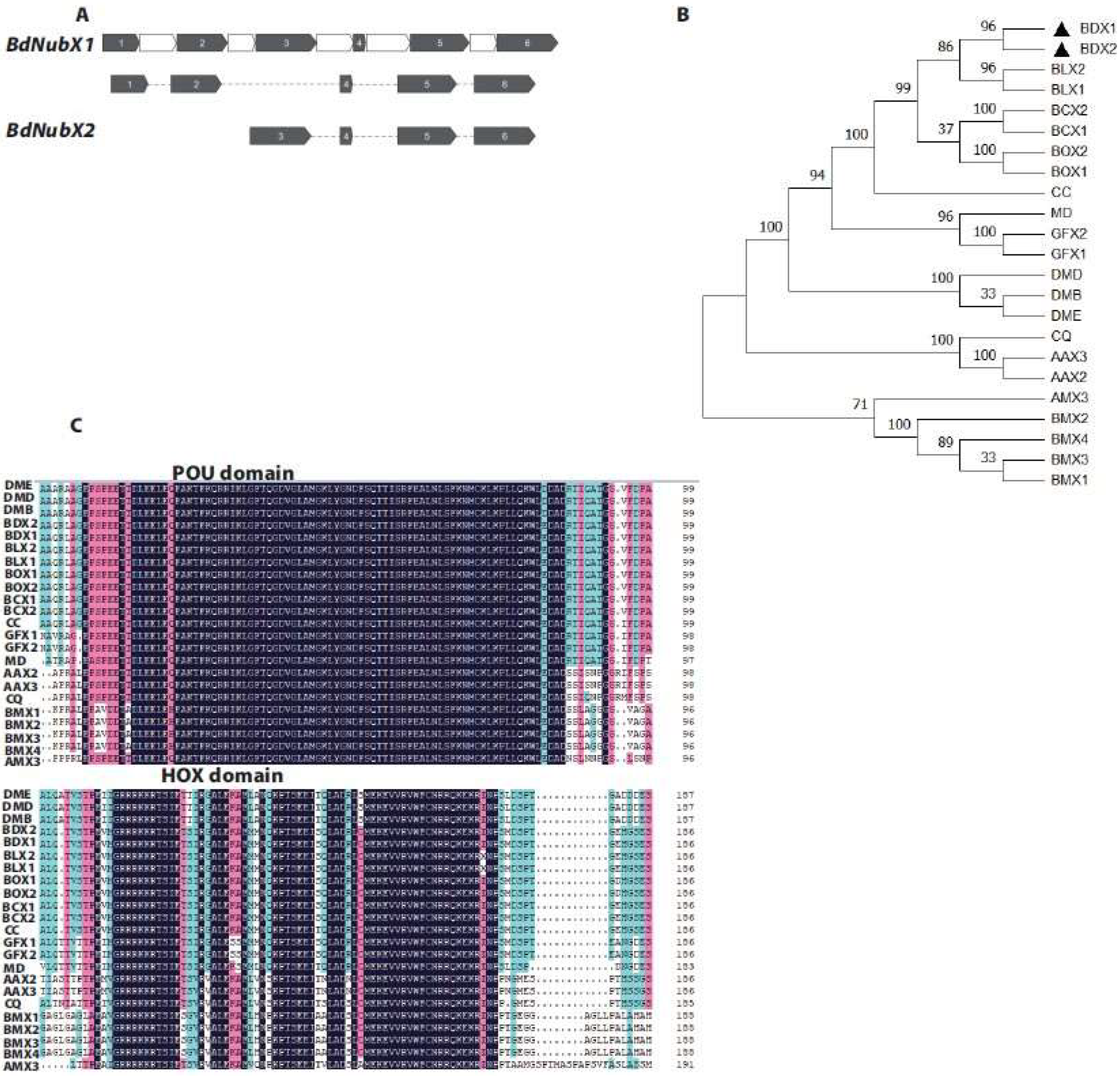
We found only one Nub gene (accession number: NW_011876379) in B. dorsalis by searching the NCBI database based on protein sequence homology using BLASTp. BdNub has two different transcripts, which we named BdNubX1 and BdNubX2, respectively. BdNub consists of 6 exons. BdNubX1 contains five exons, including transcript-specific exons 1 and 2, which are missing in BdNubX2, while BdNubX2 has four exons, including transcript-specific exon 3 (Figure 1A). BdNubX1 encodes 833 amino acids, and BdNubX2 encodes 567 amino acids. The two transcripts share the same 529 amino acids. Sequence alignment showed that BdNubX1 has 304 transcript-specific amino acids, while BdNubX2 has 38 transcript-specific amino acids. The Nub belongs to the OCT/POU family genes, evolutionarily conserved from arthropods to mammals. We performed the phylogenetic analysis on insects, including B. dorsalis, D. melanogaster, Musca domestica, Ceratitis capitata, Bombyx mori, and Aedes aegypti. The results confirmed that the Nub gene is conserved in insects. The results also revealed that despite sequence differences, two Nub isoforms clustered in the same branch. BdNub was closely related to Bactrocera capsicum (Figure 1B).
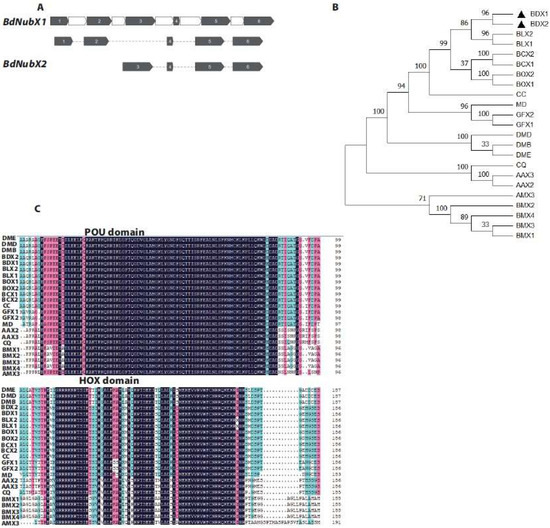
Figure 1.
Identification of BdNub gene. (A): The alternative splicing of BdNub. Exons are indicated by grey color, Introns are indicated by white color. (B): Phylogenetic analysis of the Nub gene, BdNub were aligned with Nub genes from 11 other insect species. Gene accession numbers were given in parentheses. Black triangle(the location of the target gene). (C): Alignment of POU and HOX domain amino acid sequence. Identical sequences were shown in black. 75% conserved amino acids were shown on pink background, and 50% conserved amino acids were shown on blue background. (B,C). BDX1, Bactrocera dorsalis nubbin X1 (XP 011207745.1); BDX2, Bactrocera dorsalis nubbin X2 (XP 011207746.1); BLX1, Bactrocera latifrons nubbin X1 (XP 018783925.1); BLX2, Bactrocera latifrons nubbin X 2(XP 018783926.1); BCX2, Bactrocera cucurbitae nubbin X2(XP 011178505.1) ;BCX1, Bactrocera cucurbitae nubbin X1 (XP 011178504.1); BOX2, Bactrocera oleae nubbin X2 (XP 014086366.1); BOX1, Bactrocera oleae nubbin X2; CC, Ceratitis capitata nubbin (XP 004530324.1); MD, Musca domestica nubbin (XP 019892278.1); GFX2, Glossina fuscipes nubbin X2 (XP 037882400.1); GFX1, Glossina fuscipes nubbin X1 (XP 037882399.1); DMB, Drosophila melanogaster nubbin B (NP 001097153.1); DMD, Drosophila melanogaster nubbin D (NP 476659.1) ; DME, Drosophila melanogaster nubbin E(NP 001285876.1) ; CQ, Culex quinquefasciatus nubbin (XP 001844054.1); AAX3, Aedes aegypti nubbin X3(XP 021704008.1); AAX2, Aedes aegypti nubbin X2 (XP 021704007.1); AMX3, Apis mellifera nubbin X3 (XP 006558737.1); BMX2, Bombyx mori nubbin X2 (XP 037870381.1) ; BMX4, Bombyx mori nubbin X4(XP 037870383.1); BMX3, Bombyx mori nubbin X3 (XP 037870382.1); BMX1, Bombyx mori nubbin X1(XP 037870380.1).
The Nub gene has two POU/OCT family conserved domains: the POU domain and the HOX domain. Although BdNubX1 and BdNubX2 have different amino acid sequences, they both have two intact conserved domains. We compared the amino acid sequences of the POU and HOX domains of BdNubX1 and BdNubX2 (Figure 1C). The amino acid sequences of BdNub POU and HOX are highly similar to the functional domains of B. capsicum and D. melanogaster, suggesting their functional similarity in vivo. We further use three other algorithms, including SMART, SOPMA, and SWISS, to predict the conserved domain and protein structure of BdNub. We also confirmed that BdNub is a classical POU/OCT family member (Figure S1).
3.2. Temporal and Spatial Expression Patterns of the BdNub Gene
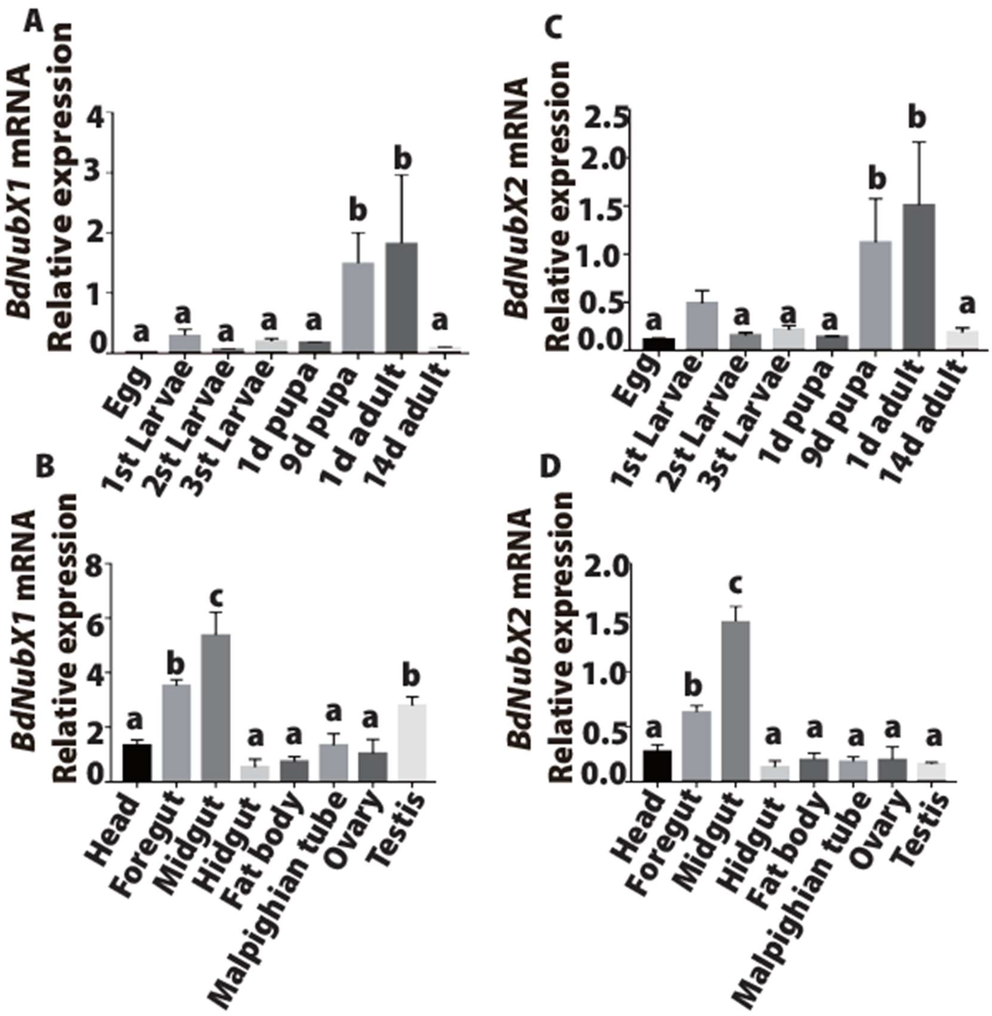
We analyzed the spatial and temporal expression patterns of BdNub gene transcripts at different periods and in different tissues. The results showed that BdNubX1 was highly expressed in 9-day-old pupa (the late-stage pupa) and the freshly emerged adults, and the expression was low in the egg, larva, and sexually mature adult stages (Figure 2A). Similarly, BdNubX2 was also highly expressed in the late pupal stage and the newly emerged adult flies (Figure 2B). Spatial analysis revealed that BdNubX1 expression was highest in the midgut, followed by the foregut and testis (Figure 2C). BdNubX2 showed a slightly different expression pattern. Although it was highly expressed in the foregut and midgut, it was much lower in the testis compared with BdNubX1 (Figure 2D). These results suggested that two BdNub transcript isoforms have similar expression patterns with high abundance in the late pupal stage, newly emerged adult flies, and in the adult gut.
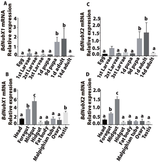
Figure 2.
Expression profiles of BdNubX1 and BdNubX2. (A): BdNubX1 Expression profile of different development stages, 20 different stage of development samples per biological replicate, three biological replicates. (B): BdNubX1 expression profile of different adult tissues, 30 different tissue samples per biological replicate, three biological replicates. (C): BdNubX2 Expression profile of different development stages, 20 different stage of development samples per biological replicate, three biological replicates. (D): BdNubX2 expression profile of different tissues of adults. Different letters indicate statistically significant differences in BdNub isoforms expression, 30 different tissue samples per biological replicate, three biological replicates. p < 0.05, Tukey’s test, One way ANOVA.
3.3. BdNub Does Not Participate in Systemic Infection of the IMD Pathway
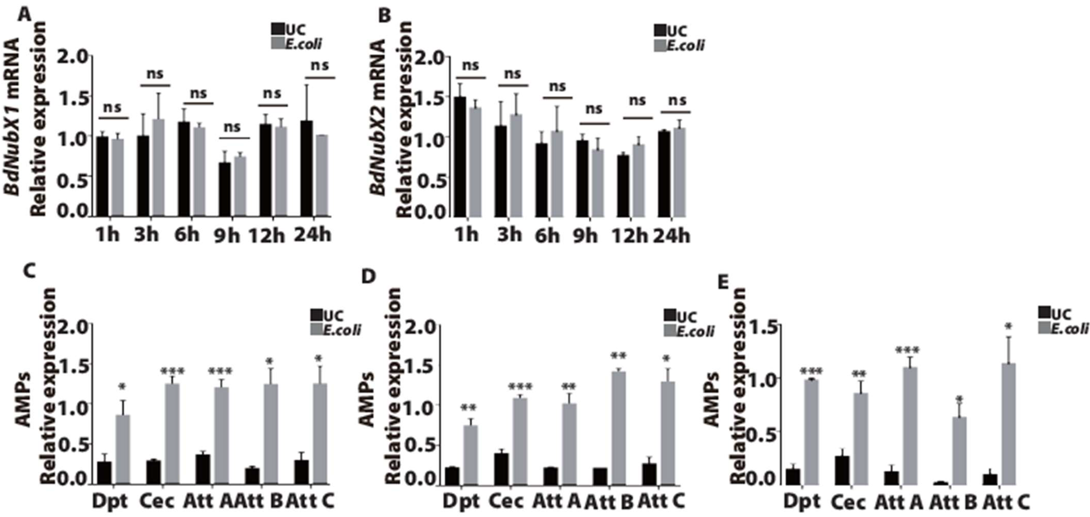
Next, we examined BdNubX1 and BdNubX2 expression at different time points after E. coli systemic infection. E. coli is a gram-negative pathogen that can elicit a robust immune response in B.dorsalis. The results showed that the BdNubX1 transcript had no significant increase after systemic infection with E. coli (Figure 3A). Similarly, the BdNubX2 transcript showed no significant change after E. coli infection (Figure 3B). These results indicated that BdNub did not play a role in the systemic infection of Gram-negative E. coli. To strengthen our conclusion, we further detected the main immune effectors of the IMD pathway, including AMPs Dpt, Cec, AttA, AttB, and AttC. The results showed that all five AMPs were significantly up-regulated at 3 h, 6 h, and 12 h after E. coli infection, proving that systemic infection works well in B. dorsalis (Figure 3C–E).
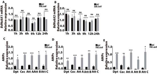
Figure 3.
The immune response of BdNub after E. coli systemic infection. (A) The relative expression level of BdNubX1 after E. coli systemic infection. (B) The relative expression level of BdNubX2 after E. coli systemic infection. (C–E) The relative expression of AMPs genes at 3 h, 6 h, 12 h after E. coli systemic infection, respectively, (A–E) 30 flies samples per biological replicate, three biological replicates. Gene expressions were normalized to the reference gene RP49. UC: unchallenged flies. * p < 0.05, ** p < 0.01, *** p < 0.001, Student’s t-test.
3.4. BdNub Regulates the Expression of Gut AMPs after Oral Infection
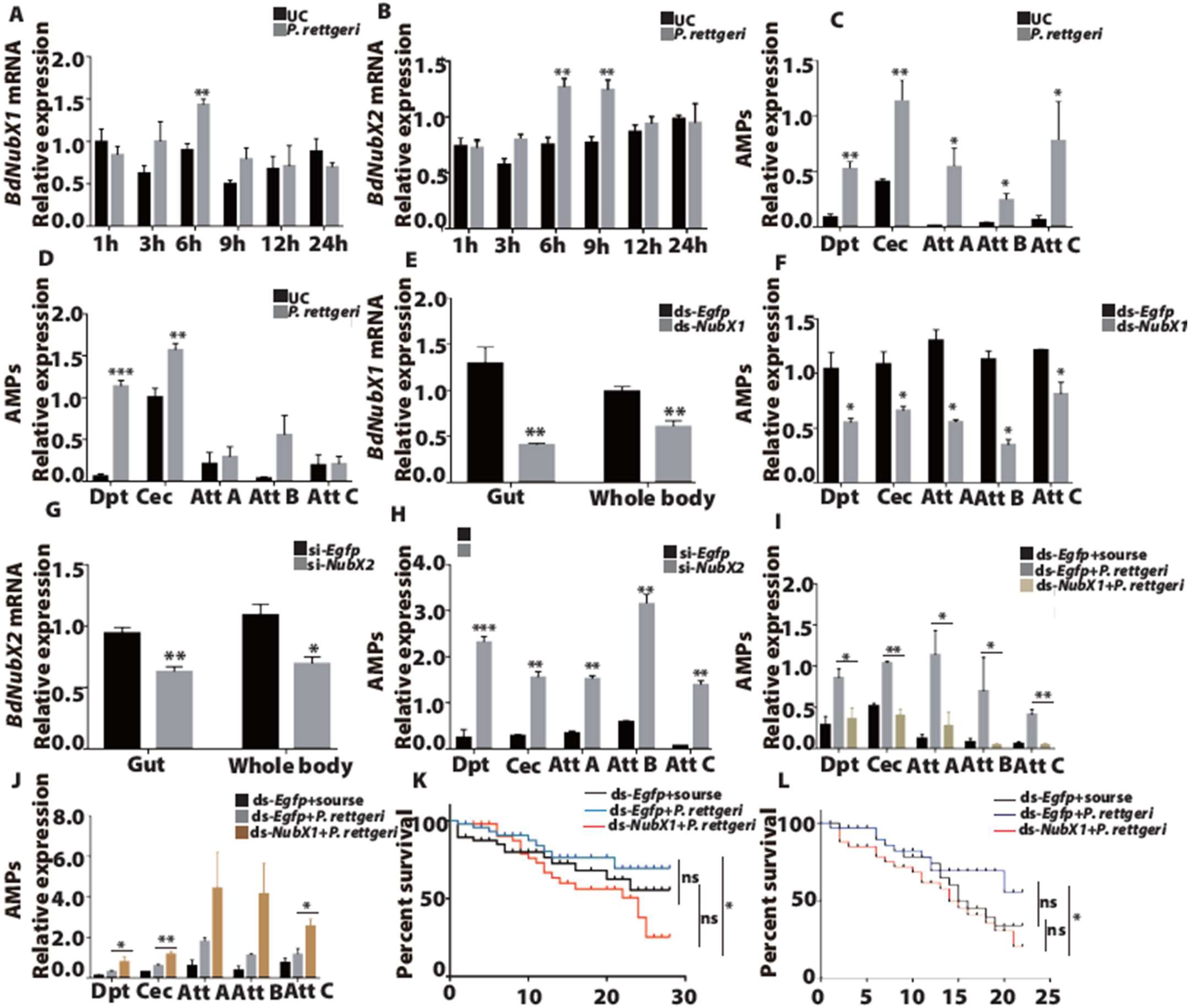
Previous data in our lab suggests that E. coli could not induce a strong and consistent gut immune response in B. dorsalis. Our screening found that P. rettgeri is a potent inducer of the gut immune response. Our results showed that BdNubX1 was significantly up-regulated at 6 h post oral infection of P. rettgeri, while there was no significant up-regulation at other time points (Figure 4A). This suggested that BdNubX1 is only transiently activated during the gut immune response. BdNubX2 showed a somewhat different expression pattern. It was significantly up-regulated at 6 h and 9 h after P. rettgeri oral infection, suggesting it played a prolonged role in gut immunity (Figure 4B). Next, to reaffirm that P. rettgeri could induce a gut immune response, we further examined AMP expression at different time points after infection. The results showed the immune effector AMPs’ expression, including Dpt, Cec, AttA, AttB, and AttC, increased at 6 h and 9 h after P. rettgeri oral infection. The results are as follows: at 6 h after oral infection, all five AMPs genes were significantly up-regulated (Figure 4C), while at 9 h after oral infection, only Dpt and Cec were significantly up-regulated (Figure 4D).
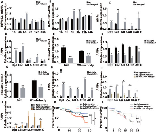
Figure 4.
The immune response of BdNub after P. rettgeri oral infection. (A,B) The relative expression level of BdNubX1 and BdNubX2 after P. rettgeri oral infection. (C,D) The relative expression of the AMPs genes at 6h and 9h after oral infection, respectively. (E) The RNAi effect of BdNubX1 dsRNA injection. (F) The expression levels of AMPs genes in BdNubX1 RNAi flies. (G) The RNAi effect of BdNubX1 siRNA injection. (H) The expression levels of AMPs genes in BdNubX2 RNAi flies. (I,J) The AMPs genes expressions at 6 h after P. rettgeri oral infection in BdNubX1 RNAi flies (I) and BdNubX2 RNAi flies (J). dsEgfp treated group was used as the control for RNAi. (K,L) The survival of BdNubX1 RNAi and BdNubX2 RNAi flies after rate after P. rettgeri oral infection. (A–L) 30 flies samples per biological replicate, three biological replicates. * p < 0.05, ** p < 0.01, *** p < 0.001, Student’s t-test. For survival assay, ns, non-significance, Log-rank (Mantel-Cox) test.
Next, we performed RNAi experiments to elucidate the function of the BdNub genes. The results showed that BdNubX1 RNAi down-regulated IMD target AMP genes, including Dpt, Cec, AttA, AttB, and AttC (Figure 4E,F). It indicated that BdNubX1 has a positive regulatory function on the IMD pathway. On the contrary, BdNubX2 RNAi leads to a significant up-regulation of the IMD-regulated AMPs expression (Figure 4G,H), suggesting that BdNubX2 has a negative regulatory function on the IMD pathway (Figure 4G,H). In order to further explore the function of the BdNub gene in gut immunity, we performed gut infection after RNAi of BdNubX1 and BdNubX2, respectively. The results showed that the expression levels of AMPs in BdNubX1-silenced flies after gut infection were significantly down-regulated compared with those in control flies after gut infection, and the expression levels of AMPs were similar to those of non-gut infection (Figure 4I). On the contrary, the expression levels of AMPs were significantly up-regulated in the BdNubX2-silenced flies after gut infection compared with the control flies after gut infection (Figure 4J).
Furthermore, to determine whether BdNubX1 and BdNubX2 RNAi affect B. dorsalis survival, we examined the survival rate of adult flies after P. rettgeri oral infection. The results showed that BdNubX1 RNAi flies fed on P. rettgeri died faster than the control group, which was provided with only sucrose. This demonstrated that P. rettgeri is indeed a pathogenic bacterium in B. dorsalis. On day 20, BdNubX1 RNAi flies showed a much lower survival rate than the Egfp RNAi control group (Figure 4K). Similarly, the results showed that BdNubX2 RNAi flies fed on P. rettgeri died faster than the control group feeding only sucrose. On day 19, BdNubX2 RNAi flies showed a lower survival rate than the Egfp RNAi control group (Figure 4L).
3.5. BdNub Is Necessary to Maintain Gut Microbiota Composition and Structure
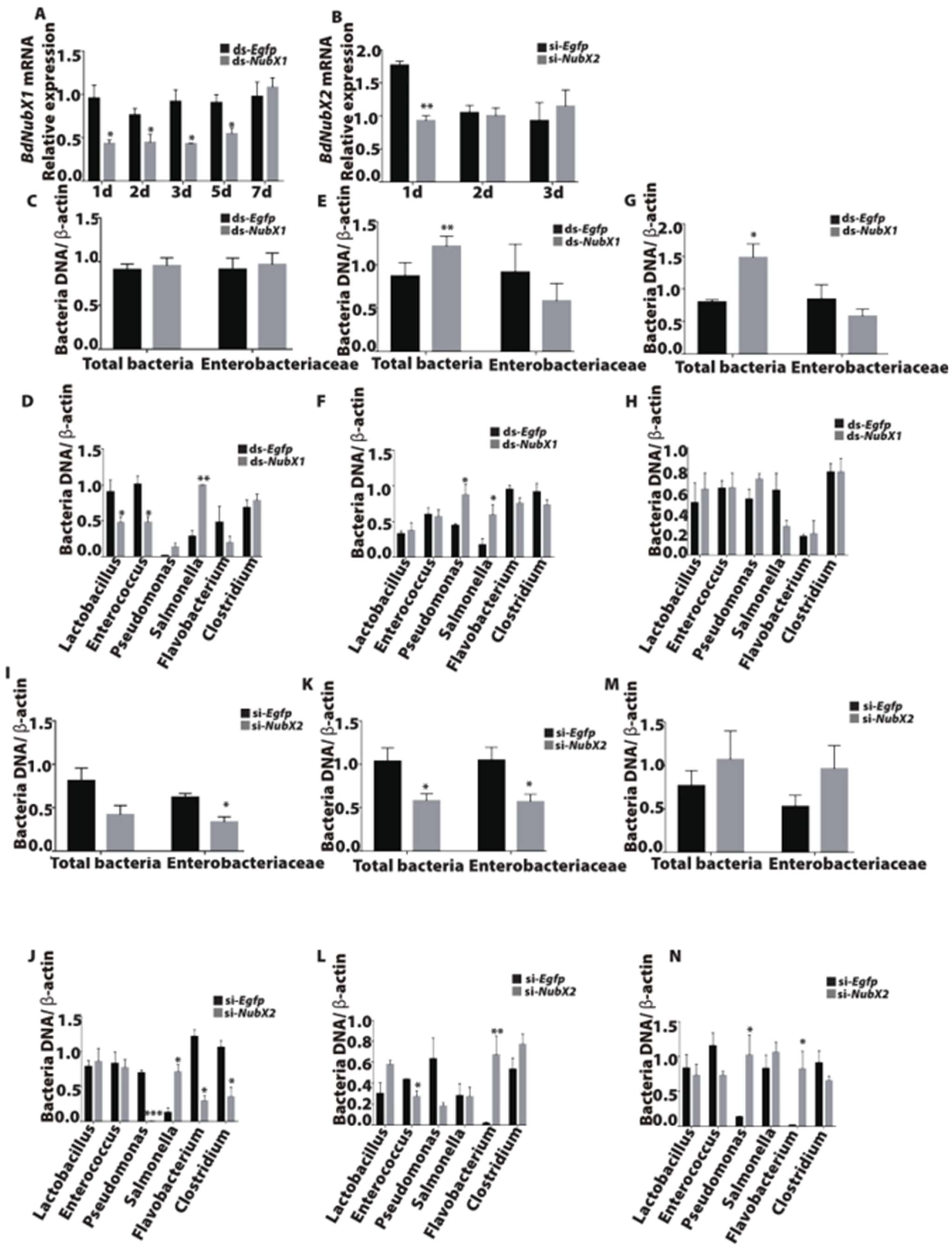
The IMD pathway is involved in insect gut microbiota regulation [2,22]. To determine the function of BdNubX1 and BdNubX2 isoforms in microbiota regulation, we examined microbiota composition using qRT-PCR in BdNubX1 and BdNubX2 RNAi flies. Our results showed that BdNubX1 RNAi disturbed gut microbiota composition and decreased microbiota abundance (Figure 5A,C,E,G). The bacterial loads of different genera have changed significantly, with strains like Lactobacillus and Enterococcus significantly down-regulated, and Pseudomonas and Salmonella significantly increased (Figure 5D,F,H). This result indicated that BdNubX1 RNAi changed gut microbiota composition and quantity. Similarly, BdNubX2 RNAi also caused significant changes in gut microbiota. The total intestinal bacteria were down-regulated on day 5 after the dsRNA injection (Figure 5B,I,K,M). Among them, the Enterobacteriaceae were significantly down-regulated on days 3 and 5. The abundance of Pseudomonas, Flavobacterium, and Salmonella also changed at different time points after RNAi (Figure 5J,L,N).
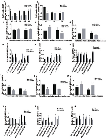
Figure 5.
Effect of BdNubX1 and BdNubX2 RNAi on gut microbiota. (A,B) BdNubX1 and BdNubX2 expression levels after dsRNA and siRNA injection, respectively. (C) The gut total bacteria and Enterobacteriaceae abundance at 3d in BdNubX1 RNAi flies. (D) The different genus bacteria abundance at 3 d in BdNubX1 RNAi flies. (E) The gut total bacteria and Enterobacteriaceae abundance at 5d in BdNubX1 RNAi flies. (F) The different genus bacteria abundance at 5 d in BdNubX1 RNAi flies. (G) The gut total bacteria and Enterobacteriaceae abundance at 7 d in BdNubX1 RNAi flies. (H) The different genus bacteria abundance at 7d in BdNubX1 RNAi flies. (I) The gut total bacteria and Enterobacteriaceae abundance at 3 d in BdNubX2 RNAi flies. (J) The different genus bacteria abundance at 3 d in BdNubX2 RNAi flies. (K) The gut total bacteria and Enterobacteriaceae abundance at 5 d in BdNubX2 RNAi flies. (L) The different genus bacteria abundance at 5 d in BdNubX2 RNAi flies. (M) The gut total bacteria and Enterobacteriaceae abundance at 7 d in BdNubX2 RNAi flies. (N) The different genus bacteria abundance at 7 d in BdNubX2 RNAi flies, (A–N) 30 flies guts samples per biological replicate, three biological replicates. * p < 0.05, ** p <0.01, *** p < 0.001, Student’s t-test.
4. Discussion
In this study, we report that BdNub encodes two isoforms, BdNubX1 and BdNubX2. They are involved in the gut immune response, not systemic immunity, possibly by regulating the gut IMD pathway. Our results also show that BdNub is essential for maintaining gut microbiota.
We identified a putative B. dorsalis Nub gene, BdNub. Similar to Drosophila, it also produces two distinct BdNub isoforms [15]. Phylogenetic tree analysis shows that the BdNub gene is closely related to B. capsicum, and the Nub gene is highly conserved in insects. This is also confirmed with the protein alignment of conserved domains, POU and HOX [33]. There are five different POU proteins in the Drosophila genome [34], regulating embryonic development and differentiation [12], immune function, and tissue homeostasis [14,15]. In addition, pdm1, a POU family gene, acts as proximal-distal growth of the wing, which has a similar function to Nub [35]. Moreover, the HOX domain regulates muscle and wing development [36,37], specifying the anterior posterior axis in all bilaterians [38]. So, this indicates the high expression of NUB genes in the old pupa stage and the day 1 adult flies.
Spatial and temporal expression pattern analysis showed that BdNubX1 and BdNubX2 were mainly expressed in the late-stage pupa, which may be related to Nub gene function in wing development [39,40]. In Drosophila, Nub mutant flies showed a severe wing size reduction [35,40,41,42], indicating that BdNub may also have an indispensable role in B. dorsalis wing formation. This might relate to the HOX domain of BdNub. Furthermore, we also observed high BdNub expression in newly emerged adults and guts. B. dorsalis must crawl out of the soil to accomplish eclosion. Thus, newly merged adult flies are exposed to various microorganisms from the soil and environment. High BdNub expression at this stage suggested its role in regulating gut immune balance in this process [43].
Our results showed that E. coli systemic infection induced the AMP genes’ expression but not BdNubX1 and BdNubX2. It suggests that the BdNub gene is not involved in E. coli-induced systemic immunity. This result is consistent with their high expression in the gut and low expression in the fat body, the major systemic immune organ. However, immunostaining reveals that Nub protein is present in the fat body of Drosophila [14]. This suggests Nub could be functional in regulating Drosophila’s systemic immune response.
In contrast, Gram-negative bacteria P. rettgeri oral infection induced a strong immune response and a strong BdNubX1 and BdNubX2 up-regulation, suggesting that BdNub was involved in the gut immune response. Furthermore, we showed that BdNubX1 positively regulated gut AMP expression, while BdNubX2 inhibited AMP expression. The fact that BdNubX2 is an immunosuppressor of IMD pathway activity is also in line with the Nub gene function in the Drosophila gut [7,43]. In Drosophila, Nub-PD RNAi increased AMP gene expression. Similarly, BdNubX2 RNAi also increased AMP gene expression. Apart from Nub, the IMD signaling pathway is regulated by many other factors. For example, the Pirk gene encodes the protein binding PGRP-LC. It is regulated by the IMD pathway itself. Nevertheless, it establishes a negative feedback loop adjusting IMD pathway activity [44]. PGRP-SB and PGRP-LB are secreted proteins with an amidase activity that scavenges DAP-type peptidoglycan [6]. They negatively regulate the IMD pathway. A recent study also shows that PGRP-SB and PGRP-LB are negative regulators of the gut IMD pathway in B. dorsalis [23,45,46]. There may also be other unknown factors that regulate the gut IMD pathway activity that have yet to be identified.
In addition, both BdNubX1 and BdNubX2 RNAi could make the flies more sensitive to P. rettgeri infection. Nevertheless, there was no significant difference in the mortality rate, which was consistent with the results of Drosophila [15]. This may be due to the fact that RNAi could not achieve a stable and long-lasting silence effect in B. dorsalis. Furthermore, the efficiency of RNAi varies considerably among insects, although RNAi can reach 90% efficacy in Coleopterans [47]. Dipteran species are not very sensitive to RNAi [48]. Studies using null mutants should be carried out to further elucidate the Nub function in gut immunity in the future. Moreover, unlike Drosophila, our screening did not find any strong lethal pathogenic bacteria for the gut infection. Although P. rettgeri could induce a strong gut immune response, it kills the wild-type flies very slowly, which might be an immune tolerance phenotype rather than an immune resistance phenotype. It indicates that the high adaptability of B. dorsalis may be related to its strong immune system.
Since BdNubX1 and BdNubX2 transcript isomers play an important role in intestinal immunity, it is plausible that they also participate in controlling the microbiota. Many reports show that changes in immune-related genes will lead to changes in intestinal microbial community structure [21,23]. BdNubX1 knockdown leads to significantly up-regulated total bacterial abundance, with increased Pseudomonas and Salmonella. This indicates BdNubX1 partially regulates gut microbiota composition and abundance through the IMD pathway. The IMD pathway is an important part of the insect gut microbiota regulation mechanism [49]. For example, PGRP-LB and PGRP-SB have high expression levels in the anterior and middle midgut, which is associated with gut commensal bacteria distribution in B. dorsalis [23]. In Drosophila, IMD-deficient flies showed a dysregulated gut microbiota and disturbed gut homeostasis [22]. Moreover, we cannot exclude the possibility that BdNubX1 also interacts with other gut immune mechanisms. For example, BdDuox regulates gut microbiota through the production of ROS [21]. Therefore, it is possible that BdNubX1 might regulate microbiota by affecting ROS production and scavenging.
On the other side, BdNubX2 knockdown leads to decreased gut microbiota and Enterobacteriaceae. It also causes changes in Pseudomonas, Flavobacterium, and Salmonella abundance. In B. dorsalis, Enterobacteriaceae bacteria account for a large proportion in the gut and are the dominant bacterial group in gut microbiota [50]. In fact, the key ROS production enzyme, Bdduox RNAi, induces a similar changes in microbiota composition, with decreased Enterobacteriaceae and a rise in secondary microbiota abundance [21]. Several studies have shown that Enterobacteriaceae bacteria are beneficial to the host [20,24]. Therefore, decreased Enterobacteriaceae in B. dorsalis is possibly detrimental to the host. Our results suggest that BdNubX2 could also be essential to host development and homeostasis through regulating Enterobacteriaceae bacteria, which could contribute to the early death of BdNubX2 RNAi flies. Actually, Drosophila Caudal mutants show constitutively activated AMP genes, leading to an increase in Gluconobacter sp., causing an early death of the hosts [49]. Altogether, we proved that the BdNub gene regulates the IMD pathway to maintain intestinal microbial homeostasis. In conclusion, BdNub plays an important role in the regulation of intestinal immunity, decreasing the host’s sensitivity to intestinal opportunistic pathogens, and regulating gut microbiota.
Supplementary Materials
The following supporting information can be downloaded at: https://www.mdpi.com/article/10.3390/insects14020178/s1, Figure S1: Protein functional domain analysis of BdNubX1 and BdNubX2.
Author Contributions
Conceptualization, H.Z., X.L. and Z.Y.; Methodology, J.G. and P.Z.; Formal analysis, J.G. and P.Z.; Investigation, J.G. and P.Z.; Data Curation, J.G. and P.Z.; Visualization, J.G. and P.Z.; Writing—original draft, J.G. and X.L.; Supervision, X.L., Z.Y. and H.Z.; Funding acquisition, H.Z.; Project administration, H.Z. All authors have read and agreed to the published version of the manuscript.
Funding
This study was supported by National Key R&D Program of China (No. 2021YFC2600400), The Major Special Science and Technology Project of Yunnan Province (NO. 202102AE090054),The Earmarked Fund for CARS (CARS-26) and Hubei Hongshan Laboratory.
Data Availability Statement
The data presented in this study are available on request from the corresponding author.
Acknowledgments
Special thanks to Qiongke Ma for insects rearing.
Conflicts of Interest
The authors declare no conflict of interest.
References
- Basset, Y.; Cizek, L.; Cuénoud, P.; Didham, R.K.; Guilhaumon, F.; Missa, O.; Novotny, V.; Ødegaard, F.; Roslin, T.; Schmidl, J.; et al. Arthropod Diversity in a Tropical Forest. Science 2012, 338, 1481–1484. [Google Scholar] [CrossRef] [PubMed]
- Engel, P.; Moran, N.A. The Gut Microbiota of Insects—Diversity in Structure and Function. FEMS Microbiol. Rev. 2013, 37, 699–735. [Google Scholar] [CrossRef] [PubMed]
- Gottar, M.; Gobert, V.; Michel, T.; Belvin, M.; Duyk, G.; Hoffmann, J.A.; Ferrandon, D.; Royet, J. The Drosophila Immune Response against Gram-Negative Bacteria Is Mediated by a Peptidoglycan Recognition Protein. Nature 2002, 416, 640–644. [Google Scholar] [CrossRef] [PubMed]
- Brennan, C.A.; Anderson, K.V. Drosophila: The Genetics of Innate Immune Recognition and Response. Annu. Rev. Immunol. 2004, 22, 457–483. [Google Scholar] [CrossRef]
- Royet, J.; Dziarski, R. Peptidoglycan Recognition Proteins: Pleiotropic Sensors and Effectors of Antimicrobial Defences. Nat. Rev. Microbiol. 2007, 5, 264–277. [Google Scholar] [CrossRef]
- Lemaitre, B.; Hoffmann, J. The Host Defense of Drosophila Melanogaster. Annu. Rev. Immunol. 2007, 25, 697–743. [Google Scholar] [CrossRef]
- Imler, J.-L.; Bulet, P. Antimicrobial Peptides in Drosophila: Structures, Activities and Gene Regulation. Mech. Epithel. Def. 2005, 86, 1–21. [Google Scholar] [CrossRef]
- Hanson, M.A.; Dostálová, A.; Ceroni, C.; Poidevin, M.; Kondo, S.; Lemaitre, B. Synergy and Remarkable Specificity of Antimicrobial Peptides in Vivo Using a Systematic Knockout Approach. eLife 2019, 8, e44341. [Google Scholar] [CrossRef]
- Horak, R.D.; Leonard, S.P.; Moran, N.A. Symbionts Shape Host Innate Immunity in Honeybees. Proc. R. Soc. B 2020, 287, 20201184. [Google Scholar] [CrossRef]
- Zhang, L.; Gallo, R.L. Antimicrobial Peptides. Curr. Biol. 2016, 26, R14–R19. [Google Scholar] [CrossRef]
- Davis, M.M.; Engström, Y. Immune Response in the Barrier Epithelia: Lessons from the Fruit Fly Drosophila Melanogaster. J. Innate Immun. 2012, 4, 273–283. [Google Scholar] [CrossRef]
- Tantin, D. Oct Transcription Factors in Development and Stem Cells: Insights and Mechanisms. Development 2013, 140, 2857–2866. [Google Scholar] [CrossRef]
- Dantoft, W.; Lundin, D.; Esfahani, S.S.; Engström, Y. The POU/Oct Transcription Factor Pdm1/Nub Is Necessary for a Beneficial Gut Microbiota and Normal Lifespan of Drosophila. J. Innate Immun. 2016, 8, 412–426. [Google Scholar] [CrossRef]
- Dantoft, W.; Davis, M.M.; Lindvall, J.M.; Tang, X.; Uvell, H.; Junell, A.; Beskow, A.; Engström, Y. The Oct1 Homolog Nubbin Is a Repressor of NF-ΚB-Dependent Immune Gene Expression That Increases the Tolerance to Gut Microbiota. BMC Biol. 2013, 11, 99. [Google Scholar] [CrossRef]
- Lindberg, B.G.; Tang, X.; Dantoft, W.; Gohel, P.; Seyedoleslami Esfahani, S.; Lindvall, J.M.; Engström, Y. Nubbin Isoform Antagonism Governs Drosophila Intestinal Immune Homeostasis. PLoS Pathog. 2018, 14, e1006936. [Google Scholar] [CrossRef]
- Dillon, R.J.; Dillon, V.M. THE GUT B ACTERIA OF INSECTS: Nonpathogenic Interactions. Annu. Rev. Entomol. 2004, 49, 71–92. [Google Scholar] [CrossRef]
- Consuegra, J.; Grenier, T.; Baa-Puyoulet, P.; Rahioui, I.; Akherraz, H.; Gervais, H.; Parisot, N.; da Silva, P.; Charles, H.; Calevro, F.; et al. Drosophila-Associated Bacteria Differentially Shape the Nutritional Requirements of Their Host during Juvenile Growth. PLoS Biol. 2020, 18, e3000681. [Google Scholar] [CrossRef]
- Storelli, G.; Strigini, M.; Grenier, T.; Bozonnet, L.; Schwarzer, M.; Daniel, C.; Matos, R.; Leulier, F. Drosophila Perpetuates Nutritional Mutualism by Promoting the Fitness of Its Intestinal Symbiont Lactobacillus Plantarum. Cell Metab. 2018, 27, 362–377.e8. [Google Scholar] [CrossRef]
- Flórez, L.V.; Scherlach, K.; Gaube, P.; Ross, C.; Sitte, E.; Hermes, C.; Rodrigues, A.; Hertweck, C.; Kaltenpoth, M. Antibiotic-Producing Symbionts Dynamically Transition between Plant Pathogenicity and Insect-Defensive Mutualism. Nat. Commun. 2017, 8, 15172. [Google Scholar] [CrossRef]
- Cai, Z.; Yao, Z.; Li, Y.; Xi, Z.; Bourtzis, K.; Zhao, Z.; Bai, S.; Zhang, H. Intestinal Probiotics Restore the Ecological Fitness Decline of Bactrocera Dorsalis by Irradiation. Evol. Appl. 2018, 11, 1946–1963. [Google Scholar] [CrossRef]
- Yao, Z.; Wang, A.; Li, Y.; Cai, Z.; Lemaitre, B.; Zhang, H. The Dual Oxidase Gene BdDuox Regulates the Intestinal Bacterial Community Homeostasis of Bactrocera Dorsalis. ISME J. 2016, 10, 1037–1050. [Google Scholar] [CrossRef] [PubMed]
- Broderick, N.A.; Buchon, N.; Lemaitre, B. Microbiota-Induced Changes in Drosophila Melanogaster Host Gene Expression and Gut Morphology. mBio 2014, 5, e01117-14. [Google Scholar] [CrossRef] [PubMed]
- Yao, Z.; Cai, Z.; Ma, Q.; Bai, S.; Wang, Y.; Zhang, P.; Guo, Q.; Gu, J.; Lemaitre, B.; Zhang, H. Compartmentalized PGRP Expression along the Dipteran Bactrocera Dorsalis Gut Forms a Zone of Protection for Symbiotic Bacteria. Cell Rep. 2022, 41, 111523. [Google Scholar] [CrossRef] [PubMed]
- Raza, M.F.; Wang, Y.; Cai, Z.; Bai, S.; Yao, Z.; Awan, U.A.; Zhang, Z.; Zheng, W.; Zhang, H. Gut Microbiota Promotes Host Resistance to Low-Temperature Stress by Stimulating Its Arginine and Proline Metabolism Pathway in Adult Bactrocera Dorsalis. PLoS Pathog. 2020, 16, e1008441. [Google Scholar] [CrossRef] [PubMed]
- Guo, Q.; Yao, Z.; Cai, Z.; Bai, S.; Zhang, H. Gut Fungal Community and Its Probiotic Effect on Bactrocera Dorsalis. Insect Sci. 2022, 29, 1145–1158. [Google Scholar] [CrossRef]
- Livak, K.J.; Schmittgen, T.D. Analysis of Relative Gene Expression Data Using Real-Time Quantitative PCR and the 2\textbackslashtextminusΔΔCT Method. Methods 2001, 25, 402–408. [Google Scholar] [CrossRef]
- Guo, X.; Xia, X.; Tang, R.; Wang, K. Real-Time PCR Quantification of the Predominant Bacterial Divisions in the Distal Gut of Meishan and Landrace Pigs. Anaerobe 2008, 14, 224–228. [Google Scholar] [CrossRef]
- Bartosch, S.; Fite, A.; Macfarlane, G.T.; McMurdo, M.E.T. Characterization of Bacterial Communities in Feces from Healthy Elderly Volunteers and Hospitalized Elderly Patients by Using Real-Time PCR and Effects of Antibiotic Treatment on the Fecal Microbiota. Appl. Environ. Microbiol. 2004, 70, 3575–3581. [Google Scholar] [CrossRef]
- Rinttila, T.; Kassinen, A.; Malinen, E.; Krogius, L.; Palva, A. Development of an Extensive Set of 16S RDNA-Targeted Primers for Quantification of Pathogenic and Indigenous Bacteria in Faecal Samples by Real-Time PCR. J. Appl. Microbiol. 2004, 97, 1166–1177. [Google Scholar] [CrossRef]
- Abell, G.C.J.; Bowman, J.P. Ecological and Biogeographic Relationships of Class Flavobacteria in the Southern Ocean. Fems Microbiol. Ecol. 2005, 51, 265–277. [Google Scholar] [CrossRef]
- Ahmed, W.; Huygens, F.; Goonetilleke, A.; Gardner, T. Real-Time PCR Detection of Pathogenic Microorganisms in Roof-Harvested Rainwater in Southeast Queensland, Australia. Appl. Environ. Microbiol. 2008, 74, 5490–5496. [Google Scholar] [CrossRef]
- Matsuda, K.; Tsuji, H.; Asahara, T.; Matsumoto, K.; Takada, T.; Nomoto, K. Establishment of an Analytical System for the Human Fecal Microbiota, Based on Reverse Transcription-Quantitative PCR Targeting of Multicopy RRNA Molecules. Appl. Environ. Microbiol. 2009, 75, 1961–1969. [Google Scholar] [CrossRef]
- Bürglin, T.R.; Affolter, M. Homeodomain Proteins: An Update. Chromosoma 2016, 125, 497–521. [Google Scholar] [CrossRef]
- Tang, X.; Engström, Y. Regulation of Immune and Tissue Homeostasis by Drosophila POU Factors. Insect Biochem. Mol. Biol. 2019, 109, 24–30. [Google Scholar] [CrossRef]
- Cifuentes, F.J.; García-Bellido, A. Proximo–Distal Specification in the Wing Disc of Drosophila by the Nubbin Gene. Proc. Natl. Acad. Sci. USA 1997, 94, 11405–11410. [Google Scholar] [CrossRef]
- Poliacikova, G.; Maurel-Zaffran, C.; Graba, Y.; Saurin, A.J. Hox Proteins in the Regulation of Muscle Development. Front. Cell Dev. Biol. 2021, 9, 731996. [Google Scholar] [CrossRef]
- Paul, R.; Giraud, G.; Domsch, K.; Duffraisse, M.; Marmigère, F.; Khan, S.; Vanderperre, S.; Lohmann, I.; Stoks, R.; Shashidhara, L.S.; et al. Hox Dosage Contributes to Flight Appendage Morphology in Drosophila. Nat. Commun. 2021, 12, 2892. [Google Scholar] [CrossRef]
- Mallo, M. Reassessing the Role of Hox Genes during Vertebrate Development and Evolution. Trends Genet. 2018, 34, 209–217. [Google Scholar] [CrossRef]
- Fernandez-Nicolas, A.; Ventos-Alfonso, A.; Kamsoi, O.; Clark-Hachtel, C.; Tomoyasu, Y.; Belles, X. Broad Complex and Wing Development in Cockroaches. Insect Biochem. Mol. Biol. 2022, 147, 103798. [Google Scholar] [CrossRef]
- Ng, M.; Diaz-Benjumea, F.J.; Cohen, S.M. Nubbin Encodes a POU-Domain Protein Required for Proximal-Distal Patterning in the Drosophila Wing. Development 1995, 121, 589–599. [Google Scholar] [CrossRef]
- Zirin, J.D.; Mann, R.S. Nubbin and Teashirt Mark Barriers to Clonal Growth along the Proximal–Distal Axis of the Drosophila Wing. Dev. Biol. 2007, 304, 745–758. [Google Scholar] [CrossRef] [PubMed]
- Neumann, C.J.; Cohen, S.M. Boundary Formation in Drosophila Wing: Notch Activity Attenuated by the POU Protein Nubbin. Science 1998, 281, 409–413. [Google Scholar] [CrossRef] [PubMed]
- Buddika, K.; Huang, Y.-T.; Ariyapala, I.S.; Butrum-Griffith, A.; Norrell, S.A.; O’Connor, A.M.; Patel, V.K.; Rector, S.A.; Slovan, M.; Sokolowski, M.; et al. Coordinated Repression of Pro-Differentiation Genes via P-Bodies and Transcription Maintains Drosophila Intestinal Stem Cell Identity. Curr. Biol. 2022, 32, 386–397.e6. [Google Scholar] [CrossRef] [PubMed]
- Kleino, A.; Myllymäki, H.; Kallio, J.; Vanha-aho, L.-M.; Oksanen, K.; Ulvila, J.; Hultmark, D.; Valanne, S.; Rämet, M. Pirk Is a Negative Regulator of the Drosophila Imd Pathway. J. Immunol. 2008, 180, 5413–5422. [Google Scholar] [CrossRef] [PubMed]
- Mellroth, P.; Steiner, H. akan PGRP-SB1: An N-Acetylmuramoyl l-Alanine Amidase with Antibacterial Activity. Biochem. Biophys. Res. Commun. 2006, 350, 994–999. [Google Scholar] [CrossRef] [PubMed]
- Zaidman-Rémy, A.; Hervé, M.; Poidevin, M.; Pili-Floury, S.; Kim, M.-S.; Blanot, D.; Oh, B.-H.; Ueda, R.; Mengin-Lecreulx, D.; Lemaitre, B. The Drosophila Amidase PGRP-LB Modulates the Immune Response to Bacterial Infection. Immunity 2006, 24, 463–473. [Google Scholar] [CrossRef]
- Joga, M.R.; Zotti, M.J.; Smagghe, G.; Christiaens, O. RNAi Efficiency, Systemic Properties, and Novel Delivery Methods for Pest Insect Control: What We Know So Far. Front. Physiol. 2016, 7, 553. [Google Scholar] [CrossRef]
- Araujo, R.N.; Santos, A.; Pinto, F.S.; Gontijo, N.F.; Lehane, M.J.; Pereira, M.H. RNA Interference of the Salivary Gland Nitrophorin 2 in the Triatomine Bug Rhodnius Prolixus (Hemiptera: Reduviidae) by DsRNA Ingestion or Injection. Insect Biochem. Mol. Biol. 2006, 36, 683–693. [Google Scholar] [CrossRef]
- Ryu, J.-H.; Kim, S.-H.; Lee, H.-Y.; Bai, J.Y.; Nam, Y.-D.; Bae, J.-W.; Lee, D.G.; Shin, S.C.; Ha, E.-M.; Lee, W.-J. Innate Immune Homeostasis by the Homeobox Gene Caudal and Commensal-Gut Mutualism in Drosophila. Science 2008, 319, 777–782. [Google Scholar] [CrossRef]
- Wang, H.; Jin, L.; Zhang, H. Comparison of the Diversity of the Bacterial Communities in the Intestinal Tract of Adult Bactrocera Dorsalis from Three Different Populations. J. Appl. Microbiol. 2011, 110, 1390–1401. [Google Scholar] [CrossRef]
Disclaimer/Publisher’s Note: The statements, opinions and data contained in all publications are solely those of the individual author(s) and contributor(s) and not of MDPI and/or the editor(s). MDPI and/or the editor(s) disclaim responsibility for any injury to people or property resulting from any ideas, methods, instructions or products referred to in the content. |
© 2023 by the authors. Licensee MDPI, Basel, Switzerland. This article is an open access article distributed under the terms and conditions of the Creative Commons Attribution (CC BY) license (https://creativecommons.org/licenses/by/4.0/).

