Article

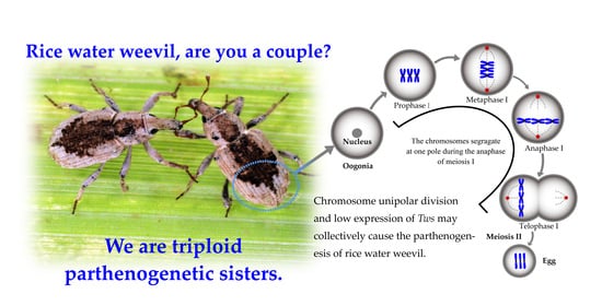
Chromosome Unipolar Division and Low Expression of Tws May Cause Parthenogenesis of Rice Water Weevil (Lissorhoptrus oryzophilus Kuschel)

1
Key Laboratory of Zoological Systematics and Evolution, Institute of Zoology, Chinese Academy of Sciences, Beijing 100101, China
2
College of Life Sciences, University of Chinese Academy of Sciences, Beijing 100049, China
3
Department of Entomology, Guizhou University, Guiyang 550025, Guizhou, China
*
Author to whom correspondence should be addressed.
Insects 2021, 12(4), 278; https://doi.org/10.3390/insects12040278
Submission received: 6 February 2021 / Revised: 22 March 2021 / Accepted: 22 March 2021 / Published: 24 March 2021
(This article belongs to the Section Insect Molecular Biology and Genomics)

Abstract

:

Simple Summary

Rice water weevil (RWW), Lissorhoptrus oryzophilus (Kuschel) (Coleoptera: Brachyceridae), is one of the main pests of rice. In 1959, the parthenogenetic RWW was first reported in California and invaded Asia in 1978. It is known that sexually reproducing RWW is a diploid organism, while parthenogenetic RWW is a triploid organism and its distribution range is much larger than in sexually reproducing populations. We provide valuable molecular evidence for parthenogenesis and analyze the cellular basic process of parthenogenetic RWW. Results show in particular that the parthenogenetic RWW has chromosome unipolar division during meiosis, Tws (a regulatory subunit of protein phosphatase 2A) has low expression in ovarian tissue, and REC8 (paralogous gene of Verthandi/Rad21) is normally expressed. Our study indicates that parthenogenetic RWW produces triploid egg cells via chromosome unipolar division, while low expression of Tws within ovarian tissues may be associated with parthenogenesis, thus providing the first molecular genetic evidence for the parthenogenetic RWW.

Abstract

Rice water weevil (RWW) is divided into two types of population, triploid parthenogenesis and diploid bisexual reproduction. In this study, we explored the meiosis of triploid parthenogenesis RWW (Shangzhuang Town, Haidian District, Beijing, China) by marking the chromosomes and microtubules of parthenogenetic RWW oocytes via immunostaining. The immunostaining results show that there is a canonical meiotic spindle formed in the triploid parthenogenetic RWW oocytes, but chromosomes segregate at only one pole, which means that there is a chromosomal unipolar division during the oogenesis of the parthenogenetic RWW. Furthermore, we cloned the conserved sequences of parthenogenetic RWW REC8 and Tws, and designed primers based on the parthenogenetic RWW sequence to detect expression patterns by quantitative PCR (Q-PCR). Q-PCR results indicate that the expression of REC8 and Tws in ovarian tissue of bisexual Drosophila melanogaster is 0.98 and 10,000.00 times parthenogenetic RWW, respectively (p < 0.01). The results show that Tws had low expression in parthenogenetic RWW ovarian tissue, and REC8 was expressed normally. Our study suggests that the chromosomal unipolar division and deletion of Tws may cause parthenogenesis in RWW.

Graphical Abstract

1. Introduction

Parthenogenesis is a reproductive mode in which female animals produce offspring from unfertilized eggs [1]. It has been reported that about 600 species of beetles in 20 families in Coleoptera are able to produce offspring by parthenogenesis [2,3]. While parthenogenesis exists in many taxa, the molecular mechanism and meiotic process of parthenogenesis are still poorly understood. Rice water weevil (RWW), one of the main pests of rice (Oryza sativa L.), is native to North America and widely distributed in the southeastern United States [4]. The parthenogenetic RWW was first discovered in California in 1959 [5]. In 1978, RWW was discovered in Aichi Prefecture Japan, and was identified as a triploid parthenogenetic population via karyotype analysis; it then spread to South Korea and China [6,7]. In 2005, it was reported that the parthenogenetic RWW was also found in Italy [8]. Currently, bisexual RWWs only exist in North America [9], while the parthenogenetic form is widely distributed in various countries, showing stronger environmental adaptability and invasive ability [10]. Previous studies have distinguished the chromosome ploidy of RWW between bisexual reproduction and parthenogenesis (2 n = 22 and 3 n = 33, respectively) [6,11], and suggested that both chromosome polyploidization and parthenogenesis could increase the environmental adaptability and invasiveness of organisms [12,13]. Here, we studied the meiotic process, the expression pattern of REC8 (a paralogous gene of Verthandi/Rad21 in insects, connecting homologous chromatid to maintain synaptic complex structures) and Tws (encoding a regulatory subunit of protein phosphatase 2A) gene in parthenogenetic RWW ovarian tissues, providing important insight for further study of the cellular and molecular mechanisms in parthenogenetic organisms.
In comparison to bisexual reproduction, little is known about the molecular mechanism and meiotic process of parthenogenesis; in particular, there is little research concerning the triploid parthenogenetic RWW. The most critical part of parthenogenesis is that the unfertilized oocyte lacks the chromosome set from sperm. Parthenogenetic organisms adopt various tactics to restore parental chromosome number in the oocyte to ensure normal development [14,15] (pp. 63–74); that can be reconciled with two main strategies: Changing the process of meiosis and automixis. In Rickettsia-infected Neochrysocharis formosa (Westwood) parasitoids, only a single equational division followed by the expulsion of a single polar body takes place [16]. On the other hand, some lineages of Daphnia pulex produce diploid resting eggs instead of meiosis via a mechanism that is genetically equivalent to mitosis [17,18]. Parthenogenetic Periplaneta americana [19], Drosophila mercatorum [20], Reticulitermes speratus [21], Extatosoma tiaratum [22], Drosophila albomicans [23], Periplaneta americana [19] and Bombyx mori [24] restore chromosome ploidy by automixis of oocyte. However, it is not clear so far how the triploid parthenogenetic RWW oocytes restore chromosome ploidy and chromosome behavior during meiosis.
In meiosis, through two consecutive cell divisions, haploid germ cells are produced from a diploid chromosomal parent cell. The chromosomes are subject to strict regulative processes during the two divisions to ensure accurate separation into progeny cells (the homologous chromosomes in meiosis I and the sister chromatids in meiosis II). In meiosis, chromosome behavior not only depends on centromere dynamics but it is also linked to the temporal and regional control of sister chromatid cohesion [25,26,27]. At the metaphase of meiosis I, homologous chromosomes are paired through synaptic complexes, and sister centromeres in each homologous chromosome are restricted as functional units [26]. At the anaphase of meiosis I, when the spindle tries to pull the homologous chromosomes to the opposite pole, the cohesion between the sister chromatids keeps the homologous chromosomes together. This mechanism triggers the mechanism of homologous separation [28]. REC8 encodes meiosis-specific cluster protein subunits that comprise SMC (chromosome structural maintenance), which provides cohesion between sister chromatid arms during meiosis [29]. SMC is cleaved by an endopeptidase called Separase during the metaphase to anaphase transition period [30]. During meiosis I, Shugoshin protein (Sgo) recruits protein phosphatase 2A (PP2A)/mts-Tws in the centromeric region, thus keeping REC8 locally in a cleavage-resistant unphosphorylated state [31,32,33]. Consequently, sister centromeres remain paired throughout meiosis I and the second burst of separase activity destroys pericentromeric cohesion before anaphase meiosis II to permit their bi-orientation during meiosis II. [34,35]. The synaptic complexes of triploid organisms generally coexist as trivalent and bivalent during meiosis I [36]. The instability of chromosome pairing in meiosis I metaphase leads to their random separation, which often results in a high incidence of miscarriage and easy reproductive isolation. Therefore, triploidy is considered the end of biological evolution to a certain extent [37]. Polyploid organisms can achieve stable meiotic behavior and genome stability only after a mechanism to ensure homologous chromosome recombination and separation is established [37]. Rice with REC8 mutation can produce unfertilized diploid oocytes [38]. Parthenogenetic Daphnia pulex carries a REC8 allele containing a 1239 bp element into a 604 bp site upstream of a transposable element as well as a frameshift mutation, both of which are completely absent from sexual lineages [39]. The GB45239 gene, which encodes the SMC protein, is almost not expressed in the ovaries of parthenogenetic honeybees (Apis mellifera capensis) [40]. Knockdown of the maternal PP2A catalytic subunit in Oikopleura dioica caused unfertilized eggs to spontaneously release polar bodies after spawning, and then start pseudo-cleavages without fertilization, namely, parthenogenesis [41]. We want to explore whether the REC8 and PP2A-Tws are also involved in the parthenogenesis of the RWW.
In this study, we demonstrate the meiosis process of the parthenogenetic RWW and detect the expression patterns of REC8 and Tws in ovarian tissues. Our research found that the chromosomes of the triploid parthenogenetic RWW segregate at one pole of the meiotic spindle in the anaphase of meiosis I. Chromosomal unipolar division is a new strategy to restore chromosomal numbers in parthenogenetic species. The expression of REC8 in RWW ovarian tissue is normal, but Tws is almost not expressed. Therefore, we recognize the absence of Tws and chromosomal unipolar division may account for RWW parthenogenesis.

2. Materials and Methods

2.1. Rice Water Weevil and Fly Stocks

The parthenogenetic RWW was collected at Shangzhuang Town, Haidian District, Beijing (40.1125 N, 116.1953 E), China, in June 2019. The abdomen was immediately dissected to obtain ovarian tissue, immunostaining experiments were performed immediately and the remaining tissues were stored in liquid nitrogen for RNA extraction. Flies were raised in a standard cornmeal molasses medium at 25 °C, 60% humidity in a 12 h:12 h light:dark cycle. The ISOCS (Wild-type Drosophila melanogaster) was a gift from Professor Chuan Zhou (Institute of Zoology, Chinese Academy of Sciences).

2.2. Immunostaining

Ovarian tissue fixation was performed according to standard methods [42,43]. The parthenogenetic RWW ovarian tissue was fixed with a solution containing heptane (200 μL of PBS containing 2% EM-grade formaldehyde and 0.5% Nonidet P-40, mixed with 600 μL heptane). The ovaries were incubated in the primary antibody at 4 °C for 12 h. Primary and secondary antibodies were diluted in PBS (21-040-CVR, Corning, Glendale, AZ, USA) containing 1.5% BSA, 0.3% Tween-20 and 0.2% Triton X-100. Fluorescent images were acquired using a Carl Zeiss LSM710 confocal microscope (Carl Zeiss, Oberkochen, Germany).
The antibody used for immunostaining is Rat anti-α-Tubulin (1:100, MAB1864, Sigma-Aldrich, Burlington, MA, USA). The concentration of DAPI (C0065, Solarbio Life Science, Beijing, China) is 0.5 μg/mL. For detailed experimental procedures, please refer to Supplementary Table S1—Reagent and immunostaining experimental procedure.

2.3. Total RNA Extraction and Reverse Transcription

Total RNA was isolated from the collected ovarian tissue using Trizol reagent (Invitrogen Life Technologies, Carlsbad, CA, USA) according to the manufacturer’s instructions. Samples were treated with RNase-free DNase (6215B, Takara Biotechnology Co. Ltd., Dalian, China) to remove any traces of genomic DNA. We measured the purity and concentration of total RNA at 260 nm and 280 nm with a spectrophotometer (Nanodrop 2000, Thermo Fisher Scientific, Shanghai, China). The absorbance (260/280 nm) of all samples was between 1.8 and 2.0. Then, 1 µg of RNA was used for reverse transcription to obtain cDNA (6215 B, Takara Biotechnology Co. Ltd., Dalian, China), following the Takara instructions.

2.4. Gene Cloning and Q-PCR

Primers for cloning the REC8 and Tws genes were designed on the sequences of the rice weevil Sitophilus oryzae (REC8: XM_030908656.1; Tws: XM_030903493.1) and the mountain pine beetle Dendroctonus ponderosae (REC8: XM_019917130.1; Tws: XM_019916086.1). PCR conditions were according to the manufacturer’s protocol (RR001, Takara Biotechnology Co. Ltd., Dalian, China). Using cDNA as a template for PCR, the obtained nucleic acid was ligated to pMD18-T vector (6011, Takara Biotechnology Co. Ltd., Dalian, China) using TA cloning technology and sequenced by BGI (Shenzhen, China). The sequences were analyzed by BLAST (BLAST+ 2.3.0 NCBI).
Total RNA was re-extracted from the RWW ovarian tube and reverse transcribed to synthesize cDNA. Q-PCR was performed in StepOnePlusTM Real-time PCR system (Thermo Fisher Scientific, Shanghai, China). TB Green Premix Ex Taq kits (RR420, Takara Biotechnology, Dalian, China) were used for Q-PCR with the internal control of β-actin. Data were analyzed with the classic 2−ΔΔCt method. All primers were designed and synthetized by BGI (Beijing, China), and the primer sequences are given in Supplementary Table S2—gene cloning and Q-PCR primer sequence—and Supplementary Table S3—gene cloning and Q-PCR experimental procedure.

2.5. Statistical Analysis

The Q-PCR results are presented as mean ± standard error of the mean (SEM) based on at least three independent experiments. Statistical test (i.e., one-way analysis of variance (ANOVA)) for all data, as well as results visualization, were performed using Prism 8 (v8.3.1) software. p < 0.05 was considered as a statistically significant difference among means.

3. Results

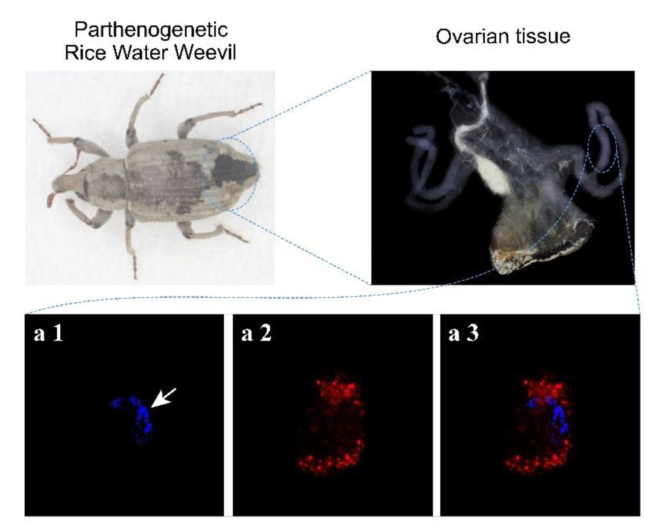
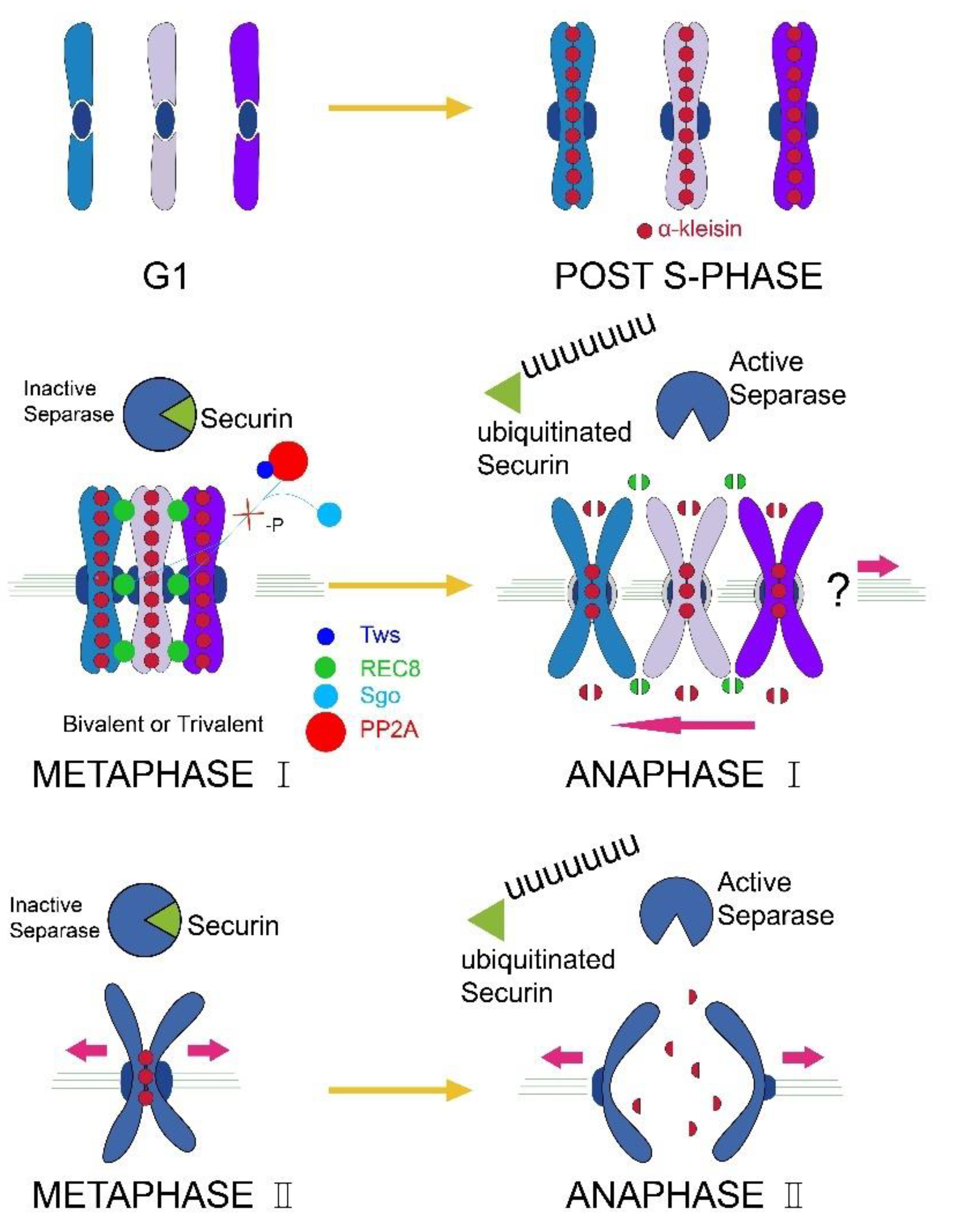
3.1. The Chromosomes of Parthenogenetic RWW Segregate at One Pole of the Meiotic Spindle during the Meiosis I Anaphase

RWW has a telotrophic type ovary; thus, we dissected the ovary of parthenogenetic RWW and individual ovarian tube tissues were immunostained for microtubules and chromosomes. The results showed that a canonical meiotic spindle is formed, while chromosomes segregate at only one pole of the anaphase cell, and there is no chromosome on the other pole (Figure 1). The white arrow in Figure 1 refers to the chromosomes moving towards one pole of the spindle. This result suggests that parthenogenesis RWW produces triploid eggs via chromosome unipolar division. The meiotic process of parthenogenetic RWW (Figure 2) therefore involves unipolar segregation of all chromosomes during anaphase I followed by a traditional meiotic second division leading to the production of triploid eggs.

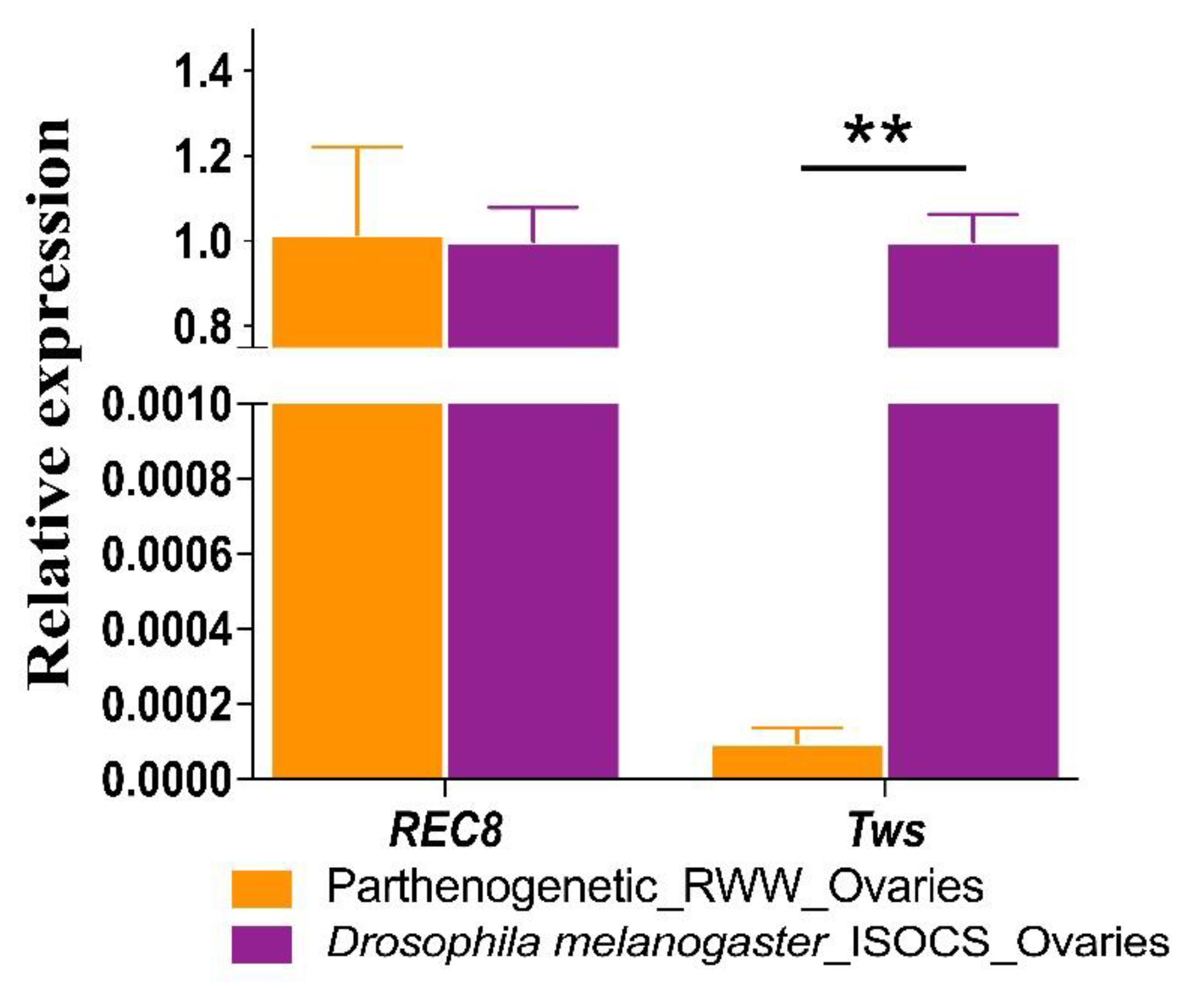
3.2. Tws and REC8 Expression Levels

To explore the genes that cause meiosis I anaphase chromosomal behavior abnormalities in parthenogenetic RWW, we cloned the conservative sequences of parthenogenetic RWW REC8 and Tws genes, and the GeneBank accession number: REC8: MW560171; Tws: MW560172. The REC8 and Tws sequences are given in Supplementary Figure S1—Sequences of gene cloning. To investigate whether the parthenogenesis of RWW is related to these genes, we detected the expression levels of REC8 and Tws by Q-PCR. The Q-PCR results showed that the expressions of REC8 and Tws in ovarian tissues of bisexual Drosophila melanogaster (ISOCS) are 0.98 (1.00/1.02 ≈ 0.98) and 10,000.00 (1.00/9.07 × 10–5 ≈ 10,000.00) times parthenogenetic RWW, respectively (p < 0.01). The results showed that the expression pattern of REC8 in the ovarian tubes of parthenogenetic RWW was the same as for sexual reproductive flies, and the expression of Tws was extremely low (p < 0.01; Figure 3, Table 1). The results indicate that PP2A-Tws cannot maintain the dephosphorylation state of the REC8 protein due to the low expression of Tws in the ovarian tissue of parthenogenetic RWW. Therefore, REC8 is cleaved by the isolating enzyme in the anaphase of meiosis I. We speculated that the low expression of the Tws may be one of the reasons for the parthenogenesis of the RWW.

4. Discussion

Parthenogenesis is a research hotspot in the field of developmental biology and plays an important role in the evolution of insect sexuality and reproductive strategies. The triploid parthenogenetic RWW has chromosomal unipolar division during oogenesis. It is known that the RWW has a telotrophic type of ovary [11], and only the cells in the meiosis I have nutritive cord [44,45]. According to the results of immunostaining and transmission electron microscope investigation, the anaphase meiotic I oocytes with nutritive cord only have chromosomes at one pole of the cell [10,44,45,46], which is consistent with our experimental results. It is confirmed that chromosomal unipolar division occurs in meiosis I. The parthenogenetic RWW maintains the parental chromosome number via a modification of the meiotic process of meiosis, a novel strategy different from the eulophid parasitoid Neochrysocharis formosa [15,16] and rice [38]; these meiotic cells undergo only a single equational division. This study also found that the Tws has low expression in the ovarian tissue of the parthenogenetic RWW, which may be one of the reasons for the parthenogenesis of the RWW. Nonetheless, the molecular mechanism that causes the unipolar division of chromosomes and the molecular mechanism of how the downstream genes controlled by the Tws cause parthenogenesis remain to be studied.
There are various reasons and methods for biological parthenogenesis. In ploidy restoration of parthenogenetic animals, meiosis is either preserved in some form (automixis) or is largely suppressed (parthenogenesis) [14]. Obligate parthenogenesis RWW is a triploid organism, which is different from silkworms (Bombyx mori) and aphids (Acyrthosiphum pisum) [24,47,48,49,50,51,52,53,54] [55] (pp. 199–257) whose reproductive strategies can be changed by environmental factors. Moreover, parthenogenetic RWW does not require sperm to participate in the entire life cycle, which is different from nematodes (Mesorhabditis belari) [56] that require sperm to activate unfertilized eggs for parthenogenesis. These results imply that diploid parthenogenetic organisms retain the complete meiotic process and meiotic genes [20,53]. In triploid parthenogenetic male Cacopsylla myrtilli meiosis, all univalent chromosomes divide during the first meiotic division resulting in the incomplete second meiotic division and formation of diploid sperms [57,58,59]. These studies collectively show that triploid parthenogenetic species are prone to modify meiosis processes, so genes related to meiosis will also change. In this study, immunostaining technology was used to observe the behavior of chromosomes and microtubules during the first division of parthenogenetic RWW meiosis. Our results show that the parthenogenetic RWW restored the parental chromosome number via chromosome unipolar split division, which is different from other parthenogenetic species. With the improvement of insect egg cell culture technology in vitro, the entire process of meiosis can be tracked and observed, which can better explain the cytological mechanism of parthenogenetic insects [60].
In meiosis, chromosomes are controlled by the behavior of the centromere and the time and area of sister chromatid cohesion during the two divisions to ensure accurate separation into daughter cells [25,26]. A unique proteic structure, the synaptonemal complex, is formed during the early stages of prophase I between the homologs [61,62]. The Drosophila melanogaster with Rad21 (REC8 homologous gene) mutation produces diploid unfertilized eggs (1/83), indicating that REC8 may be related to the chromosome accurate separation of female meiosis I [27]. Levels of REC8 protein in Tws mutant individual were reduced to about 50% compared with the wild type, which indicates that PP2A is required mainly to maintain the level of REC8 protein but not REC8 mRNA expression [31]. PP2A is composed of a trimeric core enzyme composed of structural A, catalytic C and regulatory B subunits [63], and plays a very important role in many physiological processes, especially in cell division [64]. Mutations in both Tws and PP2A can cause abnormal spindle assembly, resulting in an abnormal chromosomal arrangement, but the chromosomes can be separated normally to the two poles of the cell [32,65]. These results indicate that PP2A-Tws and REC8 can maintain the stability of the chromosome structure during meiosis. In Drosophila melanogaster cells, PP2A-Tws promotes nuclear envelope reformation during mitotic exit by dephosphorylating barrier-to-autointegration factor [66]. In Oikopleura dioica the knockdown of the maternal PP2A catalytic subunit caused unfertilized eggs to spontaneously release polar bodies after spawning, and then start pseudo-cleavages without fertilization, namely, parthenogenesis. On the other hand, egg cells with completely inactivated PP2A have aberrations in cleavage and cannot develop into a new individual [40]. These results illustrate that PP2A-Tws play an important role in the process of meiosis, and the complete inactivation of PP2A is detrimental to egg development. In parthenogenetic RWW, Tws and REC8 expression levels suggest that Tws can cause REC8 protein to be cleaved prematurely, which may be the cause of chromosome unipolar division. When Tws has low expression, some maternal proteins could be over-phosphorylated, which may lead to parthenogenesis of RWW.
In summary, we find that all chromosomes of parthenogenetic RWW segregate at one pole of the meiotic spindle in meiosis I anaphase and the Tws in the parthenogenetic RWW ovarian tube tissue had low expression. We conclude that the chromosome unipolar division and low expression of Tws together caused parthenogenesis of RWW. Triploid parthenogenesis in RWW shows a new way of meiosis, but it also confirms that parthenogenesis is prone to change in the biological meiosis process. It has been reported that Tws is the regulatory subunit of PP2A, which not only plays an important role in meiosis but also participates in the Wg/Wnt signaling pathway to regulate the development of Drosophila wings [67,68,69]. While we have elucidated the meiosis process of triploid parthenogenesis rice water weevil, the relationship between Tws and chromosome monopolar division has not been found yet. Our study opens an avenue to elucidate the unique chromosomal segregation mechanism of meiosis for parthenogenetic organisms. The specific function of PP2A-Tws needs to be further studied to unravel the relationship between Tws and parthenogenesis.

Supplementary Materials

The following are available online at https://www.mdpi.com/article/10.3390/insects12040278/s1. Table S1: Reagent and Immunostaining experimental procedure, Table S2: Gene cloning and Q-PCR primer sequence, Table S3: Gene cloning and Q-PCR experimental procedure, Figure S1 Sequences of gene cloning.

Author Contributions

Conceptualization and review, R.Z.; software and original draft, P.W.; writing-original draft preparation, P.W.; data curation, F.Y. and Z.M., visualization, F.Y. and Z.M.; review and editing, R.Z. All authors have read and agreed to the published version of the manuscript.

Funding

This research was supported by the Strategic Priority Research Program of the Chinese Academy of Sciences (XDA19050204).

Institutional Review Board Statement

Not applicable.

Data Availability Statement

All data generated or analyzed during this study are included in this published article.

Acknowledgments

We are very grateful to Chuan Zhou, professor of Institute of Zoology, Chinese Academy of Sciences, for his assistance with the instruments and ISCOS of this research. We are also grateful to Chunyan Jiang from the Institute of Zoology, Chinese Academy of Sciences and Zhaochun Jiang from the Plant Protection and Inspection Station of Guizhou for his assistance on samples collection.

Conflicts of Interest

The authors declare no conflict of interest. The funders had no role in the design of the study; in the collection, analyses, or interpretation of data; in the writing of the manuscript, or in the decision to publish the results.

References

  1. Kearney, M. Hybridization, glaciation and geographical parthenogenesis. Trends Ecol. Evol. 2005, 20, 495–502. [Google Scholar] [CrossRef]
  2. Vershinina, A.O.; Kuznetsova, V.G. Parthenogenesis in Hexapoda: Entognatha and non-holometabolous insects. J. Zool. Syst. Evol. Res. 2016, 54, 257–268. [Google Scholar] [CrossRef]
  3. Gokhman, V.E.; Kuznetsova, V.G. Parthenogenesis in Hexapoda: Holometabolous insects. J. Zool. Syst. Evol. Res. 2017, 56, 23–34. [Google Scholar] [CrossRef]
  4. Bowling, C.C. Rice Water Weevil Resistance to Aldrin in Texas. J. Econ. Èntomol. 1968, 61, 1027–1030. [Google Scholar] [CrossRef]
  5. Lange, W.; Grigarick, A. Rice water weevil: Beetle pest in rice growing areas of southern states discovered in California. Calif. Agric. 1959, 13, 10–11. [Google Scholar]
  6. Takenouchi, Y. A chromosome study of the parthenogenetic rice water weevil, Lissorhoptrus oryzophilus Kuschel (Coleoptera: Curculionidae), in Japan. Cell. Mol. Life Sci. 1978, 34, 444–445. [Google Scholar] [CrossRef]
  7. Huang, Y.; Ao, Y.; Jiang, M. Reproductive Plasticity of an Invasive Insect Pest, Rice Water Weevil (Coleoptera: Curculionidae). J. Econ. Èntomol. 2017, 110, 2381–2387. [Google Scholar] [CrossRef] [PubMed]
  8. Giantsis, I.A.; Sierra, J.C.; Chaskopoulou, A. The distribution of the invasive pest, rice water weevil Lissorhoptrus oryzophilus Kuschel (Coleoptera: Curculionidae), is expanding in Europe: First record in the Balkans, confirmed by CO1 DNA barcoding. Phytoparasitica 2017, 45, 147–149. [Google Scholar] [CrossRef]
  9. Saito, T.; Hirai, K.; Way, M.O. The rice water weevil, Lissorhoptrus oryzophilus Kuschel (Coleoptera: Curculionidae). Appl. Èntomol. Zool. 2005, 40, 31–39. [Google Scholar] [CrossRef]
  10. Jiang, C. Taxonomy of Mecinini and Bagoini (Coleoptera: Curculionidae) and Spread of Rice Water Weevil in China. Ph.D. Thesis, University of Chinese Academy of Sciences, Beijing China, 2018. [Google Scholar]
  11. Yang, P. Geographical Parthenogenesis of Rice Water Weevil. Ph.D. Thesis, Zhejiang University, Hangzhou, China, 2008. [Google Scholar]
  12. Kirchheimer, B.; Wessely, J.; Gattringer, A.; Hülber, K.; Moser, D.; Schinkel, C.C.F.; Appelhans, M.; Klatt, S.; Caccianiga, M.; Dellinger, A.; et al. Reconstructing geographical parthenogenesis: Effects of niche differentiation and reproductive mode on Holocene range expansion of an alpine plant. Ecol. Lett. 2018, 21, 392–401. [Google Scholar] [CrossRef]
  13. Lee, C.-C.; Hsu, S.-F.; Yang, C.; Lin, C.-C. Thelytokous parthenogenesis in the exotic dacetine ant Strumigenys rogeri (Hymenoptera: Formicidae) in Taiwan. Èntomol. Sci. 2018, 21, 28–33. [Google Scholar] [CrossRef]
  14. Galis, F.; Van Alphen, J.J.M. Parthenogenesis and developmental constraints. Evol. Dev. 2020, 22, 205–217. [Google Scholar] [CrossRef]
  15. Stenberg, P.; Saura, A. Cytology of Asexual Animals. In Lost Sex; Schön, I., Martens, K., Dijk, P., Eds.; Springer: Dordrecht, The Netherlands, 2009; pp. 63–74. [Google Scholar] [CrossRef]
  16. Adachi-Hagimori, T.; Miura, K.; Stouthamer, R. A new cytogenetic mechanism for bacterial endosymbiont-induced parthenogenesis in Hymenoptera. Proc. R. Soc. B Biol. Sci. 2008, 275, 2667–2673. [Google Scholar] [CrossRef]
  17. Lynch, M.; Seyfert, A.; Eads, B.; Williams, E. Localization of the Genetic Determinants of Meiosis Suppression in Daphnia pulex. Genetics 2008, 180, 317–327. [Google Scholar] [CrossRef] [PubMed]
  18. Innes, D.J.; Hebert, P.D.N. The Origin and Genetic Basis of Obligate Parthenogenesis in Daphnia pulex. Evolution 1988, 42, 1024. [Google Scholar] [CrossRef] [PubMed]
  19. Tanaka, M.; Daimon, T. First molecular genetic evidence for automictic parthenogenesis in cockroaches. Insect Sci. 2019, 26, 649–655. [Google Scholar] [CrossRef]
  20. Eisman, R.C.; Kaufman, T.C. Cytological investigation of the mechanism of parthenogenesis in Drosophila mercatorum. Fly 2007, 1, 317–329. [Google Scholar] [CrossRef] [PubMed]
  21. Matsuura, K.; Fujimoto, M.; Goka, K. Sexual and asexual colony foundation and the mechanism of facultative parthenogenesis in the termite Reticulitermes speratus (Isoptera, Rhinotermitidae). Insectes Sociaux 2004, 51, 325–332. [Google Scholar] [CrossRef]
  22. Alavi, Y.; Van Rooyen, A.; Elgar, M.A.; Jones, T.M.; Weeks, A.R. Novel microsatellite markers suggest the mechanism of parthenogenesis in Extatosoma tiaratum is automixis with terminal fusion. Insect Sci. 2018, 25, 24–32. [Google Scholar] [CrossRef] [PubMed]
  23. Chang, C.-C.; Ting, C.-T.; Chang, C.-H.; Fang, S.; Chang, H.-Y. The Persistence of Facultative Parthenogenesis in Drosophila albomicans. PLoS ONE 2014, 9, e113275. [Google Scholar] [CrossRef] [PubMed]
  24. Legay, J.M. Morphogenesis of the silkworm egg. J. Embryol. Exp. Morphol. 1976, 36, 13–18. [Google Scholar] [CrossRef]
  25. Zhang, Y.-L.; Zhang, H.; Gao, Y.-J.; Yan, L.-L.; Yu, X.-Y.; Yang, Y.-H.; Xu, W.-Y.; Pu, C.-X.; Sun, Y. Protein Phosphatase 2A B’α and B’β Protect Centromeric Cohesion during Meiosis I. Plant Physiol. 2019, 179, 1556–1568. [Google Scholar] [CrossRef] [PubMed]
  26. Petronczki, M.; Siomos, M.F.; Nasmyth, K. Un Ménage à Quatre: The Molecular Biology of Chromosome Segregation in Meiosis. Cell 2003, 112, 423–440. [Google Scholar] [CrossRef]
  27. Urban, E.; Nagarkar-Jaiswal, S.; Lehner, C.F.; Heidmann, S.K. The Cohesin Subunit Rad21 Is Required for Synaptonemal Complex Maintenance, but Not Sister Chromatid Cohesion, during Drosophila Female Meiosis. PLoS Genet. 2014, 10, e1004540. [Google Scholar] [CrossRef] [PubMed]
  28. Rieder, C.L.; Cole, R. Chromatid cohesion during mitosis: Lessons from meiosis. J. Cell Sci. 1999, 112, 2607–2613. [Google Scholar]
  29. Watanabe, Y.; Nurse, P. Cohesin Rec8 is required for reductional chromosome segregation at meiosis. Nature 1999, 400, 461–464. [Google Scholar] [CrossRef]
  30. Schleiffer, A.; Kaitna, S.; Maurer-Stroh, S.; Glotzer, M.; Nasmyth, K.; Eisenhaber, F. Kleisins: A Superfamily of Bacterial and Eukaryotic SMC Protein Partners. Mol. Cell 2003, 11, 571–575. [Google Scholar] [CrossRef]
  31. Buonomo, S.B.; Clyne, R.K.; Fuchs, J.; Loidl, J.; Uhlmann, F.; Nasmyth, K. Disjunction of Homologous Chromosomes in Meiosis I Depends on Proteolytic Cleavage of the Meiotic Cohesin Rec8 by Separin. Cell 2000, 103, 387–398. [Google Scholar] [CrossRef]
  32. Kim, L.-H.; Hong, S.-T.; Choi, K.-W. Protein phosphatase 2A interacts with Verthandi/Rad21 to regulate mitosis and organ development in Drosophila. Sci. Rep. 2019, 9, 1–12. [Google Scholar] [CrossRef]
  33. Kitajima, T.S.; Sakuno, T.; Ishiguro, K.-I.; Iemura, S.-I.; Natsume, T.; Kawashima, S.A.; Watanabe, Y. Shugoshin collaborates with protein phosphatase 2A to protect cohesin. Nat. Cell Biol. 2006, 441, 46–52. [Google Scholar] [CrossRef]
  34. Riedel, C.G.; Katis, V.L.; Katou, Y.; Mori, S.; Itoh, T.; Helmhart, W.; Gálová, M.; Petronczki, M.; Gregan, J.; Cetin, B.; et al. Protein phosphatase 2A protects centromeric sister chromatid cohesion during meiosis I. Nat. Cell Biol. 2006, 441, 53–61. [Google Scholar] [CrossRef]
  35. Horner, V.L.; Wolfner, M.F. Mechanical stimulation by osmotic and hydrostatic pressure activates Drosophila oocytes in vitro in a calcium-dependent manner. Dev. Biol. 2008, 316, 100–109. [Google Scholar] [CrossRef]
  36. Loidl, J. Meiotic chromosome pairing in triploid and tetraploid Saccharomyces cerevisiae. Genetics 1995, 139, 1511–1520. [Google Scholar] [CrossRef]
  37. Köhler, C.; Scheid, O.M.; Erilova, A. The impact of the triploid block on the origin and evolution of polyploid plants. Trends Genet. 2010, 26, 142–148. [Google Scholar] [CrossRef]
  38. Khanday, I.; Skinner, D.; Yang, B.; Mercier, R.; Sundaresan, V. A male-expressed rice embryogenic trigger redirected for asexual propagation through seeds. Nat. Cell Biol. 2019, 565, 91–95. [Google Scholar] [CrossRef] [PubMed]
  39. Eads, B.D.; Tsuchiya, D.; Andrews, J.; Lynch, M.; Zolan, M.E. The spread of a transposon insertion in Rec8 is associated with obligate asexuality in Daphnia. Proc. Natl. Acad. Sci. USA 2012, 109, 858–863. [Google Scholar] [CrossRef]
  40. Yagound, B.; Dogantzis, K.A.; Zayed, A.; Lim, J.; Broekhuyse, P.; Remnant, E.J.; Beekman, M.; Allsopp, M.H.; Aamidor, S.E.; Dim, O.; et al. A Single Gene Causes Thelytokous Parthenogenesis, the Defining Feature of the Cape Honeybee Apis mellifera capensis. Curr. Biol. 2020, 30, 2248–2259.e6. [Google Scholar] [CrossRef]
  41. Matsuo, M.; Onuma, T.A.; Omotezako, T.; Nishida, H. Protein phosphatase 2A is essential to maintain meiotic arrest, and to prevent Ca2+ burst at spawning and eventual parthenogenesis in the larvacean Oikopleura dioica. Dev. Biol. 2020, 460, 155–163. [Google Scholar] [CrossRef] [PubMed]
  42. Khetani, R.S.; Bickel, S.E. Regulation of meiotic cohesion and chromosome core morphogenesis during pachytene in Drosophila oocytes. J. Cell Sci. 2007, 120, 3123–3137. [Google Scholar] [CrossRef] [PubMed]
  43. Demarco, R.S.; Eikenes, Å.H.; Haglund, K.; Jones, D.L. Investigating spermatogenesis in Drosophila melanogaster. Methods 2014, 68, 218–227. [Google Scholar] [CrossRef] [PubMed]
  44. Kurihara, M.; Matsuzaki, M. Ovarian structure and oogenesis in the rice water weevil, Lissorphoptrus oryzophilus (Coleoptera, Curculionidae). Jpn. J. Enotomol. 1989, 57, 666–680. [Google Scholar]
  45. Büning, J. The trophic tissue of telotrophic ovarioles in polyphage coleoptera. Zoomorphology 1979, 93, 33–50. [Google Scholar] [CrossRef]
  46. Huebner, E.; Anderson, E. A cytological study of the ovary ofRhodnius prolixus. II. Oocyte differentiation. J. Morphol. 1972, 137, 385–415. [Google Scholar] [CrossRef] [PubMed]
  47. Blackman, R. Early development of the parthenogenetic egg in three species of aphids (homoptera: Aphididae). Int. J. Insect Morphol. Embryol. 1978, 7, 33–44. [Google Scholar] [CrossRef]
  48. Wilson, A.C.C.; Sunnucks, P.; Hales, D.F. SHORT PAPER Random loss of X chromosome at male determination in an aphid, Sitobion near fragariae, detected using an X-linked polymorphic microsatellite marker. Genet. Res. 1997, 69, 233–236. [Google Scholar] [CrossRef]
  49. Stevens, N.M. A study of the germ cells of Aphis rosæ and Aphis œnotheræ. J. Exp. Zool. 1905, 2, 313–333. [Google Scholar] [CrossRef]
  50. Stevens, N.M. Studies on the Germ Cell of Aphids (No. 51); Carnegie: Washington, DC, USA, 1906. [Google Scholar] [CrossRef]
  51. Morgan, T.H. A biological and cytological study of sex determination in phylloxerans and aphids. Dev. Genes Evol. 1910, 29, 201–203. [Google Scholar] [CrossRef]
  52. Morgan, T.H. Sex determination and parthenogenesis in phylloxerans and aphids. Science 1909, 29, 234–237. [Google Scholar] [CrossRef] [PubMed]
  53. Srinivasan, D.G.; Abdelhady, A.; Stern, D.L. Gene Expression Analysis of Parthenogenetic Embryonic Development of the Pea Aphid, Acyrthosiphon pisum, Suggests That Aphid Parthenogenesis Evolved from Meiotic Oogenesis. PLoS ONE 2014, 9, e115099. [Google Scholar] [CrossRef]
  54. Astaurov, B.L. Artificial parthenogenesis and experimental polyploidy in silkworm. J. Sericult. Sci. Jpn. 1967, 36, 277–285. [Google Scholar] [CrossRef]
  55. Astaurov, B.L. Experimental alterations of the developmental cytogenetic mechanisms in mulberry silkworms: Artificial parthenogenesis, polyploidy, gynogenesis, and androgenesis. In Advances in Morphogenesis; Abercrombie, M., Brachet, J., Eds.; Elsevier: Amsterdam, The Netherlands, 1967; Volume 6, pp. 199–257. [Google Scholar] [CrossRef]
  56. Grosmaire, M.; Launay, C.; Siegwald, M.; Brugière, T.; Estrada-Virrueta, L.; Berger, D.; Burny, C.; Modolo, L.; Blaxter, M.; Meister, P.; et al. Males as somatic investment in a parthenogenetic nematode. Science 2019, 363, 1210–1213. [Google Scholar] [CrossRef] [PubMed]
  57. Nokkala, C.; Kuznetsova, V.G.; Nokkala, S. Rare diploid females coexist with rare males: A novel finding in triploid parthenogenetic populations in the psyllid Cacopsylla myrtilli (W. Wagner, 1947) (Hemiptera, Psylloidea) in northern Europe. Genetic 2015, 143, 589–595. [Google Scholar] [CrossRef] [PubMed]
  58. Nokkala, C.; Kuznetsova, V.G.; Nokkala, S. Meiosis in rare males in parthenogenetic Cacopsylla myrtilli (Wagner, 1947) (Hemiptera, Psyllidae) populations from northern Europe. Comp. Cytogenet. 2013, 7, 241–251. [Google Scholar] [CrossRef]
  59. Nokkala, S.; Kuznetsova, V.G.; Nokkala, C. Characteristics of parthenogenesis in Cacopsylla ledi (Flor, 1861) (Hemiptera, Sternorryncha, Psylloidea): Cytological and molecular approaches. Comp. Cytogenet. 2017, 11, 807–817. [Google Scholar] [CrossRef] [PubMed]
  60. Peters, N.C.; Berg, C.A. In Vitro Culturing and Live Imaging of Drosophila Egg Chambers: A History and Adaptable Method. Breast Cancer 2016, 1457, 35–68. [Google Scholar] [CrossRef]
  61. Leonard, A.C.; Grimwade, J.E. Chromosome Replication and Segregation. Bacteriol Rev. 2019, 30, 3–32. [Google Scholar] [CrossRef]
  62. Nicholson, J.M.; Cimini, D. How Mitotic Errors Contribute to Karyotypic Diversity in Cancer. Adv. Cancer Res. 2011, 112, 43–75. [Google Scholar] [CrossRef]
  63. Cho, U.S.; Xu, W. Crystal structure of a protein phosphatase 2A heterotrimeric holoenzyme. Nat. Cell Biol. 2006, 445, 53–57. [Google Scholar] [CrossRef]
  64. Mayer-Jaekel, R.E.; Ohkura, H.; Gomes, R.; Sunkel, C.E.; Baumgärtner, S.; Hemmings, B.A.; Glover, D.M. The 55 kd regulatory subunit of Drosophila protein phosphatase 2A is required for anaphase. Cell 1993, 72, 621–633. [Google Scholar] [CrossRef]
  65. Snaith, H.A.; Armstrong, C.G.; Guo, Y.; Kaiser, K.; Cohen, P.T. Deficiency of protein phosphatase 2A uncouples the nucle-ar and centrosome cycles and prevents attachment of microtubules to the kinetochore in Drosophila microtubule star (mts) embryos. J. Cell Sci. 1996, 109, 3001–3012. [Google Scholar]
  66. Mehsen, H.; Boudreau, V.; Garrido, D.; Bourouh, M.; Larouche, M.; Maddox, P.S.; Swan, A.; Archambault, V. PP2A-B55 promotes nuclear envelope reformation after mitosis in Drosophila. J. Cell Biol. 2018, 217, 4106–4123. [Google Scholar] [CrossRef]
  67. Uemura, T.; Shiomi, K.; Togashi, S.; Takeichi, M. Mutation of twins encoding a regulator of protein phosphatase 2A leads to pattern duplication in Drosophila imaginal discs. Genes Dev. 1993, 7, 429–440. [Google Scholar] [CrossRef] [PubMed]
  68. Bajpai, R.; Makhijani, K.; Rao, P.R.; Shashidhara, L.S. Drosophila Twins regulates Armadillo levels in response to Wg/Wnt signal. Development 2004, 131, 1007–1016. [Google Scholar] [CrossRef] [PubMed]
  69. Brownlee, C.W.; Klebba, J.E.; Buster, D.W.; Rogers, G.C. The Protein Phosphatase 2A regulatory subunit Twins stabilizes Plk4 to induce centriole amplification. J. Cell Biol. 2011, 195, 231–243. [Google Scholar] [CrossRef] [PubMed]
Figure 1. Observation of parthenogenetic ovarian tissue staining. Chromosomes and microtubule organization in rice water weevil (RWW) oocyte were stained with DAPI (white arrow, a1) and anti-Tubulin antibodies (in red, a2), respectively. Chromosomes move to one end of the cell during the meiosis I anaphase (a3).
Insects 12 00278 g001
Figure 2. Triploid parthenogenetic RWW meiotic pattern diagram. Parthenogenetic RWW has three chromosome sets (G1). In S phase, chromosome duplication (POST S-PHASE). The low expression of Tws prevents the PP2A protein from maintaining the low phosphorylation state of the chromosome (METAPHASE I), which causes the REC8 protein to be cleaved by Separase earlier. All chromosomes segregate at one pole of the meiotic spindle during the anaphase of meiosis I (ANAPHASE I). Subsequently, the oocyte undergoes a standard meiosis II to produce triploid eggs (METAPHASE II and ANAPHASE II).
Insects 12 00278 g002
Figure 3. Quantification of REC8 and Tws in RWW ovarian tissues with bisexual reproductive ISOCS as reference. The y-axis indicates the relative expression (for details see Table 1, β-actin as internal reference). Error bars are s.d., n = 3. ** p < 0.01 (t-test).
Insects 12 00278 g003
Table 1. Q-PCR results.
SpeciesRelative Expression
REC8Tws
Lissorhoptrus oryzophilus-P 11.02 ± 0.209.70 × 10−5 ± 0.4 × 10⁻⁵
Drosophila melanogaster1.00 ± 0.081.00 ± 0.0627
1Lissorhoptrus oryzophilus-P: Parthenogenetic RWW. All data retain three significant figures; each experiment was repeated three times (n = 3). The relative gene expression level is based on the standardized bisexual reproduction of Drosophila melanogaster (ISOCS) as a reference.
Publisher’s Note: MDPI stays neutral with regard to jurisdictional claims in published maps and institutional affiliations.

