Article

Functional Characterization of a Trehalose-6-Phosphate Synthase in Diaphorina citri Revealed by RNA Interference and Transcriptome Sequencing

1
College of Life Science, Gannan Normal University, Ganzhou 341000, China
2
National Navel Orange Engineering Research Center, Ganzhou 341000, China
*
Authors to whom correspondence should be addressed.
Insects 2021, 12(12), 1074; https://doi.org/10.3390/insects12121074
Submission received: 26 October 2021 / Revised: 24 November 2021 / Accepted: 29 November 2021 / Published: 30 November 2021
(This article belongs to the Section Insect Physiology, Reproduction and Development)

Abstract

:

Simple Summary

Trehalose-6-phosphate synthase (TPS) is a key enzyme in regulating trehalose content in the insect hemolymph. The loss or dysfunction significantly affects the growth and development of insects. Diaphorina citri is a notorious phloem sap-sucking pest that can spread huanglongbing between the diseased tree and the healthy tree. The control of huanglongbing mainly depends the management of D. citri. So far, the management of D. citri populations has depended on using chemical pesticides, though pesticide abuse has caused serious problems. Therefore, it necessary to find new targets for D. citri control. In this paper, we identified a TPS gene from Diaphorina citri, and named it DcTPS1. Silencing of DcTPS1 induced an abnormal phenotype, and inhibited chitin metabolism and fatty acid metabolism. Moreover, the mortality and malformation rate significantly increased, and the molting rate decreased after inhibition of DcTPS1. KEGG analysis revealed that upregulated DEGs were mainly responsible for oxidative phosphorylation, whereas downregulated DEGs were mainly related to lysosome and ribosome. Overall, our data suggested that DcTPS1 might play a crucial role for the growth and development of D. citri.

Abstract

Trehalose-6-phosphate synthase (TPS) plays an important role in the synthesis of trehalose. In the current study, a TPS gene was obtained from Diaphorina citri, and named as DcTPS1 which encoded a protein of 833 amino acid residues. Real-time quantitative PCR (qPCR) analysis revealed that DcTPS1 had the highest expression level in the midgut and fifth-instar nymph stage. Knockdown of DcTPS1 by RNA interference (RNAi) induced an abnormal phenotype and increased mortality and malformation rate with a decreased molting rate. In addition, silencing of DcTPS1 significantly inhibited D. citri chitin metabolism and fatty acid metabolism, while the expression levels of fatty acid decomposition-related genes were downregulated. Furthermore, comparative transcriptomics analysis revealed that 791 differentially expressed genes (DEGs) were upregulated and 678 DEGs were downregulated when comparing dsDcTPS1 groups with dsGFP groups. Bioinformatics analysis showed that upregulated DEGs were mainly involved in oxidative phosphorylation, whereas downregulated DEGs were mainly attributed to the lysosome and ribosome. These results indicated that DcTPS1 played an important role in the growth and development of D. citri.

1. Introduction

Trehalose is a nonreducing disaccharide which is widely distributed in various organisms, including bacteria, fungi, insects, invertebrates and plants [1]. In insects, trehalose exists mainly in the hemolymph, and plays an important role as an instant energy source, facilitating carbohydrate absorption, and as a starting substrate for chitin biosynthesis [2,3,4]. In insects, biosynthesis of trehalose is catalyzed by trehalose-6-phosphate synthase (TPS) and trehalose-6-phosphate phosphatase (TPP), and then is transported across the cellular membrane into the hemolymph through trehalose transporter [5,6]. To date, TPS genes have been identified from different insect species, and play crucial functions in regulating insect sugar metabolism, chitin metabolism, and stress reactions [7,8,9,10,11]. However, the TPP genes have not been found in many insect species, but TPS genes that encode proteins with both TPS and TPP domains were identified in the same species [12]. In Drosophila, a mutation of the TPS1 gene failed to produce trehalose and exhibited severe growth defects on a low-protein diet [13]. Shi et al. revealed that knockdown of Leptinotarsa decemlineata TPS (LdTPS) gene decreased trehalose and chitin content [10]. In Dendroctonus ponderosae, TPS expression levels are high in the autumn, while they are significantly lower in the spring. The results indicated that TPS is mainly involved in the synthesis of trehalose for D. ponderosae survival during cold periods [14]. In previous research, Liu et al. characterized a TPS gene from Diaphorina citri, and silencing of DcTPS by RNAi significantly reduced trehalose content [15]. However, the specific biological functions of TPS gene in D. citri are still unclear.
Diaphorina citri is a phloem sap-sucking insect which feeds on citrus and is distributed worldwide [16]. D. citri nymphs and adults feed on phloem sap of rutaceous plants, consequently cause leaf distortion, curling and yellowing [17]. Furthermore, because the phloem sap is rich in various sugars, D. citri excretes large amounts of honeydew causing citrus fuliginous disease [18]. Additionally, D. citri transmits the bacteria Candaidatus Liberbacter asiaticus, causing huanglongbing (HLB). HLB is a plant disease, which causes serious losses for the citrus industry every year [19]. Nowadays, chemical pesticides play a dominant role in the prevention and control of D. citri, including pyrethroid, organophosphate, and neonicotinoid classes [20]. Aggressive application of insecticides has caused many problems, such as environment pollution, human poisoning and insecticide resistance [21,22]. Therefore, there is a need for treatments with new modes of action for the control of D. citri, such as RNAi.
RNAi has been developed as an useful tool for functional gene research, which was triggered by double-stranded RNA (dsRNA) [23,24]. Therefore, RNAi has shown great potential for pest control [25]. The RNAi mechanism was described for the first time in the nematode, following which it was discovered to be a common phenomenon in eukaryotic organisms, including protozoans, invertebrates, vertebrates, fungi, algae and plants [26]. However, many factors limiting RNAi efficiency has been reported, such as incomplete dsRNA internalization, instability of dsRNA, impaired systemic spreading of the RNAi signal, and refractory target genes [27]. Therefore, efficient dsRNA delivery method and selection of appropriate target genes are essential prerequisites for pest management. Additionally, concentration and length of dsRNA are further factors contributing to a change in RNAi penetrance [28]. RNAi has been successfully applied to research the gene functions in various insects, including lepidopteran, hemipteran, coleoptera and dipteran [29,30,31]. In D. citri, for different target genes, the dsRNA delivery methods need to be changed accordingly. Yuan et al. revealed that silenced D. citri NADPH-cytochrome P450 reductase (DcCPR) increased the sensibility of D. citri to imidacloprid and thiamethoxam by RNAi using the parafilm feeding method [32]. According to topical feeding with dsRNA-AChE, the sensitivity of D. citri nymphs to chlorpyrifos and carbaryl was significantly increased [33]. Using an artificial diet mixed with dsRNA, knockdown of DcGSTd1 and DcGSTe2 significantly increased the mortalities of thiamethoxam-treated psyllid [34]. Using to the parafilm feeding method, silencing of DcCHS significantly reduced the expression levels of chitin metabolism-related genes, which were significantly downregulated [5].
In the present research, we identified a TPS gene from D. citri genome database. qPCR was performed to analyze the expression profiles of DcTPS1 in different tissues and at different developmental stages. In addition, the functions of DcTPS1 were described by RNAi and transcriptome sequencing. These results will provide a new target for further control of D. citri.

2. Materials and Methods

2.1. Insect Rearing and Sample Prepartion

The D. citri were obtained from a citrus germplasm resource nursery located in Gannan Normal University, Ganzhou, China. The D. citri were continuously reared using Murraya exotica. The rearing conditions were 26 ± 1 °C, 60 ± 5% relative humidity under a 14:10 dark light cycle. To keep the consistency of D. citri growth, the mated D. citri females were released into the flourishing Murraya exotica with many bud breaks in an insect rearing cage. After 48 h, all D. citri adults were removed using a portable aspirator. According to morphological features (the length of cohort size from egg to fifth-instar nymph stage are 0.253, 0.304, 0.46, 0.675, 1.038 and 1.563, respectively), D. citri at seven different stages were collected under a stereomicroscope with a camel hairbrush. The psyllid eggs were harvested using a sterilized blade, and then pooled together. All collected samples were kept at −80 °C. Each group of samples contained three biological replicates.

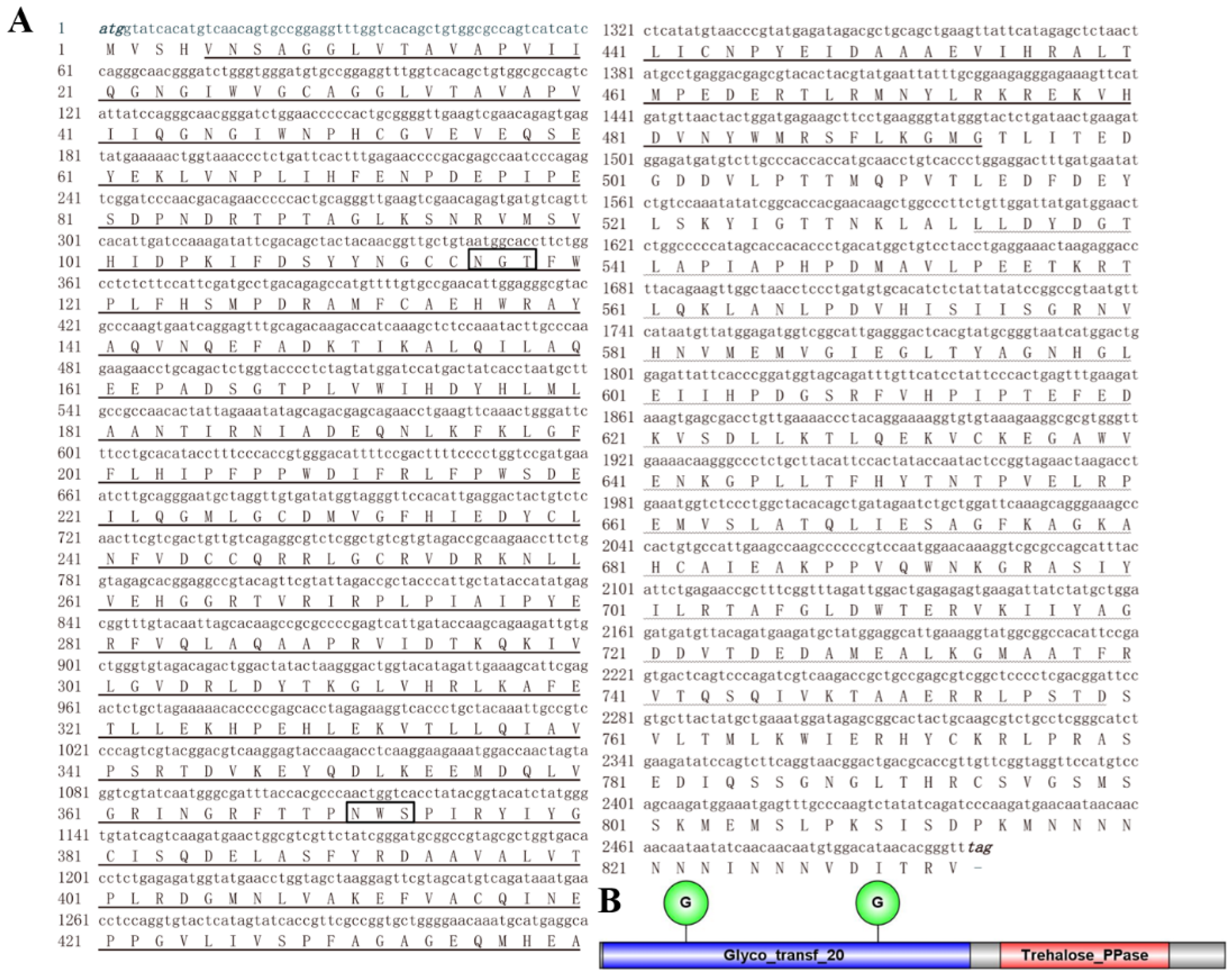
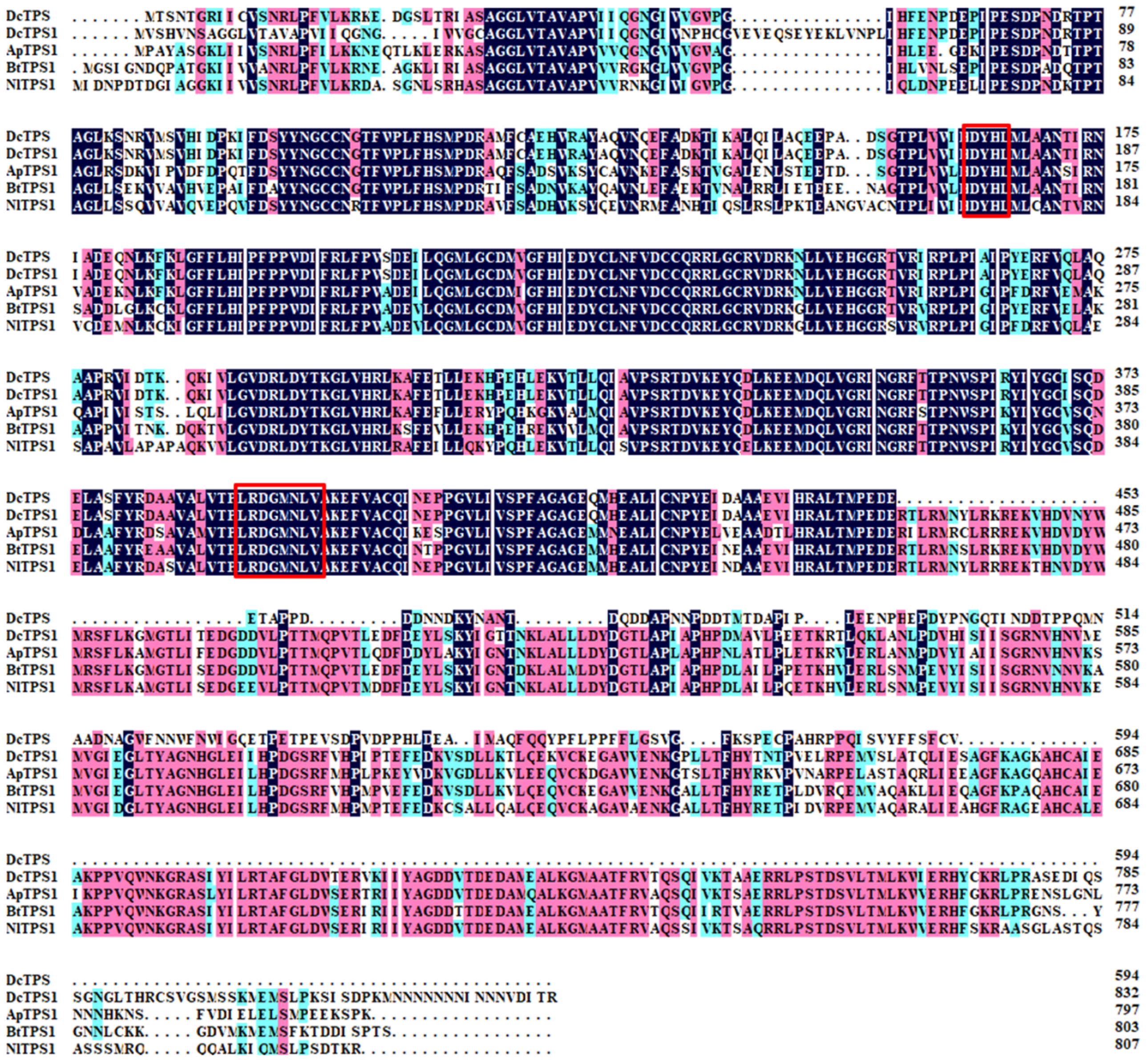
2.2. Cloning of DcTPS1 and Bioinformatic Analysis

The sequence of TPS genes from Nilaparvata lugens, Acyrthosiphon pisum and Drosophila melanogaster were downloaded and used for blasting against the D. citri genome database. After sequence assembly, splicing and sequencing, DcTPS1 with a complete open reading frame was identified and amplified according to PCR. The purified product was linked to pMD19-T and sequenced by a biotechnology company (Sangon Biotech, Shanghai, China).
The amino acid sequence of DcTPS1 was analyzed by using DNASTAR software. The molecular weight (MW) and isogenic point (pI) were predicted by ExPASy (http://web.expasy.org/compute_pi (accessed on 1 May 2021)). The structural domain was identified by using SMART online software (http://smart.embl-heidelberg.de/ (accessed on 20 May 2021)). The phylogenetic tree was constructed using MEGA 7.0 with the neighbor-joining method with 1000 replicates. In addition, the glycosylation sites were predicted according to NetNGlyc 1.0 Server (https://www.cbs.dtu.dk/services/NetNGlyc/ (accessed on 1 May 2021)).

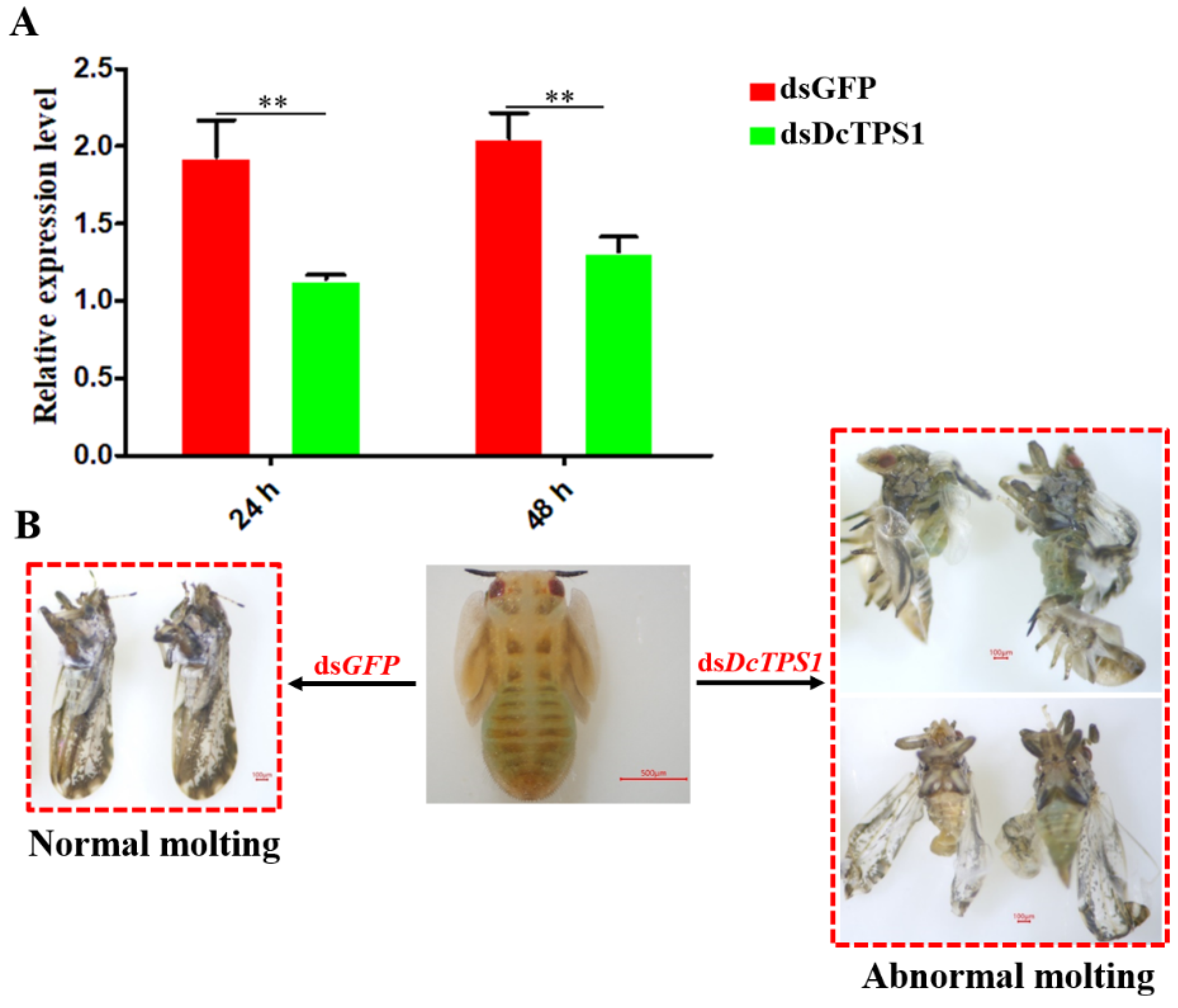
2.3. dsRNA Synthesis and DcTPS1 RNA Interference Analysis

For RNA interference, specific primers of DcTPS1 (accession No: MZ888936) and GFP (accession No: X83959) with T7 promoters were designed and are presented in Table S1. The dsDcTPS1 and dsGFP were synthesized using the T7 RioMAXTM Express RNAi System (Promega, CarIsbad, CA, USA), and the delivery of dsRNA was performed based on a previous protocol [5]. In brief, the length of the amplified sequence for dsDcTPS1 synthesis is 462 bp (dsDcTPS1 fragment from 1419 bp to 1880 bp), and the purified products were linked to pMD19-T for obtaining the recombinant plasmid. The target sequence was further amplified using primers with T7 promoters, and then added to RiboMAXTM Express T7 2× Buffer, nuclease-free water and Enzyme mix T7 express. The mixture was incubated for 4 h at 37 °C and 10 min at 70 °C. The RNase solution and RQ RNase-free DNase were added and incubated for 30 min at 37 °C. Finally, the synthetic dsDcTPS1 was diluted to 500 ng/µL using RNase-free water containing 15% sucrose and 0.1% red food dye and added between two layers of stretched parafilm which was fixed on a glass double pipe. In total, 180 fifth-instar D. citri nymphs were divided into three groups and placed to the glass double pipe for 24 h, and then transferred onto the fresh M. exotica seedlings. All experiments contained three biological replicates. All the living D. citri were collected at 24 h and 48 h after dsRNA treatment. The effects of dsDcTPS1 on gene expression were analyzed using qPCR.

2.4. qPCR Analysis of DcTPS1

The total RNA was extracted from D. citri at different tissues (head, leg, midgut, fat body and wing) and nymph at different instars (egg, first-, second-, third-, fourth-, fifth-instar nymphs) using TRIzol reagent (Invitrogen). All samples consisted of three biological replicates. The purity and concentration of RNA were measured by a spectrophotometer (NanoDrop 2000, Thermo Fisher Scientific, New York, NY, USA). The cDNA was synthesized using the PrimeScriptTM RT Reagent Kit with gDNA Eraser (TaKaRa, Dalian, China) based on a previous protocol.
qPCR was conducted to analyze the expression levels of DcTPS1 in different tissues and developmental stages. The primers are presented in Table S1. The reaction procedures were set as follows: 40 cycles at 95 °C for 10 s and 60 °C for 10 s. The reactions were performed with a LightCycler® 96 PCR detection system (Roche, Basel, Switzerland). The relative expression levels were calculated using 2−ΔΔCt method. The reference gene was glyceraldehyde-3-phosphate dehydrogenase (GAPDH). All experiments contained three biological replicates.

2.5. cDNA Libaray Preparation and Illumina Sequencing

The cDNA library preparation and Illumina sequencing were performed at Novogene Biological Information Technology Co., Ltd. (Tianjin, China). Approximately 50 D. citri were collected from each treatment group (treated with dsDcTPS1) and control groups (treated with dsGFP) at 48 h after ingestion of dsRNA. All experiments contained three biological replicates. RNA concentration and purity were measured according to a Qubit RNA Assay Kit in a Qubit®2.0 Fluorometer (Life Technologies, CA, USA). In total, 1 µg RNA was used to construct cDNA library by TruSeq RNA Sample Preparation Kit v2 (Illumina, San Diego, CA, USA) according to the manufacturer’s instructions.
The prepared cDNA library was sequenced by the Illumina HiSeq platform, generating 150 bp paired-end reads. The clean reads were obtained by removing reads containing the adapter from raw data. Additionally, the Q20, the Q30 and the GC-content of the clean data were calculated.

2.6. Transcriptome Analsysis after Silencing of DcTPS1

The transcriptome data were mapped to D. citri reference genome (ftp://ftp.citrusgreening.org/annotation/OSGv2.0 (accessed on 1 January 2021)) using Hisat2 (version 2.0.5; https://anaconda.org/biobuilds/hisat2 (accessed on 1 January 2021)) aligner. This generated a database of splice junctions based on the gene model annotation file. The expression levels of these genes were calculated using reads per kilobase of exon per million reads mapped. Differential expression analyses of genes between dsGFP and dsDcTPS1 groups were performed using the DESeq2 R package. p-values were adjusted using the Benhamini-Hochberg method to control for the false-discovery rate. A corrected p-value of 0.05 and an absolute |log2 (fold change)| (Fold change > 1) of 0 were set as the thresholds for significantly different gene expression. The hierarchical cluster analysis of DEGs was conducted using Genesis software (http://genome.tugraz.at/genesisclient_download.shtml (accessed on 1 January 2021)).
Gene ontology (GO) is a tool used for gene annotation by collecting a defined, structured and controlled vocabulary. The topGO R package, which implements the GO terms, was used for the enrichment analysis of length-corrected DEGs. Kyoto Encyclopedia of Genes and Genomes (KEGG) is a database that can be used to understand the high-level functions and utilities of biological systems, such as cells, organisms and ecosystems from molecular level. A KEGG pathway enrichment analysis for DEGs was performed using KOBAS. A p-value of <0.01 was set as the threshold.

3. Results

3.1. Analysis of the cDNA and Protein Sequence of DcTPS1

A TPS gene was identified and named DcTPS1 (GenBank accession:MZ888936). Bioinformatic analysis revealed that the ORF of DcTPS1 is 2502 bp, encoding a protein with 833 amino acids. The predicted MW is 93.60 kDa and pI is 5.73 (Figure 1A). SMART software analysis suggested that DcTPS1 contained one Glyco_transf_20 domain (5–494) and one Trehalose_PPase domain (534–759) (Figure 1B). The multiple sequence alignment of TPSs form different insects revealed that DcTPS1 protein sequence had 74.38%, 71.60% and 70.84% identities with those of B. tabaci, A. pisum and N. lugens, respectively, and two signature motifs of HDYHL (174–178) and DGMNLV (404–409) were also found (Figure 2). In addition, multiple sequence alignment analysis suggested that DcTPS1 protein sequence had 53.83% identifies with DcTPS protein and exhibited a significant difference at the C-terminal. The DcTPS1 gene sequence had 58.77% similarities to the DcTPS gene sequence (Figure 2). In addition, there were two glycosylation sites (NGT and NWS) found in DcTPS1 (Figure 1A). The phylogenetic analysis showed that TPS can be divided into two categories, including TPS1 and TPS2. DcTPS1 had a close relationship with the TPS1 of sap-sucking hemipteran, B. tabaci, A. pisum and N. lugens, but it kept a distant relationship with the TPS2 (Figure S1).

3.2. Tissue Distribution and Developmental Stages Expression Patterns of DcTPS1

The expression patterns of DcTPS1 in different tissues and at different developmental stages were analyzed by qPCR. The results showed that the DcTPS1 expression could be detected in all tissues, including head, leg, midgut, fat body and wing (Figure 3). Higher expression of DcTPS1 was found in the midgut and wing tissues, whereas it had low expression levels in the leg and fat body (Figure 3). The expression level of DcTPS1 in the midgut was 18.3 times of that in the fat body. Additionally, the expression level of DcTPS1 was significantly downregulated from egg to nymph stages. However, the expression level of DcTPS1 was constantly observed without significant differences from first-instar nymph to fourth-instar nymph (Figure 3). DcTPS1 expression level was upregulated from the fourth-instar nymph to fifth-instar nymph stages. Interestingly, the expression levels of DcTPS1 in the female adults were higher than in the male adults (Figure 3).

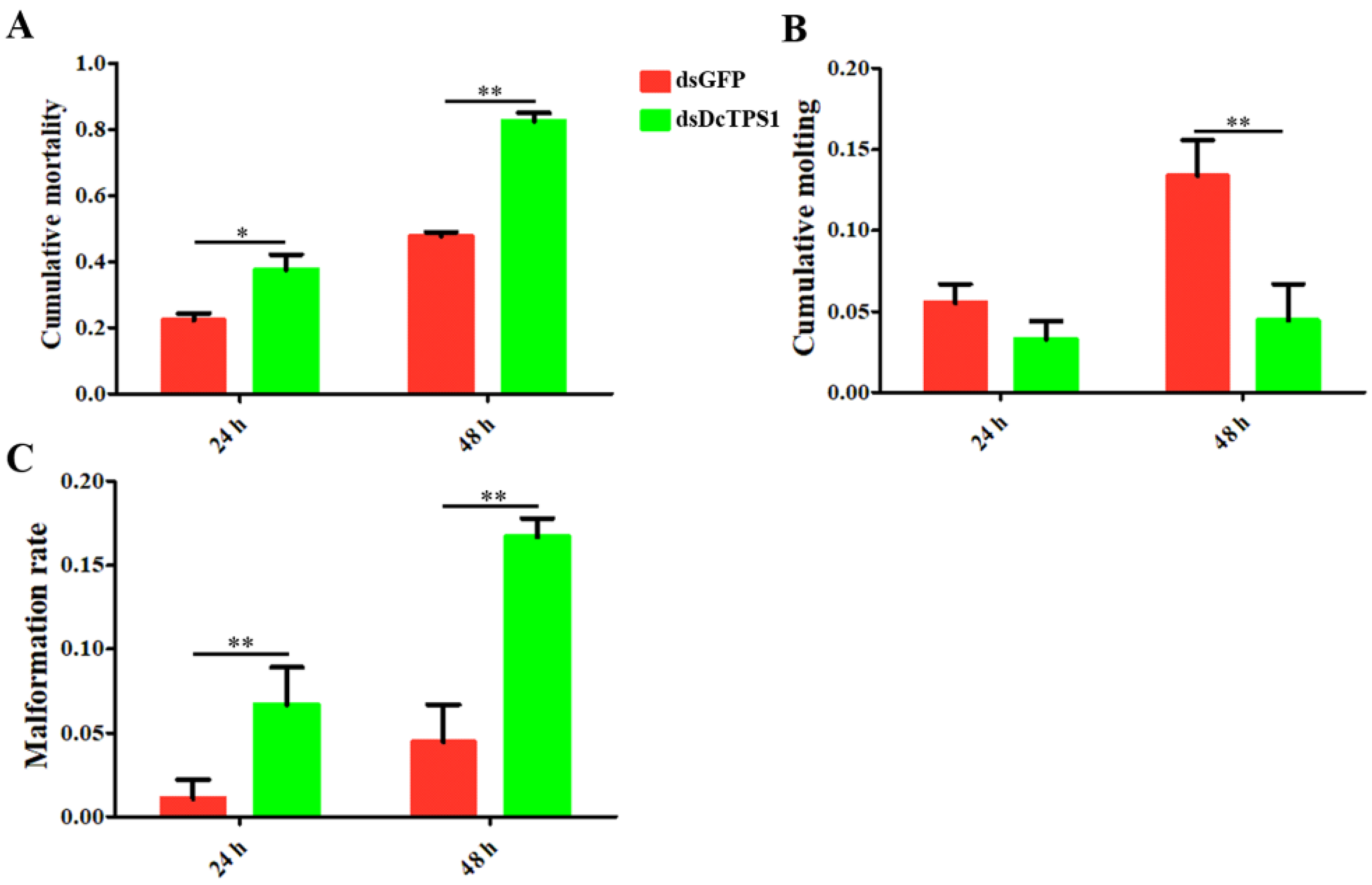
3.3. Analysis of Mortality, Molting and Malformation Rate after Inhibition of DcTPS1

RNAi was performed to determine the biological functions of DcTPS1 in the development of D. citri. The results showed that DcTPS1 expression level was significantly downregulated at 24 h and 48 h after ingestion of dsRNA (Figure 4A). However, the differences in expression levels between dsDcTPS1 group and dsGFP group had no significant change at 24 h and 48 h. Regarding phenotype, the transition from fifth-instar nymph to adult was disrupted in the dsDcTPS1 treatment group, and the emerged D. citri adult exhibited two abnormal phenotypes. Phenotypic observation showed that the treated nymphs molted into adults with abnormal dorsal tergites or malformed wings. Wings were irregular and curled at the distal end or smaller in size. Legs were curled and could not be stretched. Some nymphs failed to completely molt. However, in the dsGFP control group, the nymphs could molt normally (Figure 4B).
The malformation rates and cumulative mortality significantly increased after silencing of DcTPS1 at 24 h and 48 h. The cumulative mortality in the treatment group (dsDcTPS1) was 37.6%, while it was 22.2% in the control group (dsGFP) at 24 h, and the mortality reached 82.5% at 48 h (Figure 5A). The malformation rate in the dsGFP control group was 1.1%, and had no significant change from 24 hpt to 48 hpt, while the malformation rate in the dsDcTPS1 treatment group increased from 6.7% to 16.7% during this period (Figure 5B). In contrast, for the rate of the cumulative molting between 24 hpt and 48 hpt, no significant difference was observed in the dsDcTPS1-treated D. citri, while it increased from 5.6% to 13.4% in the control group (Figure 5C). These results indicated that silencing of DcTPS1 impaired the molting process of fifth-instar D. citri nymphs.

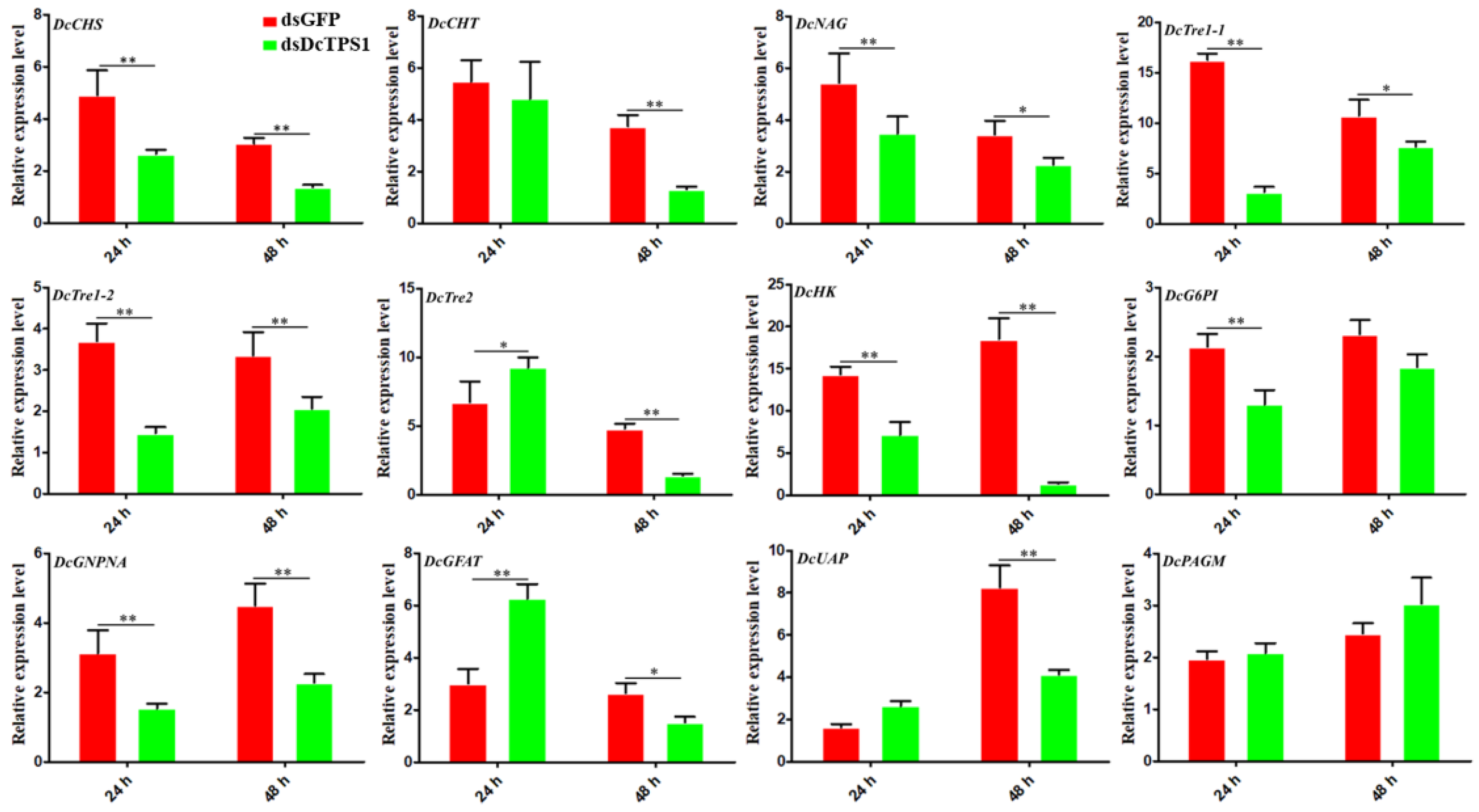
3.4. Analysis of the Effect on Chitin Metabolism after Silencing DcTPS1

The results showed that the relative expression levels of D. citri chitin synthase (DcCHS), D. citri beta-N-acetylglucosaminidase (DcNAG), D. citri trehalose 1-1 (DcTre1-1), D. citri trehalose 1-2 (DcTre1-2), D. citri hexokinase (DcHK) and D. citri glucosamine-phosphate N-acetyltransferase (DcGNPNA) were downregulated at 24 h and 48 h after silencing of DcTPS1 (Figure 6). The relative expression levels of D. citri chitinase (DcCHT) and D. citri UDP-N-acetylglucosamine pyrophosphorylase (DcUAP) were downregulated at 48 h after silencing of DcTPS1, but it had no obvious change at 24 h (Figure 6). Additionally, two genes (DcTre2 and D. citri fructose-6-phosphate transaminase (DcGFAT)) showed upregulation at 24 h after knockdown of DcTPS1, whereas their expression levels were downregulated at 48 h (Figure 6).

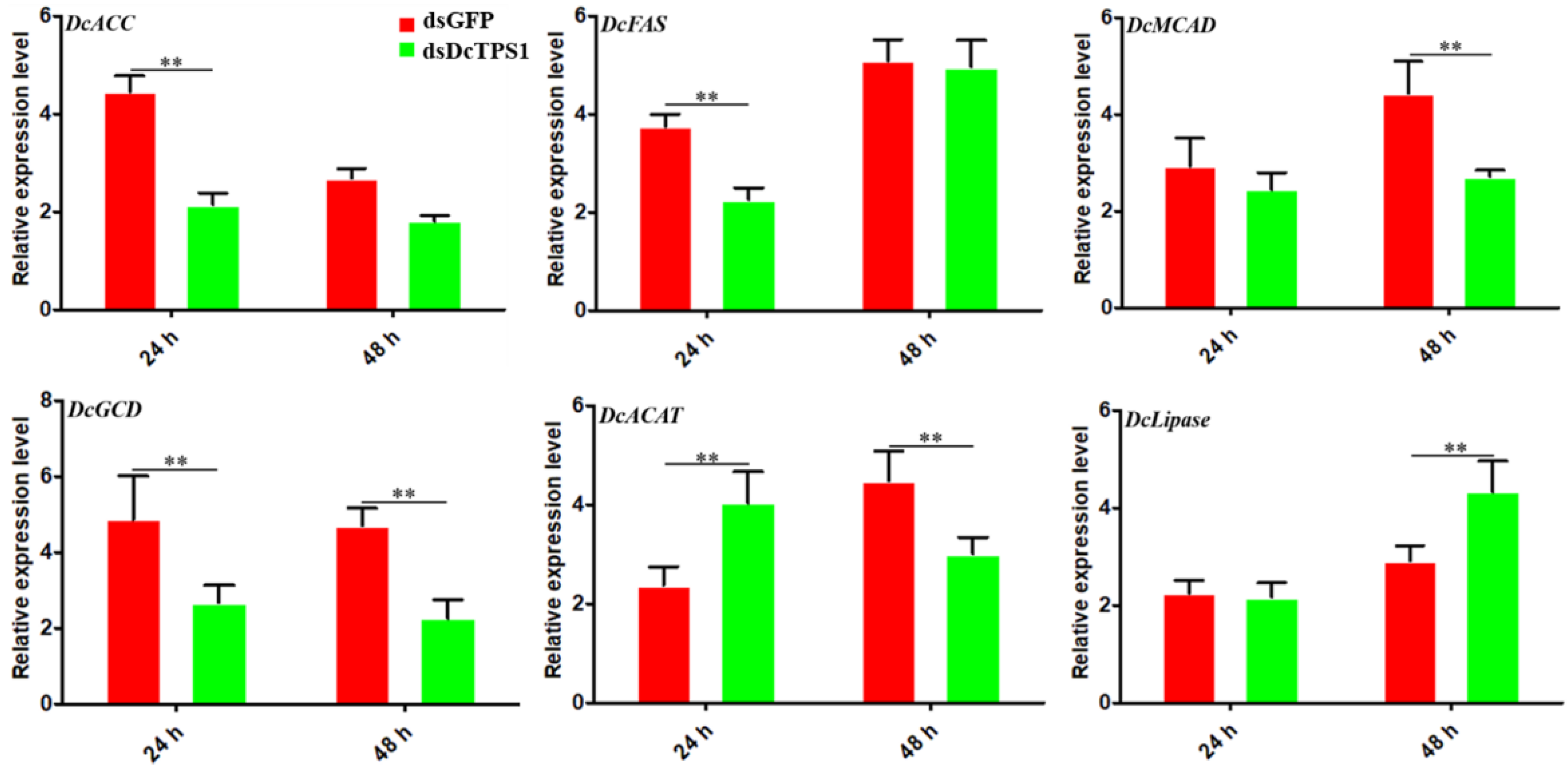
3.5. Analysis of the Effect on Fatty Acid Metabolism after Silencing of DcTPS1

Acetyl-CoA produced form glycolysis can be utilized to form lipids. In order to analyze the effect of DcTPS1 on D. citri fatty acid metabolism, a total of six genes involved in synthesis and degradation of fatty acid were analyzed at 24 h and 48 h after knockdown of DcTPS1. The results suggested that two genes associated with fatty acid synthesis were significantly downregulated at 24 h after silencing of DcTPS1, whereas they had no significant change between dsDcTPS1 group and dsGFP group at 48 h, including acetyl-CoA carboxylase-like (DcACC) and fatty acid synthase-like (DcFAS) (Figure 7). In addition, three genes involved in oxidative decomposition of fatty acids exhibited similar expression patterns, and they were downregulated at 24 h or 48 h after silencing of DcTPS1, including medium-chain-specific acyl-CoA dehydrogenase (DcMCAD), glutaryl-CoA dehydrogenase (DcGCD) and acetyl-CoA acetyltransferase (DcACAT). The expression level of DcLipase was significantly increased at 48 h after silencing of DcTPS1, but it had no obvious change at 24 h (Figure 7).

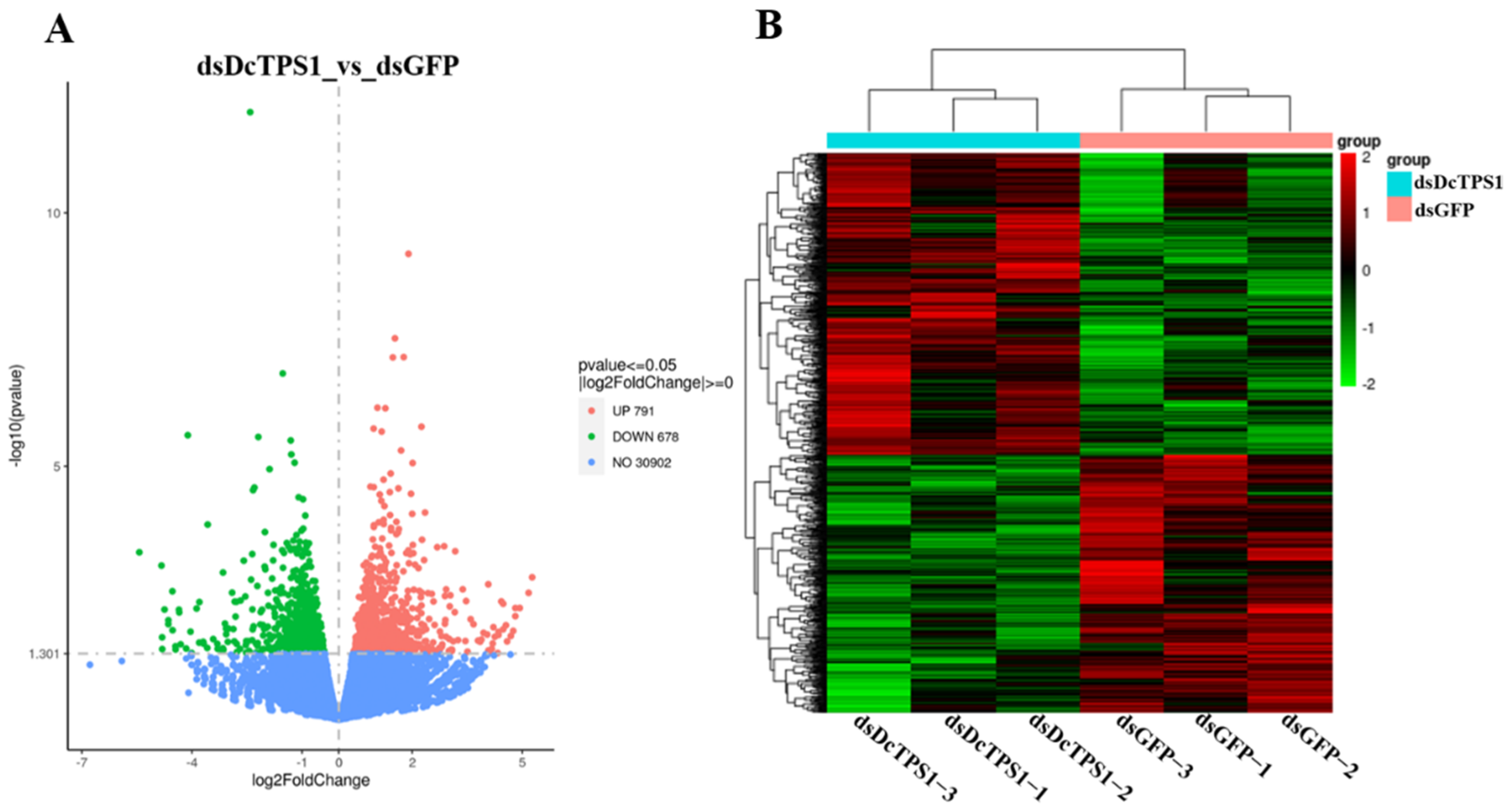
3.6. Transcriptome Sequencing and Reads Assembly

After removing the redundant reads, a total of 51,460,996 (97.8%), 47,166,336 (97.6%), and 42,176,502 (97.9%) clean reads from the treatment groups (dsDcTPS1); 40,384,536 (98.0%), 49,969,160 (98.0%), and 42,794,132 (97.8%) clean reads from the control groups (dsGFP) were obtained. The raw reads from the six libraries were submitted to the sequence read archive (SRA) of NCBI (BioProject: PRJNA782056). The values of Q20 and Q30 were approximately 97% and 93%, respectively. The values of GC content in different samples were about 40% (Table S2). Furthermore, 41,276,108 (80.2%), 37,944,770 (80.4%) and 34,460,721 (81.7%) clean reads from the treatment groups (dsDcTPS1); 32,409,083 (80.3%), 40,246,014 (80.5%) and 34,339,514 (80.2%) clean reads from control groups (dsGFP) were successfully mapped to the D. citri genome (Table S3).

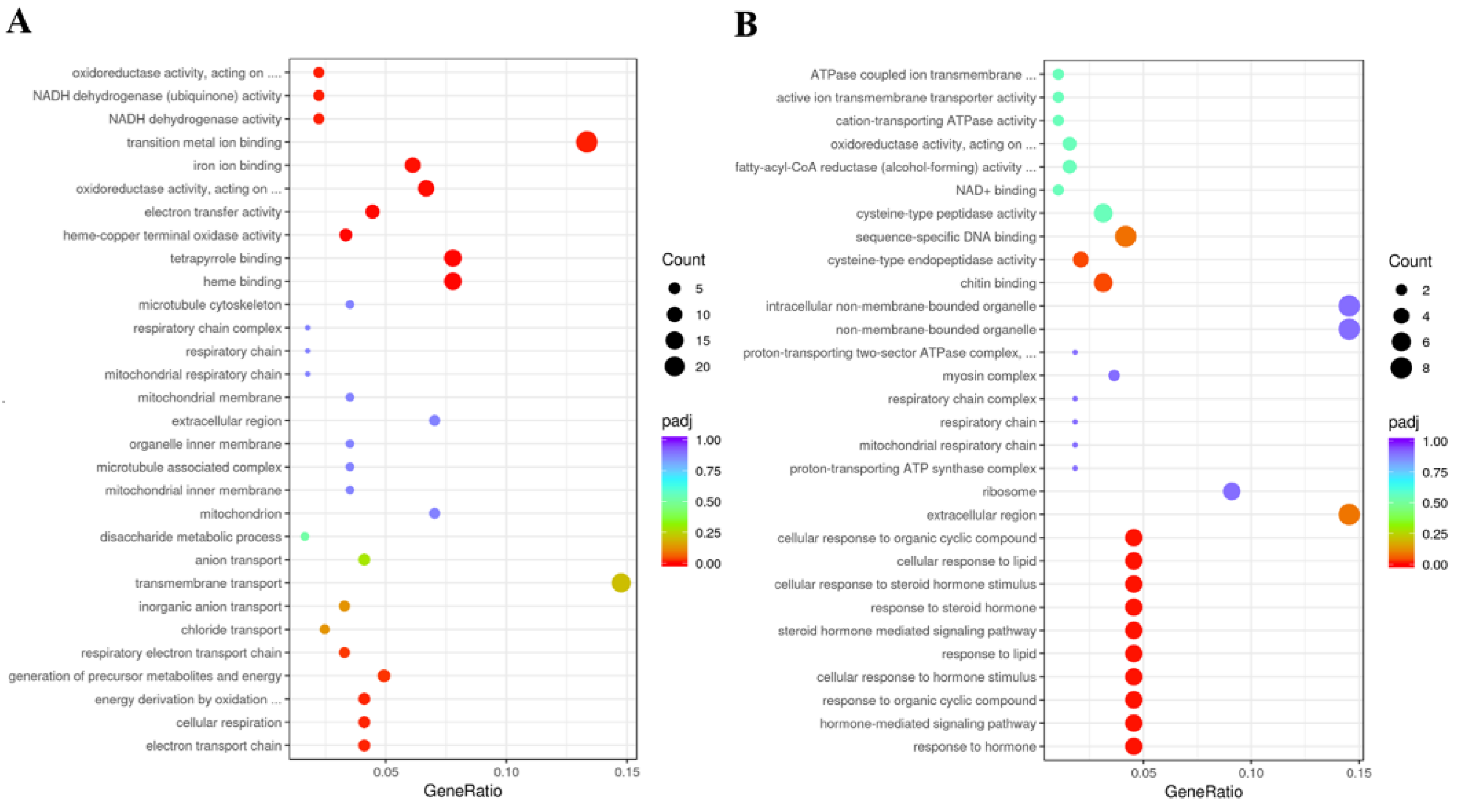
3.7. Identification of DEGs and Functional Prediction

According to use the DESeq method, DEGs were identified between control groups (dsGFP) and treatment groups (dsDcTPS1). In total, 1469 DEGs were identified in dsDcTPS1 groups compared with dsGFP groups, among which 791 DEGs were upregulated, and 678 DEGs were downregulated (Figure 8A; Table S4). The hierarchical clustering revealed that DEGs with similar expression patterns were clustered together, and further showed good repeatability among the three biological replicates (Figure 8B).
GO enrichment analyses suggested that upregulated DEGs were mainly involved in transition metal ion binding and transmembrane transport, and DEGs of downregulation were involved in chitin binding and sequence-specific DNA binding in the dsDcTPS1_vs_dsGFP groups (Figure 9; Table S5). KEGG enrichment revealed that upregulated DEGs were mainly involved in oxidative phosphorylation, and downregulated DEGs were associated with the lysosome and ribosome (Figure 10; Table S6). To further validate the expression levels of DEGs in the control groups and treatment groups, we selected nine DEGs involved with the ribosome, oxidative phosphorylation and the lysosome. The results showed that the express trend of nine genes remained consistent between qPCR and transcriptome data (Figure 11). A total of three DEGs related to ribosome were downregulated after silencing of dsDcTPS1, including 39S ribosomal protein L2 (DcRPL2), 40S ribosomal protein S10 (DcRPS10) and 60S ribosomal protein L11 (DcRPL11). A total of three DEGs related to oxidative phosphorylation were upregulated after silencing of dsDcTPS1, including NADH-ubiquinone oxidoreductase chain 4 (DcND4), ATP synthase lipid-binding protein (DcAslp) and cytochrome c oxidase subunit 3 (DcCox3). Additionally, three DEGs associated with lysosome were downregulated after silencing of dsDcTPS1, including cathepsin B-like cysteine proteinase (DcCath-B), uncharacterized LOC103505824 (Loc103505824) and alpha-mannosidase At3g26720 (DcMan).

4. Discussion

Trehalose is the principal sugar circulating in the hemolymph of most insects and is synthesized in the fat body. The stored energy reserve in the form of trehalose is hydrolyzed by trehalase to meet the energy demands for flight and development [6,35]. Chitin is a polymer of N-acetyl glucosamine that forms the protective exoskeleton of all arthropods and is replaced during growth and development [36]. The insect chitin biosynthetic pathway starts with trehalose and involves several key enzymes [2]. Therefore, accurate regulation of trehalose concentration is crucial for normal growth and development of insects. In insects, trehalose forms the major hemolymph sugar and is synthesized in a way that involves two enzymes, including trehalose-6-phosphate synthase and trehalose 6-phosphate phosphatase [37]. In the current study, a TPS gene was identified from the D. citri genome database. Bioinformatic analysis revealed that DcTPS1 encoded a total of 833 amino acids (Figure 1). In previous research, Liu et al. also identified a TPS gene from D. citri which encoded a protein of 594 amino acid residues [15]. Tang et al. revealed that the insect TPS gene encoded an 820–850 amino acid protein with two conserved domains—TPS and TPP—corresponding to OtsA and OtsB genes in yeast [38]. Additionally, phylogenetic tree analysis suggested that DcTPS1 kept a close relationship with hemipteran insects, including B. tabaci, A. pisum and N. lugens. Interestingly, we also found that DcTPS1 had a close relationship with DcTPS reported by Liu et al. [15]. Therefore, we considered that DcTPS1 belonged to a TPS gene in D. citri. The number of TPS genes varies among various insects. In Blattella germanica, a total of two TPS genes were cloned, including BgTPS1 and BgTPS2 [39]. In N. lugens, a total of three TPS genes were identified, and these TPS genes had been found to encode proteins with two conserved TPS and TPP domains. However, we did not find TPS2 or TPS3 homolog sequences from the D. citri genome and transcriptome databases.
Insects store energy reserves in the form of glycogen and triglycerides in the adipocytes, the main fat body cell [40]. Furthermore, insect fat body is an important tissue for production of trehalose [41]. In this study, DcTPS1 had a high expression in the midgut and wing, while it had a low expression in the fat body. Most insects express the TPS gene mainly in the fat body, including Helicoverpa armigera, N. lugens and Bactrocera minax [2,37,42]. We speculated that DcTPS1 was mainly involved in trehalose synthesis in the midgut. High expression of TPS genes in the midgut was also found in some other insects. In Leptinotarsa decemlineata, LdTPS showed a high expression level in the fat body, foregut and hindgut, involving in the synthesis of trehalose [10]. Furthermore, we also considered that midgut tissue might have been doped with fat body during the extraction process. In addition, we also found that DcTPS1 exhibited a high expression at the egg, fifth-instar nymph and adult stages. In D. citri, DcTPS1 expression level in female adults was significantly higher than that of male adults. In previous report, Liu et al. also found that DcTPS1 exhibited a high expression in the D. citri adult stage [15]. Based on the high expression of DcTPS1 in the egg and female adults, we speculated that DcTPS1 might be involved in reproduction of D. citri eggs. In D. citri, the fifth-instar nymph stage is a critical period that involves progressing from the nymph stage into adult [43]. After molting of nymph, D. citri adults are required to synthesize more trehalose to maintain flight and bounce. Moreover, during the D. citri molting period, synthesis of more trehalose promotes chitin formation to maintain the rigid structure of new cuticle. Interestingly, we also found that DcTPS1 had a high expression in the D. citri egg. We speculated that DcTPS1 might be involved in chitin synthesis on the embryonic cuticle of D. citri. In Rhodnius prolixus, reduction of chitin synthase gene transcripts by RNAi significantly affected chitin deposition and eclosion of the first-instar nymph [44].
RNAi is now widely used as a useful tool for discovering or validating gene functions. At 24 h and 48 h after ingestion of dsDcTPS1, the relative expression levels of DcTPS1 were significantly downregulated in treatment groups compared with control groups (dsGFP treatment), which indicated that DcTPS1 was effectively silenced. Furthermore, the mortality and malformation rates increased after silencing DcTPS1, and the fifth-instar nymphs could not completely molt and die. From the phenotype, we found that D. citri wing exhibited curl, and abdominal cuticle could not completely molt after treatment with dsDcTPS1. Our results are similar to those previously reported by Liu et al. [15]. In Tribolium castaneum, silencing TPS genes lead to molting deformities and high mortality rates via regulation of gene expression in the chitin biosynthesis pathway [11]. Yang et al. revealed that silencing N. lugens TPS genes induced insects displaying aberrant phenotypes [45]. In addition, we also found that knockdown of DcTPS1 gene reduced the expression levels of chitin metabolism-related genes. Therefore, we considered that inhibition of DcTPS1 gene disrupted D. citri chitin synthesis, resulting in abnormal phenotypes.
Trehalose represents the main hemolymph sugar in many insects, which is an energy source that meets the demands of flight muscles and other energy-consuming organs [35]. In these energy-consuming organs, the energy is ultimately derived from lipid. The relative expression levels of two fatty acid metabolism-related genes were significantly downregulated at 24 h after silencing of DcTPS1, including DcACC and DcFAS. Acetyl-CoA carboxylase (ACC) is a major rate-limiting enzyme of fatty acid biosynthesis; its product, malonyl-CoA, also contributes to the regulation of fatty acid oxidation and elongation [46]. Fatty acid synthase (FAS) is a multifunctional enzyme involved in the formation of fatty acids [47]. In a previous report, Shi et al. revealed that L. decemlineata TPS RNAi survivors consumed a greater amount of foliage. On the contrary, silencing of the DcTPS1 gene lead to a smaller polypide. The results indicated that inhibition of DcTPS1 disrupts the synthesis of fatty acid. According to transcriptome sequencing, GO enrichment analysis showed that downregulated DEGs were significantly involved in chitin metabolism. KEGG enrichment analysis showed that upregulated DEGs were significantly involved in oxidative phosphorylation. In biological cells, oxidative phosphorylation is primarily involved in the synthesis of ATP and is also associated with the oxidation of NADH. We found that knockdown of the DcTPS1 gene increased the expression levels of DEGs associated with oxidative phosphorylation. Therefore, we speculated that a reduction in DcTPS1 expression level significantly suppress energy metabolism. When energy synthesis is insufficient, D. citri will activate oxidative phosphorylation through its negative feedback mechanism. Additionally, DEGs involved with the ribosome and lysosome were significantly downregulated after silencing of DcTPS1. Lysosomes play a central role in the degradation of extracellular and intracellular macromolecules [48]. The expression level of D. citri cathepsin B (DcCath-B) significantly decreased after inhibition of DcTPS1. The results indicated that knockdown of DcTPS1 gene inhibited protein hydrolysis.

5. Conclusions

A TPS gene was identified from D. citri genome database. DcTPS1 showed high expression in the midgut and fifth-instar nymph stage. Additionally, silencing of DcTPS1 led to an abnormal phenotype. Knockdown of DcTPS1 significantly reduced the expression levels of chitin metabolism-related genes and fatty acid synthesis-related genes, while the expression levels of fatty acid decomposition-related genes were downregulated. Furthermore, transcriptome sequencing analysis revealed that DEGs involved in oxidative phosphorylation were significantly upregulated, whereas DEGs attributed to the lysosome and ribosome were downregulated.

Supplementary Materials

The following are available online at https://www.mdpi.com/article/10.3390/insects12121074/s1, Figure S1: Multiple sequence alignment of the conserved domain of the TPS1 from four insect species, Table S1: Primers used in this study, Table S2: A summary of the transcriptomes in the different treatments in D. citri, Table S3: A summary of reads mapped to D. citri genome in different treatments, Table S4: Identification of differentially expressed genes in different treatments, Table S5: GO enrichment analysis of DEGs, Table S6: KEGG enrichment analysis of DEGs.

Author Contributions

Conceptualization, J.-C.S. and H.-Z.Y.; methodology, L.Y.; software, J.-C.S. and H.-Z.Y.; validation, J.-C.S.; formal analysis, H.-Z.Y.; investigation, L.Y.; resources, H.-Z.Y. and Z.-J.L.; data curation, Z.-J.L.; writing—original draft preparation, J.-C.S. and H.-Z.Y.; writing—review and editing, Z.-J.L.; visualization, H.-Z.Y.; supervision, H.-Z.Y.; project administration, Z.-J.L. and H.-Z.Y.; funding acquisition, Z.-J.L. and H.-Z.Y. All authors have read and agreed to the published version of the manuscript.

Funding

This research was funded by the Major Science and Technology R&D Program of Jiangxi Province (20194ABC28007), Natural Science Foundation of Jiangxi Province (20202BAB215004), the National Natural Science Foundation of China (31960116; 32160625) and Key R & D plan of Jiangxi Province (20192BBF60057).

Institutional Review Board Statement

Not applicable.

Informed Consent Statement

Not applicable.

Data Availability Statement

All published data are available upon formal request.

Conflicts of Interest

The authors declare no conflict of interest.

References

  1. Chen, Q.F.; Haddad, G.G. Role of trehalose phosphate synthase and trehalose during hypoxia: From flies to mammals. J. Biol. Chem. 2004, 207, 3125–3129. [Google Scholar] [CrossRef] [Green Version]
  2. Xiong, K.C.; Wang, J.; Li, J.H.; Deng, Y.Q.; Pu, P.; Fan, H.; Liu, Y.H. RNA interference of a trehalose-6-phosphate synthase gene reveals its roles during larval-pupal metamorphosis in Bactrocera minax (Diptera: Tephritidae). J. Insect Physiol. 2016, 91–92, 84–92. [Google Scholar] [CrossRef] [PubMed]
  3. Shoji, K.; Akiyama, Y.; Suzuki, M.; Hoshino, T.; Nakamura, N.; Ohno, H.; Morishima, K. Insect biofuel cells using trehalose included in insect hemolymph leading to an insect-mountable biofuel cell. Biomed. Microdevices 2012, 14, 1063–1068. [Google Scholar] [CrossRef]
  4. Karpova, E.K.; Eremina, M.A.; Pirozhkova, D.S.; Gruntenko, N.E. Stress-related hormones affect carbohydrate metabolism in Drosophila females. Arch. Insect Biochem. Physiol. 2019, 101, e21540. [Google Scholar] [CrossRef]
  5. Yu, H.Z.; Huang, Y.L.; Lu, Z.J.; Zhang, Q.; Su, H.N.; Du, Y.M.; Yi, L.; Zhong, B.L. Inhibition of trehalase affects the trehalose and chitin metabolism pathways in Diaphorina citri (Hemiptera: Psyllidae). Insect Sci. 2021, 28, 718–734. [Google Scholar] [CrossRef]
  6. Shukla, E.; Thorat, L.J.; Nath, B.B.; Gaikwad, S.M. Insect trehalase: Physiological significance and potential applications. Glycobiology 2015, 25, 357–367. [Google Scholar] [CrossRef] [Green Version]
  7. Wang, S.S.; Li, G.Y.; Liu, Y.K.; Luo, Y.J.; Xu, C.D.; Li, C.; Tang, B. Regulation of carbohydrate metabolism by trehalose-6-phosphate synthase 3 in the brown planthopper, Nilaparvata lugens. Front. Physiol. 2020, 11, 575485. [Google Scholar] [CrossRef]
  8. Wang, G.; Gou, Y.P.; Guo, S.F.; Zhou, J.J.; Liu, C.Z. RNA interference of trehalose-6-phosphate synthase and trehalase genes regulates chitin metabolism in two color morphs of Acyrthosiphon pisum Harris. Sci. Rep. 2021, 11, 948. [Google Scholar] [CrossRef]
  9. Tang, B.; Chen, J.; Yao, Q.; Pan, Z.Q.; Xu, W.H.; Wang, S.G.; Zhang, W.Q. Characterization of a trehalose-6-phosphate synthase gene from Spodoptera exigua and its function identification through RNA interference. J. Insect Physiol. 2010, 56, 813–821. [Google Scholar] [CrossRef]
  10. Shi, J.F.; Xu, Q.Y.; Sun, Q.K.; Meng, Q.W.; Mu, L.L.; Guo, W.C.; Li, G.Q. Physiological roles of trehalose in Leptinotarsa larvae revealed by RNA interference of trehalose-6-phosphate synthase and trehalase genes. Insect Biochem. Mol. Biol. 2016, 77, 52–68. [Google Scholar] [CrossRef] [PubMed]
  11. Chen, Q.W.; Jin, S.; Zhang, L.; Shen, Q.D.; Wei, P.; Wei, Z.M.; Wang, S.G.; Tang, B. Regulatory functions of trehalose-6-phosphate synthase in the chitin biosynthesis pathway in Tribolium castaneum (Coleoptera: Tenebrionidae) revealed by RNA interference. Bull. Entomol. Res. 2018, 108, 388–399. [Google Scholar] [CrossRef]
  12. Kern, C.; Wolf, C.; Bender, F.; Berger, B.; Noack, S.; Schmalz, S.; Ilg, T. Trehalose-6-phosphate synthase from the cat flea Ctenocephalides felis and Drosophila melanogaster: Gene identification, cloning, heterologous functional expression and identification of inhibitors by highthroughput screening. Insect Mol. Biol. 2012, 21, 456–471. [Google Scholar] [CrossRef]
  13. Matsuda, H.; Yamada, T.; Yoshida, M.; Nishimura, T. Flies without trehalose. J. Biol. Chem. 2015, 290, 1244–1255. [Google Scholar] [CrossRef] [Green Version]
  14. Bonnett, T.R.; Robert, J.A.; Pitt, C.; Fraser, J.D.; Keeling, C.I.; Bohlmann, J.; Huber, D.P.W. Global and comparative proteomic profiling of overwintering and developing mountain pine beetle, Dendroctonus ponderosae (Coleoptera: Curculionidae), larvae. Insect Biochem. Mol. Biol. 2012, 42, 890–901. [Google Scholar] [CrossRef] [PubMed] [Green Version]
  15. Liu, X.Y.; Zou, Z.W.; Zhang, C.; Liu, X.; Wang, J.; Xin, T.R.; Xia, B. Knockdown of the trehalose-6-phosphate synthase gene using RNA interference inhibits synthesis of trehalose and increases lethality rate in Asian citrus psyllid, Diaphorina citri (Hemiptera: Psyllidae). Insects 2020, 11, 605. [Google Scholar] [CrossRef] [PubMed]
  16. Hosseinzadeh, S.; Higgins, S.A.; Ramsey, J.; Howe, K.; Griggs, M.; Castrillo, L.; Heck, M. Proteomic polyphenism in color morphotypes of Diaphorina citri, insect vector of citrus greening disease. J. Proteome Res. 2021, 20, 2851–2866. [Google Scholar] [CrossRef] [PubMed]
  17. EI-Shesheny, I.; EI-Hawary, I.; Mesbah, I.; Killiny, N. Comparative proteomic analysis between fifth-instar nymphs and adults of Asian citrus psyllid Diaphorina citri. Physiol. Entomol. 2016, 41, 162–184. [Google Scholar] [CrossRef]
  18. Hijaz, F.; Lu, Z.J.; Killiny, N. Effect of host-plant and infection with ‘Candidatus Liberibacter asiaticus’ on honeydew chemical composition of the Asian citrus psyllid, Diaphorina citri. Entomol. Exp. Appl. 2016, 158, 34–43. [Google Scholar] [CrossRef]
  19. Qureshi, J.A.; Kostyk, B.C.; Stansly, P.A. Insecticidal suppression of Asian citrus psyllid Diaphorina citri (Hemiptera: Liviidae) vector of huanglongbing pathogens. PLoS ONE 2014, 9, e112331. [Google Scholar]
  20. Tiwari, S.; Mann, R.S.; Rogers, M.E.; Stelinski, L.L. Insecticide resistance in field populations of Asian citrus psyllid in Florida. Pest Manag. Sci. 2011, 67, 1258–1268. [Google Scholar] [CrossRef]
  21. Kishk, A.; Anber, H.A.I.; AbdEI-Raof, T.K.; EI-Sherbeni, A.-H.D.; Hamed, S.; Gowda, S.; Killiny, N. RNA interference of carboxyesterases causes nymph mortality in the Asian citrus psyllid, Diaphorina citri. Arch. Insect Biochem. Physiol. 2017, 94, e21377. [Google Scholar] [CrossRef]
  22. Tiwari, S.; Clayson, P.J.; Kuhns, E.H.; Stelinski, L.L. Effects of buprofezin and diflubenzuron on various developmental stages of Asian citrus psyllid, Diaphorina citri. Pest Manag. Sci. 2012, 68, 1405–1412. [Google Scholar] [CrossRef] [PubMed]
  23. Fire, A.; Xu, S.; Montgomery, M.K.; Kostas, S.A.; Driver, S.E.; Mello, C.C. Potent and specific genetic interference by double-stranded RNA in Caenorhabditis elegans. Nature 1998, 391, 806–811. [Google Scholar] [CrossRef]
  24. Zha, W.J.; Peng, X.X.; Chen, R.Z.; Du, B.; Zhu, L.L.; He, G.C. Knockdown of midgut genes by dsRNA-transgenic plant-mediated RNA interference in the hemipteran insect. PLoS ONE 2011, 6, e20504. [Google Scholar] [CrossRef] [PubMed] [Green Version]
  25. Price, D.R.G.; Gatehouse, J.A. RNAi-mediated crop protection against insects. Trends Biotechnol. 2008, 26, 393–400. [Google Scholar] [CrossRef]
  26. Zhu, K.Y.; Palli, S.R. Mechanisms, applications, and challenges of insect RNA interference. Annu. Rev. Entomol. 2020, 65, 293–311. [Google Scholar] [CrossRef] [PubMed] [Green Version]
  27. Cooper, A.M.W.; Silver, K.; Zhang, J.Z.; Park, Y.; Zhu, K.Y. Molecular mechanisms influencing efficiency of RNA interference in insects. Pest Manag. Sci. 2019, 75, 18–28. [Google Scholar] [CrossRef] [Green Version]
  28. Guan, R.B.; Li, H.C.; Miao, X.X. Prediction of effective RNA interference targets and pathway-related genes in lepidopteran insects by RNA sequencing analysis. Insect Sci. 2018, 25, 356–367. [Google Scholar] [CrossRef]
  29. Ghosh, S.K.B.; Hunter, W.B.; Park, A.L.; Gundersen-Rindal, D.E. Double-stranded RNA oral delivery methods to induce RNA interference in phloem and plant-sap-feeding hemipteran insects. JoVE-J. Vis. Exp. 2018, 135, 57390. [Google Scholar] [CrossRef] [Green Version]
  30. Yoshiyama, N.; Tojo, K.; Hatakeyama, M. A survey of the effectiveness of non-cell autonomous RNAi throughoutdevelopment in the sawfly, Athalia rosae (Hymenoptera). J. Insect Physiol. 2013, 59, 400–407. [Google Scholar] [CrossRef]
  31. Rangasamy, M.; Siegfried, B.D. Validation of RNA interference in western corn rootworm Diabrotica virgifera virgifera LeConte (Coleoptera: Chrysomelidae) adults. Pest Manag. Sci. 2012, 68, 587–591. [Google Scholar] [CrossRef] [PubMed]
  32. Yuan, C.Y.; Jing, T.X.; Li, W.; Liu, X.Q.; Liu, T.Y.; Liu, Y.; Chen, M.L.; Jiang, R.X.; Yuan, G.R.; Dou, W.; et al. NADPH-cytochrome P450 reductase mediates the susceptibility of Asian citrus psyllid Diaphorina citri to imidacloprid and thiamethoxam. Pest Manag. Sci. 2021, 77, 677–685. [Google Scholar] [CrossRef]
  33. Kishk, A.; Hijaz, F.; Anber, H.A.; AbdEI-Raof, T.K.; EI-Sherbeni, A.-H.D.; Hamed, S.; Killiny, N. RNA interference of acetylcholinesterase in the Asian citrus psyllid, Diaphorina citri, increases its susceptibility to carbamate and organophosphate insecticides. Pestic. Biochem. Phys. 2017, 143, 81–89. [Google Scholar] [CrossRef] [PubMed] [Green Version]
  34. Yu, X.D.; Killiny, N. RNA interference of two glutathione S-transferase genes, DcGSTe2 and DcGSTd1, increases the susceptibility of Asian citrus psyllid (Hemiptera: Liviidae) to the pesticides, fenpropathrin and thiamethoxam. Pest Manag. Sci. 2018, 74, 638–647. [Google Scholar] [CrossRef]
  35. Thompson, S.N. Trehalose-the insect ‘blood’ sugar. Adv. Insect Physiol. 2003, 31, 205–285. [Google Scholar]
  36. Doucet, D.; Retnakaran, A. Insect chitin: Metabolism, genomics and pest management. Adv. Insect Physiol. 2012, 43, 437–511. [Google Scholar]
  37. Xu, J.; Bao, B.; Zhang, Z.F.; Yi, Y.Z.; Xu, W.H. Identification of a novel gene encoding the trehalose phosphate synthase in the cotton bollworm, Helicoverpa armigera. Glycobiology 2009, 19, 250–257. [Google Scholar] [CrossRef] [PubMed] [Green Version]
  38. Tang, B.; Shen, Q.D.; Zeng, B.P.; Xiao, Z.J.; Qiu, L.Y.; Pan, B.Y.; Li, K.; Zhang, D.W. Characteristics, developmental expression and RNAi effect analysis of a novel trehalose-6-phosphate synthase gene in Nilaparvata lugens. Sci. Agric. Sin. 2019, 52, 466–477. [Google Scholar]
  39. Chen, J.; Zhang, D.W. Molecular cloning, tissue distribution and temperature-induced expression of two trehalose-6-phosphate synthase genes in Blattella germanica (Blattodea: Blattellidae). Acta Entomol. Sin. 2015, 58, 1046–1053. [Google Scholar]
  40. Arrese, E.L.; Soulages, J.L. Insect fat body: Energy, metabolism, and regulation. Annu. Rev. Entomol. 2010, 55, 207–225. [Google Scholar] [CrossRef] [Green Version]
  41. Gans, U.; Subramanian, V.; Tan, B.H. Selective phagocytosis: A new concept in protein catabolism. Science 1968, 159, 107–110. [Google Scholar] [CrossRef] [PubMed]
  42. Chen, J.; Zhang, D.; Yao, Q.; Zhang, X.; Dong, H.; Tian, H.; Chen, J.; Zhang, W. Feeding-based RNA interference of a trehalose phosphate synthase gene in the brown planthopper, Nilaparvata lugens. Insect Mol. Biol. 2010, 19, 777–786. [Google Scholar] [CrossRef] [PubMed]
  43. Boina, D.R.; Rogers, M.E.; Wang, N.; Stelinski, L.L. Effect of pyriproxyfen, a juvenile hormone mimic, on egg hatch, nymph development, adult emergence and reproduction of the Asian citrus psyllid, Diaphorina citri. Pest Manag. Sci. 2010, 66, 349–357. [Google Scholar] [CrossRef]
  44. Souza-Ferreira, P.S.; Mansur, J.F.; Berni, M.; Moreira, M.F.; Dos Santos, R.E.; Araijo, H.M.M.; de Souza, W.; Ramos, I.B.; Masuda, H. Chitin deposition on the embryonic cuticle of Rhodnius prolixus: The reduction of CHS transcripts by CHS–dsRNA injection in females affects chitin deposition and eclosion of the first instar nymph. Insect Biochem. Mol. Biol. 2014, 51, 101–109. [Google Scholar] [CrossRef]
  45. Yang, M.M.; Zhao, L.N.; Shen, Q.D.; Xie, G.Q.; Wang, S.G.; Tang, B. Knockdown of two trehalose-6-phosphate synthases severely affects chitin metabolism gene expression in the brown planthopper Nilaparvata lugens. Pest Manag. Sci. 2017, 73, 206–216. [Google Scholar] [CrossRef]
  46. Bianchi, A.; Evans, J.L.; Iverson, A.J.; Nordlund, A.C.; Watts, T.D.; Witters, L.A. Identification of an isozymic form of acetyl-CoA carboxylase. J. Biol. Chem. 1990, 265, 1502–1509. [Google Scholar] [CrossRef]
  47. Moriconi, D.E.; Dulbecco, A.B.; Juarez, M.P.; Calderon-Fernandez, G.M. A fatty acid synthase gene (FASN3) from the integument tissue of Rhodnius prolixus contributes to cuticle water loss regulation. Insect Mol. Biol. 2019, 28, 850–861. [Google Scholar] [CrossRef] [PubMed]
  48. Dunn, W.A., Jr. Autophagy and related mechanisms of lysosome-mediated protein degradation. Trends Cell Biol. 1994, 4, 139–143. [Google Scholar] [CrossRef]
Figure 1. Sequence analysis of DcTPS1 cDNA sequence. (A) Nucleotide and amino acid sequence analysis of the DcTPS1 cDNA sequence. Numbers on the left side indicate the position of nucleotide and amino acid. The initiation codon (ATG) and termination codon (TAG) are showed in bold italics. The black solid line indicates the Glyco_transf_20 domain. The black break line indicates the Trehalose_PPase domain. The black box represents two glycosylation sites. (B) Structural domain analysis of DcTPS1 by SMART software. The blue box represents Glyco_transf_20 domain. The red box represents Trehalose_PPase domain. Two green circles indicate the glycosylation sites.
Insects 12 01074 g001
Figure 2. Multiple sequence alignment of the conserved domain of the TPS1 from four insect species, including Diaphrina citri TPS1 (MZ888936), Diaphorina citri TPS (QOU11567), Acyrthosiphon pisum TPS1 (XP_001945523), Bemisia tabaci TPS1 (XP_018916964) and Nilaparvata lugens TPS1 (ACV20871). Signature motifs (HDYHL and DGMNLV) unique to TPS was presented with red box. The conserved amino acid residues are highlighted in black, and similar amino acid residues are labelled in pink and blue.
Insects 12 01074 g002
Figure 3. The spatiotemporal expression patterns of DcTPS1 in different developmental stages (A) and different tissues (B) of Diaphorina citri. Relative expression levels of DcTPS1 were analyzed using qPCR. Data were normalized using glyceraldehyde-3-phosphate dehydrogenase (GAPDH) and are represented as the means ± standard errors of the means from three independent experiments. The 2−∆∆Ct method was used to calculate the relative expression level. Statistical analysis was conducted using SPSS software. Different letters indicate significant differences, for example, a, b, c and d (p < 0.05).
Insects 12 01074 g003
Figure 4. Detection of DcTPS1 expression levels and phenotypic observation after treatment with dsDcTPS1 and dsGFP. (A) Analysis of DcTPS1 expression levels after treatment with dsDcTPS1 and dsGFP. The 2−∆∆Ct method was adopted to calculate the relative expression level. The SPSS software was used to conduct statistical analysis. The asterisks indicate the significance differences by ** p < 0.01. (B) Phenotypic observation of D. citri adult at 48 h.
Insects 12 01074 g004
Figure 5. Statistical analysis of D. citri mortality, malformation and molting after inhibition of DcTPS1. (A) Determination of D. citri mortality after inhibition of DcTPS1. The dsGFP treatment group was used as a control; (B) Detection of malformation rate of D. citri at 24 h and 48 h after RNAi of DcTPS1; (C) Detection of cumulative molting of D. citri at 24 h and 48 h after inhibition of DcTPS1. The asterisks indicate the significance differences by * p < 0.05 and ** p < 0.01. The SPSS software was used to conduct statistical analysis.
Insects 12 01074 g005
Figure 6. Analysis of expression levels of chitin metabolism-related genes after inhibition of DcTPS1. The dsGFP treatment group was used as a control. The mean expression level represents three biological replicates. The asterisks indicate significant differences by * p < 0.05 and ** p < 0.01. The SPSS software was used to conduct statistical analysis.
Insects 12 01074 g006
Figure 7. Effects of RNAi on key genes in Diaphorina citri fatty acid metabolism pathway. The dsGFP treatment group was used as a control. The mean expression level represents three biological replicates. The asterisks indicate significance differences by ** p < 0.01. The SPSS software was used to conduct statistical analysis.
Insects 12 01074 g007
Figure 8. Identification and hierarchical cluster analysis of DEGs among different samples. (A) A volcano diagram for each gene. The red and green points indicate upregulated genes and downregulated genes, respectively. The blue point indicates no significant difference between dsDcTPS1 and DcGFP groups. (B) Hierarchical clustering of DEGs.
Insects 12 01074 g008
Figure 9. GO enrichment analysis of DEGs between dsDcTPS1 groups and dsGFP groups. The sizes of the bubble indicate the number of DEGs enriched in the GO term. The color of the bubble indicates the Q value. (A) Upregulated DEGs. (B) Downregulated DEGs.
Insects 12 01074 g009
Figure 10. KEGG enrichment analysis of DEGs between dsDcTPS1 groups and dsGFP groups. The sizes of the bubble indicate the number of DEGs enriched in the KEGG pathway. The color of the bubble indicates the Q value. (A) Upregulated DEGs. (B) Downregulated DEGs.
Insects 12 01074 g010
Figure 11. Validation of DEGs involved with the ribosome, oxidative phosphorylation and lysosome, respectively. The mean expression level represents three biological replicates. The asterisks indicate the significance differences by * p < 0.05 and ** p < 0.01. The SPSS software was used to conduct statistical analysis.
Insects 12 01074 g011
Publisher’s Note: MDPI stays neutral with regard to jurisdictional claims in published maps and institutional affiliations.

