

Review

The Gut–Skin Axis in Atopic Dermatitis and Inflammatory Bowel Disease: Mechanisms, Microbiota, and Therapeutic Implications


1
Pediatrics Children’s Research Institute, University of North Carolina at Chapel Hill, Chapel Hill, NC 27599, USA
2
Department of Dermatology, University of North Carolina at Chapel Hill, Chapel Hill, NC 27599, USA
3
Department of Pediatrics, University of North Carolina at Chapel Hill, Chapel Hill, NC 27599, USA
4
Laboratory of Animal Food Function, School of Agricultural Science, Tohoku University, Sendai 980-8572, Japan
5
Warren Alpert Medical School of Brown University, Providence, RI 02912, USA
6
Department of Pediatric Surgery, School of Medicine, Juntendo University, Tokyo 113-8421, Japan
7
Faculty of Medical Sciences, Newcastle University, Newcastle upon Tyne NE2 4HH, UK
*
Authors to whom correspondence should be addressed.
These authors contributed equally to this work.
J. Pers. Med. 2026, 16(9), 482; https://doi.org/10.3390/jpm16090482 (registering DOI)
Submission received: 20 August 2026 / Revised: 9 September 2026 / Accepted: 13 September 2026 / Published: 18 September 2026
(This article belongs to the Special Issue Personalized Management of Inflammatory Bowel Diseases)

Abstract

The gut–skin axis is a bidirectional communication network linking the gastrointestinal tract, skin, microbiota, and immune system. As major barrier organs, the gut and skin harbor complex microbial communities that contribute to tissue homeostasis, immune regulation, and protection from environmental insults. Increasing evidence indicates that dysbiosis of the gut and skin microbiota is associated with inflammatory diseases through interconnected microbial, metabolic, and immune pathways. Clinical observations further reveal strong associations between gastrointestinal disorders, including inflammatory bowel disease (IBD) and celiac disease, and cutaneous manifestations. Atopic dermatitis (AD) is characterized by epithelial barrier dysfunction, immune dysregulation, microbial imbalance, and environmental influences. Patients with AD frequently exhibit reduced gut microbial diversity, depletion of beneficial short-chain fatty acid (SCFA)-producing bacteria, and enrichment of potentially pathogenic microorganisms. Cutaneous dysbiosis, particularly expansion of Staphylococcus aureus, can further impair the skin barrier and sustain inflammation. Microbial metabolites, including SCFAs, tryptophan-derived aryl hydrocarbon receptor ligands, and bile acid metabolites, may mediate gut–skin communication by regulating epithelial integrity, immune tolerance, and inflammatory signaling. IBD is also increasingly recognized as a systemic disorder involving alterations in skin microbiota and cutaneous immunity. Intestinal inflammation may disrupt immune tolerance to skin commensals and promote cutaneous inflammation through cytokine signaling and immune-cell trafficking, while emerging evidence suggests reciprocal skin-to-gut effects. Epidemiologic, genetic, and clinical studies indicate an association between AD and IBD, potentially reflecting shared genetic susceptibility, barrier dysfunction, dysbiosis, and immune pathways. This review summarizes current evidence linking the gut–skin axis to AD and IBD.

1. Introduction

Inflammatory bowel disease (IBD), including Crohn’s disease (CD) and ulcerative colitis (UC), is a chronic, recurrent, inflammatory condition that arises from a combination of genetic, environmental, intestinal microflora, epithelial barrier dysfunction, and immune mechanisms [1,2]. Although CD and UC share many pathogenic mechanisms, they exhibit distinct pathological patterns. CD can affect any region of the gastrointestinal (GI) tract and is characterized by discontinuous “skip” lesions and transmural inflammation, which predisposes patients to strictures, fistulas, and abscesses. In contrast, UC is restricted to the colon and rectum and typically produces continuous inflammation beginning in the rectum and extending proximally, with inflammation predominantly involving the mucosa and submucosa. Histologically, CD may demonstrate deep fissuring ulcers and non-caseating granulomas, whereas UC is characterized by mucosal ulceration, crypt abscesses, and pseudopolyps. These differences contribute to distinct clinical manifestations and complications and reflect partially divergent immune mechanisms, including prominent Th1/Th17-associated inflammation in CD and atypical Th2/Th9-associated responses in UC [1,2]. IBD can also cause systemic inflammation and extraintestinal manifestations affecting organs and systems outside the digestive tract in 25% to 50% of patients.
IBD pathogenesis reflects genetic susceptibility (e.g., NODs, ATG16L1, IL23R), epithelial barrier dysfunction, and intestinal dysbiosis, which drive the dysregulated Th1/Th17 (CD) and atypical Th2 (UC) responses described above [3]. Management has evolved from corticosteroids, 5-aminosalicylates, and immunomodulators toward targeted biologics (anti-TNF agents, vedolizumab, IL-12/23 or IL-23-selective inhibitors) and oral small molecules (JAK inhibitors, S1P modulators), with surgery remaining important for refractory or complicated disease [4].
The gut–organ axis should be regarded as a bidirectional communication system between the GI tract and other organs [5,6]. The gut–organ axis is formed by a complex network of interactions between the GI tract and its microbiota with distant organs that is best understood as an interconnected network of individual two-link connections. Intestinal bacteria, their metabolites, and immune cells traffic to distant organs and modulate their function. Short-chain fatty acids (SCFAs), bile acids, tryptophan metabolites, lipopolysaccharides (LPS), cytokines, and immune cells are common molecules involved in the communication between the gut and other organs. Moreover, neural and endocrine connections contribute to the bidirectional nature of the gut–organ axis. Thus, intestinal dysbiosis and barrier dysfunction can lead to a state of increased inflammation that involves the brain [7,8], liver [9,10], lungs [11,12,13], kidneys [14,15], and pancreas [16]. On the other hand, malfunction of these organs can affect the intestines and change the microbiome and immunity of the gut. These findings provide an integrated view of mechanisms by which IBD can cause extraintestinal manifestations. They also suggest that treatments that target the microbiota, gut immunity, and the production of beneficial metabolites by the microbiota might have beneficial effects beyond the intestine.
The concept of the gut–skin axis has emerged as a central framework for understanding the bidirectional interactions between the GI tract, skin, microbiota, and immune system. The gut and skin are major barrier organs that are constantly exposed to environmental insults and depend on resident microbiota to maintain their integrity. Microbial imbalance in either the gut or skin can promote local and systemic inflammation. Environmental stimuli, especially air pollutants, cause oxidative stress and inflammation by disrupting the gut microflora. Redox mechanisms may contribute to gut dysbiosis, inflammation, and downstream effects on the skin, underscoring the role of oxidative stress and microbial communication in gut–skin axis dysfunction [17].
Associations between GI and cutaneous diseases are well-supported by a previous study [18]. Notably, 10% to 25% of patients with GI diseases such as IBD and celiac disease also experience skin pathology, including psoriasis (characterized by scaly plaques and hypertrophic skin lesions) and cutaneous ulcers [18,19]. Celiac disease, which is characterized by intestinal malabsorption, commonly co-presents with dermatitis and psoriasis as cutaneous manifestations, whereas patients with rosacea (a chronic inflammatory skin condition that causes reddened skin and a rash) frequently exhibit small intestinal bacterial overgrowth (SIBO) [20]. Peutz-Jeghers Syndrome, which is characterized by GI polyposis and malignancy, often features perioral hyperpigmentation as a common cutaneous manifestation [19]. Additionally, IBD patients frequently experience skin manifestations such as skin ulcers, vasculitis, hair loss, erythema folliculitis, and psoriasis [18,19], with some of these conditions linked to the severity of the patient’s GI inflammation [21].
One important mechanism of gut-to-skin communication involves microbial metabolites, which have been revealed to mediate the dialogue between the two organs. Indeed, beneficial metabolites like SCFAs and tryptophan derivatives mediate immune tolerance, induce regulatory T-cell differentiation, and induce anti-inflammatory cytokines IL-10 and TGF-β, among other processes. Some microbial metabolites may also influence keratinocytes and epidermal barrier regulation after entering the bloodstream. In other words, harmful metabolites such as phenolics could promote skin inflammation and disruption of the epidermal barrier. In summary, the manipulation of microbial metabolites as a treatment approach for skin diseases like atopic dermatitis (AD) via probiotics and prebiotics seems promising [22].
Recent advances in knowledge have gone beyond the classical paradigm of gut-to-skin interaction by developing a bidirectional model of the gut–skin axis. Numerous review articles exist regarding the gut–skin axis and skin diseases, such as atopic dermatitis [23,24,25,26,27]. In contrast, there are few papers discussing the impact of the gut–skin axis on enteritis [28,29]. While the impact of gut microbiota on skin integrity is well established, recent findings indicate that signaling pathways related to innate immunity, vitamin D receptors, and aryl hydrocarbon receptor (AhR) could play a role in maintaining skin and gut homeostasis. Interestingly, it was found that ultraviolet B (UVB) irradiation of the skin is associated with favorable changes in gut microbiome and physiology [30]. These findings suggest that the gut and skin should be viewed as interconnected organs within a shared immune–microbial network rather than as isolated systems [31].
From a personalized medicine perspective, differences in microbiome composition, host genetics, and immune responses may contribute to variation in disease susceptibility, severity, and therapeutic response. Integrating these features may therefore enable improved patient stratification, biomarker development, and selection of microbiota- or immune-targeted therapies. Unlike prior reviews focused primarily on gut microbiota or gut-to-skin effects in individual diseases, the present review considers evidence across both AD and IBD and highlights the emerging role of eosinophils in bidirectional gut–skin communication.

2. Methods

We have clarified the literature search and selection strategy used for this review. The review was developed through a comprehensive literature retrieval focusing on the gut–skin axis, with particular attention to the relationship between gut microbiota, immune and skin inflammation, and translational dermatology. We searched PubMed/MEDLINE, Google Scholar, and Web of Science for relevant peer-reviewed literature published from 1985 through 2026.
The search strategy incorporated combinations of keywords and, where applicable, MeSH terms related to the major themes of this review, including “gut–skin axis,” “microbiome,” “microbiota,” “inflammatory bowel disease,” and “atopic dermatitis,”. Representative search combinations included (“gut–skin axis”) AND (“microbiome” OR “microbiota”), (“inflammatory bowel disease”) AND (“microbiome” OR “microbiota”), (“inflammatory bowel disease”) AND (“atopic dermatitis”), and (“atopic dermatitis”) AND (“microbiome” OR “microbiota”).
The literature considered in this review included peer-reviewed original research articles, reviews, observational studies, clinical studies, clinical trials, animal studies, and relevant in vitro investigations. Particular emphasis was placed on studies providing empirical evidence for bidirectional communication among the gut and skin; alterations in the intestinal or cutaneous microbiota; immune and inflammatory pathways; microbial metabolites and metabolic signaling; and microbiota-targeted or other translational interventions relevant to inflammatory skin diseases. Relevant case reports were also considered when they provided clinically informative observations pertinent to the topics discussed.
The retrieved literature was screened for relevance to the scope of the review, and eligible studies were organized and critically evaluated according to their contribution to the mechanistic and translational framework. Literature that was unrelated to the gut–skin axis or the dermatological conditions and mechanisms discussed in the review was not considered. Rather than representing a formal systematic review or meta-analysis, this article was designed as a comprehensive narrative review integrating findings from diverse experimental and clinical literature. We have revised the Section 2 accordingly to make the search strategy, scope, and nature of the evidence synthesis more transparent.

3. Microbiota

3.1. Gut Microbiota

Gut microbiota refers to a diverse and dynamic community of microorganisms, including bacteria, viruses, fungi, and archaea, that inhabit the GI tract and function as an integrated ecosystem within the host. Far from being a mere passive collection of microbes, it acts as a functional organ that co-evolves with the host, playing an essential role in digestion, immune regulation, metabolism, and the maintenance of overall physiological homeostasis [32,33].
Gut microbiota contributes to nutrient processing and the regulation of metabolism by breaking down dietary components that are otherwise indigestible, such as complex carbohydrates and dietary fiber, and producing bioactive metabolites that influence host physiology. These metabolites include SCFAs, bile acid derivatives, and amino acid-derived metabolites, which modulate energy balance, epithelial function, and immune signaling pathways [33,34]. Furthermore, the gut microbiota is involved in the synthesis of vitamins, such as vitamin K and B vitamins, thereby serving as a metabolic partner in host physiological processes [32].
One of the primary functions of gut microbiota is its role in the development and regulation of the immune system. Microbial communities constantly interact with innate and adaptive immune cells, promoting immune system maturation and enabling responses to pathogens while maintaining immune tolerance toward harmless antigens. A disruption of this balance can lead to immune-mediated diseases, including chronic inflammation and IBD [33,35].
Importantly, microbial metabolites act directly on immune cell populations, such as dendritic cells, macrophages, and T cells, thereby linking microbiota composition to systemic immune responses [33]. The gut microbiota also plays a crucial role in maintaining the integrity of the intestinal barrier and ecological stability. Beneficial microbes compete with pathogens, reinforce epithelial defense mechanisms, and support mucosal barrier function, thereby preventing microbial translocation and inflammation [32]. At an ecosystem level, the structure of the microbiota is shaped by ecological processes such as host-mediated environmental filtering, nutrient availability, colonization history, and inter-microbial competition. The interplay of these processes determines the assembly and stability of the microbiota in adulthood [34]. Furthermore, microbial communities are not uniformly distributed within the gut; instead, they occupy spatially distinct niches, influenced by oxygen gradients, the mucus layer, and site-specific physiological characteristics, to form a highly structured biogeographical system [36].
The gut microbiota exerts systemic effects that extend beyond the intestinal tract, influencing distant organs and disease pathology. Through host–microbiota signaling pathways, such as the gut–brain and gut–immune axes, bacterial metabolites and signaling molecules can impact metabolism, cardiovascular health, neurobehavioral functions, and inflammatory responses [32,33]. Regarding disease associations, an imbalance in the gut microbiota is strongly linked to IBD such as CD and UC; reduced microbial diversity and altered compositional profiles are recognized as consistent features in the pathogenesis of these conditions [37]. In the specific case of CD, bacterial translocation into mesenteric adipose tissue may contribute to the formation of “creeping fat,” suggesting a link between microbiota alterations and processes such as tissue remodeling and fibrosis [38].
Importantly, the gut microbiota exhibits a high degree of plasticity and responsiveness to environmental factors. This plasticity is particularly pronounced during early infancy, when the microbiota becomes established, yet it remains subject to change across all age groups. Diet is a primary determinant of microbiota composition, while factors such as antibiotics, stress, sleep patterns, and environmental exposures can also profoundly influence its community structure and function [32,34]. This plasticity underpins the growing interest in the gut microbiota as a novel therapeutic target. Research is currently underway on interventions, including probiotics, prebiotics, dietary therapies, and fecal microbiota transplantation, targeting a wide range of conditions, from IBD and necrotizing enterocolitis to immune-mediated diseases [33,39,40].

3.2. Skin Microbiota

The human skin microbiota is a complex and dynamic ecosystem composed of bacteria, fungi, viruses, and microeukaryotes that work in concert to maintain skin homeostasis and contribute to host defense. In healthy individuals, this microbial community is shaped by distinct skin niches, which are influenced by local physiological conditions such as sebum levels, moisture content, pH, and temperature, resulting in the formation of site-specific microbiota. On healthy skin, resident microorganisms are not merely a passive layer of microbes; rather, they function as integral components of skin biology, contributing to the maintenance of barrier function, the education of the immune system, and resistance against pathogen colonization [41,42].
Advances in sequencing technologies, particularly amplicon and shotgun metagenomic analyses, have enabled detailed characterization of the skin microbiome at both community and strain levels, revealing significant inter-individual variation and ecological complexity [41]. These studies highlight that microbial composition is shaped not only by anatomical site but also by host-specific factors such as genetics, age, sex, immune status, and environmental exposures [42]. Crucially, the skin microbiome is highly dynamic, reflecting constant interactions among microbial species and between microbes and host tissues; these communities form tightly regulated ecosystems that contribute to skin resilience and homeostasis [43]. While skin health relies on balanced microbial interactions, dysbiosis is associated with various skin diseases. In AD, reduced microbial diversity and the overgrowth of Staphylococcus aureus are characteristic findings, with disease severity closely linked to microbial imbalances and strain-specific virulence factors [44,45]. Furthermore, disruptions of microbiota in early life can influence immune system development and potentially increase susceptibility to AD and food allergies, underscoring the importance of the skin microbiome during the period of immune maturation [46]. Similar patterns of dysbiosis have been implicated in other skin conditions, such as acne, psoriasis, seborrheic dermatitis, and alopecia areata, underscoring the broad relevance of microbiome alterations in cutaneous pathology [47].
Recent research has shifted the focus beyond mere compositional changes to emphasize the functional and ecological interactions within the skin microbiome. Commensal bacteria, such as Staphylococcus epidermidis, Cutibacterium acnes, and the genus Roseomonas, inhibit pathogens through antimicrobial activity and the suppression of virulence, while simultaneously modulating the host immune response via interactions with keratinocytes and immune cells [45,48]. These interactions involve the production of bacteriocins, proteases, and metabolites that suppress pathogens like Staphylococcus aureus and regulate the expression of virulence genes, revealing an intricate network of microbial competition and cooperation [48]. In this context, the skin microbiome functions as an ecological barrier that actively defends against pathogen invasion while maintaining immune homeostasis.

4. AD and Gut–Skin Axis

AD is now known as a complex immune-mediated systemic disease with intricate relationships between genetic predisposition, impaired skin barrier function, immune dysfunction, dysbiosis, and metabolism. Disruption of the gut–skin axis plays an important role in the development of chronic inflammation, sensitization, and immune dysfunction typical of AD [49,50,51,52]. Peng et al. revealed enrichment of some gut microbiota (e.g., Bacteroides plebeius, Bacteroides thetaiotaomicron, Bacteroides xylanisolvens, and Parabacteroides merdae) in mild-to-moderate pediatric AD [53].
An emerging trend in the recent literature is considering gut microbial dysbiosis as a pivotal factor in the development of AD. Many patients with AD have abnormal gut microbiomes, which are defined by an increased presence of pathogenic microflora, namely Clostridium difficile, Escherichia coli, Staphylococcus aureus, as well as lower levels of commensal microorganisms like Bifidobacterium, Bacteroides, and other producers of SCFAs [54]. The impairment of intestinal barrier function, systemic immune response, and chronic cutaneous inflammation may be caused by intestinal dysbiosis. At the same time, the skin microbiota of patients with AD is characterized by decreased diversity with overgrowth of S. aureus. Thus, intestinal and skin dysbiosis appear to be interrelated processes.
Microbial metabolites are considered essential mediators involved in the interaction between gut microbes and the skin immune system. SCFAs, including butyrate, acetate, and propionate, help maintain skin epithelial barrier integrity, regulate Treg development, and inhibit the activity of signaling pathways involved in triggering an inflammatory reaction. Thus, low levels of SCFAs in AD can be regarded as one of the contributing factors to immune tolerance disruption and inflammatory reactions [54,55]. Besides SCFAs, tryptophan AhR agonists and bile acid-dependent FXR/TGR5 signaling pathways are increasingly recognized as key modulators of epithelial stability and immune regulation [24,55]. Altered production of microbial metabolites may trigger abnormal Th2-driven inflammation accompanied by IL-4/IL-13 pathway stimulation, contributing to the impairment of skin barrier function observed in AD [55]. Metagenomics-, metabolomics-, and transcriptomics-based multi-omics studies have recently identified unique endotypes of AD, defined by microbial and metabolic characteristics. Specifically, Bacteroides-dominant enterotypes were associated with lipopolysaccharide-induced inflammation, while Prevotella-containing ones were associated with AhR activation and skin regeneration [24]. Therefore, AD can be considered a heterogeneous disease with multiple endotypes.
Recent studies have also highlighted the importance of immune-cell trafficking along the gut–skin axis. Aberrant migration of immune cells between intestinal and cutaneous tissues may facilitate propagation of inflammation across distant organs [56]. Under inflammatory conditions, gut-primed immune cells may acquire altered homing receptor expression and migrate to the skin, thereby contributing to chronic dermatologic inflammation [57,58,59,60,61,62,63,64]. Conversely, cutaneous immune activation may influence intestinal immune responses through reciprocal trafficking mechanisms [56,65]. Dysregulated migration of dendritic cells, T cells, and other immune populations may therefore represent an important mechanism linking intestinal and skin inflammation beyond microbial and metabolic communication alone. However, the molecular signals governing immune-cell reprogramming, tissue-specific homing, and dendritic-cell egress remain incompletely understood.
The growing recognition of microbiota-mediated immunometabolic regulation has stimulated interest in microbiome-targeted therapeutic strategies in AD. Probiotics, particularly multi-strain formulations dominated by Lactobacillus and Bifidobacterium species, have demonstrated modest but relatively consistent clinical benefits in pediatric AD, including improvements in disease severity, pruritus, and inflammatory biomarkers [26]. Mechanistically, probiotics may enhance SCFA production, suppress toll-like receptor (TLR)2/TLR4-NF-κB inflammatory signaling, and promote IL-10- and TGFβ-mediated immune tolerance. Nevertheless, clinical evidence remains heterogeneous due to differences in probiotic strains, dosages, study populations, and outcome measures. Consequently, probiotics are currently considered adjunctive therapies rather than replacements for standard dermatologic treatments.
Experimental studies further support the therapeutic relevance of microbial metabolites in AD. Colon-targeted delivery of butyrate using butyrylated Smilax glabra starch (BSGS) significantly alleviated cutaneous inflammation in murine AD models by restoring intestinal barrier integrity, modulating gut microbiota composition, and suppressing NF-κB signaling [66]. BSGS treatment increased beneficial microbial taxa such as Bacteroides and Prevotellaceae while reducing inflammation-associated bacteria, thereby reinforcing the concept that modulation of gut microbial metabolism can improve systemic and cutaneous immune homeostasis. These findings highlight the therapeutic potential of microbiota-derived metabolites and colon-targeted metabolic interventions for chronic inflammatory skin diseases.
Emerging precision-medicine approaches aim to integrate host genetics, microbiome composition, metabolomics, and immune profiling to improve disease stratification and therapeutic selection in AD [24]. Future therapeutic strategies may include genotype-guided biologics, engineered probiotics, bacteriophage therapy, postbiotics, fecal microbiota transplantation, and precision nutrition approaches targeting individualized microbial and metabolic abnormalities. However, substantial challenges remain, including heterogeneity among studies, limited longitudinal microbiome data, insufficient mechanistic validation, and the need for standardized multi-omics profiling. Future investigations integrating gut and skin microbiome analyses, immune-cell trafficking studies, metabolomic profiling, and long-term clinical outcomes will be essential for establishing microbiome-based precision therapies.

5. IBD and Gut–Skin Axis

IBD is increasingly recognized as a systemic disease that affects not only the gut microbiota but also the skin microbial ecosystem. Direct analysis of skin bacterial communities has revealed that patients with CD and UC possess skin microbiota profiles distinct from those of healthy individuals, providing evidence that dysbiosis extends beyond the GI tract [67]. The most pronounced changes were observed in the retroauricular crease, where CD patients exhibited significantly higher microbial diversity compared to UC patients and healthy controls. Common microbial shifts observed in both CD and UC included an increase in Corynebacterium and Pseudomonas, alongside a decrease in commensal genera such as Cutibacterium, Actinomyces, Lawsonella, Prevotella, and Streptococcus. However, disease-specific characteristics were also identified; Corynebacterium, Bacteroides, and Methylobacterium methylo-rubrum were abundant in CD, whereas Cutibacterium, Delftia, and Psychrobacter were relatively more prevalent in UC [67]. These findings suggest that distinct subtypes of IBD are associated with unique skin microbiota profiles. Evidence from experimental studies indicates that intestinal inflammation can directly alter the host immune response to skin-resident microbes. Research using mouse models of colitis demonstrated that intestinal inflammation impairs immune tolerance to the skin commensal Staphylococcus epidermidis by reducing commensal-specific regulatory T cells (Tregs) in both the skin and skin-draining lymph nodes, thereby increasing neutrophilic inflammation in the skin [68]. These changes were driven by IL-1-dependent signaling and increased damage-associated molecular patterns (DAMPs)+ T-cell trafficking between gut-associated and skin-associated lymphoid tissues. This study provides mechanistic evidence that intestinal inflammation remodels cutaneous immune homeostasis and alters host–microbiota interactions at a distant barrier site, thereby supporting a functional gut–skin axis [68].
Genetic studies further substantiate the biological link between intestinal and cutaneous inflammation. Genome-wide analyses have revealed a substantial shared genetic architecture between IBD and psoriasis, including evidence of positive genetic correlation and causality, as well as 43 shared susceptibility loci enriched in immune-related tissues such as blood, spleen, and lymphocytes [28]. Similarly, Mendelian randomization analyses demonstrated that genetically predicted IBD increases the risk of hidradenitis suppurativa (HS), whereas HS does not appear to exert a causal effect on IBD; this suggests that intestinal inflammation may promote the development of certain inflammatory skin diseases, rather than the reverse [69,70]. Meanwhile, Dokoshi et al. identified a previously unrecognized “skin-to-gut axis” in mice, wherein skin injury actively disrupts intestinal immunity and the gut microbiota [65]. They demonstrated that skin damage, caused by physical wounds or elevated dermal hyaluronidase activity, triggers the systemic release of hyaluronic acid fragments acting as DAMPs. These signals elicit a robust intestinal epithelial response characterized by the upregulation of genes associated with mucus and antimicrobial substances (specifically the Muc2 and Reg3 families), thereby reinforcing the host’s basal defense mechanisms in the colon.
The link between IBD, skin disorders, and imbalance in microbial flora is further highlighted by research into follicular skin diseases such as HS. A systematic review concluded that, in addition to alterations in gut and skin microbiota, shared inflammatory cytokine networks and T-cell responses likely contribute to the frequent comorbidity of HS and IBD [71]. Specific microbial groups, including Enterococcus and Veillonella, are associated with disease severity, and dietary interventions that alter gut microbiota composition may also influence disease outcomes. These findings are consistent with a potential role for microbiota-mediated immune dysregulation in linking intestinal and cutaneous inflammation [71]. Although changes in the skin microbiota may have potential as biomarkers of disease status or treatment response, further studies are needed to determine their clinical utility, including their ability to predict treatment-related complications in IBD. Patients with CD who subsequently developed cutaneous adverse events associated with anti-TNF agents exhibited characteristic patterns in their skin microbiota profiles prior to the initiation of treatment. Specifically, these patterns were characterized by a reduction in commensal bacteria such as Cutibacterium and Staphylococcus hominis, alongside an increase in genera including Corynebacterium, Micrococcus, Enhydrobacter, Dietzia, Gemella, and Kocuria [67]. Although longitudinal analyses did not confirm consistent microbiota changes during the process of lesion formation, these pre-treatment microbiota characteristics suggest that skin dysbiosis may predispose certain patients to cutaneous toxicity during biologic therapy.

6. IBD and AD: The Bidirectional Relationship

A large body of epidemiologic, genetic, and clinical evidence suggests a complex and partially bidirectional association between AD and IBD, although the magnitude and even the direction of this relationship vary across study design, population, and analytical approach (Table 1). Early population-based cohort data from Denmark indicated that adult AD was not associated with an increased risk of incident IBD after adjustment for confounders (HR for CD 0.69, UC 0.94) [72], suggesting that previously observed associations might reflect prevalence or detection bias rather than true causality. Similarly, a nationwide Taiwanese cohort found no increased long-term risk of IBD in patients with AD [73], reinforcing the possibility that adult-onset AD may not independently drive IBD development in some populations.
Several large observational cohorts and meta-analyses have reported a consistent positive association between AD and IBD. A landmark German cohort study demonstrated increased risk of CD and UC in patients with AD, with risk ratios of 1.34 and 1.25, respectively [74]. More recent large-scale databases, including the UK-based THIN cohort, showed a 34% increased risk of IBD in adults with AD and a 44% increased risk in children, with a clear severity-dependent relationship [80]. Similarly, a Scandinavian birth cohort confirmed that early-life AD (by age 3) is associated with later IBD (aHR 1.46), affecting both CD and UC [81]. A US-based nationwide database further supported this association, reporting approximately twofold increased odds of IBD in individuals with AD [82]. A Taiwanese case–control study even reported a markedly elevated odds ratio (OR 5.73), particularly in moderate-to-severe AD [85], although the wide confidence intervals and design suggest possible inflation of effect size.
Meta-analyses have generally confirmed a modest but statistically robust association between AD and IBD. A 2025 meta-analysis including over 61 million participants found a pooled odds ratio of 1.37 for IBD, with consistent associations for CD (OR 1.51) and UC (OR 1.33), albeit with substantial heterogeneity in subtype analyses [83,84]. Earlier meta-analyses similarly demonstrated bidirectional associations, with roughly 30–80% increased relative risks depending on direction and outcome definition [75,76]. Collectively, these data support a reproducible epidemiologic link, though effect sizes remain modest and heterogeneous.
Genetic and causal inference studies, however, provide a more nuanced interpretation of directionality. One Mendelian randomization analysis suggested a causal effect of AD on IBD (OR 1.11) [78], whereas another found the opposite direction, implicating IBD as a potential causal factor for AD rather than vice versa [77]. These conflicting results highlight the limitations of genetic instruments and heterogeneity in genome-wide association study (GWAS) datasets, leaving the causal direction unresolved. Additional mechanistic hypotheses propose shared immune dysregulation across Th2 (AD) and Th17/Th1 (IBD) pathways, supporting the idea that both diseases may arise from overlapping immune architecture rather than strict causality in one direction.
Importantly, AD may also influence the clinical course of IBD rather than merely its incidence. A multicenter observational study found that patients with both AD and IBD had shorter biologics-free survival, particularly in ulcerative colitis, suggesting more aggressive disease requiring earlier escalation of therapy (HR~1.83) [79]. This supports the concept that AD may serve as a marker of systemic inflammatory burden or immune dysregulation influencing disease severity.
Therapeutically, the intersection of AD and IBD is further highlighted by paradoxical and overlapping treatment responses. Anti-TNFα therapy for IBD has been associated with the development of de novo AD, likely reflecting immune deviation toward Th2 responses [86]. In such cases, targeting IL-4/IL-13 signaling with dupilumab has been shown to be effective without exacerbating IBD activity. Additionally, emerging dual-biologic strategies targeting both Th2 and Th17 pathways have demonstrated feasibility and clinical benefit in small cohorts of patients with overlapping AD, psoriasis, and IBD [87], underscoring the shared immunological network underlying these conditions.
Finally, interpretive analyses emphasize that although relative risks are often statistically significant, the absolute risk increase is small, with very high numbers needed to harm, suggesting limited clinical impact at the individual level [88]. This is particularly important given the low baseline incidence of IBD in the general population. Therefore, while AD is consistently associated with IBD at a population level, routine clinical screening for IBD in patients with AD is not currently supported by evidence.
Despite the overall evidence supporting an association between AD and IBD, several limitations should be considered when interpreting these findings. Most epidemiologic studies are observational and therefore remain susceptible to selection bias, surveillance bias, and residual confounding. Patients with AD may have more frequent healthcare encounters and specialist evaluations, potentially increasing the likelihood of IBD detection compared with individuals without AD. Conversely, patients with established IBD may have greater healthcare utilization and increased opportunities for recognition of concomitant dermatologic disease. Medication exposure may represent another important source of confounding, as corticosteroids, immunomodulators, biologic therapies, and other systemic treatments can affect the clinical manifestations and detection of both AD and IBD. Shared referral patterns between dermatology and gastroenterology, differences in healthcare access, and variation in diagnostic practices across countries and healthcare systems may further influence reported associations. Although several studies attempted to adjust for healthcare utilization and other potential confounders, unmeasured or residual confounding cannot be excluded. Furthermore, study quality varies across this literature. Several of the meta-analyses cited above assessed methodological quality using the Newcastle–Ottawa Scale [75,83,84], while the primary cohort studies differ in diagnostic method (administrative or claims-based diagnosis versus clinician-confirmed diagnosis) and degree of covariate adjustment. Differences in study design, quality, and population characteristics suggest that the magnitude of the association should be interpreted cautiously. Mendelian randomization studies provide additional evidence regarding potential causality, but their findings have been directionally discordant, emphasizing that the causal relationship between AD and IBD remains incompletely established.

7. Roles of Eosinophils in IBD and AD Along the Gut–Skin Axis

Eosinophils are critical effector cells in type 2 inflammation and contribute to both cutaneous and GI immune responses. In AD, elevated circulating cell numbers of eosinophils and eosinophil-derived granule proteins have long been associated with disease activity, while eosinophils accumulate in lesional skin and release cytotoxic mediators that can contribute to tissue injury and persistent inflammation. Type 2 cytokines, particularly IL-5, promote eosinophilopoiesis, recruitment, activation, and prolonged survival, providing a mechanistic link between Th2 responses and eosinophil-mediated tissue damage in AD [89,90]. Therapeutic strategies targeting type 2 cytokine pathways may also modify eosinophil recruitment and activity in AD, further supporting the clinical relevance of eosinophil-associated inflammatory networks [90].
Eosinophils also have prominent roles in the GI tract beyond their classical function as inflammatory effector cells. GI tract eosinophils play a role in mucosal homeostasis, epithelial barrier regulation, tissue development, repair, and protection against pathogens, as well as interactions with other immune and stromal cells. Their functional heterogeneity likely means that intestinal eosinophils can have protective or pathological effects depending on tissue infiltration and activation state [91,92]. In allergic GI tract inflammation, eosinophils and mast cells have been established as important mediators of mucosal responses, and eosinophil-derived mediators might serve as potential biomarkers of intestinal allergic inflammation [93]. These findings are applicable to IBD, where eosinophils can be activated in the intestinal mucosa; however, eosinophils are not established as key drivers of CD or UC. They support the general idea that eosinophils participate in intestinal immune regulation and may contribute to inflammatory processes in susceptible individuals [91,93].
The role of eosinophils in the gut–skin axis is demonstrated by experiments showing that an inflammatory stimulus at one epithelial site may also affect the recruitment and/or activation of eosinophils at a remote site [94]. For example, inhibition of LTB4 in OVA-sensitized mice profoundly reduced eosinophil infiltration into the skin and intestine, whereas anti-IL-5 strongly suppressed infiltration of eosinophils into the skin, indicating that the recruitment of eosinophils to the intestine and to the skin can be controlled by common inflammatory pathways [94].
The link between intestinal eosinophils, microbiota, and AD is of particular interest. The gut microbiome regulates immune development and systemic immune homeostasis, whereas intestinal dysbiosis can influence skin immunity and barrier function [27,95]. Experimental AD models provide evidence that cutaneous allergen exposure can induce eosinophil accumulation and inflammatory changes in the small intestine. In one model, epicutaneous allergen challenge increased intestinal eosinophils, whereas eosinophil deficiency or depletion of intestinal bacteria attenuated inflammatory responses in both the intestine and skin, supporting a bidirectional relationship among cutaneous inflammation, intestinal eosinophils, microbiota, and skin disease [96]. Thus, intestinal eosinophils may function not only as downstream effectors but also as intermediaries through which intestinal immune and microbial signals influence systemic and cutaneous inflammation.
A similar mechanism in a psoriasis model has been demonstrated and adds another piece of evidence for eosinophil-mediated skin-to-gut communication. TLR7 activation via imiquimod led to eosinophil degranulation in the small intestine, disruption of intestinal barrier integrity, and induction of intestinal inflammatory responses. Transfer of eosinophils corrected the impairment in intestinal inflammation in eosinophil-deficient mice, which had decreased skin and intestinal inflammation [97]. This indicates that TLR7-dependent activation can convey messages from the skin to the intestine, but the inflammation in the intestine can also boost the cutaneous disease [97]. Although it is psoriasis rather than AD or IBD that is at the center of the study, it is still valuable insight into the mechanisms of how eosinophils can participate in a bidirectional dialogue between the two organs.

8. Personalized Medicine

The gut–skin axis is a rapidly evolving field, but several important limitations currently restrict our understanding of the relationship between AD, IBD, microbiota, and immune regulation. Addressing these challenges will be essential for translating mechanistic discoveries into clinical applications.

8.1. Predominantly Associative Rather than Causal Evidence

Most clinical studies demonstrate a correlation between microbial dysbiosis and disease phenotypes, rather than a direct causal link. Alterations in gut or skin microbiota may not be primary drivers of pathology but rather consequences of inflammation, medication use, dietary changes, or disease severity. While animal models support a causal relationship between microbiota and immune dysfunction, translating these findings to human disease remains challenging. Future research must integrate prospective cohort studies with mechanistic investigations to determine whether microbial alterations precede disease onset or arise as a secondary consequence of inflammation. Large-scale birth cohorts may prove particularly valuable in identifying early-life microbial factors that increase the risk of developing AD or IBD.

8.2. Heterogeneity Among Patients and Studies

AD and IBD are highly heterogeneous conditions characterized by diverse clinical phenotypes, genetic backgrounds, environmental exposures, and treatment histories. Microbiome studies often report inconsistent results due to variations in patient populations, sampling methods, sequencing platforms, and bioinformatic analysis techniques. Consequently, a universal microbial signature has yet to be identified for either disease. Integrating metagenomics, meta-transcriptomics, metabolomics, proteomics, epigenomics, and single-cell sequencing promises a more comprehensive understanding of host–microbe interactions. Such approaches could reveal key microbial metabolites, signaling pathways, and immune cell populations that mediate gut–skin crosstalk. These advances may pave the way for personalized nutritional interventions, microbiota-targeted dietary strategies, and tailored probiotic therapies aimed at restoring immune homeostasis and improving clinical outcomes [98].

8.3. Limited Integration of Multi-Organ Microbiome Analyses

Many studies have focused on investigating either the gut microbiota or the skin microbiota in isolation. Simultaneous analysis of microbial communities across the gut, skin, and other mucosal surfaces within the same individual remains relatively rare. Consequently, the mechanisms governing microbial exchange and interactions among these barrier organs are not yet fully understood. Future research should aim to identify biomarkers capable of predicting gut and skin inflammation; such biomarkers include microbial taxa, blood cytokines, microbial metabolites, markers of epithelial barrier function, and immune cell signatures. These biomarkers could facilitate early diagnosis and patient stratification.

8.4. Insufficient Longitudinal and Interventional Studies

Many studies are cross-sectional, providing only a snapshot of microbiota composition at a single point in time. Longitudinal studies tracking microbiota dynamics before disease onset, during progression, and after treatment are limited. Similarly, there have been relatively few controlled clinical trials evaluating microbiota-targeted interventions in patients with co-occurring AD and IBD.

8.5. Limitations of Experimental Models

Animal models do not fully recapitulate the complexities of the human microbiome, environmental exposures, diet, or immune responses. Furthermore, since the physiology of mouse skin and the intestinal tract differs significantly from that of humans, the validity of extrapolating certain findings to humans may be limited. Human intestinal and skin organoids, organ-on-a-chip systems [99], and integrated gut–skin co-culture platforms could serve as more physiologically relevant models for studying host–microbe interactions and evaluating therapeutic interventions.

8.6. Investigation of Microbial Metabolites as Therapeutic Targets

SCFAs, AhR ligands, secondary bile acids, indole derivatives, and other microbiota-derived metabolites are promising therapeutic candidates. SCFAs support epithelial barrier integrity, promote Treg differentiation, and suppress inflammatory signaling [54], whereas AhR and bile acid-dependent FXR/TGR5 signaling regulate epithelial stability and immune response [24]. Probiotics may act further upstream by enhancing SCFA production, suppressing TLR2/TLR4-NF-κB signaling, and promoting IL-10- and TGF-β-mediated immune tolerance [26]. Microbiota-targeted strategies can therefore act either by modifying microbial communities or by delivering specific microbial metabolites [26]. Metabolite-directed approaches may offer more predictable effects because the active compounds and mechanisms are better defined, whereas ecological approaches such as probiotics may have broader but more variable effects. The two strategies may also be complementary.

8.7. Elucidation of Immune-Cell Trafficking Between Barrier Organs

Further research is needed to elucidate how T cells, innate lymphoid cells, dendritic cells, macrophages, and B cells traffic between gut-associated lymphoid tissue and skin-associated lymphoid tissue. Unraveling these pathways could lead to the identification of novel mechanisms linking intestinal inflammation and skin inflammation.

8.8. Exploring Epigenetic Regulation of the Gut–Skin Axis

An accumulating body of evidence suggests that microbial metabolites can influence DNA methylation, histone modifications, and chromatin accessibility in immune and epithelial cells. Future research is needed to elucidate how epigenetic regulators (e.g., TET enzymes, DNA methyltransferases, histone deacetylases, and non-coding RNAs) contribute to gut–skin crosstalk and disease susceptibility.

9. Conclusions

Under physiological conditions, the intestinal epithelium barrier and mucosal immune system establish tolerance to commensal microbes while providing protection to the host from pathogens. The integrity of the intestinal barrier, appropriate interactions with the microbiota, and protective immune responses are altered by genetic and environmental factors in IBD. Dysregulated intestinal immunity leads to structural damage and loss of function of the intestinal barrier, which, in turn, promotes altered microbiota composition and dysregulated immune responses to the microbiota. Therefore, IBD results from a complex interaction between the host’s immune system, intestinal barrier, and microbiota, as well as environmental factors, rather than an isolated abnormal immune response [1,100,101]. Therapeutic strategies for IBD have consequently evolved from nonspecific suppression of inflammation toward mechanism-based and targeted therapies [2,100,102] (Figure 1).
Future research should move beyond descriptive microbiome analyses toward mechanistic, longitudinal, and interventional studies that integrate microbial, metabolic, immunologic, and epigenetic data. A deeper understanding of the gut–skin axis will likely reveal novel biomarkers and therapeutic targets, ultimately enabling personalized medicine approaches for patients with AD, IBD, and other immune-mediated inflammatory disorders.

Author Contributions

Conceptualization: Y.Y. and L.H.; Data: K.N., O.G.C. and K.T.; Writing—Original Draft Preparation: E.A., E.V.B., Y.Y., K.N., O.G.C. and K.T.; Writing—Review and Editing: E.A., E.V.B., Y.Y., O.G.C., K.T. and L.H. All authors have read and agreed to the published version of the manuscript.

Funding

This research received no external funding.

Institutional Review Board Statement

Not applicable.

Informed Consent Statement

Not applicable.

Data Availability Statement

No new data were created or analyzed in this study. Data sharing is not applicable to this article.

Acknowledgments

ChatGPT GPT-5.6 Luna (OpenAI, San Francisco, CA, USA; https://chatgpt.com/, accessed on 1 August 2026) was used to assist with grammatical and language editing of the manuscript and with preparation/refinement of figures 1 August 2026. The authors reviewed and approved all AI-assisted content and remain fully responsible for the accuracy, interpretation, and final presentation of the manuscript and figures.

Conflicts of Interest

Lei Huang is the owner of Zeal BioScience Group Limited, Hong Kong. The company had no role in the design of the study, data collection, data analysis, interpretation of results, manuscript preparation, or the decision to publish. The remaining authors declare that they have no competing interests. The other authors confirm that there was no commercial or financial relationship involved in the research that could be perceived as a potential conflict of interest.

Abbreviations

The following abbreviations are used in this manuscript:
ADAtopic dermatitis
AhRAryl hydrocarbon receptor
ATG16L1Autophagy-related 16-like 1
BSGSButyrylated Smilax glabra starch
CDCrohn’s disease
CD4Cluster of differentiation 4
DAMPsDamage-associated molecular patterns
DNADeoxyribonucleic acid
FXRFarnesoid X receptor
GIGastrointestinal
GWASGenome-wide association study
HRHazard ratio
HSHidradenitis suppurativa
IBDInflammatory bowel disease
IL-1Interleukin-1
IL-4Interleukin-4
IL-5Interleukin-5
IL-10Interleukin-10
IL-12Interleukin-12
IL-13Interleukin-13
IL-23Interleukin-23
IL-23RInterleukin-23 receptor
JAKJanus kinase
LTB4Leukotriene B4
Muc2Mucin 2
NF-κBNuclear factor kappa-light-chain-enhancer of activated B cells
NOD2Nucleotide-binding oligomerization domain containing 2
OROdds ratio
OVAOvalbumin
Reg3Regenerating family member 3
RNARibonucleic acid
SCFAShort-chain fatty acid
SIBOSmall intestinal bacterial overgrowth
S1PSphingosine-1-phosphat
TETTen-eleven translocation
TGF-βTransforming growth factor-beta
TGR5Takeda G protein-coupled receptor 5
Th1Helper cell 1
Th2Helper cell 2
Th9Helper cell 9
Th17Helper cell 17
THINThe health improvement network
TLR2Toll-like receptor 2
TLR4Toll-like receptor 4
TLR7Toll-like receptor 7
TNFTumor necrosis factor
TNFαTumor necrosis factor-alpha
TregsRegulatory T cells
UCUlcerative colitis
UVBUltraviolet B
VDRVitamin D receptor

References

  1. Hendrickson, B.A.; Gokhale, R.; Cho, J.H. Clinical aspects and pathophysiology of inflammatory bowel disease. Clin. Microbiol. Rev. 2002, 15, 79–94. [Google Scholar] [CrossRef] [Scilit] [PubMed]
  2. Kim, D.H.; Cheon, J.H. Pathogenesis of Inflammatory Bowel Disease and Recent Advances in Biologic Therapies. Immune Netw. 2017, 17, 25–40. [Google Scholar] [CrossRef] [Scilit] [PubMed]
  3. Lopetuso, L.R.; Murgiano, M.; Mantuano, E.; Schiavone, V.; Costa, A.; Mascianà, G.; Bezzerri, V.; Costa, G. The Molecular Landscape of Inflammation in Inflammatory Bowel Disease (IBD): Targets for Precision Medicine. Biomedicines 2025, 13, 2738. [Google Scholar] [CrossRef] [Scilit] [PubMed]
  4. Hashash, J.G.; Limdi, J.K.; Shapiro, J.M.; Shah, S.A. Medical management of inflammatory bowel diseases. BMJ 2025, 391, e079050. [Google Scholar] [CrossRef] [Scilit] [PubMed]
  5. Saxami, G.; Kerezoudi, E.N.; Eliopoulos, C.; Arapoglou, D.; Kyriacou, A. The Gut-Organ Axis within the Human Body: Gut Dysbiosis and the Role of Prebiotics. Life 2023, 13, 2023. [Google Scholar] [CrossRef] [Scilit] [PubMed]
  6. Fukasawa, N.; Tsunoda, J.; Sunaga, S.; Kiyohara, H.; Nakamoto, N.; Teratani, T.; Mikami, Y.; Kanai, T. The gut-organ axis: Clinical aspects and immune mechanisms. Allergol. Int. 2025, 74, 197–209. [Google Scholar] [CrossRef] [Scilit] [PubMed]
  7. Yuan, C.; He, Y.; Xie, K.; Feng, L.; Gao, S.; Cai, L. Review of microbiota gut brain axis and innate immunity in inflammatory and infective diseases. Front. Cell. Infect. Microbiol. 2023, 13, 1282431. [Google Scholar] [CrossRef] [Scilit] [PubMed]
  8. Petracco, G.; Faimann, I.; Reichmann, F. Inflammatory bowel disease and neuropsychiatric disorders: Mechanisms and emerging therapeutics targeting the microbiota-gut-brain axis. Pharmacol. Ther. 2025, 269, 108831. [Google Scholar] [CrossRef] [Scilit] [PubMed]
  9. Li, W.; Wang, X.; Fu, Y.; Xie, J.; Cao, J.; Yu, L.; Miao, R.; Ou, M.; Mei, L. Multidimensional intestinal barrier repair strategies for alleviating inflammatory bowel disease and gut-liver axis-associated metabolic liver disease. Biomaterials 2026, 335, 124409. [Google Scholar] [CrossRef] [Scilit] [PubMed]
  10. Park, J.; Miller, A.S.; Pore, G.; Banginwar, M.; Lee, S.; Li, J.; Jung, E.; Wagner, A.; Smith, J.; Malone, C.; et al. Oral exposure to perfluorooctanoic acid disrupts the microbiota-gut-liver axis and enhances the severity of chemically induced colitis in mice. Environ. Pollut. 2026, 409, 128915. [Google Scholar] [CrossRef] [Scilit] [PubMed]
  11. Liu, X.; Yang, S.; Yan, Y.; Zhang, L.; Yang, X.; Liu, L. The gut-lung axis: Pathological crosstalk and inter-organ communication in chronic obstructive pulmonary disease and inflammatory bowel disease. Front. Immunol. 2026, 17, 1796944. [Google Scholar] [CrossRef] [Scilit] [PubMed]
  12. Mu, Z.; Zhou, R.; Hao, W.; Chen, Y.; Zhu, X.; Gu, L.; Lin, Q. Pulmonary injury in inflammatory bowel disease: Intestinal barrier disruption, gut-lung axis remodeling, and treatment-related lung toxicity. Front. Immunol. 2026, 17, 1883855. [Google Scholar] [CrossRef] [Scilit] [PubMed]
  13. Zheng, M.; Huang, L.; Yuan, H.; Li, Z.; Wang, Y.; Su, Y.; Deng, Z.; Chen, A.; Zhao, W.; Wang, W.; et al. Investigating the Mechanism of Qifenggubiao Granules in COPD Treatment: An Integrated Exploration of Ferroptosis and the Gut-Lung Axis. Biomater. Res. 2025, 29, 0263. [Google Scholar] [CrossRef] [Scilit] [PubMed]
  14. de Sire, R.; La Mantia, A.; Bonacci, L.; Testa, A.; Guarino, A.D.; Rispo, A.; Nardone, O.M.; Castiglione, F. Inflammatory Bowel Diseases and Nephropathies: Exploring the Gut-Kidney Axis. Life 2024, 14, 1541. [Google Scholar] [CrossRef] [Scilit] [PubMed]
  15. Zhang, L.; Du, Y.; Dong, X.; Gao, R.; Wang, W.; Li, X.; Xiao, G.; Liu, S.; Xue, Y.; Aigerim, T.; et al. Self-Propelling WO(3) Nanomotors Restore the Gut-Kidney Axis by Modulating Intestinal Homeostasis in Inflammatory Bowel Disease. Adv. Healthc. Mater. 2026, e71639. [Google Scholar] [CrossRef] [Scilit] [PubMed]
  16. Kirsoy, F.; Yalniz, M.; Bahçecioğlu, i.H.; Artaş, H.; Türkoğlu, S.; Solmaz, O.; Tawheed, A. The gut-pancreas axis: Investigating the relationship between microbiota metabolites and pancreatic steatosis. Intern. Emerg. Med. 2024, 19, 1887–1896. [Google Scholar] [CrossRef] [Scilit] [PubMed]
  17. Ferrara, F.; Valacchi, G. Role of microbiota in the GUT-SKIN AXIS responses to outdoor stressors. Free Radic. Biol. Med. 2024, 225, 894–909. [Google Scholar] [CrossRef] [Scilit] [PubMed]
  18. O’Neill, C.A.; Monteleone, G.; McLaughlin, J.T.; Paus, R. The gut-skin axis in health and disease: A paradigm with therapeutic implications. Bioessays 2016, 38, 1167–1176. [Google Scholar] [CrossRef] [Scilit] [PubMed]
  19. Thrash, B.; Patel, M.; Shah, K.R.; Boland, C.R.; Menter, A. Cutaneous manifestations of gastrointestinal disease: Part II. J. Am. Acad. Dermatol. 2013, 68, 211.e1–211.e33. [Google Scholar] [CrossRef] [Scilit] [PubMed]
  20. Drago, F.; De Col, E.; Agnoletti, A.F.; Schiavetti, I.; Savarino, V.; Rebora, A.; Paolino, S.; Cozzani, E.; Parodi, A. The role of small intestinal bacterial overgrowth in rosacea: A 3-year follow-up. J. Am. Acad. Dermatol. 2016, 75, e113–e115. [Google Scholar] [CrossRef] [Scilit] [PubMed]
  21. Greuter, T.; Navarini, A.; Vavricka, S.R. Skin Manifestations of Inflammatory Bowel Disease. Clin. Rev. Allergy Immunol. 2017, 53, 413–427. [Google Scholar] [CrossRef] [Scilit] [PubMed]
  22. Rios-Carlos, M.; Cervantes-García, D.; Córdova-Dávalos, L.E.; Bermúdez-Humarán, L.G.; Salinas, E. Unraveling the gut-skin axis in atopic dermatitis: Exploiting insights for therapeutic strategies. Gut Microbes 2024, 16, 2430420. [Google Scholar] [CrossRef] [Scilit] [PubMed]
  23. Boldeanu, L.; Ghenea, A.E.; Novac, M.B.; Galatis, V.; Pădureanu, R.; Assani, M.Z.; Pădureanu, V.; Mitroi, G.G.; Boicea Camen, A.R.; Boldeanu, M.V. Gut Microbiome Dysbiosis in Atopic Dermatitis: Pathogenic Mechanisms, Gut-Skin Axis Disruption, and Emerging Microbiota-Targeted Therapies. Biomedicines 2026, 14, 1711. [Google Scholar] [CrossRef] [Scilit] [PubMed]
  24. Xie, M.; Kong, L.; Hou, L.; Chen, Y.; Hou, J. Atopic dermatitis: Multi-omics insights into microbiota-driven modulation of the gut-skin axis. Microb. Pathog. 2026, 216, 108504. [Google Scholar] [CrossRef] [Scilit] [PubMed]
  25. Peng, R.; Huang, X.; Bao, Y.; Luo, G.; Zeng, J.; Tang, L. Atopic dermatitis and the gut-skin axis: A review from dysbiosis to novel targeted therapeutic strategies. Front. Immunol. 2026, 17, 1895580. [Google Scholar] [CrossRef] [Scilit] [PubMed]
  26. Micu, A.E.; Popescu, I.A.; Halip, I.A.; Mocanu, M.; Vâță, D.; Hulubencu, A.L.; Gheucă-Solovăstru, D.F.; Gheucă-Solovăstru, L. From Gut Dysbiosis to Skin Inflammation in Atopic Dermatitis: Probiotics and the Gut-Skin Axis-Clinical Outcomes and Microbiome Implications. Int. J. Mol. Sci. 2025, 27, 365. [Google Scholar] [CrossRef] [Scilit] [PubMed]
  27. Yang, L.; Xia, J.N. Beyond the Skin: Exploring the Gut-Skin Axis and Metabolic Pathways in Atopic Dermatitis Pathogenesis. Int. J. Gen. Med. 2025, 18, 6123–6136. [Google Scholar] [CrossRef] [Scilit] [PubMed]
  28. Guo, J.; Luo, Q.; Li, C.; Liang, H.; Cao, Q.; Li, Z.; Chen, G.; Yu, X. Evidence for the gut-skin axis: Common genetic structures in inflammatory bowel disease and psoriasis. Ski. Res. Technol. 2024, 30, e13611. [Google Scholar] [CrossRef] [Scilit] [PubMed]
  29. Wang, F.Y.; Chi, C.C. Rosacea, Germs, and Bowels: A Review on Gastrointestinal Comorbidities and Gut-Skin Axis of Rosacea. Adv. Ther. 2021, 38, 1415–1424. [Google Scholar] [CrossRef] [Scilit] [PubMed]
  30. Bosman, E.S.; Albert, A.Y.; Lui, H.; Dutz, J.P.; Vallance, B.A. Skin Exposure to Narrow Band Ultraviolet (UVB) Light Modulates the Human Intestinal Microbiome. Front. Microbiol. 2019, 10, 2410. [Google Scholar] [CrossRef] [Scilit] [PubMed]
  31. Jimenez-Sanchez, M.; Celiberto, L.S.; Yang, H.; Sham, H.P.; Vallance, B.A. The gut-skin axis: A bi-directional, microbiota-driven relationship with therapeutic potential. Gut Microbes 2025, 17, 2473524. [Google Scholar] [CrossRef] [Scilit] [PubMed]
  32. Luo, J.; Liang, S.; Jin, F. Gut microbiota and healthy longevity. Sci. China Life Sci. 2024, 67, 2590–2602. [Google Scholar] [CrossRef] [Scilit] [PubMed]
  33. Yu, Y.J.; Liu, X.D.; Liao, C.; Yu, R.; Wang, X.; Li, M.; Wang, Y. Targeting gut microbiota for immunotherapy of diseases. Arch. Toxicol. 2024, 98, 2429–2439. [Google Scholar] [CrossRef] [Scilit] [PubMed]
  34. Chavez-Arroyo, A.; Radlinski, L.C.; Bäumler, A.J. Principles of gut microbiota assembly. Trends Microbiol. 2025, 33, 718–726. [Google Scholar] [CrossRef] [Scilit] [PubMed]
  35. Danne, C.; Skerniskyte, J.; Marteyn, B.; Sokol, H. Neutrophils: From IBD to the gut microbiota. Nat. Rev. Gastroenterol. Hepatol. 2024, 21, 184–197. [Google Scholar] [CrossRef] [Scilit] [PubMed]
  36. McCallum, G.; Tropini, C. The gut microbiota and its biogeography. Nat. Rev. Microbiol. 2024, 22, 105–118. [Google Scholar] [CrossRef] [Scilit] [PubMed]
  37. Vestergaard, M.V.; Allin, K.H.; Eriksen, C.; Zakerska-Banaszak, O.; Arasaradnam, R.P.; Alam, M.T.; Kristiansen, K.; Brix, S.; Jess, T. Gut microbiota signatures in inflammatory bowel disease. United Eur. Gastroenterol. J. 2024, 12, 22–33. [Google Scholar] [CrossRef] [Scilit] [PubMed]
  38. Quaglio, A.E.; Magro, D.O.; Imbrizi, M.; De Oliveira, E.C.; Di Stasi, L.C.; Sassaki, L.Y. Creeping fat and gut microbiota in Crohn’s disease. World J. Gastroenterol. 2025, 31, 102042. [Google Scholar] [CrossRef] [Scilit] [PubMed]
  39. Ozbey, D.; Saribas, S.; Kocazeybek, B. Gut microbiota in Crohn’s disease pathogenesis. World J. Gastroenterol. 2025, 31, 101266. [Google Scholar] [CrossRef] [Scilit] [PubMed]
  40. Pei, Q.; Zhang, M.; Lei, M.; Hou, R.; Zhang, L.; Liu, W.; Wu, H.; Fan, H. Gut microbiota in necrotizing enterocolitis: A bibliometric and visualization analysis. Pediatr. Res. 2025, 100, 350–362. [Google Scholar] [CrossRef] [Scilit] [PubMed]
  41. Byrd, A.L.; Belkaid, Y.; Segre, J.A. The human skin microbiome. Nat. Rev. Microbiol. 2018, 16, 143–155. [Google Scholar] [CrossRef] [Scilit] [PubMed]
  42. MacGibeny, M.A.; Adjei, S.; Pyle, H.; Bunick, C.G.; Ghannoum, M.; Grada, A.; Harris-Tryon, T.; Tyring, S.K.; Kong, H.H. The human skin microbiome in health. J. Am. Acad. Dermatol. 2025, 93, 329–336. [Google Scholar] [CrossRef] [Scilit] [PubMed]
  43. Smythe, P.; Wilkinson, H.N. The Skin Microbiome: Current Landscape and Future Opportunities. Int. J. Mol. Sci. 2023, 24, 3950. [Google Scholar] [CrossRef] [Scilit] [PubMed]
  44. Edslev, S.M.; Agner, T.; Andersen, P.S. Skin Microbiome in Atopic Dermatitis. Acta Derm. Venereol. 2020, 100, adv00164. [Google Scholar] [CrossRef] [Scilit] [PubMed]
  45. Koh, L.F.; Ong, R.Y.; Common, J.E. Skin microbiome of atopic dermatitis. Allergol. Int. 2022, 71, 31–39. [Google Scholar] [CrossRef] [Scilit] [PubMed]
  46. Tham, E.H.; Chia, M.; Riggioni, C.; Nagarajan, N.; Common, J.E.A.; Kong, H.H. The skin microbiome in pediatric atopic dermatitis and food allergy. Allergy 2024, 79, 1470–1484. [Google Scholar] [CrossRef] [Scilit] [PubMed]
  47. Carmona-Cruz, S.; Orozco-Covarrubias, L.; Sáez-de-Ocariz, M. The Human Skin Microbiome in Selected Cutaneous Diseases. Front. Cell. Infect. Microbiol. 2022, 12, 834135. [Google Scholar] [CrossRef] [Scilit] [PubMed]
  48. Glatthardt, T.; Lima, R.D.; de Mattos, R.M.; Ferreira, R.B.R. Microbe Interactions within the Skin Microbiome. Antibiotics 2024, 13, 49. [Google Scholar] [CrossRef] [Scilit] [PubMed]
  49. Papapostolou, N.; Xepapadaki, P.; Gregoriou, S.; Makris, M. Atopic Dermatitis and Food Allergy: A Complex Interplay What We Know and What We Would Like to Learn. J. Clin. Med. 2022, 11, 4232. [Google Scholar] [CrossRef] [Scilit] [PubMed]
  50. Liu, C.X.; Li, L.; Zeng, Y.P. The Role of Air Pollution in the Pathogenesis of Atopic Dermatitis, With a Focus on Oxidative Stress. Clin. Transl. Allergy 2025, 15, e70104. [Google Scholar] [CrossRef] [Scilit] [PubMed]
  51. DeVore, S.B.; Gonzalez, T.; Sherenian, M.G.; Herr, A.B.; Khurana Hershey, G.K. On the surface: Skin microbial exposure contributes to allergic disease. Ann. Allergy Asthma Immunol. 2020, 125, 628–638. [Google Scholar] [CrossRef] [Scilit] [PubMed]
  52. Fyhrquist, N.; Muirhead, G.; Prast-Nielsen, S.; Jeanmougin, M.; Olah, P.; Skoog, T.; Jules-Clement, G.; Feld, M.; Barrientos-Somarribas, M.; Sinkko, H.; et al. Microbe-host interplay in atopic dermatitis and psoriasis. Nat. Commun. 2019, 10, 4703. [Google Scholar] [CrossRef] [Scilit] [PubMed]
  53. Peng, J.; Li, Z.; Wu, W.; Sun, N.; Yang, X.; Liu, Q.; Li, H. Non-invasive detection of pediatric atopic dermatitis based on fecal microbiota and metabolite profiles: A diagnostic approach. Front. Immunol. 2026, 17, 1836716. [Google Scholar] [CrossRef] [Scilit] [PubMed]
  54. Pessôa, R.; Clissa, P.B.; Sanabani, S.S. The Interaction between the Host Genome, Epigenome, and the Gut-Skin Axis Microbiome in Atopic Dermatitis. Int. J. Mol. Sci. 2023, 24, 14322. [Google Scholar] [CrossRef] [Scilit] [PubMed]
  55. Andrzejczak, K.; Kucharczyk, E.; Sternak, A.; Busłowicz, T.; Ponikowska, M. Gut Dysbiosis and the Molecular Landscape of the Gut-Skin Axis: Comparative Insights and Therapeutic Implications for Atopic Dermatitis and Psoriasis. Cells 2026, 15, 594. [Google Scholar] [CrossRef] [Scilit] [PubMed]
  56. Zhang, J.; Yao, Z. Immune cell trafficking: A novel perspective on the gut-skin axis. Inflamm. Regen. 2024, 44, 21. [Google Scholar] [CrossRef] [Scilit] [PubMed]
  57. Picker, L.J.; Kishimoto, T.K.; Smith, C.W.; Warnock, R.A.; Butcher, E.C. ELAM-1 is an adhesion molecule for skin-homing T cells. Nature 1991, 349, 796–799. [Google Scholar] [CrossRef] [Scilit] [PubMed]
  58. Campbell, J.J.; Haraldsen, G.; Pan, J.; Rottman, J.; Qin, S.; Ponath, P.; Andrew, D.P.; Warnke, R.; Ruffing, N.; Kassam, N.; et al. The chemokine receptor CCR4 in vascular recognition by cutaneous but not intestinal memory T cells. Nature 1999, 400, 776–780. [Google Scholar] [CrossRef] [Scilit] [PubMed]
  59. Fuhlbrigge, R.C.; Kieffer, J.D.; Armerding, D.; Kupper, T.S. Cutaneous lymphocyte antigen is a specialized form of PSGL-1 expressed on skin-homing T cells. Nature 1997, 389, 978–981. [Google Scholar] [CrossRef] [Scilit] [PubMed]
  60. Homey, B.; Alenius, H.; Müller, A.; Soto, H.; Bowman, E.P.; Yuan, W.; McEvoy, L.; Lauerma, A.I.; Assmann, T.; Bünemann, E.; et al. CCL27-CCR10 interactions regulate T cell-mediated skin inflammation. Nat. Med. 2002, 8, 157–165. [Google Scholar] [CrossRef] [Scilit] [PubMed]
  61. Islam, S.A.; Chang, D.S.; Colvin, R.A.; Byrne, M.H.; McCully, M.L.; Moser, B.; Lira, S.A.; Charo, I.F.; Luster, A.D. Mouse CCL8, a CCR8 agonist, promotes atopic dermatitis by recruiting IL-5+ T(H)2 cells. Nat. Immunol. 2011, 12, 167–177. [Google Scholar] [CrossRef] [Scilit] [PubMed]
  62. Schaerli, P.; Ebert, L.; Willimann, K.; Blaser, A.; Roos, R.S.; Loetscher, P.; Moser, B. A skin-selective homing mechanism for human immune surveillance T cells. J. Exp. Med. 2004, 199, 1265–1275. [Google Scholar] [CrossRef] [Scilit] [PubMed]
  63. Trifari, S.; Kaplan, C.D.; Tran, E.H.; Crellin, N.K.; Spits, H. Identification of a human helper T cell population that has abundant production of interleukin 22 and is distinct from T(H)-17, T(H)1 and T(H)2 cells. Nat. Immunol. 2009, 10, 864–871. [Google Scholar] [CrossRef] [Scilit] [PubMed]
  64. Clark, R.A.; Chong, B.; Mirchandani, N.; Brinster, N.K.; Yamanaka, K.; Dowgiert, R.K.; Kupper, T.S. The vast majority of CLA+ T cells are resident in normal skin. J. Immunol. 2006, 176, 4431–4439. [Google Scholar] [CrossRef] [Scilit] [PubMed]
  65. Dokoshi, T.; Chen, Y.; Cavagnero, K.J.; Rahman, G.; Hakim, D.; Brinton, S.; Schwarz, H.; Brown, E.A.; O’Neill, A.; Nakamura, Y.; et al. Dermal injury drives a skin to gut axis that disrupts the intestinal microbiome and intestinal immune homeostasis in mice. Nat. Commun. 2024, 15, 3009. [Google Scholar] [CrossRef] [Scilit] [PubMed]
  66. Wang, N.; Wu, L.; Su, Y.; Zhang, C.; Chen, X.; Yuan, H. Butyrylated Smilax glabra starch relieves atopic dermatitis through gut-skin axis modulation via colon-targeted delivery. Acta Pharm. Sin. B 2025, 15, 6587–6606. [Google Scholar] [CrossRef] [Scilit] [PubMed]
  67. Reiss, Z.; Rob, F.; Kolar, M.; Schierova, D.; Kreisinger, J.; Jackova, Z.; Roubalova, R.; Coufal, S.; Mihula, M.; Thon, T.; et al. Skin microbiota signature distinguishes IBD patients and reflects skin adverse events during anti-TNF therapy. Front. Cell. Infect. Microbiol. 2022, 12, 1064537. [Google Scholar] [CrossRef] [Scilit] [PubMed]
  68. Merana, G.R.; Dwyer, L.R.; Dhariwala, M.O.; Weckel, A.; Gonzalez, J.R.; Okoro, J.N.; Cohen, J.N.; Tamaki, C.M.; Han, J.; Tasoff, P.; et al. Intestinal inflammation alters the antigen-specific immune response to a skin commensal. Cell Rep. 2022, 39, 110891. [Google Scholar] [CrossRef] [Scilit] [PubMed]
  69. Bao, B.; Zhu, C.; Shi, J.; Lu, C. Causal association between inflammatory bowel disease and hidradenitis suppurativa: A two-sample bidirectional Mendelian randomization study. Front. Immunol. 2023, 14, 1071616. [Google Scholar] [CrossRef] [Scilit] [PubMed]
  70. Mpakogiannis, K.; Fousekis, F.S.; Karampinis, E.; Mastoridou, E.; Gaitanis, G.; Katsanos, K.H. Unravelling the Link Between Hidradenitis Suppurativa and Inflammatory Bowel Disease: A Literature Review. Biomedicines 2025, 13, 1833. [Google Scholar] [CrossRef] [Scilit] [PubMed]
  71. Fleshner, L.; Roster, K.; Farabi, B.; Hirani, R.; Tepper, K.; Pitchumoni, C.S.; Safai, B.; Marmon, S. Follicular Skin Disorders, Inflammatory Bowel Disease, and the Microbiome: A Systematic Review. Int. J. Mol. Sci. 2024, 25, 10203. [Google Scholar] [CrossRef] [Scilit] [PubMed]
  72. Egeberg, A.; Wienholtz, N.; Gislason, G.H.; Skov, L.; Thyssen, J.P. New-onset inflammatory bowel disease in adults with atopic dermatitis. J. Eur. Acad. Dermatol. Venereol. 2017, 31, e363–e365. [Google Scholar] [CrossRef] [Scilit] [PubMed]
  73. Weng, Y.C.; Juan, C.K.; Ho, H.J.; Chang, Y.L.; Wu, C.Y.; Chen, Y.J. Atopic dermatitis does not increase the risk of inflammatory bowel disease: A nationwide cohort study. J. Dermatol. 2021, 48, 168–174. [Google Scholar] [CrossRef] [Scilit] [PubMed]
  74. Schmitt, J.; Schwarz, K.; Baurecht, H.; Hotze, M.; Fölster-Holst, R.; Rodríguez, E.; Lee, Y.A.E.; Franke, A.; Degenhardt, F.; Lieb, W.; et al. Atopic dermatitis is associated with an increased risk for rheumatoid arthritis and inflammatory bowel disease, and a decreased risk for type 1 diabetes. J. Allergy Clin. Immunol. 2016, 137, 130–136. [Google Scholar] [CrossRef] [Scilit] [PubMed]
  75. Lee, H.; Lee, J.H.; Koh, S.J.; Park, H. Bidirectional relationship between atopic dermatitis and inflammatory bowel disease: A systematic review and meta-analysis. J. Am. Acad. Dermatol. 2020, 83, 1385–1394. [Google Scholar] [CrossRef] [Scilit] [PubMed]
  76. Shi, X.; Chen, Q.; Wang, F. The Bidirectional Association between Inflammatory Bowel Disease and Atopic Dermatitis: A Systematic Review and Meta-Analysis. Dermatology 2020, 236, 546–553. [Google Scholar] [CrossRef] [Scilit] [PubMed]
  77. Gu, X.; Ke, X.; Shen, M.; Zhang, M.; Su, J. Association Between Inflammatory Bowel Disease and Atopic Dermatitis: A Two-Sample Mendelian Randomization Study. Inflamm. Bowel Dis. 2022, 28, e27–e28. [Google Scholar] [CrossRef] [Scilit] [PubMed]
  78. Meisinger, C.; Freuer, D. Causal Association Between Atopic Dermatitis and Inflammatory Bowel Disease: A 2-Sample Bidirectional Mendelian Randomization Study. Inflamm. Bowel Dis. 2022, 28, 1543–1548. [Google Scholar] [CrossRef] [Scilit] [PubMed]
  79. Kim, K.W.; Koh, S.J.; Kang, H.W.; Park, H.; Ha, H.; Park, J.; Kim, K.; Jun, Y.; Han, Y.M.; Lee, H.J.; et al. Atopic dermatitis is associated with the clinical course of inflammatory bowel disease. Scand. J. Gastroenterol. 2023, 58, 1115–1121. [Google Scholar] [CrossRef] [Scilit] [PubMed]
  80. Chiesa Fuxench, Z.C.; Wan, J.; Wang, S.; Syed, M.N.; Shin, D.B.; Abuabara, K.; Gelfand, J.M. Risk of Inflammatory Bowel Disease in Patients With Atopic Dermatitis. JAMA Dermatol. 2023, 159, 1085–1092. [Google Scholar] [CrossRef] [Scilit] [PubMed]
  81. Lerchova, T.; Størdal, K.; Andersson, B.; Ludvigsson, J.; Mårild, K. Atopic Dermatitis in Early Childhood and Risk of Inflammatory Bowel Disease: A Scandinavian Birth Cohort Study. J. Pediatr. 2024, 270, 114027. [Google Scholar] [CrossRef] [Scilit] [PubMed]
  82. Joel, M.Z.; Damsky, W.; Cohen, J.M.; Wride, M. Association between atopic dermatitis and inflammatory bowel disease among US adults in the All of Us Research Program. Clin. Exp. Dermatol. 2024, 49, 390–392. [Google Scholar] [CrossRef] [Scilit] [PubMed]
  83. Yu, P.; Lin, L.; Xue, K.; Yang, J.; Wang, S.; An, Y.; Yang, S. The Association Between Atopic Dermatitis and Inflammatory Bowel Disease Risk: A Meta-Analysis of Longitudinal Studies. JGH Open 2024, 8, e70077. [Google Scholar] [CrossRef] [Scilit] [PubMed]
  84. Wan, M.; Yang, X. Does Atopic Dermatitis Increase the Risk of Inflammatory Bowel Disease? A Meta-Analysis of Data from 61 Million Participants. Int. Arch. Allergy Immunol. 2025, 186, 607–618. [Google Scholar] [CrossRef] [Scilit] [PubMed]
  85. Tseng, C.F.; Wu, M.C.; Wang, Y.H.; Huang, J.Y.; Lin, H.C.; Tseng, T.C.; Yeh, C.J.; Wei, J.C. Inflammatory Bowel Disease in Patients With Atopic Dermatitis and Allergic Rhinitis: A Nationwide Case-Control Study. Int. J. Rheum. Dis. 2026, 29, e70614. [Google Scholar] [CrossRef] [Scilit] [PubMed]
  86. Pagan, A.D.; Ghalili, S.; Cices, A.; Facheris, P.; Tan, K.; Ungar, B.; Guttman-Yassky, E. Atopic dermatitis induced during anti-TNF-α therapy for inflammatory bowel disease: Potential for Th2 inhibition with dupilumab. J. Allergy Clin. Immunol. Pract. 2023, 11, 2235–2238.e1. [Google Scholar] [CrossRef] [Scilit] [PubMed]
  87. Hren, M.G.; Guenin, S.; Khattri, S. Use of dual biologic therapy targeting the Th2 and Th17 axes simultaneously to treat patients with atopic dermatitis and concomitant psoriasis, psoriatic arthritis, or inflammatory bowel disease. J. Am. Acad. Dermatol. 2024, 91, 138–140. [Google Scholar] [CrossRef] [Scilit] [PubMed]
  88. Gantz, H.Y.; Greenzaid, J.D.; Feldman, S.R. Atopic dermatitis may not confer clinically meaningful risk of inflammatory bowel disease or renal malignancy. J. Am. Acad. Dermatol. 2024, 91, 586–588. [Google Scholar] [CrossRef] [Scilit] [PubMed]
  89. Simon, D.; Braathen, L.R.; Simon, H.U. Eosinophils and atopic dermatitis. Allergy 2004, 59, 561–570. [Google Scholar] [CrossRef] [Scilit] [PubMed]
  90. Cheng, G.; Sun, S.; Deng, G.; Luo, Y.; Li, M.; Zhao, H.; Yang, X.; Wang, R.; Kuai, L.; Zhang, Y.; et al. The Role of and Therapeutic Strategies for Eosinophils in Atopic Dermatitis. Biomedicines 2026, 14, 1212. [Google Scholar] [CrossRef] [Scilit] [PubMed]
  91. Chen, X.; Li, K.; Jin, X.; He, X.; Du, Z.; Liu, S.; Jiang, S.; Ou, C.; Li, J.; Li, J. Comprehensive insights into the roles of eosinophils in the mucosal immunity of gastrointestinal tract: Defenders, destructors, and modulators. Front. Immunol. 2026, 17, 1856746. [Google Scholar] [CrossRef] [Scilit] [PubMed]
  92. Salvo-Romero, E.; Rodiño-Janeiro, B.K.; Albert-Bayo, M.; Lobo, B.; Santos, J.; Farré, R.; Martinez, C.; Vicario, M. Eosinophils in the Gastrointestinal Tract: Key Contributors to Neuro-Immune Crosstalk and Potential Implications in Disorders of Brain-Gut Interaction. Cells 2022, 11, 1644. [Google Scholar] [CrossRef] [Scilit] [PubMed]
  93. Bischoff, S.C. Mucosal allergy: Role of mast cells and eosinophil granulocytes in the gut. Baillieres Clin. Gastroenterol. 1996, 10, 443–459. [Google Scholar] [CrossRef] [Scilit] [PubMed]
  94. Hakugawa, J.; Bae, S.J.; Vinuesa, M.A.; Tanaka, Y.; Katayama, I. The inhibitory effect of LTB4 antagonist on eosinophil infiltration in cutaneous and gut late phase response in BALB/C mice sensitized with ovalubumin (OVA). Arerugi 1997, 46, 42–48. [Google Scholar] [PubMed]
  95. De Pessemier, B.; Grine, L.; Debaere, M.; Maes, A.; Paetzold, B.; Callewaert, C. Gut-Skin Axis: Current Knowledge of the Interrelationship between Microbial Dysbiosis and Skin Conditions. Microorganisms 2021, 9, 353. [Google Scholar] [CrossRef] [Scilit] [PubMed]
  96. Olbrich, C.L.; Bivas-Benita, M.; Xenakis, J.J.; Maldonado, S.; Cornwell, E.; Fink, J.; Yuan, Q.; Gill, N.; Mansfield, R.; Dockstader, K.; et al. Remote allergen exposure elicits eosinophil infiltration into allergen nonexposed mucosal organs and primes for allergic inflammation. Mucosal Immunol. 2020, 13, 777–787. [Google Scholar] [CrossRef] [Scilit] [PubMed]
  97. Kim, H.J.; Jang, J.; Na, K.; Lee, E.H.; Gu, H.J.; Lim, Y.H.; Joo, S.A.; Baek, S.E.; Roh, J.Y.; Maeng, H.J.; et al. TLR7-dependent eosinophil degranulation links psoriatic skin inflammation to small intestinal inflammatory changes in mice. Exp. Mol. Med. 2024, 56, 1164–1177. [Google Scholar] [CrossRef] [Scilit] [PubMed]
  98. Liu, S.; Chen, Q.; Gu, Y.; Lei, H.; Li, B.; Qin, Q. Microorganisms, Microbial Metabolites and Precision Nutrition: Targeting the Gut-Skin Axis for Immune Microenvironment Remodeling in Atopic Dermatitis. Clin. Rev. Allergy Immunol. 2025, 68, 102. [Google Scholar] [CrossRef] [Scilit] [PubMed]
  99. Lee, H.R.; Sung, J.H. Multiorgan-on-a-chip for realization of gut-skin axis. Biotechnol. Bioeng. 2022, 119, 2590–2601. [Google Scholar] [CrossRef] [Scilit] [PubMed]
  100. Yeshi, K.; Ruscher, R.; Hunter, L.; Daly, N.L.; Loukas, A.; Wangchuk, P. Revisiting Inflammatory Bowel Disease: Pathology, Treatments, Challenges and Emerging Therapeutics Including Drug Leads from Natural Products. J. Clin. Med. 2020, 9, 1273. [Google Scholar] [CrossRef] [Scilit] [PubMed]
  101. Yang, X.; Guo, H.; Zou, M. Inflammatory bowel diseases: Pathological mechanisms and therapeutic perspectives. Mol. Biomed. 2026, 7, 2. [Google Scholar] [CrossRef] [Scilit] [PubMed]
  102. Laffusa, A.; Burti, C.; Viganò, C.; Poggi, F.; Grieco, L.; Occhipinti, V.; Greco, S.; Orlando, S. Inflammatory Bowel Disease: Understanding Therapeutic Effects of Distinct Molecular Inhibitors as the Key to Current and Future Advanced Therapeutic Strategies. Biomedicines 2025, 13, 2667. [Google Scholar] [CrossRef] [Scilit] [PubMed]
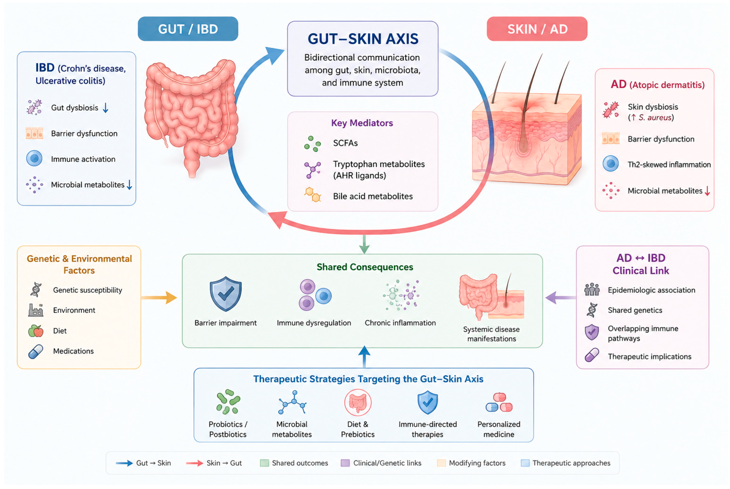
Figure 1. Proposed mechanisms linking atopic dermatitis (AD) and inflammatory bowel disease (IBD) through the gut–skin axis. The gut and skin communicate through microbial, immune, and metabolic pathways. Dysbiosis, barrier dysfunction, altered microbial metabolites, and immune dysregulation contribute to the pathogenesis of both AD and IBD. Bidirectional signaling between the gut and skin promotes chronic inflammation and may explain the epidemiologic and mechanistic association between these disorders. Targeting the gut–skin axis represents a promising therapeutic strategy for inflammatory diseases affecting both organs.
Jpm 16 00482 g001
Table 1. Epidemiologic and genetic evidence for the association between IBD and AD.
StudyPopulation/Sample SizeDesignAssociation ExaminedEffect Estimate (95% CI)Direction/Principal Finding
Schmitt et al., 2016 [74]German nationwide health-insurance cohort; 655,815 individuals ≤ 40 years, including 49,847 with ADRetrospective cohortAD → incident IBDCD: RR 1.34 (1.11–1.61); UC: RR 1.25 (1.03–1.53)Positive association; AD associated with increased risk of incident CD and UC
Egeberg et al., 2017 [72]Denmark; 7032 adults with AD vs. 3,587,974 population controlsNationwide registry-based cohortAD → incident IBDCD: HR 0.69 (0.34–1.30); UC: HR 0.94 (0.61–1.43)No significant association with incident CD or UC, despite higher baseline IBD prevalence among patients with AD
Lee et al., 2020 [75]95,291,110 participants from 10 observational studiesSystematic review and meta-analysisBidirectional AD ↔ IBDIBD → AD: OR 1.39 (1.28–1.50); AD → IBD prevalence: OR 1.35 (1.05–1.73); incident IBD: RR 1.46 (0.98–2.17)Overall bidirectional association; evidence for incident IBD was less definitive
Shi et al., 2020 [76]14 eligible observational studiesSystematic review and meta-analysis; Newcastle–Ottawa Scale used for quality assessmentBidirectional AD ↔ IBDIBD → AD: RR 1.83 (1.39–2.40); CD → AD: RR 2.06 (1.61–2.64); UC → AD: RR 1.66 (1.23–2.24); AD associated with 48% higher likelihood of IBD, 44% of CD, and 38% of UCBidirectional positive association; both IBD and its major subtypes were associated with AD
Weng et al., 2021 [73]Taiwan; 36,400 patients with AD and 364,000 matched controlsNationwide retrospective cohortAD → incident IBDCumulative IBD incidence: 0.047% vs. 0.047%; p = 0.973No significant association between AD and subsequent IBD
Gu et al., 2022 [77]IBD GWAS: 34,652 subjects; AD GWAS: 212,036 subjectsTwo-sample bidirectional Mendelian randomizationIBD ↔ ADIBD → AD: β = 0.159, p = 0.01 (IVW); reverse MR: p = 0.43Possible IBD → AD causal effect; no evidence for AD → IBD in this analysis
Meisinger & Freuer, 2022 [78]AD GWAS: 10,788 cases/30,047 controls; IBD outcomes from UK Biobank and an independent European cohortTwo-sample bidirectional Mendelian randomizationAD ↔ IBDAD → IBD: OR 1.11 (1.04–1.18) in combined analysis; IBD → AD: not significantPossible AD → IBD causal effect; opposite direction from Gu et al.
Kim et al., 2023 [79]Korea; 61 patients with IBD + AD vs. 122 matched IBD controlsMulticenter retrospective observational studyAD → IBD clinical courseAll IBD: HR 1.83 (1.02–3.27); UC: HR 3.50 (1.07–11.48); CD: HR 1.54 (0.72–3.30)AD associated with shorter biologics-free survival, particularly in UC
Chiesa Fuxench et al., 2023 [80]UK THIN; 409,431 children and 625,083 adults with AD plus matched controlsPopulation-based matched cohortAD → incident IBDChildren: IBD HR 1.44 (1.31–1.58); CD 1.74 (1.54–1.97); adults: IBD 1.34 (1.27–1.40); CD 1.36 (1.26–1.47); UC 1.32 (1.24–1.41)Positive association; risk increased with AD severity
Lerchova et al., 2024 [81]Sweden/Norway prospective birth cohorts; 83,311 childrenProspective population-based birth cohortEarly childhood AD → later IBDIBD: aHR 1.46 (1.13–1.88); CD: 1.53 (1.04–2.26); UC: 1.78 (1.15–2.75)Positive association between AD at age 3 and subsequent IBD
Joel et al., 2024 [82]US All of Us Research Program; 296,440 adultsCross-sectional observational studyAD ↔ prevalent IBDIBD: aOR 1.97 (1.75–2.21); CD: 1.91 (1.64–2.22); UC: 2.02 (1.74–2.35)Positive association with prevalent IBD, CD, and UC
Yu et al., 2024 [83]61,190,816 participants from 8 longitudinal cohort studiesMeta-analysis of longitudinal studiesAD → incident IBDIBD: OR 1.37 (1.31–1.43); CD: 1.51 (1.31–1.76); UC: 1.33 (1.13–1.56)Positive pooled association; substantial heterogeneity for CD and UC
Wan & Yang, 2025 [84]61,190,816 participants from 8 retrospective cohort studiesMeta-analysisAD → IBDIBD: OR 1.37 (1.31–1.43); CD: 1.51 (1.31–1.76); UC: 1.33 (1.13–1.56)Positive pooled association; overall effect was modest, with heterogeneity for disease subtypes
Tseng et al., 2026 [85]Taiwan; 300 incident IBD cases and 2400 matched controlsNationwide case–control studyAD → IBDOR 5.73 (1.69–19.48)Positive association; markedly elevated point estimate with wide confidence interval
Footnote: Effect estimates are reported as presented in the original studies. Because study designs, populations, outcomes, and analytic methods differed, effect estimates should not be interpreted as directly comparable across studies.
Disclaimer/Publisher’s Note: The statements, opinions and data contained in all publications are solely those of the individual author(s) and contributor(s) and not of MDPI and/or the editor(s). MDPI and/or the editor(s) disclaim responsibility for any injury to people or property resulting from any ideas, methods, instructions or products referred to in the content.


