Article

Gene Expression Profiling Provides an Improved Characterization of CD79B-Mutated Diffuse Large B-Cell Lymphomas

1
Institute of Pathology, Universitätsklinikum Erlangen, Friedrich-Alexander-Universität Erlangen-Nürnberg (FAU), Krankenhausstrasse 8-10, 91054 Erlangen, Germany
2
Institute of Pathology Kaufbeuren-Ravensburg, Julius-Probst-Strasse 4, 87600 Kaufbeuren, Germany
3
Foundation Medicine GmbH, Nonnenwald 2, 82377 Penzberg, Germany
4
Teilgemeinschaftspraxis Molekularpathologie Südbayern, Giesinger Bahnhofplatz 2, 81539 Munich, Germany
5
Institute of Pathology Rosenheim, Lilienweg 12, 83022 Rosenheim, Germany
6
Medizinisches Versorgungszentrum für Hämatologie und Onkologie Ravensburg GmbH, Elisabethenstrasse 19, 88212 Ravensburg, Germany
7
Medical Clinic and Policlinic III, Ludwig-Maximilians-Universität München, Marchioninistrasse 15, 81377 Munich, Germany
8
Institute of Pathology, Ludwig-Maximilians-Universität München, Thalkirchner Strasse 36, 80337 Munich, Germany
9
Institute of Pathology, University Hospital Salzburg, PMU Salzburg, Müllner Hauptstraße 48, A-5020 Salzburg, Austria
10
Gemeinschaftspraxis für Pathologie Traunstein, Cuno-Niggl-Strasse 3, 83278 Traunstein, Germany
*
Author to whom correspondence should be addressed.
J. Pers. Med. 2025, 15(11), 548; https://doi.org/10.3390/jpm15110548
Submission received: 8 August 2025 / Revised: 27 October 2025 / Accepted: 29 October 2025 / Published: 10 November 2025
(This article belongs to the Special Issue Molecular Pathology in Cancer Research)

Abstract

Background and Objectives: Diffuse large B-cell lymphomas (DLBCLs) are heterogeneous neoplasms. CD79B and MYD88 mutations are associated with the activated B-cell-like (ABC) subtype of DLBCL and often co-occur and lead to constitutive activation of the NF-κB pathway. Several different genetic classifications to date have recognized CD79B- and MYD88-mutated DLBCLs as a unique subtype with poor response to therapy and unfavorable survival. However, little is known about gene expression in DLBCLs with mutated CD79B (and MYD88) in comparison to their wild type counterparts. The objective of this study was to compare the gene expression in DLBCLs according to their CD79B mutational status. Methods: A total of 48 primary, treatment-naïve DLBCLs (CD79B-mutated: 35%/n = 17, CD79B-wild type: 65%/n = 31) were investigated using RNA expression profiling (770 genes), followed by immunohistochemical analysis of the up-regulated genes and survival analysis. Results: The gene expression analysis revealed that downstream of CD79B CARD11 and the NF-κB targets NFKBIZ, IL10, IL12A, PIM1 and BCL2A1 were up-regulated in CD79B-mutated DLBCLs. The strongest up-regulation was detected for ARNT2 and WNT11. Other up-regulated genes included the apoptosis-related BID and granzyme B, as well as genes of cell cycle regulation such as RUNX1, RUNX1T1 and RASGRF1. Up-regulation was also found for IL7, STAT3, MLLT4, CD14 and the HSP90B1 subunit. TP53 mutation showed an association with poorer overall survival in a secondary analysis, consistent with prior reports, while survival by CD79B/MYD88 mutation status and the differentially expressed genes showed no significant differences in this cohort. Conclusions: In conclusion, the current study identified novel up-regulated genes in CD79B-mutated DLBCLs beyond NF-κB pathway signaling, which may contribute to a better definition of potential therapeutic targets and further improves the characterization of this distinct and aggressive DLBCL subgroup.

1. Introduction

Diffuse large B-cell lymphoma (DLBCL) is the most frequent lymphoma, accounting for 40% of cases [1,2]. This class of non-Hodgkin’s lymphomas presents with manifestations in both nodal and extranodal locations and is characterized by a high degree of molecular heterogeneity that remains incompletely understood [3,4,5,6]. Based on gene expression profiling, DLBCL can be subdivided into different prognostic subgroups: germinal center B-cell-like (GCB) DLBCL, activated B-cell-like (ABC) DLBCL and unclassified [2,3,7]. Since gene arrays are costly and not practical in clinical routine, DLBCLs are often subdivided into GCB and non-GCB lymphomas using the immunohistochemical Hans classifier [8,9]. In the ABC subtype, activating mutations in CD79B and MYD88 are frequent and often occur concomitantly leading to constitutive activation of the NF-κB pathway [10,11,12,13]. NF-κB activation is considered a central mechanism in the pathogenesis of this lymphoma subtype and is associated with poor survival [10,11,14]. CD79B mutations are frequently located in the immunoreceptor tyrosine-based activation motif (ITAM) region in ABC DLBCLs, resulting in chronic activation of B-cell receptor (BCR) signaling [11]. In MYD88, an adaptor protein in interleukin-1 (IL1) and toll-like receptor signaling, the amino acid substitution L265P is predominantly found [10]. A high prevalence of CD79B and MYD88 mutations is found in extranodal DLBCLs such as in the breast, central nervous system (CNS) and testes [12,15,16,17,18,19]. Genetic subtyping of DLBCL revealed that the co-occurrence of mutations in the MYD88 and CD79B genes defines a unique molecular subtype of DLBCLs, mostly termed MCD [20,21,22,23,24]. The MCD subtype was associated with poor response to standard therapy and unfavorable survival [20,24]. CD79B mutation was identified as an unfavorable prognostic factor for DLBCL patient survival and may be an important biomarker for DLBCL disease progression [25].
However, little is known about the gene and protein expression of DLBCLs with CD79B (and MYD88) mutations in comparison to their wild type (wt) counterparts. The main objective of the current study was the comparison of gene expression of 770 cancer-associated genes in 48 primary, treatment-naïve DLBCLs according to their CD79B mutational status. For a subset of the up-regulated genes, a corresponding overexpression at the protein level was investigated by immunohistochemistry (IHC). Survival analysis was performed to assess the prognostic significance of clinical variables, mutation status and differentially expressed genes.

2. Materials and Methods

2.1. Patients

Formalin-fixed paraffin-embedded (FFPE) tissue specimens of 48 DLBCL patients were obtained from the Institutes of Pathology Kaufbeuren-Ravensburg, Rosenheim and Erlangen. All cases were newly diagnosed, primary DLBCL. The tumor tissue was collected at initial diagnosis prior to any systemic therapy. Thus, the profiled samples are treatment-naïve. Patient characteristics and first-line treatments are summarized in the supplement (Supplement Table S1). The study was conducted in accordance with the Declaration of Helsinki and was approved by the Ethics Committee of the medical faculty of the Friedrich-Alexander-Universität Erlangen-Nürnberg (Germany) (103_17 B). Informed consent was obtained from patients included in this study.

2.2. Mutation Detection Analyses

2.2.1. Mutation Detection of CD79A/B and MYD88

Three representative 7 µm sections were cut from each FFPE tissue block, deparaffinized, rehydrated in a graded ethanol series and rinsed in distilled water. FFPE sections were manually micro- or macrodissected, depending on tumor cellularity and distribution, to enrich for tumor DNA and transferred into 2 mL Eppendorf tubes. Samples were digested with Proteinase K (Roche Diagnostics GmbH, Mannheim, Germany) overnight at 56 °C on a shaking device. DNA was then purified using a column-based extraction kit (Quiagen GmbH, Hilden, Germany) and stored at 4 °C until further analysis. All tumor DNA samples were screened for mutations in the ITAM region of CD79A and CD79B as well as for the point mutation L265P of MYD88 by real-time polymerase chain reaction (real-time PCR) combined with high-resolution melting analysis (HRMA). Tumor samples with a melting curve deviating from the wild type control were further analyzed by bidirectional Sanger sequencing to verify and characterize the mutations.

2.2.2. Mutation Detection of TP53

Sample DNA was amplified with target specific primers, followed by barcode incorporation to generate barcoded libraries covering TP53. PCR was performed using the FastStartTM High Fidelity PCR-System according to the manufacturer’s instructions (Roche Diagnostics GmbH, Germany). Libraries were prepared using the Ion PGMTM Hi-QTM View Chef Kit on the Ion Chef Instrument (Thermo Fisher Scientific Inc., Waltham, MA, USA). Sequencing of multiplexed templates was performed on the Ion PGMTM System using the Ion PGMTM Hi-QTM View Sequencing Kit on Ion 314/316 chips following the manufacturer’s instructions (Thermo Fisher Scientific Inc., USA). Data analysis was performed using SeqNext (JSI medical systems GmbH, Ettenheim, Germany).

2.3. Multi-Gene Expression Analysis

At least three representative 7 µm sections were cut from the respective FFPE tissues and deparaffinized as mentioned. Following micro- or macrodissection based on tumor content, the samples were digested with Proteinase K (Roche Diagnostics GmbH, Germany) overnight at 56 °C. RNA was extracted using the RNeasy Kit according to the manufacturer’s protocol (Quiagen GmbH, Germany). DNA was digested with DNase I and after several washing steps RNA was eluted. RNA yield and purity were measured using a spectrophotometer. The RNA samples were stored at −80 °C until further analysis. Gene expression analysis was performed using the nCounter® PanCancer Pathways Panel according to the manufacturer’s instructions (NanoString Technologies Inc., Seattle, WA, USA). The panel includes 770 genes from 13 canonical pathways and selected reference genes. The input per sample was 100 ng RNA. The nCounter® Analysis System (NanoString Technologies Inc., USA) generated the raw RCC files. These files were analyzed using the nSolverTM Analysis Software 2.5 with the Advanced Analysis Module (NanoString Technologies Inc., USA). Three normal lymph nodes were used as baseline controls.

2.4. TMA and Immunohistochemistry

A tissue microarray (TMA) was constructed using 2 mm tissue cores embedded in three comparable tissue blocks. Several 2 µm sections were cut from at least two different TMA blocks and stained with hematoxylin and eosin (H&E) for morphological evaluation. Subsequent sections were deparaffinized after drying and rehydrated in a graded ethanol series. Antigen retrieval was performed by heat treatment. After the washing steps, primary antibodies were applied at the respective dilutions (Supplement Table S2). After washing, antibody binding was visualized with the ZytoChem Plus (AP) Polymer Bulk Kit for 30 min at room temperature (RT), employing Permanent AP Red (both from Zytomed Systems GmbH, Berlin, Germany) as the chromogenic substrate. Tissue sections were analyzed using an Axio Imager.A2 light microscope (Carl Zeiss AG, Oberkochen, Germany) and a MIRAX DESK slide scanner (Carl Zeiss AG, Germany) with a Pannoramic Scanner 1.22 and CaseViewer 2.4 software (3DHISTECH, Budapest, Hungary). Staining results were evaluated manually using a semi-quantitative scoring method based on the percentage of positive cells (0–100%) and the staining intensity, which was assessed using four levels: 0 = no immunoreactivity (IR); 1 = weak IR; 2 = medium IR; 3 = strong IR. For each sample, the staining intensities were multiplied by the respective percentage of positive tumor cells, resulting in an H-score ranging from 0 to 300. Protein expression was classified as negative for H-scores < 80 and positive for H-scores ≥ 80. The investigation of a corresponding protein expression for the detected up-regulated genes from the RNA expression analysis in the CD79B-mutated DLBCLs comprised the immunohistochemical analysis of granzyme B, PIM1, IL10, IL7, HSP90 and STAT3 in the lymphoma TMAs. NF-κB and p53 were also stained. Given the finite archival tissue, IHC analyses were pre-specified as exploratory and limited to a subset of up-regulated genes.

2.5. Survival Analysis

The survival analyses included univariate and multivariate examinations. The prognostic significance of clinical variables, mutation analysis results and gene expression analysis results were assessed separately. Both relapse-free survival (RFS) and overall survival (OS) were investigated using the Kaplan–Meier method (product-limit procedure), with the date of histological diagnosis as the starting point. Overall survival was defined as time to death from any cause, with censoring at last follow-up. Relapse-free survival was defined as time to first documented relapse, with censoring at death without prior relapse or at last follow-up.

2.6. Statistical Analysis

Gene expression data was analyzed using the NanoString nSolverTM Advanced Analysis Software. Genes showing a fold change of >1.5 in the comparison of the CD79B-mutated vs. non-mutated group were considered biologically relevant. Further statistical tests were conducted with SPSS Statistics 25 (IBM, Armonk, NY, USA). The association of different parameters was tested using the two-sided Fisher’s exact test. The presence of a corresponding protein overexpression for the up-regulated genes was evaluated using the one-sided Fisher’s exact test. In the survival analysis (OS and RFS) using the Kaplan–Meier method, differences between categories were tested by the log rank test. Cox regression analysis was conducted to identify independent prognostic factors. Benjamini–Yekutieli correction for multiple testing was applied to control the false discovery rate (FDR), resulting in adjusted (adj.) p-values. The significance level was α = 0.05 overall. The tables and survival figures were generated using the R 4.3.1 software (R Foundation, Vienna, Austria) in RStudio (2025.05.1+513 ‘Mariposa Orchid’, Posit PBC, Boston, MA, USA).

3. Results

3.1. Clinical Information About the DLBCL Patient Population

The patient population comprised 48 primary, treatment-naïve DLBCLs (Table 1). CD79B ITAM alterations were detected in 35% (n = 17) of DLBCLs, which mostly comprised missense mutations affecting the first tyrosine of the ITAM domain (Y196) (Supplement Table S3). In total, 65% (n = 31) of cases did not harbor a CD79B mutation. No CD79A alterations were found. A MYD88 L265P mutation was identified in 29% (n = 14) of patients. CD79B mutations frequently co-occurred with MYD88 L265P, with 71% (n = 12) of CD79B-mutated lymphomas harboring a MYD88 mutation (p < 0.05). Per Hans classifier, 54% (n = 26) of the lymphomas were categorized as non-GCB type and 42% (n = 20) as GCB type. CD79B-mutated DLBCLs were more prevalent of non-GCB type compared to GCB type—76% (n = 13) vs. 18% (n = 3). 65% (n = 31) of DLBCLs were diagnosed at an extranodal site, with 42% (n = 13) of these originating from the testicles. Notably, 59% (n = 10) of the specimens with CD79B mutations were localized in the testicles. Since p53 protein overexpression, which was detected in the CD79B-mutated group, is known to be associated with TP53 mutation, we analyzed the mutational status of TP53 in a secondary analysis [26]. In total, 25% (n = 12) of the DLBCLs showed a TP53 mutation, which were mainly missense mutations (Supplement Table S4). We could not observe an association of TP53 mutation with the MYD88 and CD79B status, nor the GCB/non-GCB type.

3.2. Gene Expression Analysis with the PanCancer Pathways Panel

In total, 48 specimens characterized by mutational analysis for CD79B and MYD88 were analyzed using the NanoString nCounter® PanCancer Pathways Panel. Profiling of RNA expression revealed several up-regulated genes in CD79B-mutated vs. non-mutated cases (adj. p < 0.05), including 18 with a biologically relevant fold change > 1.5 (Table 2).
Downstream of CD79B in the BCR signaling pathway, CARD11 was up-regulated, while NF-κB (NFKB1) RNA levels did not differ between the groups. An up-regulation of various direct NF-κB targets, such as NFKBIZ, IL10, IL12A, PIM1 and the antiapoptotic BCL2A1, was detected in the CD79B-mutated group. Further up-regulated genes involved in apoptosis were BID and granzyme B. The differential expression of many genes not considered as NF-κB targets was also found in the CD79B-mutated group, with the overall strongest up-regulation for ARNT2 and WNT11 as well as genes involved in cell cycle regulation such as RUNX1, RUNX1T1 and RASGRF1. Other up-regulated genes included IL7, STAT3, MLLT4, CD14 and the HSP90B1 subunit. TP53 showed a significant differential expression, but its fold change did not reach the threshold of 1.5. In addition, 26 down-regulated genes were identified (Supplement Table S5).

3.3. Immunohistochemical Analysis

Immunohistochemical analysis was performed for a subset of the up-regulated genes identified in the RNA expression analysis to investigate whether corresponding protein overexpression could be detected. A significant difference at the protein level between the CD79B-mutated and wild type groups was observed only in the initial analysis for p53 (p < 0.05), which was weakly up-regulated at the RNA level (Figure 1). However, this difference did not remain significant after correction for multiple testing. Granzyme B was expressed independently of the CD79B mutation status in a non-neoplastic T-cell subpopulation rather than lymphoma cancer cells.

3.4. Survival Outcomes

The survival cohort comprised 85.4% (n = 41) of the overall 48 patients. A total of 14.6% (n = 7) were excluded due to missing survival data. Descriptive survival metrics are summarized in the supplement (Supplement Table S6). We investigated the associations of various clinical and molecular parameters related to RFS and OS using the log rank test. Clinical variables, the mutation status of CD79B and MYD88, as well as the detected up- and down-regulated genes were not associated with worse OS and RFS (Supplement Figure S1). Significantly shortened OS was observed for patients with a TP53 mutation in a secondary analysis (adj. p < 0.05) (Figure 2). No independent prognostic factors were identified by Cox regression analysis.

4. Discussion

DLBCL is a non-Hodgkin’s lymphoma characterized by molecular heterogeneity [4,5,6]. DLBCLs can be subdivided into prognostic subgroups based on the cell of origin, the so-called GCB DLBCL, ABC DLBCL and unclassified cases [2,3,7]. The ABC subtype frequently harbors activating mutations in CD79B and MYD88, resulting in constitutive activation of the NF-κB pathway [10,11,12,13,14].
CD79B mutation was recently identified as an unfavorable prognostic factor for DLBCL patient survival and may be an important biomarker for DLBCL disease progression [25]. However, little is known about the gene and protein expression of DLBCLs with CD79B (and MYD88) mutations in comparison to their wild type counterparts. The current study compared the gene expression of 770 cancer-associated genes between a CD79B-mutated and a CD79B wild type primary, treatment-naïve DLBCL population. Most of the specimens with a CD79B mutation carried a concomitant MYD88 mutation and were classified as non-GCB lymphomas, which is in concordance to previous studies [10,11]. The majority of CD79B-mutated lymphomas were localized in the testicles. Thus, this DLBCL group was characterized by a large proportion of entities from immunoprivileged sites. We already reported a high frequency of CD79B and MYD88 mutations in testicular DLBCLs in a previous study [15].
RNA expression analysis detected, for several genes, a differential expression between CD79B-mutated and CD79B wild type cases, whereby we focused on the up-regulated genes. An up-regulation was found for CARD11, a downstream effector of CD79B in the BCR pathway, which is involved in the oncogenic activation of NF-κB in DLBCL [11,27]. Although the level of NF-κB (NFKB1) up-regulation in the CD79B-mutated group turned out to be minimal, we observed an altered gene expression of several direct NF-κB targets (NFKBIZ, IL10, IL12A, PIM1 and BCL2A1) [28,29,30,31,32].
A strong up-regulation was observed for NFKBIZ, the gene encoding the atypical nuclear IκB family member IκBζ, which is indispensable for nuclear NF-κB activity in ABC DLBCLs and therefore for the survival of this DLBCL subtype [33]. The important role of the cytokine IL10 in the survival and proliferation of ABC DLBCLs has already been described, in particular via STAT3 signaling [34,35]. STAT3 is a transcription factor, which is frequently constitutively activated in cancer as in ABC DLBCLs, involved in many central oncogenic processes and its gene was also up-regulated in the CD79B-mutated group [35,36,37]. Furthermore, it has been suggested that these three molecules form an autoregulatory feed-forward loop in ABC-DLBCLs, since all three proteins have been described as being overexpressed in this lymphoma subtype [33,35,37,38]. According to the literature, the constitutively active NF-κB pathway, as a result of a BCR signaling pathway mutation (e.g., CD79B), up-regulates IκBζ followed by the activation of a NF-κB target gene subset, including IL10 [33,38]. Autocrine signaling mediated by this cytokine leads to the activation of STAT3, which in turn presumably promotes IκBζ expression, resulting in a protumorigenic effect of the malignant ABC DLBCL cells by sustaining the oncogenic NF-κB activity in the nucleus [35,38]. In addition to the concomitant up-regulation of the three genes involved in our study, further research is required to prove the existence and function of this feed-forward cycle or to investigate other roles and regulatory mechanisms of these genes in CD79B-mutated DLBCLs.
PIM1 encodes a serine/threonine kinase that is known to be frequently mutated and overexpressed in DLBCL and plays an important protumorigenic role in this lymphoma [20,31,39,40]. PIM1 was up-regulated in CD79B-mutated DLBCLs vs. CD79B wild type DLBCLs, indicating an important role of PIM1 for CD79B-mutated DLBCLs. This is consistent with the characteristic mutations of the PIM1 proto-oncogene together with CD79B and MYD88 in the MCD subtype [21,23].
Our study also showed that the NF-κB target gene BCL2A1, an anti-apoptotic member of the BCL-2 family already known to be overexpressed in DLBCL, was up-regulated in CD79B-mutated DLBCLs [32,41,42]. BCL2A1 overexpression induces a protumorigenic and chemoresistant function in malignant cells and represents an attractive target in cancer [42].
We detected the up-regulation of further tumor-associated genes that are not NF-κB targets. The most strongly up-regulated gene in the CD79B-mutated group was ARNT2. ARNT2 is a transcription factor of the basic helix–loop–helix/Per-ARNT-SIM (bHLH/PAS) family, which is mainly expressed in the kidney and CNS [43,44]. The function and expression of ARNT2 in cancer seems to be tissue-dependent, and the underlying mechanisms are yet to be determined. One feature observed in hypoxic conditions, as found in the microenvironment of aggressive tumors, was the binding of the transcription factor to hypoxia-inducible factor (HIF)-1α in the nucleus, and this heterodimeric complex induced oncogenic processes in order to promote tumor growth [44,45,46]. A hypoxia-mediated activation leading to cancer progression was reported for WNT11, the second most up-regulated gene in the CD79B-mutated group [47]. This induction has been described as a result of the heterodimerization of HIF-1α with the ARNT2 homolog ARNT [47]. Although HIF-1α complexed with either ARNT or ARNT2 has been shown to promote similar gene expression profiles in hypoxic conditions, a corresponding mechanism of WNT11 activation via ARNT2 was not examined [46,47]. Neither ARNT2 nor WNT11 have, to our knowledge, been investigated in DLBCL so far. Given that they were the two most up-regulated genes in the CD79B-mutated DLBCL group in our study, further investigations are needed to determine whether they are involved in CD79B-mutated DLBCL development either separately or via a (HIF-1α)-ARNT2-mediated WNT11 activation and whether this feature can be used as a new therapeutic target in this lymphoma group.
IL7, MLLT4, RASGRF1, HSP90B1, RUNX1T1 and RUNX1 were up-regulated in CD79B-mutated DLBCLs. However, their potential involvement in DLBCL, to our knowledge, remains to be elucidated. As these genes were associated with other hematological and non-hematological cancers, further research is required to assess their oncogenic relevance in the CD79B-mutated group.
In survival analysis, TP53 mutation was significantly associated with worse OS. This finding regarding the tumor suppressor gene is consistent with the results of several studies of DLBCL patients [26,48,49,50,51].
A limitation of the study is that the investigation of a corresponding protein overexpression for the up-regulated genes in the CD79B-mutated group was limited to immunohistochemical analysis of a small subset of the gene expression profile. Thus, only preliminary conclusions can be drawn about the protein expression profile in CD79B-mutated DLBCLs, requiring further studies in this regard.

5. Conclusions

In conclusion, the obtained gene expression profile of CD79B-mutated DLBCLs improves the characterization of this lymphoma subpopulation and contributes to a better understanding of the complex molecular heterogeneity of DLBCL. The results confirm the established characteristics of DLBCL and identify novel up-regulated genes in relation to CD79B mutation status. Further studies on independent, larger cohorts are required to investigate the relevance of these previously unrecognized genes in this context, potentially uncovering new therapeutic targets.

Supplementary Materials

The following supporting information can be downloaded at https://www.mdpi.com/article/10.3390/jpm15110548/s1: Table S1: Clinical information about the patient population; Table S2: Primary antibodies used in this study for immunohistochemistry; Table S3: Detected mutations in the CD79B gene; Table S4: Detected mutations in the TP53 gene; Table S5: Down-regulated genes in CD79B-mutated DLBCL compared to CD79B wild type DLBCL; Table S6: Descriptive survival metrics; Figure S1: Survival analysis. Kaplan–Meier curves for CD79B and MYD88 mutation.

Author Contributions

Conceptualization, L.G., S.S.-F. and A.G.; data curation, A.G.; formal analysis, L.G., W.J., M.B., S.S.-F., K.S., H.B., J.H. and A.G.; funding acquisition, S.S.-F. and A.G.; investigation, L.G., W.J., M.B., K.S. and H.B.; methodology, L.G., W.J., M.B., S.S.-F., T.D. (Tobias Dechow), T.D. (Thomas Decker), M.D. and A.G.; project administration, A.G.; resources, S.B., T.R., A.H. and A.G.; supervision, M.B. and S.S.-F.; validation, W.J. and A.G.; visualization, L.G., W.J. and A.G.; writing—original draft, L.G.; writing—review and editing, W.J., S.B., S.S.-F., T.R., T.D. (Tobias Dechow), T.D. (Thomas Decker), M.D., A.H., J.H. and A.G. All authors have read and agreed to the published version of the manuscript.

Funding

This research was supported by a grant of Novartis Oncology Germany to A.G. and S.S.-F., HINC-124.

Institutional Review Board Statement

The study was conducted in accordance with the Declaration of Helsinki (6th revision 2008) and approved by the Ethics Committee of the medical faculty of the Friedrich-Alexander-Universität Erlangen-Nürnberg (Germany) (103_17 B; approved on 9 May 2017).

Informed Consent Statement

Informed consent was obtained from all subjects involved in the study.

Data Availability Statement

The datasets generated and analyzed during the current study are available from the corresponding author on reasonable request.

Acknowledgments

The present work was performed in partial fulfillment of the requirements for obtaining the degree ‘Dr. rer. biol. hum.’ for Luis Grossmann at the Friedrich-Alexander-Universität Erlangen-Nürnberg (FAU). The authors thank Rudolf Jung for TMA production, Daniel Hofmann for the NanoString preparation and Irina Reicherd, Alexandra Werner and Vanessa Grage for technical support.

Conflicts of Interest

Luis Grossmann is a part-time employee of Foundation Medicine GmbH, a wholly owned subsidiary of Roche Holding AG. Tobias Dechow is a full-time employee of Medizinisches Versorgungszentrum für Hämatologie und Onkologie Ravensburg GmbH. Thomas Decker is a full-time employee of Medizinisches Versorgungszentrum für Hämatologie und Onkologie Ravensburg GmbH. Thomas Decker has received speaker honoraria from Novartis and has served on scientific advisory boards for Novartis. Martin Dreyling has received speaker honoraria from AstraZeneca, BeiGene, BMS, Kite/Gilead, Janssen, Lilly and Roche; has served on scientific advisory boards for AbbVie, AstraZeneca, AvenCell, BeiGene, BMS, Genmab, Kite/Gilead, Incyte, Janssen, Lilly/Loxo, Novartis, Roche and SOBI; and has received institutional research funding from AbbVie, Kite/Gilead, Janssen, Lilly and Roche. Julius Honecker is a full-time employee of Foundation Medicine GmbH, a wholly owned subsidiary of Roche Holding AG. Neither the funders nor any company have had any role in the design, conduct, interpretation, or writing of the study.

References

  1. Pileri, S.A.; Tripodo, C.; Melle, F.; Motta, G.; Tabanelli, V.; Fiori, S.; Vegliante, M.C.; Mazzara, S.; Ciavarella, S.; Derenzini, E. Predictive and Prognostic Molecular Factors in Diffuse Large B-Cell Lymphomas. Cells 2021, 10, 675. [Google Scholar] [CrossRef]
  2. Shi, Y.; Xu, Y.; Shen, H.; Jin, J.; Tong, H.; Xie, W. Advances in biology, diagnosis and treatment of DLBCL. Ann. Hematol. 2024, 103, 3315–3334. [Google Scholar] [CrossRef] [PubMed]
  3. Sehn, L.H.; Salles, G. Diffuse Large B-Cell Lymphoma. N. Engl. J. Med. 2021, 384, 842–858. [Google Scholar] [CrossRef]
  4. Cutmore, N.H.; Krupka, J.A.; Hodson, D.J. Genetic Profiling in Diffuse Large B-Cell Lymphoma: The Promise and the Challenge. Mod. Pathol. 2023, 36, 100007. [Google Scholar] [CrossRef] [PubMed]
  5. Kurz, K.S.; Ott, M.; Kalmbach, S.; Steinlein, S.; Kalla, C.; Horn, H.; Ott, G.; Staiger, A.M. Large B-Cell Lymphomas in the 5th Edition of the WHO-Classification of Haematolymphoid Neoplasms-Updated Classification and New Concepts. Cancers 2023, 15, 2285. [Google Scholar] [CrossRef]
  6. Berhan, A.; Almaw, A.; Damtie, S.; Solomon, Y. Diffuse large B cell lymphoma (DLBCL): Epidemiology, pathophysiology, risk stratification, advancement in diagnostic approaches and prospects: Narrative review. Discov. Oncol. 2025, 16, 184. [Google Scholar] [CrossRef]
  7. Alaggio, R.; Amador, C.; Anagnostopoulos, I.; Attygalle, A.D.; Araujo, I.B.O.; Berti, E.; Bhagat, G.; Borges, A.M.; Boyer, D.; Calaminici, M.; et al. The 5th edition of the World Health Organization Classification of Haematolymphoid Tumours: Lymphoid Neoplasms. Leukemia 2022, 36, 1720–1748. [Google Scholar] [CrossRef]
  8. Alig, S.K.; Chapuy, B.; Ennishi, D.; Dunleavy, K.; Hodson, D.J. Evolving molecular classification of aggressive B-cell lymphoma. Histopathology 2025, 86, 94–105. [Google Scholar] [CrossRef]
  9. Chan, A.; Dogan, A. Prognostic and Predictive Biomarkers in Diffuse Large B-cell Lymphoma. Surg. Pathol. Clin. 2019, 12, 699–707. [Google Scholar] [CrossRef]
  10. Ngo, V.N.; Young, R.M.; Schmitz, R.; Jhavar, S.; Xiao, W.; Lim, K.H.; Kohlhammer, H.; Xu, W.; Yang, Y.; Zhao, H.; et al. Oncogenically active MYD88 mutations in human lymphoma. Nature 2011, 470, 115–119. [Google Scholar] [CrossRef]
  11. Davis, R.E.; Ngo, V.N.; Lenz, G.; Tolar, P.; Young, R.M.; Romesser, P.B.; Kohlhammer, H.; Lamy, L.; Zhao, H.; Yang, Y.; et al. Chronic active B-cell-receptor signalling in diffuse large B-cell lymphoma. Nature 2010, 463, 88–92. [Google Scholar] [CrossRef]
  12. Visco, C.; Tanasi, I.; Quaglia, F.M.; Ferrarini, I.; Fraenza, C.; Krampera, M. Oncogenic Mutations of MYD88 and CD79B in Diffuse Large B-Cell Lymphoma and Implications for Clinical Practice. Cancers 2020, 12, 2913. [Google Scholar] [CrossRef]
  13. Guo, Q.; Jin, Y.; Chen, X.; Ye, X.; Shen, X.; Lin, M.; Zeng, C.; Zhou, T.; Zhang, J. NF-kappaB in biology and targeted therapy: New insights and translational implications. Signal Transduct. Target. Ther. 2024, 9, 53. [Google Scholar] [CrossRef]
  14. Davis, R.E.; Brown, K.D.; Siebenlist, U.; Staudt, L.M. Constitutive nuclear factor kappaB activity is required for survival of activated B cell-like diffuse large B cell lymphoma cells. J. Exp. Med. 2001, 194, 1861–1874. [Google Scholar] [CrossRef] [PubMed]
  15. Frick, M.; Bettstetter, M.; Bertz, S.; Schwarz-Furlan, S.; Hartmann, A.; Richter, T.; Lenze, D.; Hummel, M.; Dreyling, M.; Lenz, G.; et al. Mutational frequencies of CD79B and MYD88 vary greatly between primary testicular DLBCL and gastrointestinal DLBCL. Leuk. Lymphoma 2018, 59, 1260–1263. [Google Scholar] [CrossRef]
  16. Franco, F.; Gonzalez-Rincon, J.; Lavernia, J.; Garcia, J.F.; Martin, P.; Bellas, C.; Piris, M.A.; Pedrosa, L.; Miramon, J.; Gomez-Codina, J.; et al. Mutational profile of primary breast diffuse large B-cell lymphoma. Oncotarget 2017, 8, 102888–102897. [Google Scholar] [CrossRef] [PubMed]
  17. Roschewski, M.; Phelan, J.D.; Jaffe, E.S. Primary large B-cell lymphomas of immune-privileged sites. Blood 2024, 144, 2593–2603. [Google Scholar] [CrossRef] [PubMed]
  18. Rivas-Delgado, A.; Lopez, C.; Clot, G.; Nadeu, F.; Grau, M.; Frigola, G.; Bosch-Schips, J.; Radke, J.; Ishaque, N.; Alcoceba, M.; et al. Testicular large B-cell lymphoma is genetically similar to PCNSL and distinct from nodal DLBCL. Hemasphere 2024, 8, e70024. [Google Scholar] [CrossRef]
  19. Schrader, A.M.R.; Jansen, P.M.; Willemze, R.; Vermeer, M.H.; Cleton-Jansen, A.M.; Somers, S.F.; Veelken, H.; van Eijk, R.; Kraan, W.; Kersten, M.J.; et al. High prevalence of MYD88 and CD79B mutations in intravascular large B-cell lymphoma. Blood 2018, 131, 2086–2089. [Google Scholar] [CrossRef]
  20. Schmitz, R.; Wright, G.W.; Huang, D.W.; Johnson, C.A.; Phelan, J.D.; Wang, J.Q.; Roulland, S.; Kasbekar, M.; Young, R.M.; Shaffer, A.L.; et al. Genetics and Pathogenesis of Diffuse Large B-Cell Lymphoma. N. Engl. J. Med. 2018, 378, 1396–1407. [Google Scholar] [CrossRef]
  21. Shen, R.; Fu, D.; Dong, L.; Zhang, M.C.; Shi, Q.; Shi, Z.Y.; Cheng, S.; Wang, L.; Xu, P.P.; Zhao, W.L. Simplified algorithm for genetic subtyping in diffuse large B-cell lymphoma. Signal Transduct. Target. Ther. 2023, 8, 145. [Google Scholar] [CrossRef]
  22. Wright, G.W.; Huang, D.W.; Phelan, J.D.; Coulibaly, Z.A.; Roulland, S.; Young, R.M.; Wang, J.Q.; Schmitz, R.; Morin, R.D.; Tang, J.; et al. A Probabilistic Classification Tool for Genetic Subtypes of Diffuse Large B Cell Lymphoma with Therapeutic Implications. Cancer Cell 2020, 37, 551–568.e514. [Google Scholar] [CrossRef]
  23. Lacy, S.E.; Barrans, S.L.; Beer, P.A.; Painter, D.; Smith, A.G.; Roman, E.; Cooke, S.L.; Ruiz, C.; Glover, P.; Van Hoppe, S.J.L.; et al. Targeted sequencing in DLBCL, molecular subtypes, and outcomes: A Haematological Malignancy Research Network report. Blood 2020, 135, 1759–1771. [Google Scholar] [CrossRef]
  24. Chen, R.; Zhou, D.; Wang, L.; Zhu, L.; Ye, X. MYD88(L265P) and CD79B double mutations type (MCD type) of diffuse large B-cell lymphoma: Mechanism, clinical characteristics, and targeted therapy. Ther. Adv. Hematol. 2022, 13, 20406207211072839. [Google Scholar] [CrossRef]
  25. Xu, P.P.; Shen, R.; Shi, Z.Y.; Cheng, S.; Wang, L.; Liu, Y.; Zhang, L.; Huang, R.; Ma, X.; Wu, X.; et al. The Prognostic Significance of CD79B Mutation in Diffuse Large B-Cell Lymphoma: A Meta-analysis and Systematic Literature Review. Clin. Lymphoma Myeloma Leuk. 2022, 22, e1051–e1058.e1. [Google Scholar] [CrossRef]
  26. Xu-Monette, Z.Y.; Wu, L.; Visco, C.; Tai, Y.C.; Tzankov, A.; Liu, W.M.; Montes-Moreno, S.; Dybkaer, K.; Chiu, A.; Orazi, A.; et al. Mutational profile and prognostic significance of TP53 in diffuse large B-cell lymphoma patients treated with R-CHOP: Report from an International DLBCL Rituximab-CHOP Consortium Program Study. Blood 2012, 120, 3986–3996. [Google Scholar] [CrossRef]
  27. Tkachenko, A.; Kupcova, K.; Havranek, O. B-Cell Receptor Signaling and Beyond: The Role of Igalpha (CD79a)/Igbeta (CD79b) in Normal and Malignant B Cells. Int. J. Mol. Sci. 2023, 25, 10. [Google Scholar] [CrossRef] [PubMed]
  28. Ito, T.; Morimatsu, M.; Oonuma, T.; Shiina, T.; Kitamura, H.; Syuto, B. Transcriptional regulation of the MAIL gene in LPS-stimulated RAW264 mouse macrophages. Gene 2004, 342, 137–143. [Google Scholar] [CrossRef] [PubMed]
  29. Cao, S.; Zhang, X.; Edwards, J.P.; Mosser, D.M. NF-kappaB1 (p50) homodimers differentially regulate pro- and anti-inflammatory cytokines in macrophages. J. Biol. Chem. 2006, 281, 26041–26050. [Google Scholar] [CrossRef] [PubMed]
  30. Koch, P.D.; Pittet, M.J.; Weissleder, R. The chemical biology of IL-12 production via the non-canonical NFkB pathway. RSC Chem. Biol. 2020, 1, 166–176. [Google Scholar] [CrossRef]
  31. Zhu, N.; Ramirez, L.M.; Lee, R.L.; Magnuson, N.S.; Bishop, G.A.; Gold, M.R. CD40 signaling in B cells regulates the expression of the Pim-1 kinase via the NF-kappa B pathway. J. Immunol. 2002, 168, 744–754. [Google Scholar] [CrossRef]
  32. Zong, W.X.; Edelstein, L.C.; Chen, C.; Bash, J.; Gelinas, C. The prosurvival Bcl-2 homolog Bfl-1/A1 is a direct transcriptional target of NF-kappaB that blocks TNFalpha-induced apoptosis. Genes. Dev. 1999, 13, 382–387. [Google Scholar] [CrossRef] [PubMed]
  33. Nogai, H.; Wenzel, S.S.; Hailfinger, S.; Grau, M.; Kaergel, E.; Seitz, V.; Wollert-Wulf, B.; Pfeifer, M.; Wolf, A.; Frick, M.; et al. IkappaB-zeta controls the constitutive NF-kappaB target gene network and survival of ABC DLBCL. Blood 2013, 122, 2242–2250. [Google Scholar] [CrossRef] [PubMed]
  34. Beguelin, W.; Sawh, S.; Chambwe, N.; Chan, F.C.; Jiang, Y.; Choo, J.W.; Scott, D.W.; Chalmers, A.; Geng, H.; Tsikitas, L.; et al. IL10 receptor is a novel therapeutic target in DLBCLs. Leukemia 2015, 29, 1684–1694. [Google Scholar] [CrossRef] [PubMed]
  35. Lam, L.T.; Wright, G.; Davis, R.E.; Lenz, G.; Farinha, P.; Dang, L.; Chan, J.W.; Rosenwald, A.; Gascoyne, R.D.; Staudt, L.M. Cooperative signaling through the signal transducer and activator of transcription 3 and nuclear factor-kappaB pathways in subtypes of diffuse large B-cell lymphoma. Blood 2008, 111, 3701–3713. [Google Scholar] [CrossRef]
  36. Zhu, F.; Wang, K.B.; Rui, L. STAT3 Activation and Oncogenesis in Lymphoma. Cancers 2019, 12, 19. [Google Scholar] [CrossRef]
  37. Ding, B.B.; Yu, J.J.; Yu, R.Y.; Mendez, L.M.; Shaknovich, R.; Zhang, Y.; Cattoretti, G.; Ye, B.H. Constitutively activated STAT3 promotes cell proliferation and survival in the activated B-cell subtype of diffuse large B-cell lymphomas. Blood 2008, 111, 1515–1523. [Google Scholar] [CrossRef]
  38. Krappmann, D. Shaping oncogenic NF-kappaB activity in the nucleus. Blood 2013, 122, 2146–2147. [Google Scholar] [CrossRef]
  39. Zhang, H.; Lu, Y.; Zhang, T.; Guan, Q.; Wang, X.; Guo, Y.; Li, L.; Qiu, L.; Qian, Z.; Zhou, S.; et al. PIM1 genetic alterations associated with distinct molecular profiles, phenotypes and drug responses in diffuse large B-cell lymphoma. Clin. Transl. Med. 2022, 12, e808. [Google Scholar] [CrossRef]
  40. Wang, C.; Chen, Q.; Luo, H.; Chen, R. Role and mechanism of PIM family in the immune microenvironment of diffuse large B cell lymphoma. World J. Surg. Oncol. 2023, 21, 76. [Google Scholar] [CrossRef]
  41. Mahadevan, D.; Spier, C.; Della Croce, K.; Miller, S.; George, B.; Riley, C.; Warner, S.; Grogan, T.M.; Miller, T.P. Transcript profiling in peripheral T-cell lymphoma, not otherwise specified, and diffuse large B-cell lymphoma identifies distinct tumor profile signatures. Mol. Cancer Ther. 2005, 4, 1867–1879. [Google Scholar] [CrossRef]
  42. Vogler, M. BCL2A1: The underdog in the BCL2 family. Cell Death Differ. 2012, 19, 67–74. [Google Scholar] [CrossRef]
  43. Hirose, K.; Morita, M.; Ema, M.; Mimura, J.; Hamada, H.; Fujii, H.; Saijo, Y.; Gotoh, O.; Sogawa, K.; Fujii-Kuriyama, Y. cDNA cloning and tissue-specific expression of a novel basic helix-loop-helix/PAS factor (Arnt2) with close sequence similarity to the aryl hydrocarbon receptor nuclear translocator (Arnt). Mol. Cell Biol. 1996, 16, 1706–1713. [Google Scholar] [CrossRef] [PubMed]
  44. Maltepe, E.; Keith, B.; Arsham, A.M.; Brorson, J.R.; Simon, M.C. The role of ARNT2 in tumor angiogenesis and the neural response to hypoxia. Biochem. Biophys. Res. Commun. 2000, 273, 231–238. [Google Scholar] [CrossRef] [PubMed]
  45. Harris, A.L. Hypoxia—A key regulatory factor in tumour growth. Nat. Rev. Cancer 2002, 2, 38–47. [Google Scholar] [CrossRef] [PubMed]
  46. Sekine, H.; Mimura, J.; Yamamoto, M.; Fujii-Kuriyama, Y. Unique and overlapping transcriptional roles of arylhydrocarbon receptor nuclear translocator (Arnt) and Arnt2 in xenobiotic and hypoxic responses. J. Biol. Chem. 2006, 281, 37507–37516. [Google Scholar] [CrossRef]
  47. Mori, H.; Yao, Y.; Learman, B.S.; Kurozumi, K.; Ishida, J.; Ramakrishnan, S.K.; Overmyer, K.A.; Xue, X.; Cawthorn, W.P.; Reid, M.A.; et al. Induction of WNT11 by hypoxia and hypoxia-inducible factor-1alpha regulates cell proliferation, migration and invasion. Sci. Rep. 2016, 6, 21520. [Google Scholar] [CrossRef]
  48. Landsburg, D.J.; Morrissette, J.J.; Nasta, S.D.; Barta, S.K.; Schuster, S.J.; Svoboda, J.; Chong, E.A.; Bagg, A. TP53 mutations predict for poor outcomes in patients with newly diagnosed aggressive B-cell lymphomas in the current era. Blood Adv. 2023, 7, 7243–7253. [Google Scholar] [CrossRef]
  49. Zenz, T.; Kreuz, M.; Fuge, M.; Klapper, W.; Horn, H.; Staiger, A.M.; Winter, D.; Helfrich, H.; Huellein, J.; Hansmann, M.L.; et al. TP53 mutation and survival in aggressive B cell lymphoma. Int. J. Cancer 2017, 141, 1381–1388. [Google Scholar] [CrossRef]
  50. Chiappella, A.; Diop, F.; Agostinelli, C.; Novo, M.; Nassi, L.; Evangelista, A.; Ciccone, G.; Di Rocco, A.; Martelli, M.; Melle, F.; et al. Prognostic impact of TP53 mutation in newly diagnosed diffuse large B-cell lymphoma patients treated in the FIL-DLCL04 trial. Br. J. Haematol. 2022, 196, 1184–1193. [Google Scholar] [CrossRef]
  51. Qin, Y.; Chen, H.; Liu, P.; Zhang, C.; Yang, J.; Gui, L.; He, X.; Zhou, L.; Zhou, S.; Jiang, S.; et al. Prognostic value of BCL2 and TP53 genetic alterations for diffuse large B-cell lymphoma patients treated with R-CHOP. Cancer Biol. Med. 2021, 19, 893–909. [Google Scholar] [CrossRef] [PubMed]
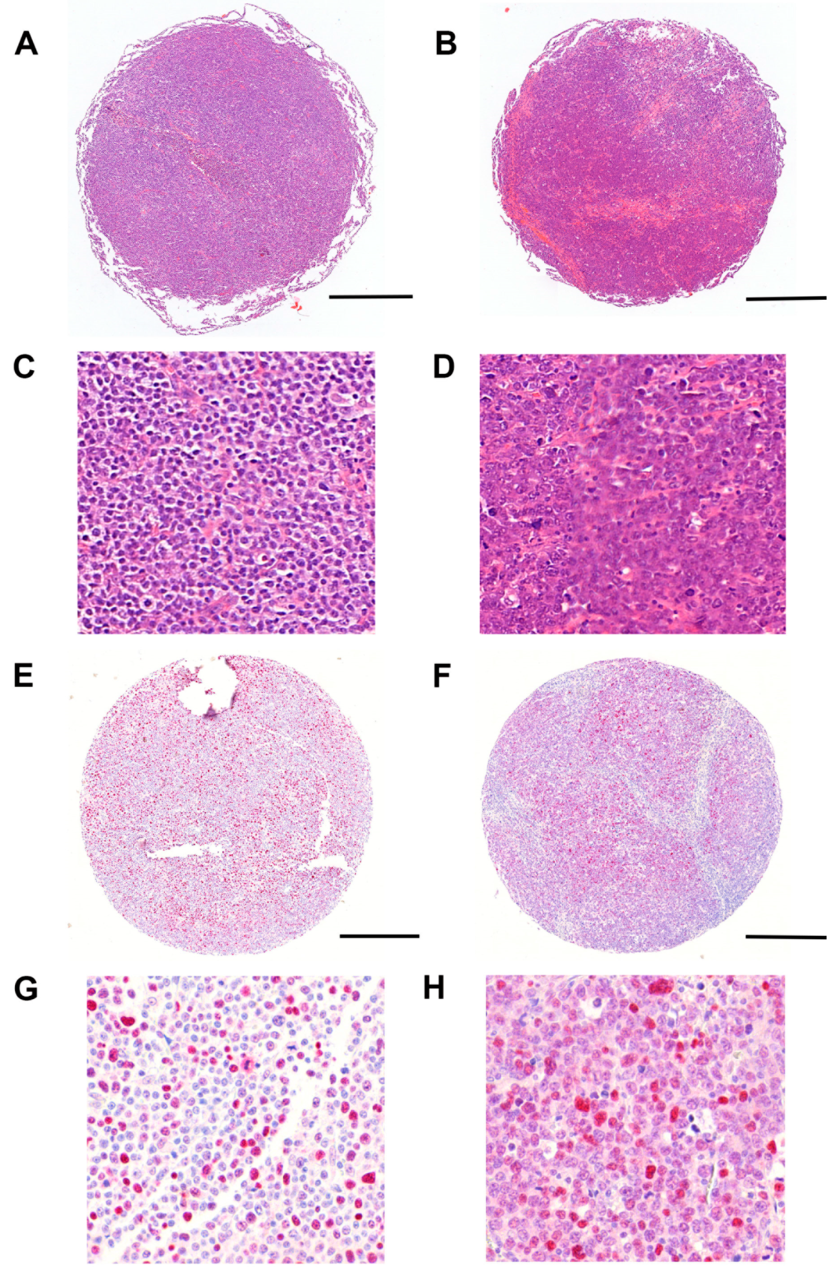
Figure 1. Representative H&E staining and p53 immunohistochemistry in CD79B-mutated and CD79B wild type (wt) DLBCLs. Low-magnification overviews of TMA cores are shown in (A) (wt, H&E), (B) (mutated, H&E), (E) (wt, p53 IHC) and (F) (mutated, p53 IHC). Corresponding high-magnification views are shown in (C) (for (A)); (D) (for (B)); (G) (for (E)) and (H) (for (F)). The cores demonstrated dense infiltrates of blastic lymphoma cells in both CD79B wild type (A,C) and mutated (B,D) DLBCLs. p53 protein expression was higher in CD79B-mutated cases (F,H) compared with wild type (E,G) (p < 0.05). This difference did not remain significant after correction for multiple testing. Scale bars: 500 µm (A,B,E,F; 5×) and 120 µm (C,D,G,H; 19.2×).
Jpm 15 00548 g001
Figure 2. Survival analysis. Survival analysis revealed that TP53 mutations were associated with a significantly worse overall survival (adj. p < 0.05).
Jpm 15 00548 g002
Table 1. Clinical information about the 48 DLBCL patients and detailed information about the CD79B-mutated and wild type groups.
Clinical Parameter Overall (n = 48)Number (%)
CD79
  CD79B-mutated17 (35%)
  CD79A-mutated0 (0%)
  Wild type31 (65%)
MYD88
  Mutated (L265P)14 (29%)
  Wild type32 (67%)
  Not informative2 (4%)
Maturation
  GCB type20 (42%)
  Non-GCB type26 (54%)
  Not informative2 (4%)
Location
  Nodal17 (35%)
  Extranodal31 (65%)
  Testicular13 (42%)
TP53
  Mutated12 (25%)
  Wild type32 (67%)
  Not informative4 (8%)
Clinical ParameterCD79B-Mutated
DLBCLs (n = 17)
CD79B Wild Type
DLBCLs (n = 31)
Number (%)Number (%)
MYD88
  Mutated (L265P)12 (71%)2 (6%)
  Wild type4 (23%)28 (90%)
  Not informative1 (6%)1 (3%)
Maturation
  GCB type3 (18%)17 (55%)
  Non-GCB type13 (76%)13 (42%)
  Not informative1 (6%)1 (3%)
Location
  Testicular10 (59%)3 (10%)
  Non-testicular7 (41%)28 (90%)
Table 2. Up-regulated genes in CD79B-mutated DLBCLs compared to CD79B wild type DLBCLs.
GeneDescriptionFold ChangeAdj. p-Value
ARNT2aryl-hydrocarbon receptor nuclear translocator 24.780.0001
WNT11wingless-type MMTV integration site family, member 113.040.0072
GZMBgranzyme B (granzyme 2, cytotoxic T-lymphocyte-associated serine esterase 1)2.890.0046
IL10interleukin 102.700.0024
IL12Ainterleukin 12A (natural killer cell stimulatory factor 1, cytotoxic lymphocyte maturation factor 1)2.620.0003
NFKBIZnuclear factor of kappa light polypeptide gene enhancer in B-cells inhibitor, zeta2.610.0001
RASGRF1Ras protein-specific guanine nucleotide-releasing factor 12.480.0011
IL7interleukin 72.150.0001
PIM1pim-1 oncogene2.110.0002
RUNX1T1runt-related transcription factor 1; translocated to, 1 (cyclin D-related)2.000.0406
CARD11caspase recruitment domain family, member 111.960.0034
BCL2A1BCL2-related protein A11.770.0307
STAT3signal transducer and activator of transcription 3 (acute-phase response factor)1.760.0001
HSP90B1heat shock protein 90 kDa beta (Grp94), member 11.760.003
MLLT4myeloid/lymphoid or mixed-lineage leukemia (trithorax homolog, Drosophila); translocated to, 41.620.0032
RUNX1runt-related transcription factor 11.590.0005
CD14CD14 molecule1.570.034
BIDBH3 interacting domain death agonist1.540.0054
TP53tumor protein p531.370.022
NFKB1nuclear factor of kappa light polypeptide gene enhancer in B-cells 11.250.0352
Disclaimer/Publisher’s Note: The statements, opinions and data contained in all publications are solely those of the individual author(s) and contributor(s) and not of MDPI and/or the editor(s). MDPI and/or the editor(s) disclaim responsibility for any injury to people or property resulting from any ideas, methods, instructions or products referred to in the content.