Review

Do Patients with Benign Paroxysmal Positional Vertigo Have a Higher Prevalence of Osteoporosis? A Systematic Review and Meta-Analysis

1
Department of Orthopaedic Surgery, Asan Medical Center, University of Ulsan College of Medicine, Seoul 05505, Republic of Korea
2
Department of Otorhinolaryngology-Head and Neck Surgery, Hallym University Kangnam Sacred Heart Hospital, Hallym University College of Medicine, Seoul 07442, Republic of Korea
*
Author to whom correspondence should be addressed.
J. Pers. Med. 2024, 14(3), 303; https://doi.org/10.3390/jpm14030303
Submission received: 8 February 2024 / Revised: 4 March 2024 / Accepted: 11 March 2024 / Published: 13 March 2024
(This article belongs to the Topic Metabolic Syndrome, Biomarkers and Lifestyles)

Abstract

:
Benign paroxysmal positional vertigo (BPPV) is a common vestibular disorder characterized by episodic vertigo. BPPV primarily affects older adults. Thus, understanding the potential relationship between BPPV and osteoporosis is clinically important. We performed a systematic search of MEDLINE (PubMed), Embase, and Cochrane Library databases for studies on the risk of osteoporosis between BPPV (+) and BPPV (−) groups up until 17 April 2023. We compared osteoporosis prevalence between groups and performed subgroup analyses for male, female, and older patients (aged ≥ 55 years). The 12 studies included 32,460 patients with BPPV and 476,304 controls. Pooled analysis showed that the BPPV (+) group had a significantly higher osteoporosis risk than the control group (odds ratio [OR], 1.73; 95% confidence interval [CI], 1.45–2.06; p < 0.01). Subgroup analyses also presented similar trends as male (OR, 2.41; 95% CI, 1.18–4.90; p = 0.02), female (OR, 2.14; 95% CI, 1.57–2.92; p < 0.001), and older patient subgroups (OR, 1.91; 95% CI, 1.47–2.49; p < 0.01) showed a higher osteoporosis risk in the BPPV (+) group than in the control group. This meta-analysis supports the hypothesis that patients with BPPV have a higher osteoporosis prevalence than those without.

1. Introduction

Benign paroxysmal positional vertigo (BPPV) is a common vestibular disorder characterized by episodic vertigo. Although the disease can occur in all age groups, it is more prevalent in older adults [1]. BPPV occurs when tiny particles called otoconia (also known as otoliths or canaliths) become loose and settle in the inner ear’s fluid-filled semicircular canals. There is no sensation when the head is still. However, otoconia shift within the fluid when the head moves, creating an imbalance that incorrectly triggers feelings of dizziness [2,3]. Therefore, patients with BPPV experience whirling-type vertigo when they tilt their head in a certain direction.
Although the exact etiology of BPPV remains unknown, various factors have been proposed, including head trauma, viral infections, and inner ear diseases [3]. Considering that otoliths are mainly composed of calcium carbonate, osteoporosis has been suggested as one of the etiologies of BPPV [4]. Recently, the association between BPPV and osteoporosis has attracted attention because several studies have suggested a potential link between these conditions [5,6,7]. However, evidence supporting this association remains inconclusive.
Osteoporosis is a systemic skeletal disorder characterized by decreased bone mass and microarchitectural deterioration, which can increase fracture risk [8]. It predominantly affects postmenopausal women and older adults; however, it can also occur in men and younger individuals with certain risk factors [9]. Considering that BPPV primarily affects older adults, understanding the potential relationship between BPPV and osteoporosis is clinically important.
Previous studies on the association between BPPV and osteoporosis have produced conflicting results [10]. Some studies have reported that patients with BPPV have a higher prevalence of osteoporosis, suggesting a possible shared pathophysiology or a common risk factor. Conversely, others have failed to demonstrate a significant association between BPPV and osteoporosis. To the best of our knowledge, no meta-analysis on this topic has been conducted. Therefore, the present study aimed to assess osteoporosis risk in patients with BPPV by conducting a meta-analysis. We hypothesized that patients with BPPV have a higher prevalence of osteoporosis than those without.

2. Materials and Methods

This study was conducted following the Revised Assessment of Multiple Systematic Reviews and Preferred Reporting Items for Systematic Reviews and Meta-Analyses guidelines [11,12]. Although this study involved human participants, neither ethical approval nor the acquisition of informed consent from participants was required because all data that were obtained were based on previously published studies and anonymously analyzed without any potential harm to the participants.

2.1. Literature Search

Studies exploring the correlation between BPPV and osteoporosis were searched across the MEDLINE (PubMed), Embase, and Cochrane Library databases. We identified articles published up until 17 April 2023, using a predetermined search strategy. This strategy incorporated synonyms and terms associated with both BPPV and osteoporosis. The complete search strategies and results for all databases are detailed in Supplement S1. Only studies published in English were included, with no limitations on the publication year. Following the initial electronic search, we conducted a manual search for pertinent articles and their references.

2.2. Study Selection

An orthopedic surgeon certified by the board and specialized in osteoporosis, along with an otologist serving as a faculty member at an academic medical center, independently chose articles for full-text review based on the titles and abstracts of the compiled studies. In cases where abstracts lacked sufficient data for decision making, the complete articles were reviewed.
The design of this meta-analysis was structured as a pairwise meta-analysis. The inclusion of studies was determined by criteria encompassing “Patient/Intervention/Comparator/Outcome/Study” criteria [13]: (1) the “population” was defined as the entire population without restrictions, (2) the “intervention” was BPPV, (3) the “comparator” was the group without BPPV, and (4) the “outcome” was the presence of osteoporosis. We only included studies on adult patients and excluded studies based on the following exclusion criteria: (1) nonoriginal articles, (2) studies that were irrelevant to the research question, and (3) duplicate studies from the same investigation group. We selected the publication with the largest population for the meta-analysis in cases where study populations overlapped.
At each stage of the article selection process, the κ-value was computed to assess inter-reviewer agreement in study selection. The agreement between reviewers was interpreted a priori using κ-values as follows: κ = 1 denoted “perfect” agreement, 1.0 > κ ≥ 0.8 indicated “almost perfect” agreement, 0.8 > κ ≥ 0.6 indicated “substantial” agreement, 0.6 > κ ≥ 0.4 indicated “moderate” agreement, 0.4 > κ ≥ 0.2 indicated “fair” agreement, and κ < 0.2 indicated “slight” agreement. Disagreements at each stage were resolved through discussion between the two investigators to achieve consensus or with the involvement of a third investigator, a board-certified otolaryngologist, in cases where consensus could not be reached.

2.3. Data Extraction

The following information and variables were extracted using a standardized form for qualitative data synthesis: (1) study design, (2) country in which the investigation was conducted, (3) number of patients in the BPPV (+) and control groups, (4) characteristics of control group, (5) patient age, (6) female sex, (7) methods used for diagnosing BPPV, (8) type of BPPV that was investigated, and (9) bone mineral density (BMD) T-score.
We only conducted a meta-analysis of variables for which data from three or more studies could be extracted. Therefore, we could only compare the presence of osteoporosis between the BPPV (+) and BPPV (−) groups. The T-scores of continuous variables could not be compared because of the lack of relevant study data. Furthermore, we conducted subgroup analyses to compare osteoporosis prevalence in the following subgroups: (1) males, (2) females, and (3) older adults (≥55 years of age).

2.4. Risk of Bias Assessment

The methodological quality of the included studies was evaluated utilizing the Methodological Index for Nonrandomized Studies (MINORS) [14], a validated tool specifically designed for assessing the quality of nonrandomized studies. The MINORS checklist comprises methodological items for nonrandomized studies (16 points) and additional criteria for comparative studies (8 points). The maximum MINORS checklist score for comparative studies is 24 points. Quality assessments were performed by two independent reviewers, and disagreements were resolved through discussion.

2.5. Data Synthesis and Statistical Analyses

The prevalence of osteoporosis between BPPV (+) patients and controls was the primary outcome of this meta-analysis. In the subgroup analyses, we analyzed osteoporosis prevalence in the following subgroups: (1) male, (2) female, and (3) older patients (aged ≥ 55 years).
For all comparisons, dichotomous data were analyzed, and the odds ratios (ORs) and 95% confidence intervals (CIs) were calculated. We assessed heterogeneity using the I2 statistic and considered 25%, 50%, and 75% as low, moderate, and high heterogeneity, respectively. Forest plots were used to present the outcomes, pooled estimates of effects, and overall summary effects of each study. Statistical significance was considered at p < 0.05. As previously recommended for studies in the medical field, we pooled all data using a random-effects model to avoid overestimating the study results [15]. The fixed-effects model operates under the assumption that the true effect size remains consistent across all included studies. Consequently, we considered the random-effects model to be a more generally appropriate choice for the present study. We tested for publication bias for the main outcome. However, we did not test for bias in the subgroup analyses because evaluations for publication bias are recommended only when at least 10 studies are included in a meta-analysis [16]. We conducted statistical analyses utilizing Review Manager (RevMan) software (version 5.3; The Nordic Cochrane Centre: Copenhagen, Denmark; The Cochrane Collaboration, 2014), along with the “Metafor” package in R (version 4.3.3; R Foundation for Statistical Computing, Vienna, Austria).

3. Results

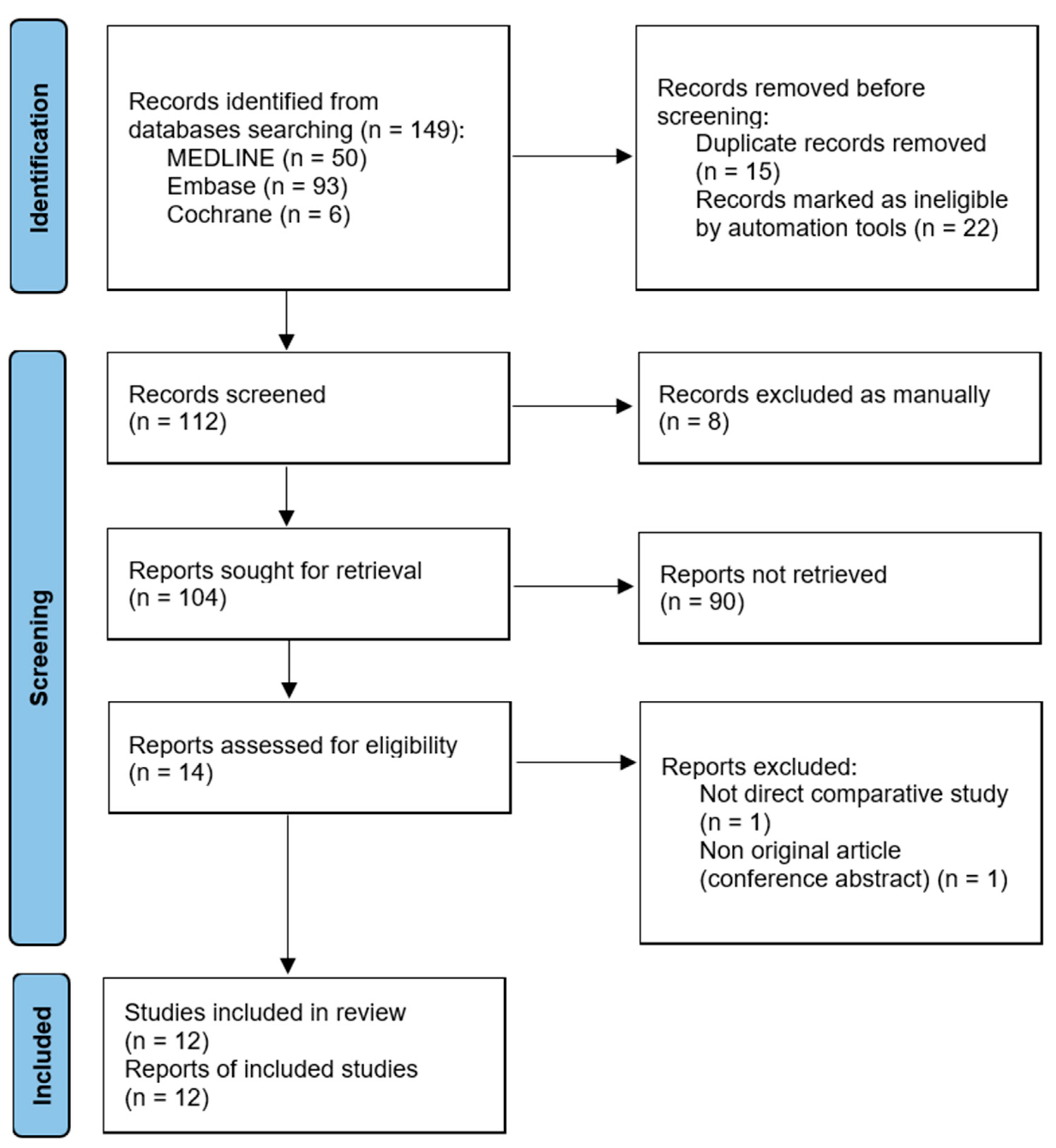
3.1. Article Identification

The present study’s identification and selection processes are outlined in Figure 1. Initially, a total of 149 articles were retrieved through the electronic literature search. Following the removal of 47 duplicates, 102 articles were retained for further review. Two additional articles were found through manual searching. Therefore, 104 articles were screened. Among them, 90 articles were excluded after screening their titles/abstracts, and two articles were excluded after a full-text review. Hence, 12 studies [4,6,7,17,18,19,20,21,22,23,24,25] met the criteria for both qualitative and quantitative data synthesis. The κ-values between the two reviewers demonstrated substantial agreement during the title review stage (κ = 0.730), nearly perfect agreement during the abstract review stage (κ = 0.891), and perfect agreement during the full-text review stage (κ = 1.000).

3.2. Study Characteristics and Qualitative Synthesis

Three studies [6,17,22] were prospective studies, and the other nine studies [4,7,18,19,20,21,23,24,25] were retrospective cohort studies. Furthermore, 10 of the 12 studies [4,6,18,19,20,21,22,23,24,25] were conducted in Asia. For the remaining studies, one was conducted in [7] North America, whereas the other [17] was conducted in South America.
The studies analyzed a total of 508,764 participants, including 32,460 patients with BPPV and 476,304 controls. With regard to participants’ mean age, the participants in most studies were in their 50s or 60s. With the exception of one study that had a male-dominant study population [23], all other studies had female-dominant study populations. The criteria used for BPPV diagnosis in each study are detailed in Table 1. Only two studies [6,24] provided details regarding BPPV subtypes. Except for two studies [7,21] that did not clearly specify their methods, all other studies appropriately diagnosed osteoporosis on the basis of BMD–dual-energy X-ray absorptiometry test results according to the World Health Organization criteria [26]. Five studies [6,22,23,24,25] described the BMD T-score in their study. However, we could not perform a meta-analysis for the T-score because of data inconsistencies. Additional details and outcomes investigated in each study are shown in Table 1.

3.3. Risk of Bias Assessment
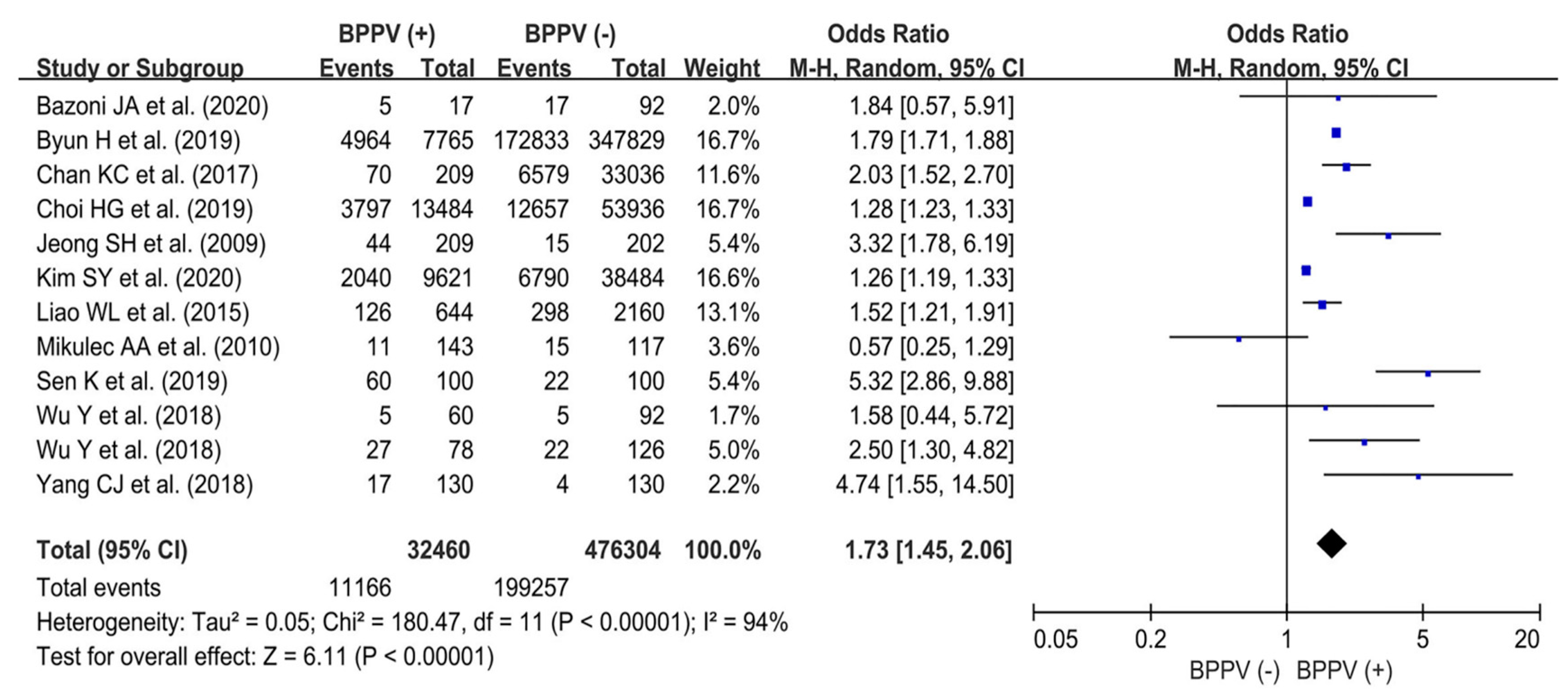
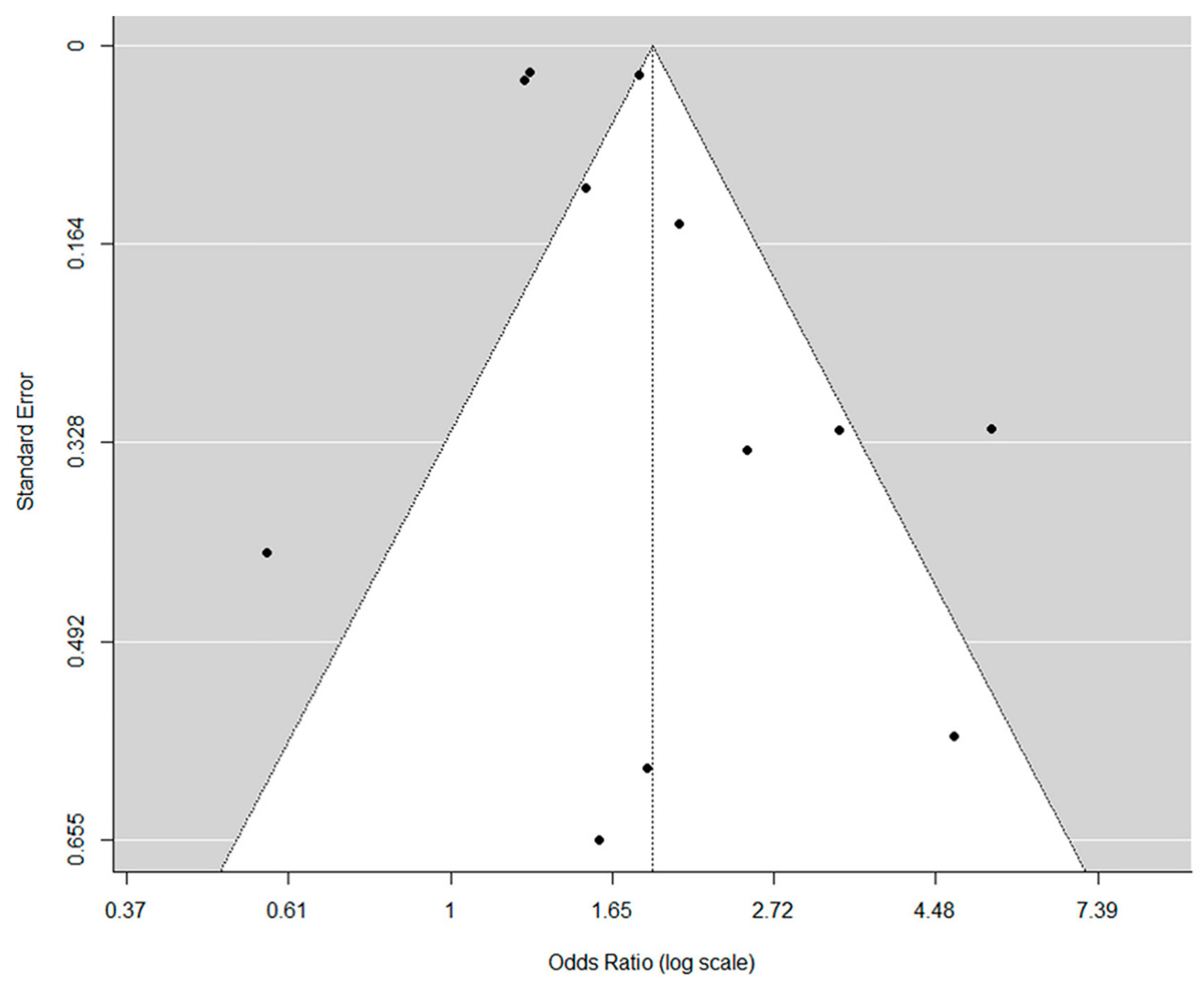
The mean MINORS score for the methodological quality assessment was 18.3/24 (range: 18–20). With regard to the eight main evaluation parameters, all included studies [4,6,7,17,18,19,20,21,22,23,24,25] clearly addressed the aim of the analysis (item 1: a clearly stated aim) and appropriately included consecutive patients (item 2: inclusion of consecutive patients). Nine of the twelve studies [4,7,18,19,20,21,23,24,25] received a point deduction for their retrospective design (item 3: prospective collection of data). All included studies [4,6,7,17,18,19,20,21,22,23,24,25] appropriately stated the aim of their study (item 4: endpoints appropriate to the aim of the study). Moreover, all included studies [4,6,7,17,18,19,20,21,22,23,24,25] did not perform a blind evaluation of the objective endpoint (item 5: unbiased assessment of the study endpoint). However, the follow-up period was appropriate (item 6: follow-up period appropriate for the aim of the study). In two studies [17,22], over 5% of patients that were initially included were lost to follow-up. Each of the included studies incurred a deduction in points due to the absence of a prospectively calculated sample size (item 8: prospective calculation of study size). However, no deductions were applied to the additional criteria domains—including adequate control group, contemporary groups, baseline equivalence of groups, and adequate statistical analyses—across all included studies.

3.4. Meta-Analysis

3.4.1. Prevalence of Osteoporosis between BPPV (+) and BPPV (−) Groups: Overall Population

The 12 included studies [4,6,7,17,18,19,20,21,22,23,24,25] investigated osteoporosis prevalence between BPPV (+) and BPPV (−) groups. A total of 11,166/32,460 patients were diagnosed with osteoporosis in the BPPV (+) group. Meanwhile, 199,257/476,304 patients were diagnosed with osteoporosis in the control group. The results of the pooled analysis showed that the BPPV (+) group had a significantly higher risk of osteoporosis than the BPPV (−) group (OR, 1.73; 95% CI, 1.45–2.06; p < 0.01), with high heterogeneity (I2 = 94%). A forest plot and further details are shown in Figure 2.
With regard to the evaluation of publication bias, the results of Egger’s test showed no publication bias upon visual assessment of the funnel plot (Figure 3) with outcomes of osteoporosis prevalence in the overall population (p = 0.25; 95% CI, 0.02–0.86).

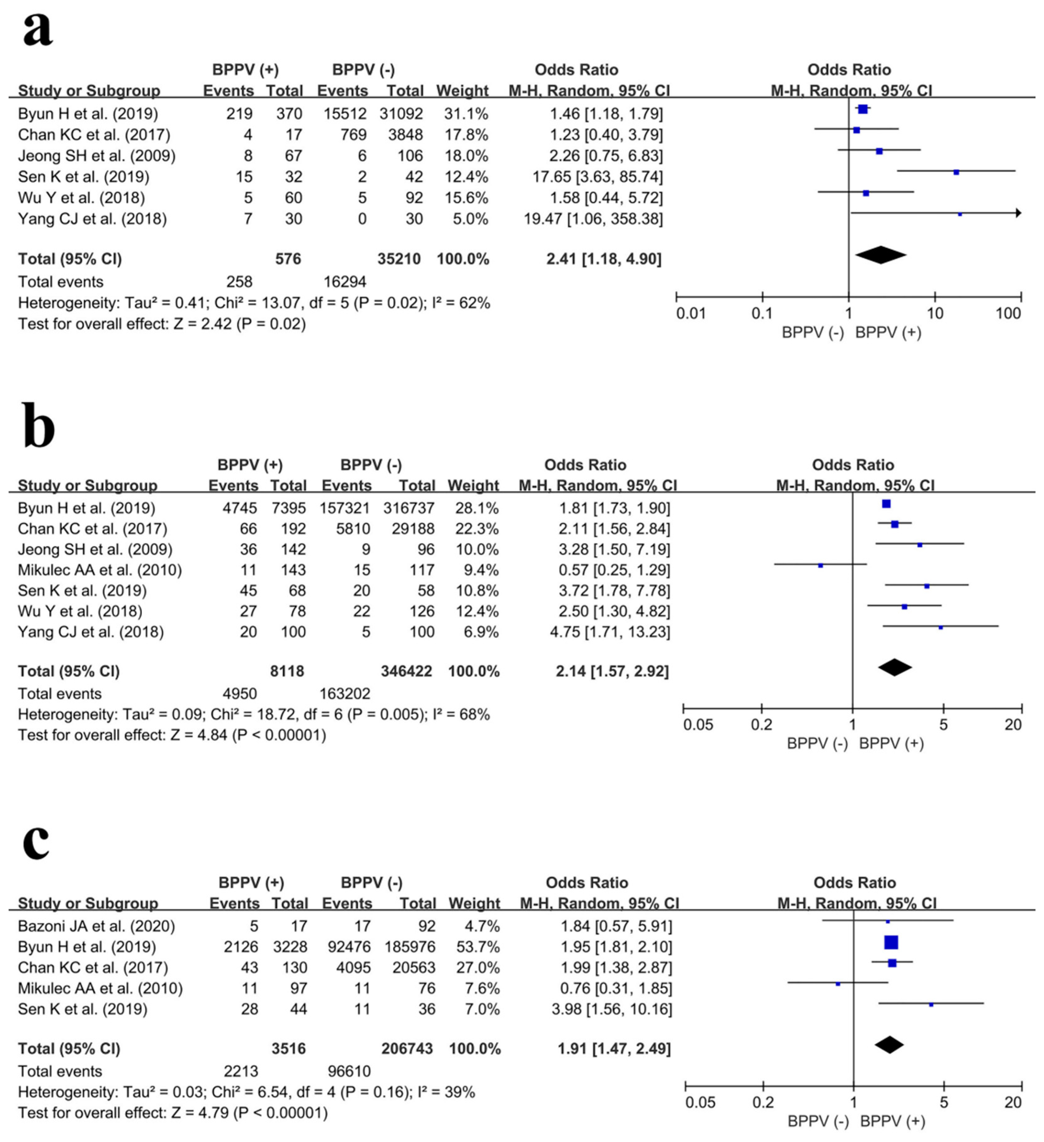
3.4.2. Subgroup Analyses: Male, Female, and Older Patient (Age ≥ 55 Years) Groups

Male Population

In the male population, data regarding osteoporosis prevalence were extracted from six studies [4,6,18,22,23,25]. Among male participants, 258/576 and 16,294/35,210 patients were diagnosed with osteoporosis in the BPPV (+) group and BPPV (−) group, respectively. The results of the meta-analysis revealed that the BPPV (+) group had a significantly higher risk of osteoporosis compared with the control group (OR, 2.41; 95% CI, 1.18–4.90; p = 0.02). Furthermore, the heterogeneity was moderate (I2 = 62%). A forest plot and further details are shown in Figure 4a.

Female Population

In the female population, data regarding osteoporosis prevalence were extracted from seven studies [4,6,7,18,22,24,25]. Among female patients, 4950/8118 and 163,202/346,422 patients were diagnosed with osteoporosis in the BPPV (+) group and BPPV (−) group, respectively. The results of the meta-analysis revealed that the BPPV (+) group also had a significantly higher risk of osteoporosis compared with the control group (OR, 2.14; 95% CI, 1.57–2.92; p < 0.001). Moreover, the heterogeneity was moderate (I2 = 68%). A forest plot and further details are shown in Figure 4b.

Older Patients (Age ≥ 55 Years)

In the older patient population (age ≥ 55 years), data regarding osteoporosis prevalence were extracted from five studies [4,7,17,18,22]. Among older adults, 2213/3516 and 96,610/206,743 patients were diagnosed with osteoporosis in the BPPV (+) group and BPPV (−) group, respectively. The results of the pooled analysis showed that the BPPV (+) group had a higher risk of osteoporosis compared to the control group (OR, 1.91; 95% CI, 1.47–2.49; p < 0.01). Moreover, the heterogeneity was low (I2 = 39%). A forest plot and further details are shown in Figure 4c.

4. Discussion

The principal finding of the present meta-analysis is that the BPPV (+) group had higher osteoporosis prevalence than the control group. A similar trend was also observed in subgroup analyses as a higher risk of osteoporosis was observed in the BPPV (+) group among male, female, and older patients (≥55 years of age).
BPPV is one of the most commonly recognized vestibular disorders encountered in neurotology clinics and accounts for approximately 20% of all cases of dizziness [27]. The overall prevalence of BPPV in the general population is 1.6% [28]; however, its prevalence is higher among older adults [29]. In terms of pathogenesis, there are two main types of BPPV: Schucknect’s “Cupulolithiasis,” in which the otoliths adhere persistently to the cupula, and Hall’s “Canalolithiasis,” in which the otoliths float in the canal [30]. Although they both explain the mechanisms of BPPV, they do not clarify why BPPV incidence progressively increases in older adults [29,31]. Furthermore, some epidemiologic studies have demonstrated that the prevalence of BPPV is higher in older adults, especially in those between 50 and 60 years of age [32,33]. Osteoporosis prevalence increases with age [34]; moreover, menopause occurs at an average age of 50 years and is associated with a significant increase in bone loss, which increases osteoporosis risk [35]. Interestingly, this also corresponds to the age group with the highest prevalence of BPPV. In a recent study by Juneja et al. [36], osteoporosis was identified as a risk factor for vestibular dysfunction and hearing deficits using vestibular-evoked myogenic potential testing. Theoretically, osteoporosis is a chronic progressive bone disease that leads to reduced mineral density in all bones. Therefore, it can decrease the density of bones, including the temporal bone, cochlear capsule, internal auditory canal, mandible, and bone in the middle ear [36]. Moreover, estrogen deficiency in postmenopausal women leads to further irreversible erosion of bone density [37,38]. BPPV predominantly occurs in female patients and those with calcium metabolism abnormalities, which suggests that there may be a positive relationship between BPPV and osteoporosis. This is also the basis of the present study. Lee et al. [39] conducted a bone turnover marker study and reported that BPPV prevalence in patients with osteoporosis was associated with vitamin D deficiency and high bone turnover rates at the systemic level, which could affect local Ca2+ homeostasis in the inner ear. Furthermore, considering that this study is a systematic review and meta-analysis, it was not possible to obtain data. However, taking into account that BPPV risk may be related to Ca2+ homeostasis in the inner ear, future extension studies should consider the relevance of vitamin D usage, serum Ca2+ levels, and osteoporosis medication in BPPV.
The existence of a likely association between BPPV and osteoporosis has been mentioned previously. In 2014, Yu et al. [10] presented a systematic review of the association between BPPV and osteoporosis. However, as only a few studies had been published, a meta-analysis could not be performed. The authors mentioned that there could be a relationship between BPPV and osteoporosis; however, there was insufficient strong evidence at the time of their study to support this hypothesis. As additional studies have been conducted, we were able to find a significant association between BPPV and osteoporosis in the present meta-analysis.
According to the results of our subgroup analyses, BPPV and osteoporosis showed significant relationships not only in female and older patients aged ≥ 55 years but also in male patients. Although osteoporosis is more prevalent in older women because of hormonal changes after menopause, our meta-analysis showed no difference in BPPV risk between male and female patients with osteoporosis. Interestingly, our study revealed that in addition to the impact of estrogen deficiency on bone quality in BPPV occurrence, there is a close association between bone quality itself and BPPV occurrence in all patients regardless of sex.
The present study has a few limitations. First, we could not perform more detailed subgroup analyses, especially for the T-score of BMD as a continuous variable or for BPPV subtype (e.g., horizontal canal, posterior canal, or anterior canal) because of a lack of study data. Nevertheless, as a meta-analysis is an appropriate method for generating a high level of evidence regarding controversial topics, our synthetic results are meaningful. Second, because of the relatively high heterogeneity of the pooled results, there is a possibility of bias in the interpretation of the meta-analysis results. Third, we could not include randomized controlled trials because of the lack of level I studies. In the future, higher level evidence from prospective randomized trials is required.
Despite these limitations, this meta-analysis supports the positive association between osteoporosis and BPPV. Furthermore, despite numerous studies conducted thus far to investigate the relationship between osteoporosis and BPPV, this study is the first meta-analysis on this topic. The findings of this study can be used in clinical settings to suggest that the presence of BPPV in patients may indicate a potential risk of osteoporosis.

Supplementary Materials

The following supporting information can be downloaded at: https://www.mdpi.com/article/10.3390/jpm14030303/s1, Supplement S1. The literature search algorithm and the results from relevant clinical studies.

Author Contributions

Methodology, data curation, validation, writing—original draft, and writing—review and editing, C.-H.K.; methodology, data curation, and writing—original draft, K.K.; formal analysis, resources, supervision, investigation, conceptualization, validation, and writing—review and editing, Y.C. All authors have read and agreed to the published version of the manuscript.

Funding

This work was supported by a grant from the Korea Evaluation Institute of Industrial Technology, funded by the Ministry of Trade, Industry and Energy (MOTIE, Korea) (No. 20016543: Development of a titanium alloy wire rod with roundness of 50 µm or less for biomedical and dental/orthopedic implant application technology).

Institutional Review Board Statement

While this study involved human participants, neither ethical approval nor the acquisition of informed consent from participants was required because all data were based on previously published studies and were anonymously analyzed without any potential harm to the participants.

Informed Consent Statement

The manuscript does not contain clinical studies or patient data. For this type of study, formal consent is not required.

Data Availability Statement

Data are available from the corresponding author upon reasonable request.

Conflicts of Interest

The authors declare no conflicts of interest.

References

  1. Balatsouras, D.G.; Koukoutsis, G.; Fassolis, A.; Moukos, A.; Apris, A. Benign paroxysmal positional vertigo in the elderly: Current insights. Clin. Interv. Aging 2018, 13, 2251–2266. [Google Scholar] [CrossRef]
  2. De Stefano, A.; Dispenza, F. Spontaneous nystagmus in horizontal canal benign paroxysmal positional vertigo. Auris Nasus Larynx 2013, 40, 117. [Google Scholar] [CrossRef]
  3. Palmeri, R.; Kumar, A. Benign Paroxysmal Positional Vertigo. In StatPearls; StatPearls Publishing: Treasure Island, FL, USA, 2023. [Google Scholar]
  4. Byun, H.; Chung, J.H.; Lee, S.H.; Park, C.W.; Kim, E.M.; Kim, I. Increased risk of benign paroxysmal positional vertigo in osteoporosis: A nationwide population-based cohort study. Sci. Rep. 2019, 9, 3469. [Google Scholar] [CrossRef]
  5. Parham, K.; Leonard, G.; Feinn, R.S.; Lafreniere, D.; Kenny, A.M. Prospective clinical investigation of the relationship between idiopathic benign paroxysmal positional vertigo and bone turnover: A pilot study. Laryngoscope 2013, 123, 2834–2839. [Google Scholar] [CrossRef]
  6. Jeong, S.H.; Choi, S.H.; Kim, J.Y.; Koo, J.W.; Kim, H.J.; Kim, J.S. Osteopenia and osteoporosis in idiopathic benign positional vertigo. Neurology 2009, 72, 1069–1076. [Google Scholar] [CrossRef]
  7. Mikulec, A.A.; Kowalczyk, K.A.; Pfitzinger, M.E.; Harris, D.A.; Jackson, L.E. Negative association between treated osteoporosis and benign paroxysmal positional vertigo in women. J. Laryngol. Otol. 2010, 124, 374–376. [Google Scholar] [CrossRef]
  8. Baek, Y.-H.; Cho, S.W.; Jeong, H.E.; Kim, J.H.; Hwang, Y.; Lange, J.L.; Shin, J.-Y. 10-Year Fracture Risk in Postmenopausal Women with Osteopenia and Osteoporosis in South Korea. Endocrinol. Metab. 2021, 36, 1178–1188. [Google Scholar] [CrossRef]
  9. Sozen, T.; Ozisik, L.; Basaran, N.C. An overview and management of osteoporosis. Eur. J. Rheumatol. 2017, 4, 46–56. [Google Scholar] [CrossRef]
  10. Yu, S.; Liu, F.; Cheng, Z.; Wang, Q. Association between osteoporosis and benign paroxysmal positional vertigo: A systematic review. BMC Neurol. 2014, 14, 110. [Google Scholar] [CrossRef]
  11. Moher, D.; Shamseer, L.; Clarke, M.; Ghersi, D.; Liberati, A.; Petticrew, M.; Shekelle, P.; Stewart, L.A.; PRISMA-P Group. Preferred reporting items for systematic review and meta-analysis protocols (PRISMA-P) 2015 statement. Syst. Rev. 2015, 4, 1. [Google Scholar] [CrossRef]
  12. Shea, B.J.; Grimshaw, J.M.; Wells, G.A.; Boers, M.; Andersson, N.; Hamel, C.; Porter, A.C.; Tugwell, P.; Moher, D.; Bouter, L.M. Development of AMSTAR: A measurement tool to assess the methodological quality of systematic reviews. BMC Med. Res. Methodol. 2007, 7, 10. [Google Scholar] [CrossRef]
  13. Moher, D.; Liberati, A.; Tetzlaff, J.; Altman, D.G.; The PRISMA Group. Preferred reporting items for systematic reviews and meta-analyses: The PRISMA statement. PLoS Med. 2009, 6, e1000097. [Google Scholar] [CrossRef]
  14. Slim, K.; Nini, E.; Forestier, D.; Kwiatkowski, F.; Panis, Y.; Chipponi, J. Methodological index for non-randomized studies (minors): Development and validation of a new instrument. ANZ J. Surg. 2003, 73, 712–716. [Google Scholar] [CrossRef]
  15. Schmidt, F.L.; Oh, I.-S.; Hayes, T.L. Fixed- versus random-effects models in meta-analysis: Model properties and an empirical comparison of differences in results. Br. J. Math. Stat. Psychol. 2009, 62, 97–128. [Google Scholar] [CrossRef]
  16. Cumpston, M.; Li, T.; Page, M.J.; Chandler, J.; Welch, V.A.; Higgins, J.P.; Thomas, J. Updated guidance for trusted systematic reviews: A new edition of the Cochrane Handbook for Systematic Reviews of Interventions. Cochrane Database Syst. Rev. 2019, 10, ED000142. [Google Scholar] [CrossRef]
  17. Bazoni, J.A.; Ciquinato, D.S.A.; de Souza Marquez, A.; Costa, V.D.S.P.; De Moraes Marchiori, G.; de Moraes Marchiori, L.L. Hypovitaminosis D, low bone mineral density, and diabetes mellitus as probable risk factors for benign paroxysmal positional vertigo in the elderly. Int. Arch. Otorhinolaryngol. 2020, 24, 272–277. [Google Scholar] [CrossRef]
  18. Chan, K.-C.; Tsai, Y.-T.; Yang, Y.-H.; Chen, P.-C.; Chang, P.-H. Osteoporosis is associated with increased risk for benign paroxysmal positional vertigo: A nationwide population-based study. Arch. Osteoporos. 2017, 12, 106. [Google Scholar] [CrossRef]
  19. Choi, H.G.; Lee, J.K.; Kong, I.G.; Lim, H.; Kim, S.Y. Osteoporosis increases the risk of benign paroxysmal positional vertigo: A nested case–control study using a national sample cohort. Eur. Arch. Oto-Rhino-Laryngol. 2019, 276, 335–342. [Google Scholar] [CrossRef]
  20. Kim, S.Y.; Kim, H.-J.; Min, C.; Choi, H.G. Association between benign paroxysmal positional vertigo and osteoporosis: Two nested case-control studies. Osteoporos. Int. 2020, 31, 2017–2024. [Google Scholar] [CrossRef]
  21. Liao, W.-L.; Chang, T.-P.; Chen, H.-J.; Kao, C.-H. Benign paroxysmal positional vertigo is associated with an increased risk of fracture: A population-based cohort study. J. Orthop. Sports Phys. Ther. 2015, 45, 406–412. [Google Scholar] [CrossRef]
  22. Sen, K.; Padiyar, B.V.; Arora, G. Association of Benign Paroxysmal Positional Vertigo with Osteoporosis and Vitamin D Deficiency—A Case-Control Study. Dubai Med. J. 2019, 1, 2–5. [Google Scholar] [CrossRef]
  23. Wu, Y.; Fan, Z.; Jin, H.; Guan, Q.; Zhou, M.; Lu, X.; Li, L.; Yan, W.; Gu, C.; Chen, C.; et al. Assessment of bone metabolism in male patients with benign paroxysmal positional vertigo. Front. Neurol. 2018, 9, 742. [Google Scholar] [CrossRef]
  24. Wu, Y.; Gu, C.; Han, W.; Lu, X.; Chen, C.; Fan, Z. Reduction of bone mineral density in native Chinese female idiopathic benign paroxysmal positional vertigo patients. Am. J. Otolaryngol.-Head Neck Med. Surg. 2018, 39, 31–33. [Google Scholar] [CrossRef]
  25. Yang, C.J.; Kim, Y.; Lee, H.S.; Park, H.J. Bone mineral density and serum 25-hydroxyvitamin D in patients with idiopathic benign paroxysmal positional vertigo. J. Vestib. Res. Equilib. Orientat. 2018, 27, 287–294. [Google Scholar] [CrossRef]
  26. Akkawi, I.; Zmerly, H. Osteoporosis: Current Concepts. Joints 2018, 6, 122–127. [Google Scholar] [CrossRef]
  27. Labuguen, R.H. Initial evaluation of vertigo. Am. Fam. Physician 2006, 73, 244–251. [Google Scholar]
  28. Neuhauser, H.K.; Lempert, T. Vertigo: Epidemiologic aspects. Semin. Neurol. 2009, 29, 473–481. [Google Scholar] [CrossRef]
  29. Oghalai, J.S.; Manolidis, S.; Barth, J.L.; Stewart, M.G.; Jenkins, H.A. Unrecognized benign paroxysmal positional vertigo in elderly patients. Otolaryngol. Head Neck Surg. 2000, 122, 630–634. [Google Scholar] [CrossRef]
  30. De Stefano, A.; Dispenza, F.; Suarez, H.; Perez-Fernandez, N.; Manrique-Huarte, R.; Ban, J.H.; Kim, M.B.; Strupp, M.; Feil, K.; Oliveira, C.A.; et al. A multicenter observational study on the role of comorbidities in the recurrent episodes of benign paroxysmal positional vertigo. Auris Nasus Larynx 2014, 41, 31–36. [Google Scholar] [CrossRef]
  31. Lee, S.H.; Kim, M.K.; Cho, K.H.; Kim, J.S. Reversal of initial positioning nystagmus in benign paroxysmal positional vertigo involving the horizontal canal. Ann. N. Y. Acad. Sci. 2009, 1164, 406–408. [Google Scholar] [CrossRef]
  32. Kim, J.-S.; Zee, D.S. Clinical practice. Benign paroxysmal positional vertigo. N. Engl. J. Med. 2014, 370, 1138–1147. [Google Scholar] [CrossRef] [PubMed]
  33. Parham, K.; Kuchel, G.A. A Geriatric Perspective on Benign Paroxysmal Positional Vertigo. J. Am. Geriatr. Soc. 2016, 64, 378–385. [Google Scholar] [CrossRef] [PubMed]
  34. Barnsley, J.; Buckland, G.; Chan, P.E.; Ong, A.; Ramos, A.S.; Baxter, M.; Laskou, F.; Dennison, E.M.; Cooper, C.; Patel, H.P. Pathophysiology and treatment of osteoporosis: Challenges for clinical practice in older people. Aging Clin. Exp. Res. 2021, 33, 759–773. [Google Scholar] [CrossRef] [PubMed]
  35. Ji, M.X.; Yu, Q. Primary osteoporosis in postmenopausal women. Chronic Dis. Transl. Med. 2015, 1, 9–13. [Google Scholar] [CrossRef]
  36. Juneja, M.K.; Munjal, S.; Sharma, A.; Gupta, A.K.; Bhadada, S. Audiovestibular functioning of post-menopausal females with osteoporosis and osteopenia. J. Otol. 2021, 16, 27–33. [Google Scholar] [CrossRef]
  37. Yang, K.; Qiu, X.; Cao, L.; Qiu, S. The role of melatonin in the development of postmenopausal osteoporosis. Front. Pharmacol. 2022, 13, 975181. [Google Scholar] [CrossRef]
  38. Baytaroglu, B.; Jafarov, S.; Erbek, S.S. The Effect of Postmenopausal Osteoporosis on Middle Ear Resonance Frequency. J. Int. Adv. Otol. 2021, 17, 526–529. [Google Scholar] [CrossRef]
  39. Lee, S.B.; Lee, C.H.; Kim, Y.J.; Kim, H.-M. Biochemical markers of bone turnover in benign paroxysmal positional vertigo. PLoS ONE 2017, 12, e0176011. [Google Scholar] [CrossRef]
Figure 1. Flow diagram illustrating the identification and selection processes of studies included in the meta-analysis.
Jpm 14 00303 g001
Figure 2. Forest plot showing osteoporosis prevalence between the BPPV (+) and BPPV (−) groups in the overall population [4,6,7,17,18,19,20,21,22,23,24,25].
Jpm 14 00303 g002
Figure 3. Funnel plot showing the absence of publication bias in the assessment of osteoporosis prevalence in the overall population. The black dots represent a single study included in the analysis.
Jpm 14 00303 g003
Figure 4. Forest plots showing osteoporosis prevalence between the BPPV (+) and BPPV (−) groups in (a) males [4,6,18,22,23,25], (b) females [4,6,7,18,22,24,25], and (c) older patients (≥55 years) [4,7,17,18,22].
Jpm 14 00303 g004
Table 1. Study design, demographic data, study details, and MINORS scores.
Author (year)Study DesignCountrySample Size (n)Characteristics of Control Participants in the Control GroupMean AgeFemale SexDiagnosis of BPPVSubtypes of BPPVDiagnosis of OsteoporosisT-Score
BPPV Group Control Group Years%
Bazoni JA et al. (2020) [17]PCSBrazil1792Volunteers of age ≥ 60, both gendersBPPV: 67.0
Control: 68.0
BPPV: 88.2
Control: 58.7
Vertigo complaints, audiological anamnesis, and D–H maneuverN/ABMDN/A
Byun H et al. (2019) [4]RCSRepublic of Korea7765347,829Matched for age, sex, social status, HTN, and DM64.5BPPV: 91.2
Control: 91.2
ICD-10 codeN/ADXA, qCT, peripheral DXA, and qUSN/A
Chan KC et al. (2017) [18]RCSTaiwan20933,036Matched for age and gender>20BPPV: 88.4
Control: 88.4
ICD-9 codeN/ADXAN/A
Choi HG et al. (2019) [19]RCSRepublic of Korea13,48453,936Matched for age, sex, socioeconomic status, HTN, DM, and dyslipidemia≥50BPPV: 69.8
Control: 69.8
ICD-10 codeN/ADXA and qCTN/A
Jeong SH et al. (2009) [6]PCSRepublic of Korea209202Volunteers without dizziness and VCF HxBPPV: 59.8
Control: 56.3
BPPV: 67.9
Control: 47.5
Vertigo complaints, typical positioning nystagmus, and no other CNS disordersHorizontal canal, posterior canal, and anterior canalDXA (LUNAR); T-score ≤ −2.5 from lumbar or femoral areas (WHO definition)BPPV: −1.7 (female); −1.1 (male).
Control: −1.0 (female); −0.7 (male).
Kim SY et al. (2020) [20]RCSRepublic of Korea962138,484Matched for age, sex, income, and region of residence>40BPPV: 62.6
Control: 62.6
ICD-10 codeN/ADXA and qCTN/A
Liao WL et al. (2015) [21]RCSTaiwan6442160Matched for age and sex; without vertigo HxBPPV: 57.1
Control: 56.7
BPPV: 63.5
Control: 63.5
ICD-9 codeN/AN/A (ICD-9 code)N/A
Mikulec AA et al. (2010) [7]RCSU.S.143117Patients without BPPV who visited two otology clinics51–80100Clinical evaluation; videonystagmography testingN/AN/AN/A
Sen K et al. (2019) [22]PCSIndia100100Healthy volunteers who want to screen for osteoporosisBPPV: 48.6
Control: 46.2
BPPV: 68.0
Control: 58.0
D-H maneuver, P-M maneuver, cephalic hyperextension, and audiological assessmentN/ADXA (T-score ≤ −2.5 from the lumbar or femoral areas)BPPV: −2.6.
Control: −1.8.
Wu Y et al. (2018) [23]RCSChina6092Matched for age; healthy controls without vertigo/dizziness historyBPPV: 59.4
Control: 62.1
0Vertigo complaints, D-H maneuver, and supine roll testN/ADXA (LUNAR); T-score ≤ −2.5 from lumbar or femoral areas (WHO definition)BPPV: 1.101 (spine); 0.966 (hip).
Control: 1.128 (spine); 1.000 (hip).
Wu Y et al. (2018) [24]RCSChina78126Matched for age; healthy controls without vertigo/dizziness historyBPPV: 58.4
Control: 58.5
100Vertigo complaints, D-H maneuver, and supine roll testHorizontal canal; posterior canalDXA (LUNAR); T-score ≤ −2.5 from lumbar or femoral areas (WHO definition)BPPV: −0.11 to −2.42 (spine); −0.04 to −1.73 (hip).
Control: 0.70 to −1.45 (spine); 0.36 to 0.96 (hip).
Yang CJ et al. (2018) [25]RCSRepublic of Korea130130Matched for age and sex; healthy controls without dizziness or fracture Hx54.9BPPV: 76.9
Control: 76.9
Vertigo complaints; D-H maneuverN/ADXA (LUNAR); T-score ≤ −2.5 from lumbar or femoral areas (WHO definition)BPPV: −1.6 (female); −0.9 (male).
Control: −1.1 (female); −0.7 (male).
Ass, assessment; BMD, bone mineral density; BPPV, benign paroxysmal positional vertigo; DXA, dual-energy X-ray absorptiometry; D–H maneuver, Dix–Hallpike diagnostic maneuver; Hx, history; N/A, nonapplicable; PCS, prospective cohort study; RCS, retrospective cohort study; WHO, World Health Organization.
Disclaimer/Publisher’s Note: The statements, opinions and data contained in all publications are solely those of the individual author(s) and contributor(s) and not of MDPI and/or the editor(s). MDPI and/or the editor(s) disclaim responsibility for any injury to people or property resulting from any ideas, methods, instructions or products referred to in the content.

