Article

Rare Mutations in CCDC7 Contribute to Early-Onset Preeclampsia by Inhibiting Trophoblast Migration and Invasion

1
Key Laboratory for Major Obstetric Diseases of Guangdong Province, Department of Obstetrics and Gynecology, The Third Affiliated Hospital of Guangzhou Medical University, Guangzhou 510150, China
2
The Medical Centre for Critical Pregnant Women in Guangzhou, Guangzhou 510150, China
3
Obstetrics & Gynecology Institute of Guangzhou, The Third Affiliated Hospital of Guangzhou Medical University, Guangzhou 510150, China
4
Department of Fetal Medicine and Prenatal Diagnosis, The Third Affiliated Hospital of Guangzhou Medical University, Guangzhou 510150, China
5
Guangdong-Hong Kong-Macao Greater Bay Area Higher Education Joint Laboratory of Maternal-Fetal Medicine, Guangzhou 510150, China
6
Guangdong Engineering and Technology Research Center of Maternal-Fetal Medicine, Guangzhou 510150, China
*
Authors to whom correspondence should be addressed.
These authors contributed equally to this work.
J. Pers. Med. 2024, 14(3), 253; https://doi.org/10.3390/jpm14030253
Submission received: 12 January 2024 / Revised: 17 February 2024 / Accepted: 24 February 2024 / Published: 27 February 2024
(This article belongs to the Section Mechanisms of Diseases)

Abstract

:
Rare gene variants have been found to play a role in complex disorders. Preeclampsia, and especially early-onset preeclampsia, has a strong genetic link. However, the role of rare variants in the offspring of mothers with preeclampsia remains unclear. In this study, whole-exome sequencing (WES) was used to identify rare pathogenic variants in two families with early-onset preeclampsia. Two heterozygous rare variants in CCDC7, c.625C>T (p.R209C) and c.1015C>T (p.R339X), were detected in two families and were cosegregated in the offspring of preeclamptic pregnancies. We examined the spatiotemporal expression pattern of CCDC7 in human placental villi and the effects of CCDC7 on migration and invasion of trophoblast cells JEG-3. The quantitative real-time PCR and Western blot results showed that the expression of CCDC7 in placental villi was the lowest during the first trimester and increased as the pregnancy progressed. The CCDC7 p.R339X variant showed a decrease in mRNA and protein expressions. Loss-of-function assays showed that knockdown of CCDC7 suppressed the migration and invasion of JEG-3 cells. In conclusion, CCDC7 is a potential susceptibility gene for preeclampsia, which is key for the migration and invasion of trophoblast cells. Rare variants of preeclampsia in offspring may play a crucial role in the pathogenesis of preeclampsia and require further research.

1. Introduction

Preeclampsia is a complex, heterogenous, pregnancy-associated disorder characterized by high blood pressure (≥140/90 mmHg) and proteinuria (≥300 mg/L per 24 h) after the 20th week of pregnancy [1]. As a leading complication of pregnancy, preeclampsia affects an estimated 4 million women each year and causes the deaths of more than 70,000 women and 500,000 newborns worldwide [2]. Preeclampsia can be divided into two groups according to the onset time: early-onset preeclampsia (EOPE, <34 weeks) and late-onset preeclampsia (LOPE, ≥34 weeks) [1]. Preeclampsia, particularly EOPE, is considered to be heritable, with an estimated heritability of about 0.50 in several populations [3,4]. Maternal and fetal contributions are estimated at 35% and 20%, respectively [5,6].
Maternal contributions to preeclampsia have attracted considerable attention over the past decades. Using genome-wide association studies (GWASs), gene variants have been reported to be associated with a high risk of preeclampsia, but most of these variants have not been replicated in different groups [7]. Some studies researched variants in the fetal genome. In multiplex Dutch preeclampsia families, variant p.Y153H of STOX1 was cosegregated in offspring from preeclamptic pregnancies [8]. This variant resulted in reduced trophoblast invasion and played a crucial role in early utero-placental development [9,10], although the association between this variant and preeclampsia was not duplicated in two different groups [11,12]. Recently, McGinnis et al. discovered a significant susceptibility locus near the FLT1 gene (rs4769613, p = 5.4 × 10⁻¹¹) between 4380 offspring from preeclamptic pregnancies and 310,238 controls [13]. These studies have noted the importance of variants of preeclampsia offspring in the pathogenesis of preeclampsia.
With the wide application of whole-exome sequencing (WES) and whole-genome sequencing (WGS), rare variants of complex disorders, such as autism spectrum disorder [14], migraine [15], and advanced age-related macular degeneration [16], have attracted great attention. Recently, several studies have revealed the effect of rare variants on pregnancy-associated diseases. Huusko et al. detected rare and likely damaging variants in HSPA1L in multiple families with recurrent spontaneous preterm births [17]. Liu et al. found that novel rare variants in ABC transporter genes were associated with intrahepatic cholestasis of pregnancy disease based on a case–control study [18]. However, far too little attention has been paid to the role of rare variants in the offspring in the pathogenesis of preeclampsia.
In this study, we identified rare variants in two families with EOPE using WES and constructed mutant cell lines to elucidate the effects of the identified variants on the function of trophoblast cells.

2. Methods

2.1. Patients

Two Han preeclampsia families were enrolled (Figure 1) in this study. The diagnosis of preeclampsia was based on the clinical features, including onset time at pregnancy, high blood pressure, proteinuria, impaired liver function, renal insufficiency, thrombocytopenia, pulmonary edema, headache, and visual symptoms, according to the ACOG practice bulletin No. 202 [1]. The onset time of all the women was after 20 weeks’ and before 34 weeks’ gestation, except that the medical record of the pregnancy of subject I-2 in family A was unknown and subject I-2 in family B only presented gestational hypertension. Subject I-2 in family A had developed hypertension at the last visit. The peripheral blood was collected from the women with preeclampsia and their relatives. Placental samples near the cord insertion site (8–10 pieces, size about 0.5 × 0.5 × 0.5 cm each) and cord blood (5–10 L) were collected within 10 min after delivery and were stored in RNAlateror liquid nitrogen for future analysis. This study was approved by the ethics committee of the Third Affiliated Hospital of Guangzhou Medical University. All of the participants enrolled in the study gave informed written consent.

2.2. Whole-Exome Sequencing and Data Analysis

Genomic DNA was extracted from placental samples, preeclampsia maternal peripheral blood, and offspring cord blood from all cases using a DNeasy Blood and Tissue Kit (Qiagen, Hilden, Germany), according to the manufacturer’s standard procedure. WES was conducted using the peripheral blood from women with preeclampsia and their relatives (cord blood was used in three newborns, III-8 in family A and III-2 and III-3 in family B, instead of peripheral blood) using next-generation sequencing platforms by iGeneTech Co., Ltd. (Beijing, China). Sequenced reads were aligned to the human reference genome GRCh37/hg19. Software including Samtools 1.9and GATK 4.0 were used to call, sort, and index variants from aligned sequence files. Annotation of genetic variants was performed using ANNOVAR. Potential maternal DNA contamination was excluded via the genotyping of short tandem repeats (STRs). The detailed information is provided in the Supplementary Materials.
This study focused on rare variants. The variants selected for downstream analysis met the following criteria: (1) minor allele frequency in population database was <0.05; (2) more than two in silico applications (Polyphen2, SIFT, or CADD) classified the variant as damaging, or it received a score ≥ 15; (3) it was cosegregated in all preeclampsia offspring from the same family. Variants selected for downstream analysis were validated via Sanger sequencing in both placental samples and offspring cord blood.

2.3. Cell Culture

The human placental choriocarcinoma JEG-3 cell line (ATCC: HTB-36) was cultured in Dulbecco’s modified Eagle medium: Nutrient Mixture F-12 (DMEM/F12) (Gibco, Grand Island, NY, USA), supplemented with 10% (v/v) fetal bovine serum (FBS; Gibco, Grand Island, NY, USA). Cells were maintained at 37 °C in an atmosphere containing 5% CO2.

2.4. Generation of Stable CCDC7 Knockdown Cell Lines

Lentiviral-based shRNA vectors and non-targeting shRNA vectors (containing puromycin and enhanced green fluorescent protein) were designed and synthesized by Genecopoeia (Guangzhou, China). The shRNA targeted sequences were as follows: CCDC7 shRNA1, 5′-CCAGTAAAGCATCTGTTGACC-3′; CCDC7 shRNA2, 5′-GCTTCGCGCCGTAGTCTTA-3′; and scrambled control, 5′-GCTTCGCGCCGTAGTCTTA-3′. Recombinant lentiviral particles were produced by co-transfecting 293Ta cells with a lentiviral expression plasmid and a Lenti-Pac™ HIV Expression Packaging Kit (GeneCopoeia, Guangzhou, China). The viral supernatants were harvested 48 h post-transfection. The JEG-3 cells were transduced with lentiviral particles in the presence of 5 μg/mL polybrene. After 48 h, the infected cells were selected with 2 μg/mL puromycin.

2.5. RNA Extraction and Quantitative Real-Time Polymerase Chain Reaction

Total RNA from the placenta samples was extracted using TRIzol reagent (Invitrogen, Carlsbad, CA, USA) following the manufacturer’s instructions. The synthesis of complementary DNA (cDNA) was performed by using a PrimeScript™ RT reagent Kit with gDNA Eraser (Takara, Tokyo, Japan). Quantitative real-time PCR (qRT-PCR) was conducted using a TB Green® Premix Ex Taq™ II Kit (Takara, Tokyo, Japan). GAPDH mRNA levels were used as the internal control, and relative gene expression levels were calculated using the comparative Ct method (2−ΔΔCT). The experiments were repeated at least three times in each placental sample, and 3–5 pieces from one placenta were used.

2.6. Protein Extraction and Western Blot Analysis

Total protein was extracted with RIPA lysis buffer supplemented with protease and phosphatase inhibitors (Sigma-Aldrich, St. Louis, MO, USA). The protein concentrations were measured with a BCA protein assay kit (Thermo Fisher Scientific, Waltham, MA, USA). After boiling for 10 min at 95 °C in Laemmli sample buffer (BioRad Laboratories, Hercules, CA, USA), equal amounts of protein (50 μg) were separated using sodium dodecyl sulfate-polyacrylamide gel-electrophoresis (SDS-PAGE) and then transferred to polyvinylidene fluoride (PVDF) membranes (Millipore, Billerica, MA, USA). The membranes were blocked for 1 hour at room temperature with 5% nonfat dry milk in phosphate-buffered saline (PBS) containing 0.1% Tween 20 (PBST) and probed with diluted primary antibody (anti-CCDC7, Abcam, Cambridge, MA, USA; 1:1000) at 4 °C overnight. Subsequently, the membranes were washed three times with PBST and incubated with a horseradish peroxidase-conjugated secondary antibody for 1 h at room temperature. The protein bands were visualized using enhanced chemiluminescence (Thermo Scientific, Waltham, MA, USA) and Image Lab V.6.1 software (BioRad Laboratories, Hercules, CA, USA).

2.7. Immunofluorescence

Sections of paraffin-embedded placental tissues (5 μm thickness) were deparaffinized and rehydrated, followed by microwave antigen retrieval in citrate buffer (pH 6.0). Then, sections were incubated in 3% H2O2 for 10 min at room temperature to remove endogenous peroxidase. and washed three times in PBS. To block nonspecific antibody binding, the sections were incubated with 10% normal goat serum for 30 min at room temperature. After overnight probing with antibody specific for CCDC7 (1:100; Novus Biologicals, Littleton, CO, USA) at 4 °C, the sections were washed in PBS and incubated in the dark with Alexa Fluor 546-conjugated antirabbit IgG (1:200; Invitrogen, Carlsbad, CA, USA) for 1 h at room temperature. The nuclei were stained with DAPI (Sigma Aldrich, St. Louis, MO, USA), and the sections were mounted in fluorescent mounting medium (Dako, Carpinteria, CA, USA). Images were captured with an inverted fluorescence microscope (Nikon, Melville, NY, USA).

2.8. Wound-Healing Assay

After a 24 h culture, the cells were grown to 80–90% confluence. The cell monolayers were scratched with a 200 μL pipette tip in a straight line to generate a wound-like gap, followed by washing with PBS to remove the cellular debris. The cells were rephotographed after 24 h for analysis of wound closure, as previously described [19]. The cell migration rate was calculated according to the formula: [(0 h wound width − 24 h wound width)/0 h wound width] × 100%. The experiments were repeated at least three times.

2.9. Transwell Invasion Assay

For the transwell invasion assay, the transwell chambers (8 µm pore size, Corning Costar, NY, USA) were coated with diluted Matrigel (300 μg/mL, BD Biosciences, Franklin Lakes, NJ, USA). The transfected cells were suspended in a serum-free medium and added to the upper chambers, and the lower chambers were filled with a medium containing 10% FBS as a chemoattractant. After incubation for 24 h at 37 °C with 5% CO2, the invasive cells in the lower chambers were fixed with 4% paraformaldehyde for 25 min and stained with 0.1% crystal violet for 15 min. Finally, the invaded cells in five random visual fields were imaged and counted under an inverted light microscope.

2.10. Statistical Analysis

Results are presented as means ± SEM. A two-way Student’s t-test was used to determine changes in mean gene expression. For analysis of demographic data, Student’s t-test, χ2, or Fisher’s exact test was used to determine significant differences in the cohorts. All statistical analyses were performed using Prism 7 software (GraphPad Software, La Jolla, CA, USA), and results were considered significant when p < 0.05.

3. Results

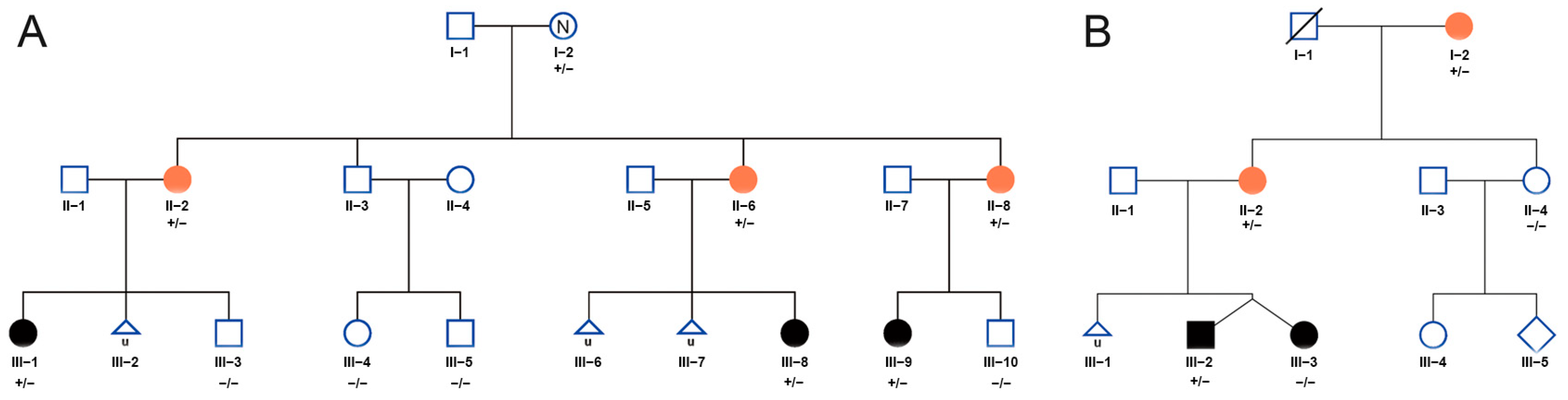
3.1. Clinical Features and Rare Variants of CCDC7 Detected in Two EOPE Families

Clinical data and blood samples were collected from two nonconsanguineous Chinese families with early-onset preeclampsia. The preeclampsia pedigrees are shown in Figure 1A,B. The mean gestational age of the subjects was 28.6 ± 5.5 years. The mean onset time was 30.3 ± 4.2 weeks of gestation. The mean time of delivery was 33.4 ± 3.4 weeks of gestation. Most of subjects presented with EOPE, except for two subjects: II-8 in family A, who had eclampsia at 32 weeks of gestation, and I-2 in family B, who presented with high blood pressure at 36 weeks of gestation but the information on the onset time of high blood pressure and other symptoms was unknown due to a lack of medical records. At the last visit, I-2 in family A presented hypertension, and other pregnant women did not develop hypertension disorders after delivery, which needed a long-term follow-up since they were young. The detailed features of the enrolled preeclampsia subjects are shown in Table 1.

3.2. Whole-Exome Sequencing Identified Rare Variants in CCDC7 in Familial Preeclampsia

The genomic DNA of the patients and their families were sent for WES analysis, and bioinformatics analysis were performed to explore the potential pathogenic variants in an unbiased way. We detected two heterozygous rare variants (c.625C>T/p.R209C and c.1015C>T/p.R339*) in the CCDC7 (NM_145023.6) gene in patients and their affected children. Then, the variants were confirmed via Sanger sequencing (Figure 2A). Both of these mutant sites were highly conserved in multiple mammalian species (Figure 2B,C). The missense variant (p.R209C) is predicted to be damaging, according to online software SIFT, Polyphen2, and CADD (score ≥ 15). The nonsense variant (p.R339*) results in a change from Arg339 to a premature stop codon, indicating a loss-of-function protein.

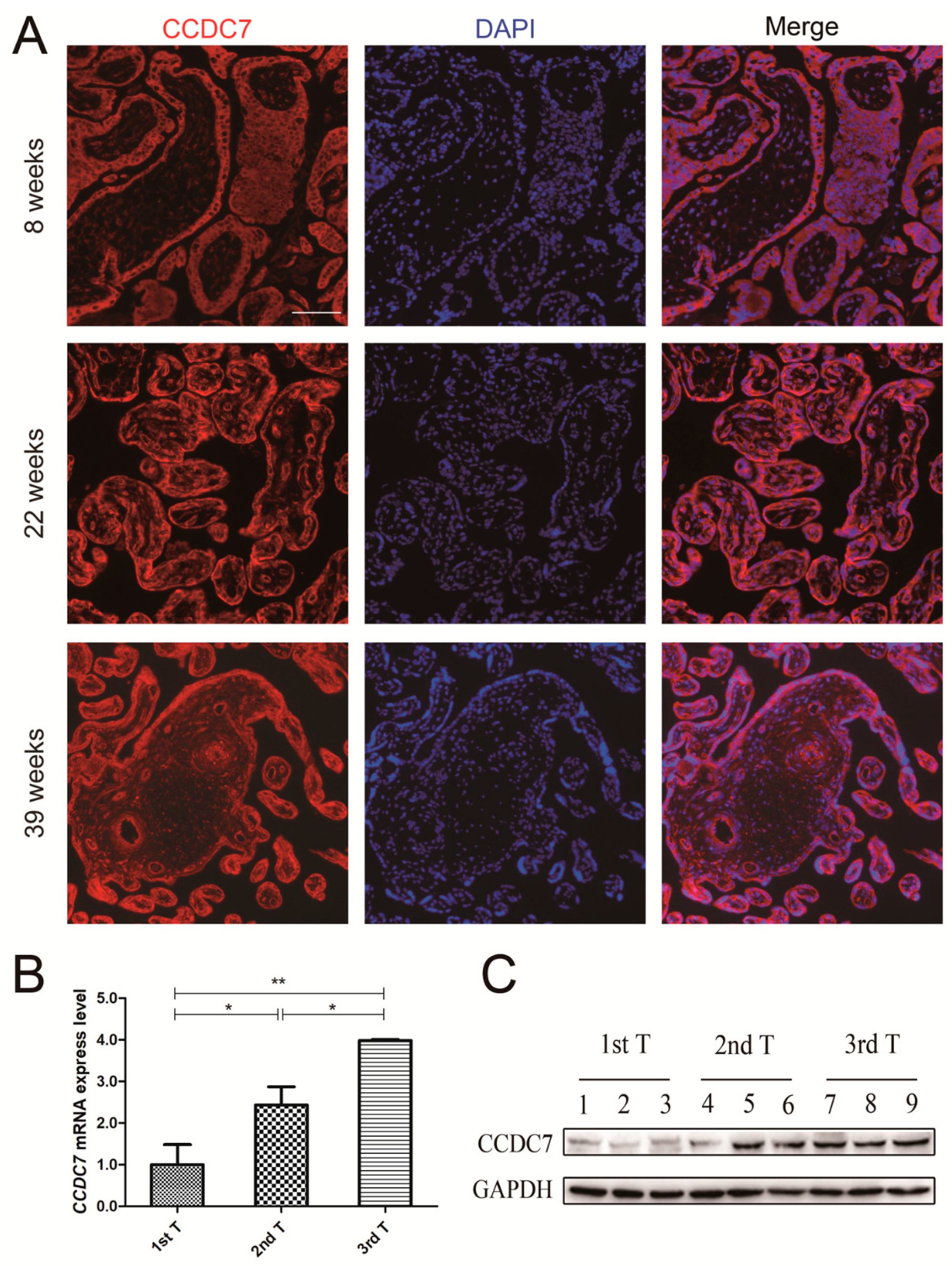
3.3. CCDC7 Spatial and Temporal Expression Patterns in the Human Placenta

To determine where CCDC7 is localized in the human placenta, we performed immunofluorescence studies on placenta tissue sections. The localization of CCDC7 within the chorionic villi structure was examined via immunofluorescence on placenta tissue from each trimester across gestation. The general localization of CCDC7 in representative samples from early (8 weeks), middle (22 weeks), and late (39 weeks) gestation is provided in Figure 3A. During the first trimester, CCDC7 was detected in syncytiotrophoblast cells, cytotrophoblast cells, and trophoblast columns. In the second and third trimesters, intense staining was still found in syncytiotrophoblast cells and cytotrophoblast cells.
In order to determine whether CCDC7 expression changes over gestation, we explored the mRNA and protein levels of CCDC7 in placental tissues at specific gestational time windows. As depicted in Figure 3B, a significant increase in CCDC7/GAPDH mRNA expression was found in placental tissues with advanced gestational age. Western blotting further confirmed that the expression of CCDC7 protein increased as pregnancy advanced (Figure 3C). These observations suggest that CCDC7 plays a key role in healthy placental development and function. The specific expression of CCDC7 in placental tissue indicated that CCDC7 plays an essential role in maintaining gestation.

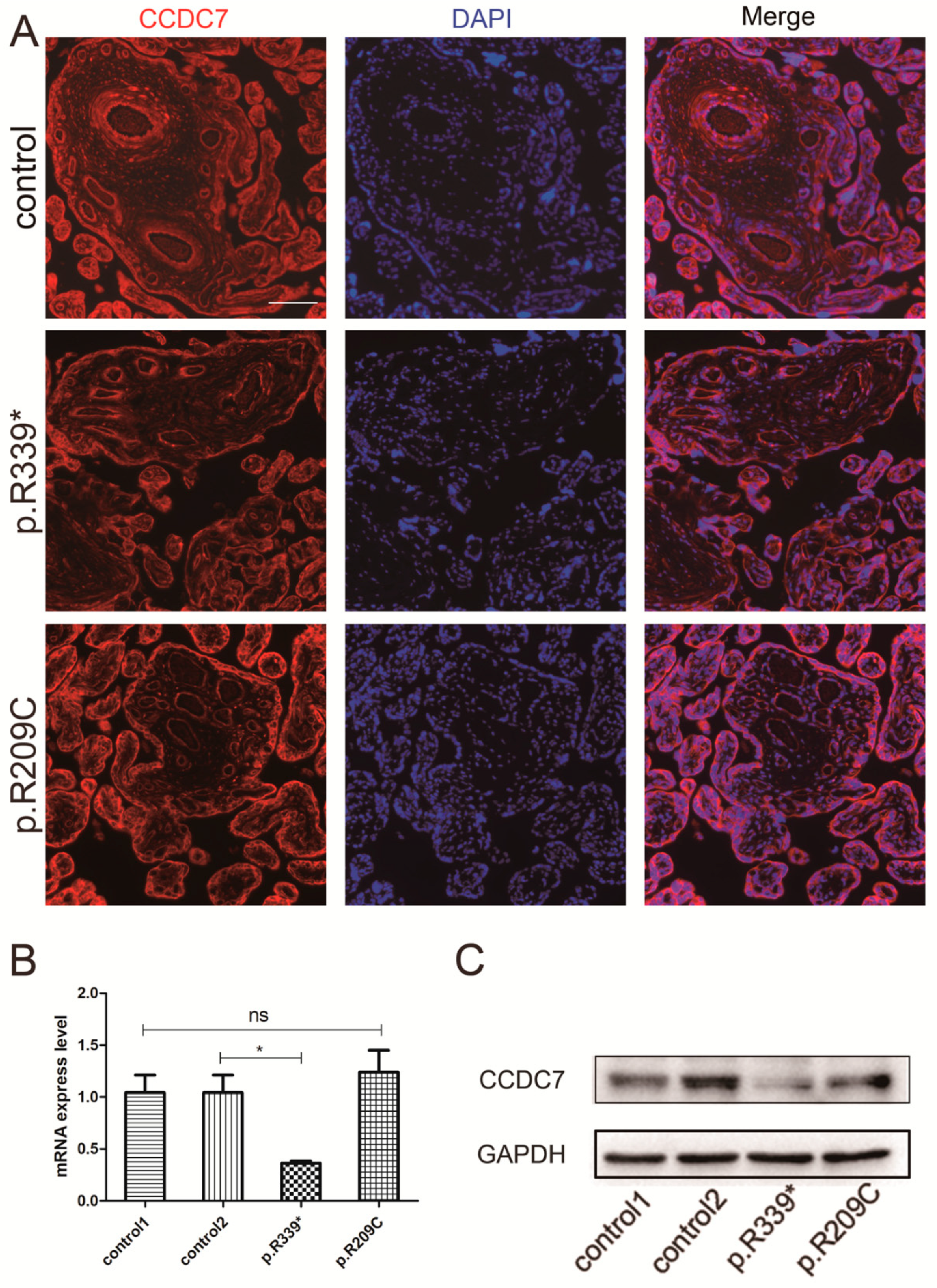
3.4. Expression and Distribution of Rare CCDC7 Variants in the Placenta

To investigate whether these variants detected in our study affected CCDC7 localization and expression, we compared CCDC7 localization and expression in the placentas from two preeclampsia families with age-matched controls. As showed by immunofluorescence, the localization of CCDC7 in the placenta did not alter in either of the two families compared with that in the controls (Figure 4A). The results of RT-qPCR and Western blotting revealed that the expression level of CCDC7 was significantly decreased in the p.R339* variant placenta compared with that in the controls. And, CCDC7 mRNA and protein expression showed little changes in the p.R209C variant placenta (Figure 4B,C). Collectively, these results imply that CCDC7 is involved in the pathology of preeclampsia.

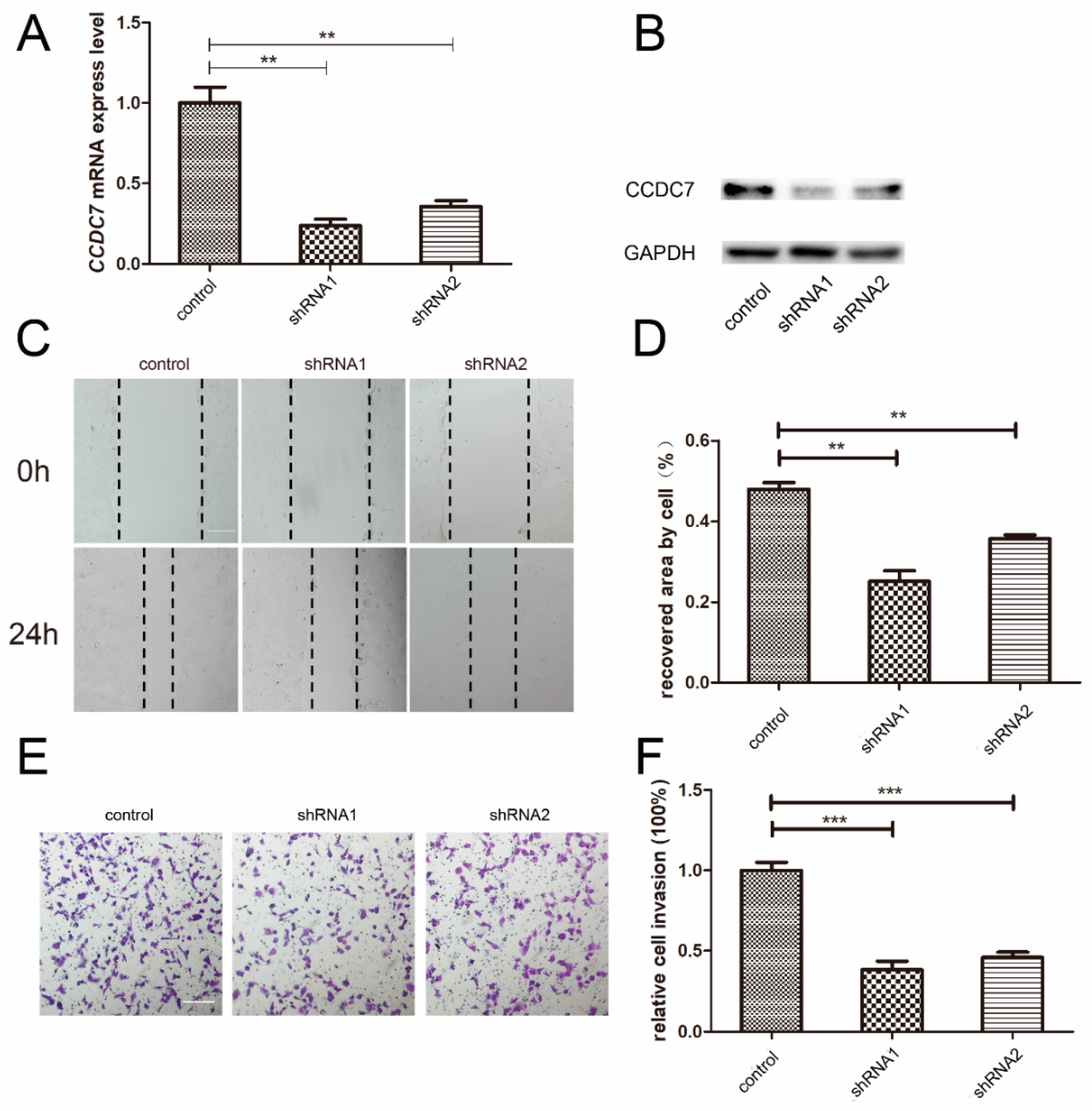
3.5. Effects of CCDC7 on JEG-3 Cell Migration and Invasion

The aberrant migration and invasion abilities of extravillous trophoblasts (EVTs) have been discovered to be involved in the pathogenesis of PE. In this study, the stable transfection of shRNAs was used to silence CCDC7 in choriocarcinoma JEG-3 cells. The results of RT-qPCR and Western blotting confirmed that the expression of CCDC7 was significantly decreased following transfection of the cells with shRNAs (Figure 5A,B). Scratch wound-healing and Matrigel invasion assays were conducted to investigate the effects of CCDC7 on the migration and invasion of trophoblast cells. As shown in Figure 5C–F, the migration and invasion capacities of JEG-3 cells were inhibited after CCDC7 silencing. These findings indicate that abnormal CCDC7 expression might contribute to abnormal trophoblast migration and invasion.

4. Discussion

In a previous study, the pathogenesis of preeclampsia was considered to involve two stages: abnormal placentation and the development of the maternal syndrome [20]. EOPE may arise due to defective placentation, while LOPE may center around interactions between the normal senescence of the placenta and a maternal genetic predisposition to cardiovascular and metabolic disease [21]. In this study, we detected two rare CCDC7 variants in the preeclampsia offspring from two unrelated families and found that CCDC7 had a crucial role in the development of preeclampsia by regulating the migration and invasion of trophoblast cells. This finding reveals the potential importance of rare variants in EOPE pathogenesis and supports the relationship between EOPE and abnormal placentation caused by genetic variation.
Complex disorders are often considered to be polygenic, with many common variants, each with modest effects that contribute to disease risk [7]. Importantly, the common variants detected to be responsible for preeclampsia have rarely been duplicated in different groups. By contrast, familial cases occurred commonly in preeclampsia. However, the probability is very low that a great number of common variants can be inherited by different offspring in one family to reach a threshold that causes a phenotype according to the law of segregation. Thus, rare variants are speculated to play a role in preeclampsia, especially familial preeclampsia, since rare variants often have greater effects than common variants, and it is more likely that a few rare variants are inherited by different offspring to reach the threshold. Recent studies have reported that rare variants contribute to preeclampsia. In women from multiplex preeclampsia families, Melton et al. [22] found a significant association between two relatively rare missense variants in C1orf35 and QRFPR and preeclampsia. The rare variant rs71324987 in MST1 identified in these families might also be associated with preeclampsia based on functional annotation and in silico prediction [23]. In our study, we found that rare variants of CCDC7 disturbed the migration and invasion of trophoblast cells and contributed to the development of preeclampsia, which further indicates a role of rare variants in the offspring of mothers with preeclampsia.
Coiled-coil domain-containing (CCDC) is a structural motif that had been identified in proteins and is involved in diverse biological processes [24]. Previous studies discovered that genes containing this motif play a role in many malignant tumors. For example, CCDC3 was found to be a nuclear tumor suppressor in breast cancers [25], and CCDC60 is a potential marker correlated with the prognosis of head and neck squamous cell carcinoma [26]. The CCDC7 gene was associated with the development of human cervical cancer [27] and colorectal cancer [28]. However, the function of CCDC7 remains unclear so far. Although cancer and placenta development are two different events, some properties and molecular pathways are shared by them, such as the ability to invade healthy tissues and the activation of VEGF to promote sustained angiogenesis [29]. Previous studies revealed that several CCDC members such as CCDC3 and CCDC85B exert a role in the migratory and invasive abilities of multiple cancer cells [30,31]. In this study, we detected two rare variants of CCDC7 in the offspring of two preeclampsia families and found that the knockdown of CCDC7 impaired the migration and invasion of trophoblast cell. Additionally, many CCDC proteins are involved in vascular stabilization and inflammation. CCDC97 and CCDC107 were found to be associated with coronary artery disease and diabetes-associated atherogenesis, respectively [32,33]. CCDC22 affects the proinflammatory response via the downregulation of NF-κB signaling [34]. CCDC134 promotes T-cell activation through the regulation of early T-cell receptor signaling [35]. Angiogenic imbalance and abnormal inflammatory are believed to underlie the pathogenesis of severe preeclampsia particularly maternal syndrome [21]. The decreased CCDC7 level may contribute to the maternal syndrome by dysregulating these cytokines. These findings could contribute to the further understanding of the role of CCDC7 in cancer and placental development associated with preeclampsia.
Although our study revealed a potential role of rare offspring variants in preeclampsia, there are some limitations. First, our study focused on rare variants of preeclampsia offspring, and both of these two rare variants of CCDC7 were maternal. Therefore, further study may be needed on the role of rare variants of CCDC7 on the maternal syndrome, although preeclampsia did not occur in all pregnancies of women carrying rare variants of CCDC7. Second, the details of the molecular mechanism by which the downregulation of CCDC7 decreased the migration and invasion ability of trophoblast cells remain unclear. Further investigations on the mechanism are needed for a better understanding of the function of CCDC7 and the molecular mechanism of preeclampsia caused by loss-of-function mutations in the CCDC7 gene. Additionally, the sample size was small in this study: two Han families. Rare variants in the CCDC7 gene screened in more families, especially of different races, are of importance for investigating the role of rare CCDC7 variants in the general population.

5. Conclusions

In this study, we detected rare loss-of-function variants of CCDC7 in two EOPE families, one of which resulted in decreased expressions of CCDC7 mRNA and protein. Further testing revealed that the downregulation of CCDC7 destroyed the migration and invasion abilities of trophoblast cells. These findings indicate that CCDC7plays a crucial role in EOPE via regulating the migration and invasion of trophoblast cells.

Supplementary Materials

The following supporting information can be downloaded at: https://www.mdpi.com/article/10.3390/jpm14030253/s1. More detail information about samples and DNA extraction, maternal DNA contamination analysis, and Whole exome sequencing.

Author Contributions

H.T., X.W., L.Y. (Lin Yu), and F.H. collected clinical data from two preeclampsia families. H.T. and L.Y. (Li Yu) carried out molecular genetic studies and functional testing. H.T., J.C., L.D. and D.C. designed and supervised the study. H.T. drafted the manuscript, and L.D. and D.C. revised it. All authors have read and agreed to the published version of the manuscript.

Funding

This study was supported by grants from the Guangdong Basic and Applied Basic Research Foundation (grant Nos. 2021A1515111180 and 2020A1515110312), the National Key Research and Development Program of China (grant No. 2017YFC1001402), and the National Natural Science Foundation of China (grant No. 81830045).

Institutional Review Board Statement

The study was conducted in accordance with the Declaration of Helsinki, and approved by the Ethics Committee of the Third Affiliated of Guangzhou Medical University (2021-242).

Informed Consent Statement

Informed consent was obtained from all subjects involved in the study.

Data Availability Statement

The data presented in this study are available on request from the corresponding author.

Acknowledgments

We thank the patients, their families, and their clinicians for their participation in this study.

Conflicts of Interest

The authors declare no conflicts of interest.

Abbreviations

CCDCCoiled-coil domain-containing
CTBscytotrophoblast cells
EOPEEarly-onset preeclampsia
GWASGenome-wide association studies
LOPELate-onset preeclampsia
MMPsMatrix metalloproteinases
PVDFPolyvinylidene fluoride
qRT-PCRQuantitative real-time polymerase chain reaction
SDS-PAGESodium dodecyl sulfate-polyacrylamide gel-electrophoresis
STBsSyncytiotrophoblasts
WESWhole-exome sequencing
WGSWhole-genome sequencing

References

  1. ACOG. Practice Bulletin No. 202: Gestational Hypertension and Preeclampsia. Obstet. Gynecol. 2019, 133, 1. [Google Scholar] [CrossRef]
  2. Dimitriadis, E.; Rolnik, D.L.; Zhou, W.; Estrada-Gutierrez, G.; Koga, K.; Francisco, R.P.V.; Whitehead, C.; Hyett, J.; Costa, F.d.S.; Nicolaides, K.; et al. Pre-eclampsia. Nat. Rev. Dis. Primers 2023, 9, 8. [Google Scholar] [CrossRef]
  3. Nilsson, E.; Ros, H.S.; Cnattingius, S.; Lichtenstein, P. The importance of genetic and environmental effects for pre-eclampsia and gestational hypertension: A family study. BJOG 2004, 111, 200. [Google Scholar] [CrossRef]
  4. Mogren, I.; Högberg, U.; Winkvist, A.; Stenlund, H. Familial occurrence of preeclampsia. Epidemiology 1999, 10, 518. [Google Scholar] [CrossRef]
  5. Parada-Niño, L.; Castillo-León, L.F.; Morel, A. Morel, Preeclampsia, Natural History, Genes, and miRNAs Associated with the Syndrome. J. Pregnancy 2022, 2022, 3851225. [Google Scholar] [CrossRef] [PubMed]
  6. Cnattingius, S.; Reilly, M.; Pawitan, Y.; Lichtenstein, P. Maternal and fetal genetic factors account for most of familial aggregation of preeclampsia: A population-based Swedish cohort study. Am. J. Med. Genet. A 2004, 130A, 365. [Google Scholar] [CrossRef] [PubMed]
  7. Gray, K.J.; Saxena, R.; Karumanchi, S.A. Genetic predisposition to preeclampsia is conferred by fetal DNA variants near FLT1, a gene involved in the regulation of angiogenesis. Am. J. Obstet. Gynecol. 2018, 218, 211. [Google Scholar] [CrossRef] [PubMed]
  8. van Dijk, M.; Mulders, J.; Poutsma, A.; Könst, A.A.M.; A Lachmeijer, A.M.; A Dekker, G.; A Blankenstein, M.; Oudejans, C.B.M. Maternal segregation of the Dutch preeclampsia locus at 10q22 with a new member of the winged helix gene family. Nat. Genet. 2005, 37, 514. [Google Scholar] [CrossRef] [PubMed]
  9. Dunk, C.E.; van Dijk, M.; Choudhury, R.; Wright, T.J.; Cox, B.; Leavey, K.; Harris, L.K.; Jones, R.L.; Lye, S.J. Functional Evaluation of STOX1 (STORKHEAD-BOX PROTEIN 1) in Placentation, Preeclampsia, and Preterm Birth. Hypertension 2021, 77, 475. [Google Scholar] [CrossRef] [PubMed]
  10. van Dijk, M.; van Bezu, J.; van Abel, D.; Dunk, C.; Blankenstein, M.A.; Oudejans, C.B.; Lye, S.J. The STOX1 genotype associated with pre-eclampsia leads to a reduction of trophoblast invasion by alpha-T-catenin upregulation. Hum. Mol. Genet. 2010, 19, 2658. [Google Scholar] [CrossRef] [PubMed]
  11. Iglesias-Platas, I.; Monk, D.; Jebbink, J.; Buimer, M.; Boer, K.; van der Post, J.; Hills, F.; Apostolidou, S.; Ris-Stalpers, C.; Stanier, P.; et al. STOX1 is not imprinted and is not likely to be involved in preeclampsia. Nat. Genet. 2007, 39, 279–280. [Google Scholar] [CrossRef]
  12. Kivinen, K.; Peterson, H.; Hiltunen, L.; Laivuori, H.; Heino, S.; Tiala, I.; Knuutila, S.; Rasi, V.; Kere, J. Evaluation of STOX1 as a preeclampsia candidate gene in a population-wide sample. Eur. J. Hum. Genet. 2007, 15, 494. [Google Scholar] [CrossRef]
  13. McGinnis, R.; The FINNPEC Consortium; Steinthorsdottir, V.; O Williams, N.; Thorleifsson, G.; Shooter, S.; Hjartardottir, S.; Bumpstead, S.; Stefansdottir, L.; Hildyard, L.; et al. Variants in the fetal genome near FLT1 are associated with risk of preeclampsia. Nat. Genet. 2017, 49, 1255. [Google Scholar] [CrossRef] [PubMed]
  14. Ghralaigh, F.N.; Gallagher, L.; Lopez, L.M. Autism spectrum disorder genomics: The progress and potential of genomic technologies. Genomics 2020, 112, 5136. [Google Scholar] [CrossRef] [PubMed]
  15. Hansen, R.D.; Christensen, A.F.; Olesen, J. Family studies to find rare high risk variants in migraine. J. Headache Pain. 2017, 18, 32. [Google Scholar] [CrossRef] [PubMed]
  16. Fritsche, L.G.; Igl, W.; Bailey, J.N.C.; Grassmann, F.; Sengupta, S.; Bragg-Gresham, J.L.; Burdon, K.P.; Hebbring, S.J.; Wen, C.; Gorski, M.; et al. A large genome-wide association study of age-related macular degeneration highlights contributions of rare and common variants. Nat. Genet. 2016, 48, 134. [Google Scholar] [CrossRef] [PubMed]
  17. Huusko, J.M.; Karjalainen, M.K.; Graham, B.E.; Zhang, G.; Farrow, E.G.; Miller, N.A.; Jacobsson, B.; Eidem, H.R.; Murray, J.C.; Bedell, B.; et al. Whole exome sequencing reveals HSPA1L as a genetic risk factor for spontaneous preterm birth. PLoS Genet. 2018, 14, e1007394. [Google Scholar] [CrossRef]
  18. Liu, X.; Lai, H.; Xin, S.; Li, Z.; Zeng, X.; Nie, L.; Liang, Z.; Wu, M.; Zheng, J.; Zou, Y. Whole-exome sequencing identifies novel mutations in ABC transporter genes associated with intrahepatic cholestasis of pregnancy disease: A case-control study. BMC Pregnancy Childbirth. 2021, 21, 110. [Google Scholar] [CrossRef] [PubMed]
  19. Canfield, J.; Arlier, S.; Mong, E.F.; Lockhart, J.; Van Wye, J.; Guzeloglu-Kayisli, O.; Schatz, F.; Magness, R.R.; Lockwood, C.J.; Tsibris, J.C.M.; et al. Decreased LIN28B in preeclampsia impairs human trophoblast differentiation and migration. Faseb. J. 2019, 33, 2759. [Google Scholar] [CrossRef] [PubMed]
  20. Phipps, E.A.; Thadhani, R.; Benzing, T.; Karumanchi, S.A. Pre-eclampsia: Pathogenesis, novel diagnostics and therapies. Nat. Rev. Nephrol. 2019, 15, 275. [Google Scholar] [CrossRef]
  21. Burton, G.J.; Redman, C.W.; Roberts, J.M.; Moffett, A. Pre-eclampsia: Pathophysiology and clinical implications. BMJ 2019, 366, l2381. [Google Scholar] [CrossRef] [PubMed]
  22. Melton, P.E.; Johnson, M.P.; Gokhale-Agashe, D.; Rea, A.J.; Ariff, A.; Cadby, G.; Peralta, J.M.; McNab, T.J.; Allcock, R.J.; Abraham, L.J.; et al. Whole-exome sequencing in multiplex preeclampsia families identifies novel candidate susceptibility genes. J. Hypertens. 2019, 37, 997. [Google Scholar] [CrossRef] [PubMed]
  23. Linhares, N.D.; Conceição, I.M.; Sandrim, V.C.; Luizon, M.R. Candidate genes identified by whole-exome sequencing in preeclampsia families: Insights into functional annotation and in-silico prediction of deleterious variants. J. Hypertens. 2020, 38, 372. [Google Scholar] [CrossRef] [PubMed]
  24. Wang, J.; Wu, X.; Dai, W.; Li, J.; Xiang, L.; Tang, W.; Lin, J.; Zhang, W.; Liu, G.; Yang, Q.; et al. The CCDC43-ADRM1 axis regulated by YY1, promotes proliferation and metastasis of gastric cancer. Cancer Lett. 2020, 482, 90. [Google Scholar] [CrossRef] [PubMed]
  25. Omari, S.; Lee, H.; Wang, J.; Zeng, S.X.; Lu, H. Extracellular and intracellular functions of coiled-coil domain containing 3. J. Mol. Cell Biol. 2023, 15, mjad037. [Google Scholar] [CrossRef] [PubMed]
  26. Liu, Z.; Chen, S.; Jia, W.; Qian, Y.; Yang, X.; Zhang, M.; Fang, T.; Liu, H. Comprehensive analysis reveals CCDC60 as a potential biomarker correlated with prognosis and immune infiltration of head and neck squamous cell carcinoma. Front Oncol. 2023, 13, 1113781. [Google Scholar] [CrossRef]
  27. Shen, Y.-M.; He, X.; Deng, H.-X.; Xie, Y.-P.; Wang, C.-T.; Wei, Y.-Q.; Zhao, X. Overexpression of the hBiot2 gene is associated with development of human cervical cancer. Oncol. Rep. 2011, 25, 75. [Google Scholar]
  28. Sun, X.-F.; Shen, Y.-M.; Arbman, G.; Sandström, P.; Gullstrand, P.; Wei, Y.-Q.; Zhang, H.; Rosell, J.; Olsson, B.; Peng, F.; et al. Novel gene hBiot2 is an independent prognostic factor in colorectal cancer patients. Oncol. Rep. 2012, 27, 376. [Google Scholar] [CrossRef]
  29. Costanzo, V.; Costanzo, V.; Bardelli, A.; Bardelli, A.; Siena, S.; Siena, S.; Abrignani, S.; Abrignani, S. Exploring the links between cancer and placenta development. Open Biol. 2018, 8, 180081. [Google Scholar] [CrossRef]
  30. Zhang, X.-F.; An, M.-Z.; Ma, Y.-P.; Lu, Y.-M. Regulatory effects of CCDC3 on proliferation, migration, invasion and EMT of human cervical cancer cells. Eur. Rev. Med. Pharmacol. Sci. 2019, 23, 3217. [Google Scholar] [CrossRef]
  31. Feng, Y.; Gao, Y.; Yu, J.; Jiang, G.; Zhang, X.; Lin, X.; Han, Q.; Rong, X.; Xu, H.; Li, Q.; et al. CCDC85B promotes non-small cell lung cancer cell proliferation and invasion. Mol. Carcinog. 2019, 58, 126. [Google Scholar] [CrossRef] [PubMed]
  32. Miller, C.L.; Pjanic, M.; Wang, T.; Nguyen, T.; Cohain, A.; Lee, J.D.; Perisic, L.; Hedin, U.; Kundu, R.K.; Majmudar, D.; et al. Integrative functional genomics identifies regulatory mechanisms at coronary artery disease loci. Nat. Commun. 2016, 7, 12092. [Google Scholar] [CrossRef] [PubMed]
  33. Huang, Q.; Deng, G.; Wei, R.; Wang, Q.; Zou, D.; Wei, J. Comprehensive Identification of Key Genes Involved in Development of Diabetes Mellitus-Related Atherogenesis Using Weighted Gene Correlation Network Analysis. Front. Cardiovasc. Med. 2020, 7, 580573. [Google Scholar] [CrossRef] [PubMed]
  34. Laulumaa, S.; Varjosalo, M. Commander Complex-A Multifaceted Operator in Intracellular Signaling and Cargo. Cells 2021, 10, 3447. [Google Scholar] [CrossRef]
  35. Zhang, T.; Shi, Q.; Gu, H.; Yu, B.; Yin, S.; Ge, Q.; Mo, X.; Liu, X.; Huang, J. CCDC134 facilitates T cell activation through the regulation of early T cell receptor signaling. Front. Immunol. 2023, 14, 1133111. [Google Scholar] [CrossRef]
Figure 1. Family pedigree of people ((A), family A; (B), family B) enrolled in this study. Orange circles represent the preeclamptic mother, and black circles and boxes represent preeclamptic offspring. “+” and “−” indicate mutant allele and wild-type allele, respectively.
Jpm 14 00253 g001
Figure 2. Rare variants of CCDC7 detected in two EOPE families and conservation analysis. (A) Variants c.625C>T (p.R209C) and c.1015C>T (p.R339*) were identified in family A and family B, respectively; (B) variants c.625C>T (p.R209C) and c.1015C>T (p.R339*) localized in exon 7 and exon 13, respectively; and (C) both of these two mutations are located in highly conservational sites across multiple species. Red represents the mutant site and “*” indicates a stop codon.
Jpm 14 00253 g002
Figure 3. CCDC7 localization and expression in human placenta of different trimesters. (A) Immunofluorescence showed that CCDC7 localized in cytoplasm of cytotrophoblast, syncytiotrophoblast, and vascular epithelial cells in the human placenta and did not change during gestation; quantitative real-time polymerase chain reaction (B) and Western blotting (C) showed that the mRNA and protein levels of CCDC7 elevated with advancing gestational age. * p < 0.05, ** p < 0.01. Scale bars, 25 μm.
Jpm 14 00253 g003
Figure 4. CCDC7 localization and expression in placenta from preeclampsia families and controls. (A) Immunofluorescence shows that the localization of CCDC7 in the placenta from the two preeclampsia families was the same as that in the controls. Quantitative real-time PCR (B) and Western blotting (C) showed that mRNA and protein levels of CCDC7 were decreased in the placenta from family B (p.R339*) and did not change in the placenta from family A (p.R209C) compared with those of age-matched controls (p.R339* vs. control2; p.R209C vs. control1). * p < 0.05; ns, not significant. Scale bars, 25 μm.
Jpm 14 00253 g004
Figure 5. Effects of CCDC7 knockdown on the proliferation, migration, and invasion of JEG3 cells. Quantitative real-time PCR (A) and Western blotting (B) showed that shRNA1 and shRNA2 significantly decreased the expression level of CCDC7 mRNA and protein. Wound-healing assay (C,D) and transwell invasion assay (E,F) showed that the migration and invasion abilities of trophoblast cells decreased after knockdown of CCDC7. ** p < 0.01, *** p < 0.001. Scale bars, 50 μm in (C,E).
Jpm 14 00253 g005
Table 1. Detail features of mothers and offspring from preeclampsia families.
Family No.AAABB
MotherII-2II-6II-8I-2II-2
Onset time at pregnancy (Week)24+127+032+0NA32+1
Gestational age at delivery (Week)34+427+432+036+436+4
High blood pressure *YesYesYesYesYes
Proteinuria *YesYesYesYesYes
Impaired liver function *NoNoNoNANo
Renal insufficiency *NoNoNoNANo
Thrombocytopenia *NoNoNoNANo
Pulmonary edema *NoNoNoNANo
Headache and/or visual symptoms *NoNoYesNANo
OffspringIII-1III-8III-9II-2III-2IIII-3
SexFemaleFemaleFemaleFemaleMaleFemale
Birth weight (g)21007501500NA23402470
Apgar score 1 min10′8′4′NA10′10′
Apgar score 5 min10′10′7′NA10′10′
Apgar score 10 min10′10′10′NA10′10′
* Diagnostic criteria for all these symptoms were referred from the ACOG practice bulletin No. 202.
Disclaimer/Publisher’s Note: The statements, opinions and data contained in all publications are solely those of the individual author(s) and contributor(s) and not of MDPI and/or the editor(s). MDPI and/or the editor(s) disclaim responsibility for any injury to people or property resulting from any ideas, methods, instructions or products referred to in the content.

