Next Article in Journal
Risk of Infertility in Males with Obstructive Sleep Apnea: A Nationwide, Population-Based, Nested Case-Control Study
Previous Article in Journal
Who to Be Treated: Nomogram Using Self-Reported Periodontal Screening Instrument among English-Speaking Adults in Multi-Ethnic Singapore
 
 
jpm-logo
Article Menu

Article Menu

Article

Common Variation in the PIN1 Locus Increases the Genetic Risk to Suffer from Sertoli Cell-Only Syndrome

by
Miriam Cerván-Martín
1,2,†,
Lara Bossini-Castillo
1,2,*,†,
Andrea Guzmán-Jimenez
1,
Rocío Rivera-Egea
3,4,
Nicolás Garrido
4,5,
Saturnino Luján
5,
Gema Romeu
5,
Samuel Santos-Ribeiro
6,7,
IVIRMA Group
,
Lisbon Clinical Group
,
José A. Castilla
2,8,9,
M. Carmen Gonzalvo
2,8,
Ana Clavero
2,8,
F. Javier Vicente
2,10,
Vicente Maldonado
11,
Sara González-Muñoz
1,
Inmaculada Rodríguez-Martín
1,12,
Miguel Burgos
1,
Rafael Jiménez
1,
Maria Graça Pinto
13,
Isabel Pereira
14,
Joaquim Nunes
14,
Josvany Sánchez-Curbelo
15,
Olga López-Rodrigo
15,
Iris Pereira-Caetano
16,
Patricia Isabel Marques
17,18,
Filipa Carvalho
17,19,
Alberto Barros
17,19,
Lluís Bassas
15,
Susana Seixas
17,18,
João Gonçalves
16,20,
Sara Larriba
21,
Alexandra M. Lopes
17,18,
F. David Carmona
1,2,§ and
Rogelio J. Palomino-Morales
2,22,§
add Show full author list remove Hide full author list
1
Departamento de Genética e Instituto de Biotecnología, Centro de Investigación Biomédica, Universidad de Granada, Armilla, 18100 Granada, Spain
2
Instituto de Investigación Biosanitaria ibs.GRANADA, 18012 Granada, Spain
3
Andrology Laboratory and Sperm Bank, IVIRMA Valencia, 46015 Valencia, Spain
4
IVI Foundation, Health Research Institute La Fe, 46026 Valencia, Spain
5
Servicio de Urología, Hospital Universitari i Politecnic La Fe e Instituto de Investigación Sanitaria La Fe (IIS La Fe), 46026 Valencia, Spain
6
IVI-RMA Lisbon, 1800-282 Lisbon, Portugal
7
Department of Obstetrics and Gynecology, Faculty of Medicine, University of Lisbon, 1649-028 Lisbon, Portugal
8
Unidad de Reproducción, UGC Obstetricia y Ginecología, HU Virgen de las Nieves, 18014 Granada, Spain
9
CEIFER-GAMETIA Biobank, 18004 Granada, Spain
10
UGC de Urología, HU Virgen de las Nieves, 18014 Granada, Spain
11
UGC de Obstetricia y Ginecología, Complejo Hospitalario de Jaén, 23007 Jaén, Spain
12
Instituto de Parasitología y Biomedicina López-Neyra, 18016 Granada, Spain
13
Centro de Medicina Reprodutiva, Maternidade Alfredo da Costa, Centro Hospitalar Universitário de Lisboa Central, 2890-045 Lisbon, Portugal
14
Departamento de Obstetrícia, Ginecologia e Medicina da Reprodução, Hospital de Santa Maria, Centro Hospitalar Universitário de Lisboa Norte, 1649-028 Lisbon, Portugal
15
Laboratory of Seminology and Embryology, Andrology Service-Fundació Puigvert, 08025 Barcelona, Spain
16
Departamento de Genética Humana, Instituto Nacional de Saúde Dr. Ricardo Jorge, 1600-609 Lisbon, Portugal
17
i3S—Instituto de Investigação e Inovação em Saúde, Universidade do Porto, 4200-135 Porto, Portugal
18
Institute of Molecular Pathology and Immunology of the University of Porto (IPATIMUP), 4200-135 Porto, Portugal
19
Serviço de Genética, Departamento de Patologia, Faculdade de Medicina da Universidade do Porto, 4200-319 Porto, Portugal
20
ToxOmics—Centro de Toxicogenómica e Saúde Humana, Nova Medical School, 1169-056 Lisbon, Portugal
21
Human Molecular Genetics Group, Bellvitge Biomedical Research Institute (IDIBELL), L’Hospitalet de Llobregat, 08908 Barcelona, Spain
22
Departamento de Bioquímica y Biología Molecular I, Universidad de Granada, 18071 Granada, Spain
*
Author to whom correspondence should be addressed.
These authors contributed equally to this work.
The full list of membership of the authors’ group lists is provided in the Supplementary File S1.
§
These authors contributed equally to this work.
J. Pers. Med. 2022, 12(6), 932; https://doi.org/10.3390/jpm12060932
Submission received: 20 April 2022 / Revised: 26 May 2022 / Accepted: 30 May 2022 / Published: 4 June 2022

Abstract

:
We aimed to analyze the role of the common genetic variants located in the PIN1 locus, a relevant prolyl isomerase required to control the proliferation of spermatogonial stem cells and the integrity of the blood–testis barrier, in the genetic risk of developing male infertility due to a severe spermatogenic failure (SPGF). Genotyping was performed using TaqMan genotyping assays for three PIN1 taggers (rs2287839, rs2233678 and rs62105751). The study cohort included 715 males diagnosed with SPGF and classified as suffering from non-obstructive azoospermia (NOA, n = 505) or severe oligospermia (SO, n = 210), and 1058 controls from the Iberian Peninsula. The allelic frequency differences between cases and controls were analyzed by the means of logistic regression models. A subtype specific genetic association with the subset of NOA patients classified as suffering from the Sertoli cell-only (SCO) syndrome was observed with the minor alleles showing strong risk effects for this subset (ORaddrs2287839 = 1.85 (1.17–2.93), ORaddrs2233678 = 1.62 (1.11–2.36), ORaddrs62105751 = 1.43 (1.06–1.93)). The causal variants were predicted to affect the binding of key transcription factors and to produce an altered PIN1 gene expression and isoform balance. In conclusion, common non-coding single-nucleotide polymorphisms located in PIN1 increase the genetic risk to develop SCO.

1. Introduction

While mature proteins require a specific 3D structure to exert their functional activities, such complex conformation is not always determined by their amino acid sequence alone [1,2]. Actually, small proteins (less than 100 amino acids) fold autonomously, but larger proteins tend to misfold [1]. Since a higher number of long proteins are required in eukaryotes compared to prokaryotes, co-translational folding strategies have been developed and specific proteins are used to control eukaryotic protein folding and to keep protein homeostasis (known as proteostasis) [1,3]. Chaperones are generally located in the endoplasmic reticulum (ER) and interact in a fine-tuned network to prevent both protein misfolding and intermediate aggregate formation [1,3]. These intermediate aggregates turn on a specific unfolded protein response (UPR) in the ER and have been identified to play a role in a number of human diseases [3].
A major aspect of protein folding is the cis/trans conformation of the peptide bond between each proline residue and its preceding amino acid [4]. The majority of peptide bonds adopt a trans-conformation, but in the case of proline a higher proportion of cis conformations are required to maintain a proper protein structure and function [4]. The proline isomerization is a slow process with high entropy and it needs the catalyzation mediated by specific enzymes known as prolyl isomerases [4]. Moreover, the coexistence of cis/trans isomers in proteins has been suggested to be linked to regulatory processes that control the functional states of proteins, although this role remains controversial and hard to prove in living cells [4].
PIN1 is one of the most studied and relevant prolyl isomerases in humans. This protein binds the phosphorylated serine or threonine residues preceding proline motifs (phospho-Ser/Thr-Pro) and catalyzes cis/trans isomerization of the peptide bonds [5]. PIN1 acts on cell cycle regulator proteins, such as cyclin-dependent kinases (CDKs), and it is involved in the fine control of their functions, stability, localization, interactions and activity [5]. Therefore, PIN1 has a central role in cell cycle progression and cancer [5]. Moreover, this isomerase has also been linked to the immune system specially in promoting inflammation and reactive-oxygen species (ROS) [5].
Spermatogenesis consists of a series of developmental steps that comprise the differentiation of a spermatogonia into a mature spermatozoon through mitosis, meiosis and spermiogenesis [6] regulated by the coordinated expression of many genes. Specifically, the generation of viable spermatozoa involves complex protein interactions and the activation of specific biological pathways that rely on a correct protein structure and activity [6]. Interestingly, Pin1 is highly expressed in adult mice testis, particularly in spermatogonia and Sertoli cells [7,8]. It has been shown that Pin1 knockout mice (Pin1−/−) are able to complete spermatogenesis in their early life but suffer a progressive spermatogonial stem cell (SSC) loss with age [7,8]. Indeed, it has been proposed that Pin1 is required to control the proliferation, survival and cellular commitment of undifferentiated spermatogonia by promoting mitosis in this cell lineage [7,8]. Additionally, Sertoli cells in Pin1−/− mice showed a reduced expression of N-Cadherin, a central protein in the blood–testis barrier (BTB) tight-junction system [9]. Thus, Pin1 has a role in controlling the integrity of the BTB, which is essential to maintain the immune privilege of the testis and to prevent a self-attack of the immune cells to the male germline [9].
Despite all the previously described connections with male infertility, mutations in PIN1 have not yet been described in human male infertility cases. Male infertility affects approximately 7% of men and represents an economic, social and psychological burden for couples that seek biological parenthood all over the world [10]. Regarding male infertility, most cases are due to defects in the spermatogenic process or SPGF. The most severe forms of SPGF comprise cases with very few or no spermatozoa in the ejaculate due to non-obstructive causes: severe oligospermia (SO) or non-obstructive azoospermia (NOA), respectively [10,11]. The histological profiles of SPGF are heterogeneous and not all patients can benefit from testicular sperm extraction (TESE) and subsequent in vitro fertilization (IVF) techniques [11]. Furthermore, a molecular cause for infertility can only be established for around 25% of the NOA cases [12]. In the remaining patients, the nature of the infertility is classified as idiopathic [12]. Unfortunately, these infertile men usually undergo testicular biopsy without a reliable estimation of the TESE success. There is growing evidence that the etiology of idiopathic NOA cases might be complex and that common genetic variants in the human genome might contribute to this condition [13].
Considering the above, this study aimed to address the association between human SPGF and common genetic variants located in the PIN1 locus in a considerably large cohort of European infertile men.

2. Materials and Methods

2.1. Patients and Clinical Definition

This study comprises the largest SPGF cohort with European ancestry recruited for a genetic association study to date. The sample set included 715 SPGF cases from the Iberian Peninsula, who were classified as either SO (n = 210) or NOA (n = 505) as described elsewhere [14,15]. A geographically matched population with similar ethnicity and age was used as a control. This control set reached 1058 men, 358 of them were unaffected and 700 were representative of the male population (with, at least, one self-reported biological child).
SO and NOA were clinically identified according to the guidelines of the World Health Organization [16] in public hospitals and private clinics in Spain and Portugal. These conditions were diagnosed by the presence of <5 million spermatozoa/mL semen (SO) or no sperm in the ejaculate after two high-speed centrifugations (NOA). Since only idiopathic SPGF cases were considered in the analyses, individuals with abnormal karyotypes, chromosome Yq deletions, testicular disorders (orchitis, testis maldevelopment, bilateral cryptorchidism, bilateral varicocele and obstruction of vas deferens) or any sign of a possible obstructive cause were not included in our study. No significant age differences were observed between the different SPGF clinical subtypes. Although quantitative data regarding hormone levels and testis reduction were not available in the majority of the cases, we did not exclude patients based on these parameters as the clinicians did consider their values for diagnosis as established in the Canadian Urological Association (CUA) [17] and the American Urological Association (AUA)/American Society for Reproductive Medicine (ASRM) guidelines [18].
Due to the clinical relevance of the testicular biopsies, the samples obtained in such interventions were used for histological classification and resulted in 3 main subtypes (Supplementary Table S1): (1) hypospermatogenesis (HS), characterized by very low numbers of mature motile sperm cells in few testicular regions, (2) meiotic arrest (MA), showing a meiotic failure at >90% of their spermatogonia or primary spermatocytes and (3) Sertoli cell-only (SCO) syndrome if no germ cells were observed and only Sertoli cells were present in the seminal tubules. The successful or unsuccessful retrieval of sperm cells in the biopsies was stated as TESE+ or TESE-, respectively [13,15].

2.2. SNP Selection and Genotyping

The PIN1 gene is located in a 14 kb long region in the human chromosome 19 (Figure 1), which is expressed in all the cellular subtypes, including somatic and germ cells, present in testis as shown in Guo et al. [19] (Supplementary Figure S1) and represents a good candidate gene to test for genetic association with male infertility. The complete PIN1 locus, including both the coding sequence and the regulatory regions (±5 kp from the gene), forms a unique linkage disequilibrium (LD) block in the included in the European cohort of the 1000 Genomes Project Phase III (1KGPh3) (https://ldlink.nci.nih.gov/?tab=ldmatrix; accessed on: 15 June 2021) [20] (Figure 1, Supplementary Table S2). Three single-nucleotide polymorphisms (SNPs) were selected to address the genetic association of this locus with SPGF: rs2287839, rs2233678 and rs62105751. Two of these variants are located in the distant 5′ upstream regulatory region (URR) in the 5′ vicinity of PIN1 promoter and the remaining variant is located in the third intron in this gene (Figure 1, note that PIN1-DT refers to PIN1 divergent transcript). We applied a SNP tagging strategy as implemented in Haploview V.4.2 (Broad Institute; Cambridge, Massachusetts, USA) [21] covering all the common genetic variation (r2 ≥ 0.8) included in the European cohort of the 1000 Genome Project Phase III (1KGPh3) (https://www.ensembl.org/Homo_sapiens/; accessed on: 15 June 2021) [22]. Therefore, the variants are representative of different minor allele frequency (MAF) ranges: high (MAF > 0.3), medium (0.1 < MAF < 0.3) and low (MAF < 0.1).
We obtained genomic DNA from peripheral blood mononuclear cells (PBMCs) using standard DNA isolation kits and carried out the genotyping of each individual for these SNPs in a 7900HT Fast Real-Time PCR System (Applied Biosystems, Foster City, California, USA) using the TaqMan™ (Thermo Fisher Scientific, Pleasanton, California, USA) allelic discrimination technology with 3 pre-designed probes (assay IDs: C__16183184_40, C___2885187_10, C__89465150_10), as previously described [15]. The genotype call rate success was over 99% for all three genetic variants.

2.3. Statistical Analyses

The statistical power of the study cohort to detect an association was estimated with the software Genetic Association Study (GAS) Power Calculator (https://doi.org/10.1101/164343; accessed on: 15 June 2021) (Supplementary Table S3). Considering a strong allelic effect (OR ≥ 1.5), the recruited cohort reached a statistical power higher than 80% for the 3 selected polymorphisms. A possible deviation from Hardy–Weinberg equilibrium (HWE) was evaluated using a Χ2 test considering a 5% significance level.
Case–control comparisons of the allele and genotype frequencies were performed between the established SPGF groups and the control population. Moreover, NOA cases with and without a specific phenotype/TESE outcome were also compared to remove the disease as a possible confounding factor. The threshold for statistical significance was set at p-value < 0.05 after multiple testing corrections by Benjamini and Hochberg False Discovery Rate (FDR) [23]. We calculated the corresponding odds ratios (ORs) and 95% confidence intervals (CI) for all the analyses. The additive, dominant, recessive and genotypic (2 degrees of freedom) association models were tested based on logistic regression using PLINK version 1.9. (https://www.cog-genomics.org/plink/1.9/credits accessed on: 15 June 2021 [24]. To control for a possible geographical origin effect, the country of origin (Spain or Portugal) was included as a covariate in all the analyses.
As mentioned above, the three analyzed polymorphisms belong to the same haplotype block. Therefore, haplotype-based logistic regression tests were performed with geographical origin included as a covariate. Allelic combinations showing a MAF < 0.01 were not considered in these analyses. In order to confirm the contribution of each SNP to the significance of the genetic association compared to the haplotypes, a likelihood ratio test was conducted in which the haplotype model was tested against each independent SNP model.
Finally, we tested the independence between the studied polymorphisms by conditional logistic regression analyses as implemented in the PLINK software [24].

2.4. In Silico SNP Functional Characterization

We took advantage of the large variety of public databases and resources that provide functional evidence to prioritize variants and propose a putative molecular mechanism for the observed associations. We extended our in silico SNP functional characterization not only to the genotyped SNPs but also to all their proxies (genetics variants showing a LD r2 ≥ 0.8 in the European subpopulation included in the 1000 Genomes Project) as implemented in LDlink (https://ldlink.nci.nih.gov/?tab=ldmatrix; accessed date: 13 January 2022) [20]. Genomic coordinates for all the reported variants and regions correspond to the GRCh38 human genome build.
The role of the different polymorphisms as cis expression and/or splicing quantitative trait loci (QTL), eQTL and sQTL, respectively, was obtained from the v8 GTEx data release (https://www.gtexportal.org/ accessed on: 25 July 2021) [25]. We prioritized those variants with a QTL effect in the testis. Their locations in the regulatory regions in testis were defined by overlap with testis specific assays in ENCODE [26]: DNase-seq hypersensitivity sites (ENCFF323BCL, ENCFF608KRZ); CTCF (ENCODE sample references: ENCFF300WML, ENCFF559LDF, ENCFF644JKD, ENCFF767LMP, ENCFF788RFY, ENCFF855EVV) and POLR2A (ENCFF535DHF, ENCFF651APG) protein ChIP-seqs; and H3K4me3 (ENCFF286DAB, ENCFF509DBT), H3K4me1 (ENCFF316MJM), H3K27ac (ENCFF610XSK, ENCFF819NRA), H3K9me3 (ENCFF711LHL) and H3K27me3 (ENCFF881OHS) histone modification ChIP-seqs. Additional functional clues per SNP were also obtained from dedicated integration databases such as SNPnexus (https://www.snp-nexus.org/, accessed on: 25 July 2021) [27], HaploReg (https://pubs.broadinstitute.org/mammals/haploreg/haploreg.php, accessed on 25 July 2021) [28] and SNP2TFBS (https://ccg.epfl.ch/snp2tfbs/ accessed on: 25 July 2021) [29]. These online tools organize the information included in: Ensembl, SIFT, Polyphen, CpG, Vista enhancers, miRbase, TarBase, TargetScan, miRNA Registry, snoRNA-LBME-DB, Roadmap, Ensembl regulatory build, CADD, DeepSEA, EIGEN, FATHMM, fitCons, FunSeq2 GWAVA, REMM and RegulomeBD [30] (Supplementary Tables S4–S5).
The online tools in the GTEx and LDmatrix portals were used for figure generation together with custom R scripts (version 4.2.0,The R foundation for Statistical Computing, https://www.r-project.org/ accessed on: 25 July 2021) [20,25].

3. Results

The three analyzed variants passed all the established quality control thresholds. Moreover, our cohort showed a very high statistical power to identify genetic associations in the range of previously reported common variant associations with SPGF [14,15] (Supplementary Table S3). No statistically significant deviation from HWE was observed either for the cases or the controls.

3.1. Testing for Association with Idiopathic Spermatogenic Failure Overall

Our analyses revealed no significant associations with SPGF neither under the additive nor under the recessive models after multiple testing corrections (Table 1, Supplementary Table S6).
Nevertheless, the less common SNP, rs2287839, which is located at the URR of this locus, showed a trend towards association with SPGF under the additive model that did not pass the FDR correction (paddadj = 0.055, ORadd = 1.38 (1.06–1.81)) (Table 1). Moreover, we observed significant genetic associations for rs2287839 with SPGF under both the dominant (pdom = 9.63 × 10⁻³, ORdom = 1.46 (1.10–1.94)) and the genotypic models (pgeno = 2.39 × 10⁻²), but only the dominant model association remained significant after multiple testing adjustments (pdomadj = 2.89 × 10⁻²) (Supplementary Table S6).
While the analysis of the SO group did not present strong evidence of association with this phenotype, the comparison between the NOA patients and the control group followed the same trends of association as the SPGF group and showed significant allelic effects under the additive (padd = 7.81 × 10⁻⁴, ORadd = 1.61 (1.22–2.13)), the dominant (pdom = 3.01 × 10⁻⁴, ORdom = 1.72 (1.28–2.32)) and the genotypic models (pgeno = 1.01 × 10⁻³) in the case of rs2287839 (Supplementary Table S6). Additionally, carriers for the rs2287839 and rs62105751 were more frequent in the NOA subgroup than in the SO patient set (padd = 5.86 × 10⁻³, ORadd = 2.08 (1.24–3.50) and padd = 4.56 × 10⁻³, ORadd = 1.48 (1.13–1.94), respectively).
When the susceptibility effects of the haplotypes were tested, we observed significant associations with NOA especially for the combinations including either two or three risk or protection alleles (Supplementary Table S7). Nevertheless, we detected that none of these combinations explained the association with NOA better than rs2287839 alone (Supplementary Table S8).

3.2. PIN1 Polymorphisms Have a Subtype-Specific Effect in SCO

We performed further analyses in order to address the association of such polymorphisms with specific histological patterns of NOA. The classification of the NOA patients into more homogeneous groups based on clinical and pathological criteria brought to light that the observed risk association was clearly biased towards an extreme NOA subphenotype, with a total absence of the germline in the seminiferous tubules, namely the SCO subset of patients (Table 1).
In the case of the SCO group, we observed significant associations for the three selected genetic variants under the additive model (ORaddrs2287839 = 1.85 (1.17–2.93), ORaddrs2233678 = 1.62 (1.11–2.36), ORaddrs62105751 = 1.43 (1.06–1.93)), which remained significant even after multiple testing corrections (Table 1). Furthermore, the minor alleles showed strong risk effects for this subset in the comparison against the control group, i.e., all the observed ORs were greater than OR = 1.4 (ORaddrs2287839 = 1.85 (1.17–2.93), ORaddrs2233678 = 1.62 (1.11–2.36), ORaddrs62105751 = 1.43 (1.06–1.93)) (Table 1). It should be noted that we also observed an increased frequency in SCO compared to the rest of the NOA subsets (Table 1).
There were significant differences between the genotype distributions for the three SNPs (pgenors2287839 = 2.24 × 10⁻², pgenors2233678 = 2.93 × 10⁻², pgenors62105751 = 3.87 × 10⁻²), but only the effects of rs2233678 and rs62105751 remained significant after multiple testing corrections were applied (Supplementary Table S6). However, the best fitting inheritance model was the recessive ones for the most frequent variants, rs2233678 and rs62105751, and the dominant model for rs2287839 (Supplementary Table S6).
The haplotype analyses revealed that the combination of risk or protection alleles was associated with the SCO group (Supplementary Table S7). However, in this case, all the polymorphisms and the haplotype explained a similar proportion of the phenotypic variance (Supplementary Table S8). Only the rarest minor allele, rs2287839-G, showed a significant association with the group of individuals with an unsuccessful sperm retrieval during TESE (padd = 4.19 × 10⁻², ORadd = 1.55 (1.02–2.37), pdom =2.05 × 10⁻², ORdom = 1.70 (1.08–2.65), data not shown), which did not reach the significance level after multiple testing corrections.
Considering that there is no recombination hotspot in the PIN1 locus (Figure 1), the possibility of one versus several independent association signals was explored. We performed multiple logistic regression analyses that combined the tested variants in pairs (Supplementary Table S9). Our results showed that all the variants lost significance when conditioned (Supplementary Table S9), thus reflecting that there is no independence between them and that they tag the same association signal.

3.3. In Silico Data from the GTEx Repository Suggest That the Genetic Variants in the PIN1 Locus Have Regulatory Functions on Gene and Isoform Expression

Our experimental design allowed us to study the influence of the genetic variants located in the PIN1 locus on the susceptibility to male infertility and specially to identify their contribution to SCO as an etiologic factor. The genetic association tests highlighted the role of lower frequency variants in the predisposition to complete lack of sperm cells in the testicles as described above. Therefore, following this lead we computationally analyzed the functional evidence and predicted effects for all the genetic variants in this locus to prioritize the most likely causal variants.
As depicted in Figure 1, the selected genomic region in chromosome 19 includes both the full PIN1 gene and a PIN1 divergent transcript, which has been characterized as a long non-coding RNA (lncRNA PIN1-DT, ENSG00000267289.1). PIN1 is highly expressed in the testis (Figure 2). Considering that no SNP in the coding region of PIN1 was tagged by the associated variants, we focused on the genetic variants that have been described to affect the mRNA expression (eQTLs) or the mRNA splicing (sQTLs) of this gene in the GTEx project [25]. Up to 38 SNPs overlapped with testis-specific assays in the ENCODE database (https://www.encodeproject.org/; data obtained in 22 April 2020) [26] and have been characterized as PIN1 sQTLs (36 SNPs) or as both PIN1 eQTLs and sQTLs (2 SNPs) in the GTEx repository testicular tissue samples. In total, 4 out of these 38 SNPs encoded DNA sequence changes that were predicted to affect spermatogenesis-related transcription factors (Supplementary Table S10).
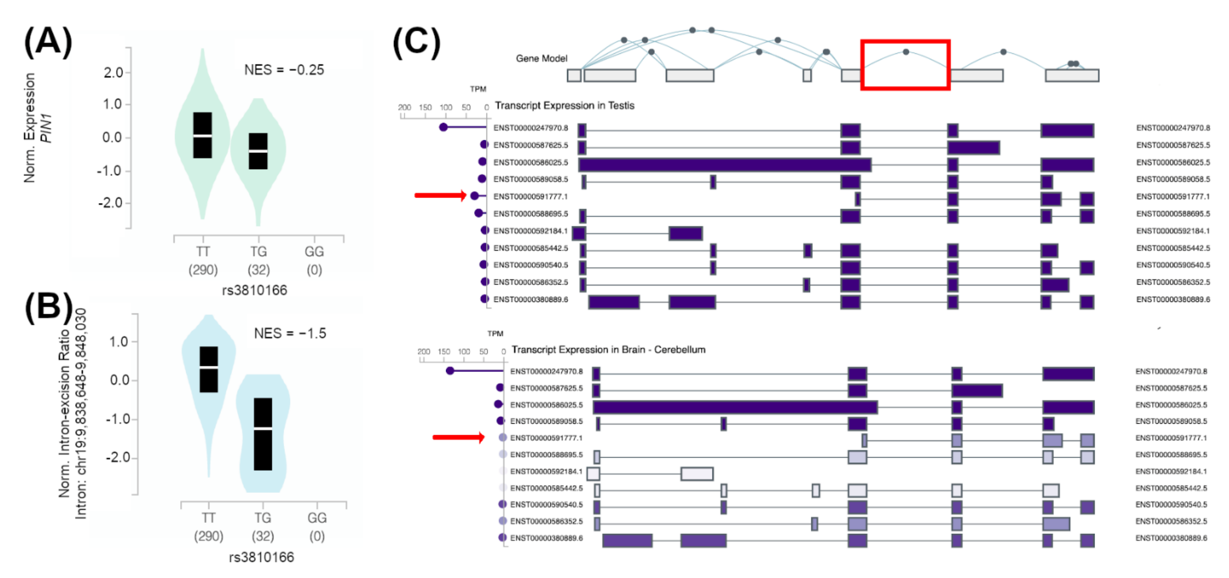
Among the prioritized variants, the rs3810166 SNP held the strongest evidence of functionality within this locus. The minor allele of this SNP, rs3810166-G, is a proxy of the observed rs2287839-G risk allele and, according to the GTEx dataset (which includes 322 individuals), it correlates with a decreased expression of PIN1 (Figure 2A) and it alters the PIN1 isoform balance in the testis (Figure 2B,C). The rs3810166 SNP is almost in complete linkage disequilibrium (r2Europeans = 0.94) with the top GTEx eQTL variant in the testicular tissue (rs138970490), with the magnitude of the reported effect (normalized effect size, NES = −0.25) corresponding to a log allelic fold change = −0.14 (i.e., a 10% decrease in PIN1 expression), which is very relevant. The individuals included in the GTEx project were healthy controls, but we hypothesize that the pathogenic effects of such changes might even be stronger in the SCO context of gene expression deregulation. Although this variant is located upstream of the PIN1 gene, it is enriched with chromatin activity, histone marks and CTCF binding sites, which supports the role of this region in the control of the expression of nearby genes (Supplementary Table S10). Additionally, the minor allele of this SNP is predicted as highly damaging by multiple functional consequence prediction algorithms and it is also predicted to alter the binding of both HDAC2, a key histone deacetylase in cell cycle progression [31], and NRSF [32], a very relevant transcriptional regulator (Supplementary Table S10). An additional prioritized SNP, rs10410379, was tagged by rs2287839 and predicted to decrease the binding of HDAC2.
The minor alleles of the two remaining polymorphisms, rs28802413 and rs10425775, have been described to affect the isoforms of PIN1 as sQTLs. They were also linked to rs2287839 and predicted to affect the binding of relevant transcription factors in the spermatogenic process such as SIN3A [33,34] and NANOG [35] (Supplementary Table S10).

4. Discussion

The analysis of common variants located in potentially relevant genes for spermatogenesis is crucial for unraveling the genetic component underlying the male infertility phenotypes with idiopathic molecular etiology [13]. Frequent mutant alleles are known to modify or disrupt gene expression subtly and, in specific contexts or if certain environmental factors are present, they might alter the correct production of male gametes [36,37]. Therefore, the reported findings should not be interpreted as highly damaging and rare mutations causing infertility, but in the context of the identification of genetic markers for a complex disease [13].
In the present study, genetic association analyses on DNA from a large and clinically well-characterized cohort were performed. Additionally, we carried out a deep in silico characterization of the prioritized polymorphisms in the PIN1 locus. Since there are no recombination hotspots in this region (Figure 1), the LD between the selected SNPs corresponded to a high D’ and a low r2 between the variants (Supplementary Table S2). We genotyped three SNPs that tag polymorphisms located in the same haplotypic block but with different MAF ranges in order to maximize the coverage of the tagging strategy.
Our results emphasized the role of PIN1 as a risk locus for male infertility. We observed associations of all the tested variants with SCO, the most severe form of NOA (Table 1). Moreover, the risk effects were also significant in the haplotype analyses (Supplementary Table S7). This phenotype is characterized by the complete absence of germ cells in the testis, in which the tubules are only composed of Sertoli cells [38]. Sertoli cells provide physical and nutritional support for the germ cells during spermatogenesis, and they form a physical barrier that prevents the immune cells from attacking the germline [39]. The present report discusses for the first time the potential association of PIN1 with SPGF, but remarkably thanks to our comprehensively characterized patient cohort, we were also able to test for subtype specific associations. In fact, the association signals observed in the SPGF, NOA and TESE- groups of patients were likely originated by the association observed in the SCO subtype. The SCO subset of patients is histologically more homogeneous and harbors the largest risk effect sizes. Therefore, it is likely that the presence of SCO patients in the SPGF, NOA and TESE- groups is responsible for the observed associations in these groups of patients.
Moreover, the association of the three selected variants, as well as the mutual dependence between them, provided us with compelling evidence for a common causal variant or variants underlying the association signal found in this region (Supplementary Tables S8 and S9). Although the most associated tag-variant, i.e., rs2287839, is located near epigenetic marks in several tissues, it is not predicted to alter the binding of relevant transcription factors in spermatogenesis. However, the strong LD patterns within the region might point towards rs2287839 as a good proxy for a putative causal risk haplotype located in the vicinity. Additionally, our results may also indicate that the causal variant/s in this locus would be in the lowest MAF ranges, as the largest effect sizes in different subsets of patients corresponded to rs2287839, which had the lowest frequency among the tested variants (MAF = 0.06).
As we aimed to prioritize plausible causal variants in this locus, we integrated a variety of functional genomic tools and datasets in an in silico approach, which led to the identification of rs3810166 as a plausible causal variant for the PIN1 association with SCO. The rs3810166 SNP seems to affect both the expression levels and the isoform balance of PIN1 in the healthy control samples of the GTEx repository. The minor allele of rs3810166-G decreases the expression of PIN1 in the testis tissue and it could additionally perturb the equilibrium between the most frequent PIN1 isoform, ENST0000247970.8, and a shorter transcript with an alternative promoter, ENST00000591777.1, which is the second most frequent PIN1 transcript in this tissue (Figure 2B,C). The alternative allele of this SNP disturbs predicted canonical binding sites of transcription factors that are essential for the maintenance of the BTB, such as NRSF [32], and SSC maintenance, such as HDAC2 [31] (Supplementary Table S10). In this functional prioritization, we found further evidence that might support variants in PIN1 as genetic risk factors to suffer from spermatogonial depletion and to develop male infertility with a SCO. Three polymorphisms tagged by the genotyped variants overlapped testis-specific epigenetic assays and showed PIN1 sQTL effects. Additionally, they were also predicted to affect the binding of other relevant transcription factors. Remarkably, the alternative allele of rs28802413 putatively influences the binding affinity of SIN3A, a key transcription factor for SSC survival (Supplementary Table S10). In line with this, the lack of Sin3a expression in mice Sertoli cells caused a microenvironment change and the loss of undifferentiated spermatogonia [33]. Furthermore, specific genetic inactivation of the Sin3A gene in the germline of adult testes led to a SCO phenotype in mice. The loss of Sin3A expression causes apoptosis of the germ cells, since they require the correct function of the Sin3–HDAC complex, but it also alters the gene expression profile in Sertoli cells [34] (Supplementary Table S10). The analysis of the Sertoli cell transcriptome has revealed that NANOG, another transcription factor that might be affected by genetic variation in PIN1 (Supplementary Table S10), is expressed in Sertoli and interstitial cells of neonatal testes (but not in the adult testes) and also in Sertoli cells from SCO patients [35].
The effect of some of these polymorphisms or a combination of them would eventually imbalance the expression of PIN1, which has been associated with male infertility in animal models. PIN1 is expressed in all the cell stages of the differentiating male germline and in Sertoli cells (Supplementary Figure S1) it has a prominent role in chromosome segregation [40] and it interacts with the androgen receptor (AR) [41]. Knockout mouse models have shown that the genetic silencing of Pin1 causes spermatogonial depletion [7], affects the Sertoli cells and disturbs the BTB [9]. Additionally, it causes a progressive loss in the progression of the mitotic cell cycle of SSC in steady state [8] and deregulates the timing of primordial germ cell proliferation during the embryonic development [42]. PIN1 has also been pinpointed as a seminal biomarker for higher fertility in porcine models [43]. Finally, we wish to highlight a recent study where the intracellular injection of PIN1 lipid nanoparticles in knockout mice (Pin1−/−) resulted in the regulation of spermatogonial proliferation and differentiation and in the restructuring of the BTB, thus rescuing fertility in male mice [44]. Therefore, we consider that our findings encourage the analysis of PIN1 as a therapeutic target to restore human male fertility.

5. Conclusions

Altogether, the selection of PIN1 as a candidate genetic risk factor for SPGF in humans and the analysis of common variation proved to be a successful strategy. We hypothesize that the nature of the majority of the cases of SPGF classified as idiopathic is complex and polygenic, with a low individual contribution of a high number of genetic risk factors, which combined with environmental factors may lead to male infertility [45,46]. However, the interrogation of the role of common genetic variation in this heterogeneous phenotype and the analysis of more homogeneous histological subsets of patients will contribute to the knowledge of the field about the disease pathogenesis. It should be noted that these approaches might eventually advance the development of a panel of genetic markers to predict TESE success and avoid surgical interventions if the odds of finding healthy spermatozoa are low, as in the case of SCO patients [13].

Supplementary Materials

The following supporting information can be downloaded at: https://www.mdpi.com/article/10.3390/jpm12060932/s1, Supplementary File S1: Authors’ group lists; Supplementary Table S1: Main clinical features of the 715 infertile men included in the study; Supplementary Table S2: Linkage disequilibrium between the genotyped SNPs; Supplementary Table S3: Estimation of the statistical power of our study for 715 patients and 1058 controls; Supplementary Table S4: Tools for generating functional prediction scores; Supplementary Table S5: RegulomeDB scoring scheme. eQTL, expression quantitative trait locus; TF, transcription factor; Supplementary Table S6: Analysis of the genotype and allele frequencies of the tested genetic variants comparing subgroups of clinical phenotypes of male infertility against fertile controls under the recessive, dominant and genotypic models; Supplementary Table S7: Pooled analysis of different allelic combinations of the PIN1 genomic region according to the specific clinical phenotypes versus controls; Supplementary Table S8: Likelihood ratio test of the associated phenotypes of male infertility versus the control cohort. Haplotype model was tested against independent SNP models; Supplementary Table S9: Conditional logistic regression analysis for the PIN1 polymorphisms considering the most associated phenotype, with is SCO; Supplementary Table S10: In silico prioritization of variants; Supplementary Figure S1: Single-cell PIN1 expression patterns in human adult testis (extracted from Guo et al. [19]).

Author Contributions

Conceptualization, R.J.P.-M. and F.D.C.; Methodology, M.C.-M., L.B.-C., A.G.-J., S.G.-M., M.B. and R.J.; Formal Analysis, M.C-M., L.B.-C, A.G.-J., M.B. and R.J.; Investigation, M.C.-M., L.B.-C., A.G.-J., S.G.-M. and I.R.-M.; Resources, R.R.-E., N.G., S.L. (Saturnino Luján), G.R., S.S.-R., J.A.C., M.C.G., A.C., F.J.V., V.M., M.G.P., I.P., J.N., J.S.-C., O.L-R., I.P.-C., P.I.M., F.C., A.B., L.B., S.S., J.G., S.L. (Sara LArriba) and A.M.L.; Data Curation, M.C.-M., L.B.-C. and A.G.-J.; Writing—Original Draft Preparation, M.C.-M. and L.B.-C.; Writing—Review and Editing, F.D.C., R.J.P.-M., A.G.-J., M.B., R.J., R.R.-E., N.G., S.L. (Saturnino Luján), G.R., S.S.-R., J.A.C., M.C.G., A.C., F.J.V., V.M., M.G.P., I.P., J.N., J.S.-C., O.L.-R., I.P.-C., P.I.M., F.C., A.B., L.B., S.S., J.G., S.L. (Sara Larriba) and A.M.L.; Visualization, R.J.P.-M. and F.D.C.; Supervision, R.J.P.-M. and F.D.C.; Funding Acquisition, R.J.P.-M. and F.D.C. All authors have read and agreed to the published version of the manuscript.

Funding

This work was supported by the Plan Andaluz de Investigación, Desarrollo e Innovación (PAIDI 2020) (ref. PY20_00212, P20_00583), and the Spanish Ministry of Economy and Competitiveness through the Spanish National Plan for Scientific and Technical Research and Innovation (ref. SAF2016–78722-R, PID2020–120157RB-I00) and the Proyectos I + D + i del Programa Operativo FEDER 2020 (ref. B-CTS-584-UGR20, B-CTS-260-UGR20). FDC was supported by the “Ramón y Cajal” program (ref. RYC-2014–16458), and LBC was supported by the Spanish Ministry of Economy and Competitiveness through the “Juan de la Cierva Incorporación” program (Grant ref. IJC2018–038026-I, funded by MCIN/AEI/10.13039/501100011033), all of them including FEDER funds. AGJ was funded by MCIN/AEI/10.13039/501100011033 and FSE “El FSE invierte en tu futuro”(grant ref. FPU20/02926). SGM was funded by a previously mentioned project (ref. PY20_00212). IPATIMUP integrates the i3S Research Unit, which is partially supported by the Portuguese Foundation for Science and Technology (FCT), financed by the European Social Funds (COMPETE-FEDER) and National Funds (projects PEstC/SAU/LA0003/2013 and POCI-01–0145-FEDER-007274). AML is funded by the Portuguese Government through FCT (IF/01262/2014). PIM is supported by the FCT post-doctoral fellowship (SFRH/BPD/120777/2016), financed from the Portuguese State Budget of the Ministry for Science, Technology and High Education and from the European Social Fund, available through the Programa Operacional do Capital Humano. ToxOmics—Centre for Toxicogenomics and Human Health, Genetics, Oncology and Human Toxicology, Nova Medical School, Lisbon, is also partially supported by FCT (Projects: UID/BIM/00009/2013 and UIDB/UIDP/00009/2020). SLarriba received support from Instituto de Salud Carlos III (grant DTS18/00101], co-funded by FEDER funds/European Regional Development Fund (ERDF)—a way to build Europe), and from “Generalitat de Catalunya” (grant 2017SGR191). SLarriba is sponsored by the “Researchers Consolidation Program” from the SNS-Dpt. Salut Generalitat de Catalunya (Exp. CES09/020). This article is related to the Ph.D. Doctoral Thesis of Miriam Cerván-Martín (grant ref. BES-2017–081222 funded by MCIN/AEI/10.13039/501100011033 and FSE “El FSE invierte en tu futuro”).

Institutional Review Board Statement

All the participants signed an informed consent form complying with the Declaration of Helsinki and received approval by the Ethics Committee “CEIM/CEI Provincial de Granada” Andalusia Spain at the session held on 26 January 2021 (approval number: 1/21). Besides, each participating center received ethical approval and complied with the requirements of their local regulatory authorities.

Informed Consent Statement

Informed consent was obtained from all subjects involved in the study.

Data Availability Statement

The generated genotyping information is available upon formal and reasonable request to the corresponding author.

Acknowledgments

We thank the National DNA Bank Carlos III (University of Salamanca, Spain) for supplying part of the control DNA samples from Spain.

Conflicts of Interest

The authors declare no conflict of interest.

References

  1. Balchin, D.; Hayer-Hartl, M.; Hartl, F.U. In vivo aspects of protein folding and quality control. Science 2016, 353, aac4354. [Google Scholar] [CrossRef] [PubMed]
  2. Stollar, E.J.; Smith, D.P. Uncovering protein structure. Essays Biochem. 2020, 64, 649–680. [Google Scholar] [CrossRef] [PubMed]
  3. Hetz, C.; Zhang, K.; Kaufman, R.J. Mechanisms, regulation and functions of the unfolded protein response. Nat. Rev. Mol. Cell Biol. 2020, 21, 421–438. [Google Scholar] [CrossRef] [PubMed]
  4. Schmidpeter, P.A.M.; Schmid, F.X. Prolyl isomerization and its catalysis in protein folding and protein function. J. Mol. Biol. 2015, 427, 1609–1631. [Google Scholar] [CrossRef] [PubMed]
  5. Cheng, C.W.; Tse, E. PIN1 in Cell Cycle Control and Cancer. Front. Pharmacol. 2018, 9, 1367. [Google Scholar] [CrossRef] [Green Version]
  6. Guzel, E.; Arlier, S.; Guzeloglu-Kayisli, O.; Tabak, M.S.; Ekiz, T.; Semerci, N.; Kayisli, U.A. Endoplasmic Reticulum Stress and Homeostasis in Reproductive Physiology and Pathology. Int. J. Mol. Sci. 2017, 18, 792. [Google Scholar] [CrossRef] [Green Version]
  7. Atchison, F.W.; Means, A.R. Spermatogonial depletion in adult Pin1-deficient mice. Biol. Reprod. 2003, 69, 1989–1997. [Google Scholar] [CrossRef] [PubMed]
  8. Kurita-Suzuki, A.; Kamo, Y.; Uchida, C.; Tanemura, K.; Hara, K.; Uchida, T. Prolyl isomerase Pin1 is required sperm production by promoting mitosis progression of spermatogonial stem cells. Biochem. Biophys. Res. Commun. 2018, 497, 388–393. [Google Scholar] [CrossRef]
  9. Islam, R.; Yoon, H.; Kim, B.S.; Bae, H.S.; Shin, H.R.; Kim, W.J.; Ryoo, H.M. Blood-testis barrier integrity depends on Pin1 expression in Sertoli cells. Sci. Rep. 2017, 7, 6977. [Google Scholar] [CrossRef]
  10. Agarwal, A.; Mulgund, A.; Hamada, A.; Chyatte, M.R. A unique view on male infertility around the globe. Reprod. Biol. Endocrinol. 2015, 13, 37. [Google Scholar] [CrossRef] [Green Version]
  11. McLachlan, R.I.; Rajpert-De Meyts, E.; Hoei-Hansen, C.E.; de Kretser, D.M.; Skakkebaek, N.E. Histological evaluation of the human testis--approaches to optimizing the clinical value of the assessment: Mini review. Hum. Reprod. 2007, 22, 2–16. [Google Scholar] [CrossRef] [PubMed]
  12. Krausz, C.; Riera-Escamilla, A. Genetics of male infertility. Nat. Rev. Urol. 2018, 15, 369–384. [Google Scholar] [CrossRef] [PubMed]
  13. Cerván-Martín, M.; Castilla, J.A.; Palomino-Morales, R.J.; Carmona, F.D. Genetic Landscape of Nonobstructive Azoospermia and New Perspectives for the Clinic. J. Clin. Med. Res. 2020, 9, 300. [Google Scholar] [CrossRef] [PubMed] [Green Version]
  14. Cerván-Martín, M.; Bossini-Castillo, L.; Rivera-Egea, R.; Garrido, N.; Luján, S.; Romeu, G.; Carmona, F.D. Evaluation of Male Fertility-Associated Loci in a European Population of Patients with Severe Spermatogenic Impairment. J. Pers. Med. 2020, 11, 22. [Google Scholar] [CrossRef]
  15. Cerván-Martín, M.; Suazo-Sánchez, M.I.; Rivera-Egea, R.; Garrido, N.; Luján, S.; Romeu, G.; Quintana, F. Intronic variation of the SOHLH2 gene confers risk to male reproductive impairment. Fertil. Steril. 2020, 114, 398–406. [Google Scholar] [CrossRef]
  16. Lu, J.C.; Huang, Y.F.; Lü, N.Q. WHO Laboratory Manual for the Examination and Processing of Human Semen: Its applicability to andrology laboratories in China. Zhonghua Nan Ke Xue 2010, 16, 867–871. [Google Scholar]
  17. Jarvi, K.; Lo, K.; Fischer, A.; Grantmyre, J.; Zini, A.; Chow, V.; Mak, V. CUA Guideline: The workup of azoospermic males. Can. Urol. Assoc. J. 2010, 4, 163–167. [Google Scholar] [CrossRef]
  18. Schlegel, P.N.; Sigman, M.; Collura, B.; De Jonge, C.J.; Eisenberg, M.L.; Lamb, D.J.; Zini, A. Diagnosis and treatment of infertility in men: AUA/ASRM guideline part I. J. Urol. 2021, 115, 54–61. [Google Scholar]
  19. Guo, J.; Grow, E.J.; Mlcochova, H.; Maher, G.J.; Lindskog, C.; Nie, X.; Cairns, B.R. The adult human testis transcriptional cell atlas. Cell Res. 2018, 28, 1141–1157. [Google Scholar] [CrossRef]
  20. Machiela, M.J.; Chanock, S.J. LDlink: A web-based application for exploring population-specific haplotype structure and linking correlated alleles of possible functional variants. Bioinformatics 2015, 31, 3555–3557. [Google Scholar] [CrossRef]
  21. Barrett, J.C. Haploview: Visualization and analysis of SNP genotype data. Cold Spring Harb. Protoc. 2009, 2009, db.ip71. [Google Scholar] [CrossRef] [PubMed]
  22. 1000 Genomes Project Consortium; Auton, A.; Brooks, L.D. A global reference for human genetic variation. Nature 2015, 526, 68–74. [Google Scholar] [CrossRef] [PubMed] [Green Version]
  23. Benjamini, Y.; Hochberg, Y. Controlling the False Discovery Rate: A Practical and Powerful Approach to Multiple Testing. J. R. Stat. Soc. Ser. B 1995, 57, 289–300. [Google Scholar] [CrossRef]
  24. Chang, C.C.; Chow, C.C.; Tellier, L.C.; Vattikuti, S.; Purcell, S.M.; Lee, J.J. Second-generation PLINK: Rising to the challenge of larger and richer datasets. Gigascience 2015, 4, 7. [Google Scholar] [CrossRef] [PubMed]
  25. GTEx Consortium. The Genotype-Tissue Expression (GTEx) project. Nat. Genet. 2013, 45, 580–585. [Google Scholar] [CrossRef] [PubMed]
  26. Luo, Y.; Hitz, B.C.; Gabdank, I.; Hilton, J.A.; Kagda, M.S.; Lam, B.; Cherry, J.M. New developments on the Encyclopedia of DNA Elements (ENCODE) data portal. Nucleic Acids Res. 2020, 48, D882–D889. [Google Scholar] [CrossRef] [PubMed]
  27. Oscanoa, J.; Sivapalan, L.; Gadaleta, E.; Dayem Ullah, A.Z.; Lemoine, N.R.; Chelala, C. SNPnexus: A web server for functional annotation of human genome sequence variation (2020 update). Nucleic Acids Res. 2020, 48, W185–W192. [Google Scholar] [CrossRef]
  28. Ward, L.D.; Kellis, M. HaploReg v4: Systematic mining of putative causal variants, cell types, regulators and target genes for human complex traits and disease. Nucleic Acids Res. 2016, 44, D877–D881. [Google Scholar] [CrossRef]
  29. Kumar, S.; Ambrosini, G.; Bucher, P. SNP2TFBS—A database of regulatory SNPs affecting predicted transcription factor binding site affinity. Nucleic Acids Res. 2017, 45, D139–D144. [Google Scholar] [CrossRef] [Green Version]
  30. Boyle, A.P.; Hong, E.L.; Hariharan, M.; Cheng, Y.; Schaub, M.A.; Kasowski, M.; Karczewski, K.J.; Park, J.; Hitz, B.C.; Weng, S.; et al. Annotation of functional variation in personal genomes using RegulomeDB. Genome Res. 2012, 22, 1790–1797. [Google Scholar] [CrossRef] [Green Version]
  31. Kofman, A.E.; Huszar, J.M.; Payne, C.J. Transcriptional analysis of histone deacetylase family members reveal similarities between differentiating and aging spermatogonial stem cells. Stem Cell Rev. Rep. 2013, 9, 59–64. [Google Scholar] [CrossRef] [PubMed] [Green Version]
  32. Wang, Y.; Lui, W.Y. Opposite effects of interleukin-1alpha and transforming growth factor-beta2 induce stage-specific regulation of junctional adhesion molecule-B gene in Sertoli cells. Endocrinology 2009, 150, 2404–2412. [Google Scholar] [CrossRef] [PubMed] [Green Version]
  33. Payne, C.J.; Gallagher, S.J.; Foreman, O.; Dannenberg, J.H.; Depinho, R.A.; Braun, R.E. Sin3a is required by sertoli cells to establish a niche for undifferentiated spermatogonia, germ cell tumors, and spermatid elongation. Stem. Cells. 2010, 28, 1424–1434. [Google Scholar] [CrossRef] [PubMed] [Green Version]
  34. Pellegrino, J.; Castrillon, D.H.; David, G. Chromatin associated Sin3A is essential for male germ cell lineage in the mouse. Dev. Biol. 2012, 369, 349–355. [Google Scholar] [CrossRef] [Green Version]
  35. Shiraishi, K.; Tabara, M.; Matsuyama, H. Transcriptome Analysis to Identify Human Spermatogonial Cells from Sertoli Cell-Only Testes. J. Urol. 2020, 203, 809–816. [Google Scholar] [CrossRef] [PubMed]
  36. Hu, Z.; Xia, Y.; Guo, X.; Dai, J.; Li, H.; Hu, H.; Sha, J. A genome-wide association study in Chinese men identifies three risk loci for non-obstructive azoospermia. Nat. Genet. 2011, 44, 183–186. [Google Scholar] [CrossRef]
  37. Cerván-Martín, M.; Bossini-Castillo, L.; Rivera-Egea, R.; Garrido, N.; Luján, S.; Romeu, G.; Correia, S. Effect and in silico characterization of genetic variants associated with severe spermatogenic disorders in a large Iberian cohort. Andrology 2021, 9, 1151–1165. [Google Scholar] [CrossRef]
  38. Ghanami Gashti, N.; Sadighi Gilani, M.A.; Abbasi, M. Sertoli cell-only syndrome: Etiology and clinical management. J. Assist. Reprod. Genet. 2021, 38, 559–572. [Google Scholar] [CrossRef]
  39. Cheng, C.Y.; Mruk, D.D. The blood-testis barrier and its implications for male contraception. Pharmacol. Rev. 2012, 64, 16–64. [Google Scholar] [CrossRef] [Green Version]
  40. Hellmuth, S.; Rata, S.; Brown, A.; Heidmann, S.; Novak, B.; Stemmann, O. Human chromosome segregation involves multi-layered regulation of separase by the peptidyl-prolyl-isomerase Pin1. Mol. Cell. 2015, 58, 495–506. [Google Scholar] [CrossRef] [Green Version]
  41. O’Hara, L.; Smith, L.B. Androgen receptor roles in spermatogenesis and infertility. Best Pract. Res. Clin. Endocrinol. Metab. 2015, 29, 595–605. [Google Scholar] [CrossRef] [PubMed] [Green Version]
  42. Atchison, F.W.; Capel, B.; Means, A.R. Pin1 regulates the timing of mammalian primordial germ cell proliferation. Development 2003, 130, 3579–3586. [Google Scholar] [CrossRef] [PubMed] [Green Version]
  43. Pérez-Patiño, C.; Parrilla, I.; Barranco, I.; Vergara-Barberán, M.; Simó-Alfonso, E.F.; Herrero-Martínez, J.M.; Roca, J. New In-Depth Analytical Approach of the Porcine Seminal Plasma Proteome Reveals Potential Fertility Biomarkers. J. Proteome. Res. 2018, 17, 1065–1076. [Google Scholar] [CrossRef] [PubMed] [Green Version]
  44. Kim, W.J.; Kim, B.S.; Kim, H.J.; Cho, Y.D.; Shin, H.L.; Yoon, H.I.; Ryoo, H.M. Intratesticular Peptidyl Prolyl Isomerase 1 Protein Delivery Using Cationic Lipid-Coated Fibroin Nanoparticle Complexes Rescues Male Infertility in Mice. ACS Nano 2020, 14, 13217–13231. [Google Scholar] [CrossRef] [PubMed]
  45. Ilacqua, A.; Izzo, G.; Emerenziani, G.P.; Baldari, C.; Aversa, A. Lifestyle and fertility: The influence of stress and quality of life on male fertility. Reprod. Biol. Endocrinol. 2018, 16, 115. [Google Scholar] [CrossRef] [PubMed]
  46. Bracke, A.; Peeters, K.; Punjabi, U.; Hoogewijs, D.; Dewilde, S. A search for molecular mechanisms underlying male idiopathic infertility. Reprod. Biomed. Online 2018, 36, 327–339. [Google Scholar] [CrossRef] [PubMed] [Green Version]
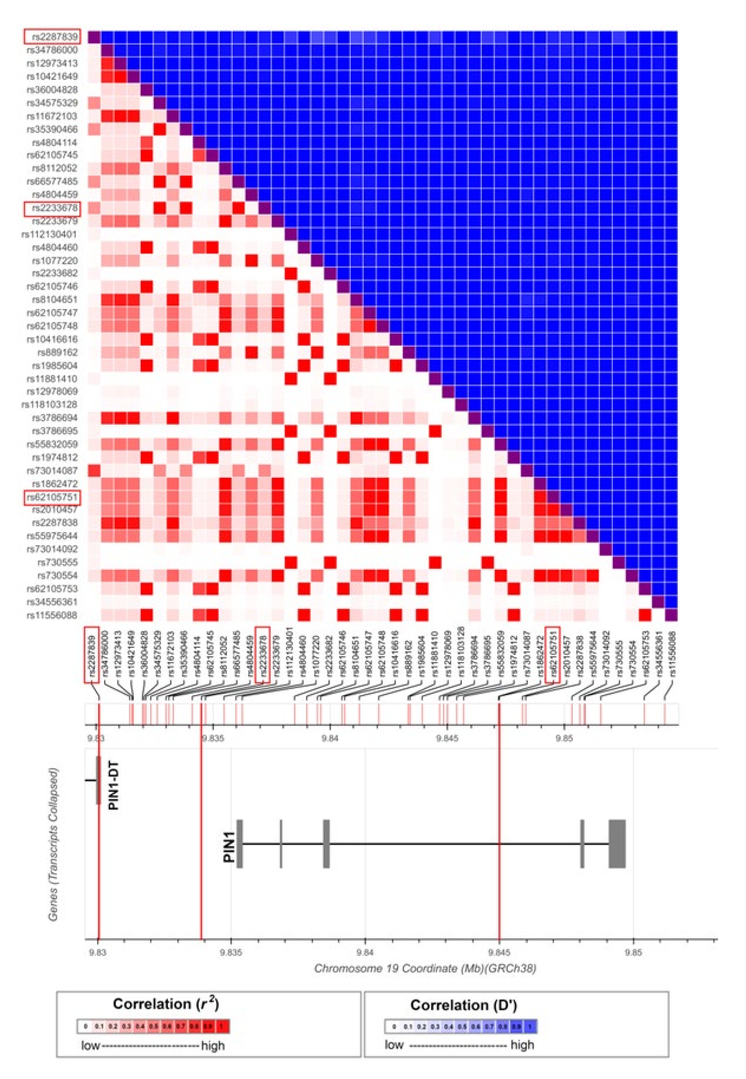
Figure 1. Genetic and functional structure of the PIN1 region. Linkage disequilibrium patterns in the European population included in the 1000 Genome Project were retrieved from the LDlink repository to design a tag-SNP study for the PIN1 locus. The selected tag-SNPs are highlighted in red. All SNP positions are reported in GRCh38. PIN1-DT: PIN1 divergent transcript.
Jpm 12 00932 g001
Figure 2. Analysis of data from the GTEx repository to detect QTL effects and isoform expression patterns in the PIN1 locus. (A) Expression-QTL (eQTL) and (B) splicing-QTL (sQTL) allele effects on PIN1 of the rs3810166 variant. (C) PIN1 transcript expression in human testis and brain tissues. The sQTL-affected intron is highlighted in red. The transcript ENST00000591777.1 is marked with a red arrow. NES: normalized effect size.
Jpm 12 00932 g002
Table 1. Genotype and allele frequency analyses of the tested genetic variants. The subgroups of clinical phenotypes of male infertility were compared against fertile controls under the additive model.
SNP (GRCh38 bp
Position)
Alleles
(1/2)
CohortGenotypes (11/12/22)MAFpAdjusted p *OR (CI 95%)
rs2287839G/CControls
(n = 1049)
6/129/9140.0672NANANA
chr19:9,830,138 SpF (n = 705)4/110/5910.08371.84 × 10−20.055221.38 (1.06–1.81)
SO (n = 205)1/17/1870.04630.1741NA0.70 (0.41–1.17)
NOA (n = 500)3/93/4040.0997.81× 10−42.34 × 10−31.61 (1.22–2.13)
SCO (n = 102)1/22/790.11768.38 × 10−31.94 × 10−21.85 (1.17–2.93)
MA (n = 52)0/8/440.07690.6242NA1.20 (0.57–2.52)
HS (n = 48)0/10/380.10420.1453NA1.66 (0.84–3.28)
rs2233678C/GControls
(n = 1050)
17/206/8270.1143NANANA
chr19:9,834,503 SpF (n = 706)13/136/5570.11470.2862NA1.13 (0.90–1.40)
SO (n = 206)2/28/1760.07770.1999NA0.76 (0.51–1.15)
NOA (n = 500)11/108/3810.130.0784NA1.23 (0.98–1.55)
SCO (n = 102)5/25/720.17161.34 × 10−21.94 × 10−21.62 (1.11–2.36)
MA (n = 52)1/10/410.11540.8202NA1.07 (0.58–1.97)
HS (n = 48)0/11/370.11460.7795NA1.09 (0.58–2.07)
rs62105751A/GControls
(n = 1052)
97/468/4870.3146NANANA
chr19:9,847,213 SpF (n = 706)72/307/3270.31940.5456NA1.05 (0.90–1.22)
SO (n = 205)14/81/1100.26590.1441NA0.82 (0.63–1.07)
NOA (n = 501)58/226/2170.34130.102NA1.15 (0.97–1.36)
SCO (n = 102)17/46/390.39221.94 × 1021.94 × 1021.43 (1.06–1.93)
MA (n = 52)6/23/230.33650.5002NA1.16 (0.76–1.77)
HS (n = 48)1/23/240.26040.3656NA0.80 [0.50–1.29]
* p adjusted is from FDR_BH.
Publisher’s Note: MDPI stays neutral with regard to jurisdictional claims in published maps and institutional affiliations.