
The Role of SNHG15 in the Pathogenesis of Hepatocellular Carcinoma

 ,
,  ,
,  ,
,  ,
,  , ,
, , 
Abstract
:1. Introduction
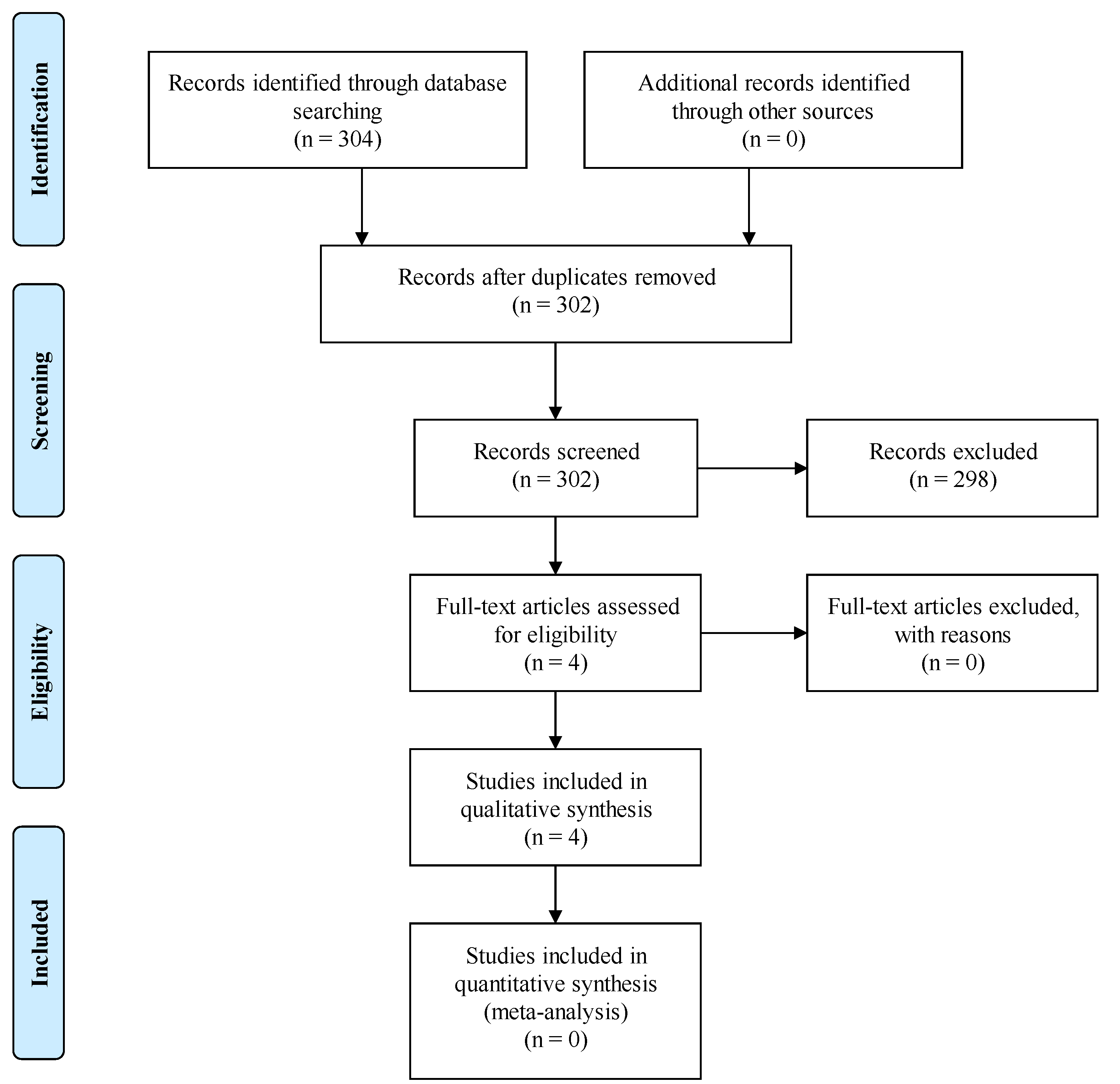
2. Materials and Methods
3. Results
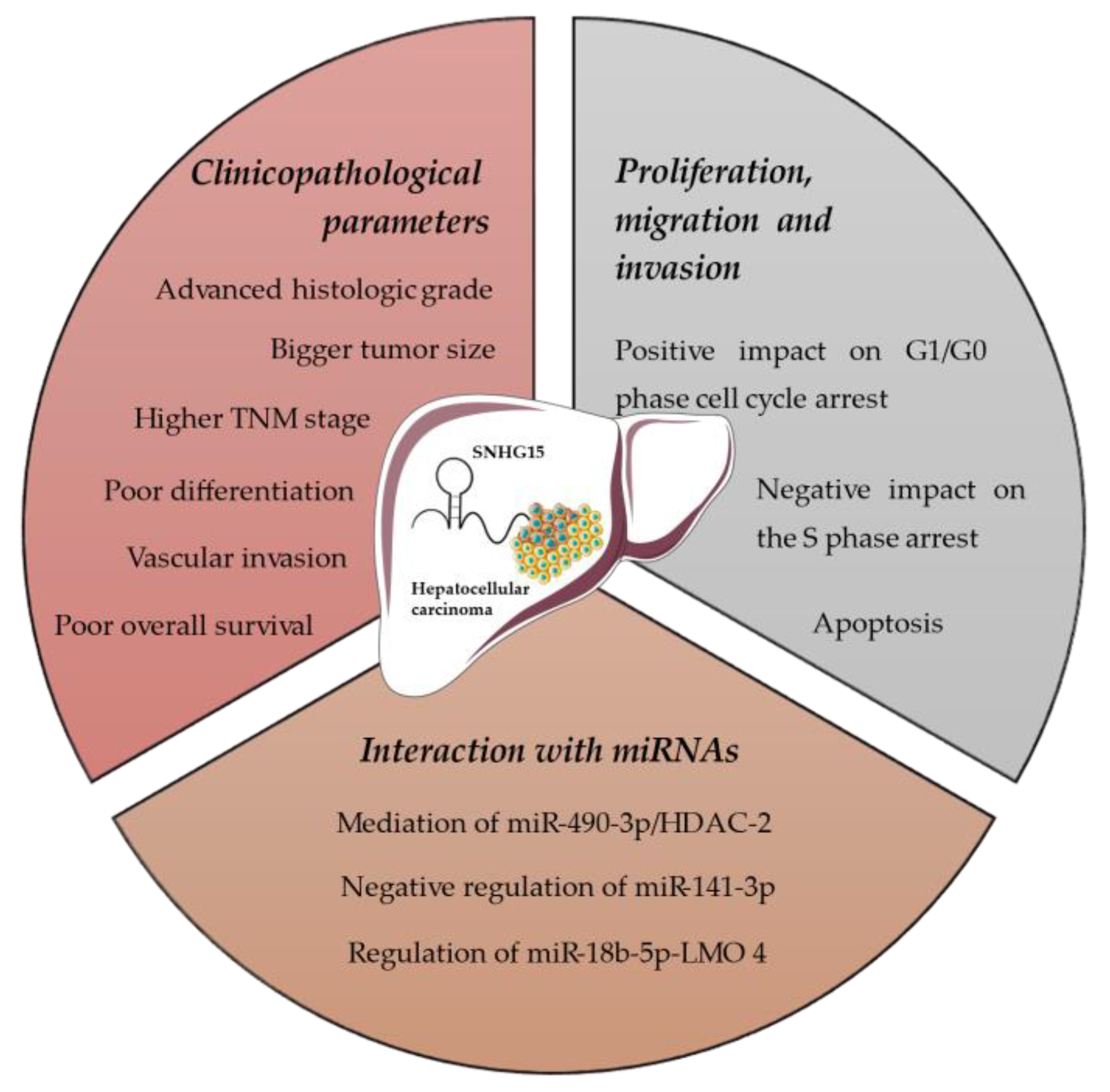
3.1. SNHG15 Expression
3.2. Proliferation, Migration and Invasion
3.3. Interaction with miRNAs
4. Discussion
5. Conclusions
Author Contributions
Funding
Institutional Review Board Statement
Informed Consent Statement
Data Availability Statement
Conflicts of Interest
References
- Bray, F.; Ferlay, J.; Soerjomataram, I.; Siegel, R.L.; Torre, L.A.; Jemal, A. Global cancer statistics 2018: GLOBOCAN estimates of incidence and mortality worldwide for 36 cancers in 185 countries. CA Cancer J. Clin. 2018, 68, 394–424. [Google Scholar] [CrossRef] [PubMed] [Green Version]
- Dimitroulis, D.; Damaskos, C.; Valsami, S.; Davakis, S.; Garmpis, N.; Spartalis, E.; Athanasiou, A.; Moris, D.; Sakellariou, S.; Kykalos, S.; et al. From diagnosis to treatment of hepatocellular carcinoma: An epidemic problem for both developed and developing world. World J. Gastroenterol. 2017, 23, 5282–5294. [Google Scholar] [CrossRef] [PubMed]
- Zhu, R.X.; Seto, W.K.; Lai, C.L.; Yuen, M.F. Epidemiology of hepatocellular carcinoma in the Asia-Pacific region. Gut Liver 2016, 10, 332–339. [Google Scholar] [CrossRef] [PubMed] [Green Version]
- Garmpis, N.; Damaskos, C.; Garmpi, A.; Georgakopoulou, V.E.; Sarantis, P.; Antoniou, E.A.; Karamouzis, M.V.; Nonni, A.; Schizas, D.; Diamantis, E.; et al. Histone deacetylase inhibitors in the treatment of hepatocellular carcinoma: Current evidence and future opportunities. J. Pers. Med. 2021, 11, 223. [Google Scholar] [CrossRef] [PubMed]
- El-Sherag, H.B.; Rudolph, K.L. Hepatocellular carcinoma: Epidemiology and molecular carcinogenesis. Gastroenterology 2007, 132, 2557–2576. [Google Scholar] [CrossRef] [PubMed]
- Luo, P.; Wu, S.; Yu, Y.; Ming, X.; Li, S.; Zuo, X.; Tu, J. Current status and perspective biomarkers in AFP negative HCC: Towards screening for and diagnosing hepatocellular carcinoma at an earlier stage. Pathol. Oncol. Res. 2020, 26, 599–603. [Google Scholar] [CrossRef]
- Elzouki, A.N.; Elkhider, H.; Yacout, K.; Al Muzrakchi, A.; Al-Thani, S.; Ismail, O. Metastatic hepatocellular carcinoma to parotid glands. Am. J. Case Rep. 2014, 15, 343–347. [Google Scholar]
- Bruix, J.; Sherman, M.; American Association for the Study of Liver Diseases. Management of hepatocellular carcinoma: An update. Hepatology 2011, 53, 1020–1022. [Google Scholar] [CrossRef]
- Hao, K.; Luk, J.M.; Lee, N.P.; Mao, M.; Zhang, C.; Ferguspn, M.D.; Lamb, J.; Dai, H.; Ng, I.O.; Sham, P.C.; et al. Predicting prognosis in hepatocellular carcinoma after curative surgery with common clinicopathologic parameters. BMC Cancer 2009, 9, 389. [Google Scholar] [CrossRef] [Green Version]
- Damaskos, C.; Kaskantamis, A.; Garmpis, N.; Dimitroulis, D.; Mantas, D.; Garmpi, A.; Sakellariou, S.; Angelou, A.; Syllaios, A.; Kostakis, A.; et al. Intensive care unit outcomes following orthotopic liver transplantation: Single-center experience and review of the literature. G. Chir. 2019, 4, 463–480. [Google Scholar]
- Chan, J.J.; Tay, Y. Noncoding RNA: RNA regulatory networks in Cancer. Int. J. Mol. Sci. 2018, 19, 1310. [Google Scholar] [CrossRef] [PubMed] [Green Version]
- Gibb, E.A.; Brown, C.J.; Lam, W.L. The functional role of long non-coding RNA in human carcinomas. Mol. Cancer 2011, 10, 38. [Google Scholar] [CrossRef] [PubMed] [Green Version]
- Zhao, W.; An, Y.; Liang, Y.; Xie, X.W. Role of HOTAIR long noncoding RNA in metastatic progression of lung cancer. Eur. Rev. Med. Pharmacol. Sci. 2014, 18, 1930–1936. [Google Scholar] [PubMed]
- Spizzo, R.; Almeida, M.I.; Colombatti, A.; Calin, G.A. Long non-coding RNAs and cancer: A new frontier of translational research? Oncogene 2012, 31, 4577–4587. [Google Scholar] [CrossRef] [Green Version]
- Vikram, R.; Ramachandran, R.; Abdul, K.S. Functional significance of long non-coding RNAs in breast cancer. Breast Cancer 2014, 21, 515–521. [Google Scholar] [CrossRef]
- Wang, L.; Zhao, Z.; Feng, W.; Ye, Z.; Dai, W.; Zhang, C.; Peng, J.; Wu, K. Long non-coding RNA TUG1 promotes colorectal cancer metastasis via EMT pathway. Oncotarget 2016, 7, 51713–51719. [Google Scholar] [CrossRef] [Green Version]
- Cheng, Y.; Jutooru, I.; Chadalapaka, G.; Corton, J.C.; Safe, S. The long non-coding RNA HOTTIP enhances pancreatic cancer cell proliferation, survival and migration. Oncotarget 2015, 6, 10840–10852. [Google Scholar] [CrossRef] [Green Version]
- Li, H.; Yu, B.; Li, J.; Su, L.; Yan, M.; Zhu, Z.; Liu, B. Overexpression of lncRNA H19 enhances carcinogenesis and metastasis of gastric cancer. Oncotarget 2014, 5, 2318–2329. [Google Scholar] [CrossRef] [Green Version]
- Ding, C.; Yang, Z.; Lv, Z.; Du, C.; Xiao, H.; Peng, C.; Cheng, S.; Xie, H.; Zhou, L.; Wu, J.; et al. Long non-coding RNA PVT1 is associated with tumor progression and predicts recurrence in hepatocellular carcinoma patients. Oncol. Lett. 2015, 9, 955–963. [Google Scholar] [CrossRef] [Green Version]
- Ma, G.; Tang, M.; Wu, Y.; Xu, X.; Pan, F.; Xu, R. LncRNAs and miRNAs: Potential biomarkers and therapeutic targets for prostate cancer. Am. J. Transl. Res. 2016, 8, 5141–5150. [Google Scholar]
- Chen, S.X.; Yin, J.F.; Lin, B.C.; Su, H.F.; Zheng, Z.; Xie, C.Y.; Fei, Z.H. Upregulated expression of long noncoding RNA SNHG15 promotes cell proliferation and invasion through regulates MMP2/MMP9 in patients with GC. Tumour Biol. 2016, 37, 6801–6812. [Google Scholar] [CrossRef] [PubMed]
- Cui, H.X.; Zhang, M.Y.; Liu, K.; Liu, J.; Zhang, Z.L.; Fu, L. LncRNA SNHG15 promotes proliferation and migration of lung cancer via targeting microRNA-211-3p. Eur. Rev. Med. Pharmacol. Sci. 2018, 22, 6838–6844. [Google Scholar] [PubMed]
- Kong, Q.; Qiu, M. Long noncoding RNA SNHG15 promotes human breast cancer proliferation, migration and invasion by sponging miR-211-3p. Biochem. Biophys. Res. Commun. 2018, 495, 1594–1600. [Google Scholar] [CrossRef] [PubMed]
- Zhang, J.H.; Wei, H.W.; Yang, H.G. Long noncoding RNA SNHG15, a potential prognostic biomarker for hepatocellular carcinoma. Eur. Rev. Med. Pharmacol. Sci. 2016, 20, 1720–1724. [Google Scholar]
- Shuai, Y.; Ma, Z.; Lu, J.; Feng, J. LncRNA SNHG15: A new budding star in human cancers. Cell. Prolif. 2020, 53, E12716. [Google Scholar] [CrossRef]
- Chen, C.; Feng, Y.; Wang, J.; Liang, Y.; Zou, W. Long non-coding RNA SNHG15 in various cancers: A meta and bioinformatic analysis. BMC Cancer 2020, 20, 1156. [Google Scholar] [CrossRef]
- Ma, Z.; Huang, H.; Wang, J.; Zhou, Y.; Pu, F.; Zhao, Q.; Peng, P.; Hui, B.; Ji, H.; Wang, K. Long non-coding RNA SNHG15 inhibits P15 and KLF2 expression to promote pancreatic cancer proliferation through EZH2-mediated H3K27me3. Oncotarget 2017, 8, 84153–84167. [Google Scholar] [CrossRef] [Green Version]
- Liu, K.; Hou, Y.; Liu, Y.; Zheng, J. LncRNA SNHG15 contributes to proliferation, invasion and autophagy in osteosarcoma cells by sponging miR-141. J. Biomed. Sci. 2017, 24, 46. [Google Scholar] [CrossRef]
- Fan, Y.; Xue, H.; Zheng, H. Systemic therapy for hepatocellular carcinoma: Current updates and outlook. J. Hepatocell. Carcinoma 2022, 9, 233–263. [Google Scholar] [CrossRef]
- Dai, W.; Dai, J.L.; Tang, M.H.; Ye, M.S.; Fang, S. lncRNA-SNHG15 accelerates the development of hepatocellular carcinoma by targeting miR-490-3p/histone deacetylase 2 axis. World J. Gastroenterol. 2019, 25, 5789–5799. [Google Scholar] [CrossRef]
- Ye, J.; Tan, L.; Fu, Y.; Xu, H.; Wen, L.; Deng, Y.; Liu, K. LncRNA SNHG15 promotes hepatocellular carcinoma progression by sponging miR-141-3p. J. Cell Biochem. 2019, 120, 19775–19783. [Google Scholar] [CrossRef] [PubMed] [Green Version]
- Chen, W.; Huang, L.; Liang, J.; Ye, Y.; Yu, S.; Zhang, Y. Long noncoding RNA small nucleolar RNA host gene 15 deteriorates liver cancer via microRNA-18b-5p/LIM-only 4 axis. IUBMB Life 2021, 73, 349–361. [Google Scholar] [CrossRef] [PubMed]
- Ji, D.; Wang, Y.; Sun, B.; Yang, J.; Luo, X. Long non-coding RNA MNX1-AS1 promotes hepatocellular carcinoma proliferation and invasion through targeting miR-218-5p/COMMD8 axis. Biochem. Biophys. Res. Commun. 2019, 513, 669–674. [Google Scholar] [CrossRef]
- Gnoni, A.; Licchetta, A.; Memeo, R.; Argentiero, A.; Solimando, A.G.; Longo, V.; Delcuratolo, S.; Brunetti, O. Role of BRAF in hepatocellular carcinoma: A rationale for future targeted cancer therapies. Medicina 2019, 55, 754. [Google Scholar] [CrossRef] [PubMed] [Green Version]
- Moon, H.; Ro, S.W. MAPK/ERK signaling pathway in hepatocellular carcinoma. Cancers 2021, 13, 3026. [Google Scholar] [CrossRef] [PubMed]
- Neuzillet, C.; Tijeras-Raballand, A.; de Mestier, L.; Cros, J.; Faivre, S.; Raymond, E. MEK in cancer and cancer therapy. Pharmacol. Ther. 2014, 141, 160–171. [Google Scholar] [CrossRef]
- Bruix, J.; Qin, S.; Merle, P.; Granito, A.; Huang, Y.H.; Bodoky, G.; Pracht, M.; Yokosuka, O.; Rosmorduc, O.; Breder; et al. RESORCE Investigators. Regorafenib for patients with hepatocellular carcinoma who progressed on sorafenib treatment (RESORCE): A randomised, double-blind, placebo-controlled, phase 3 trial. Lancet 2017, 389, 56–66. [Google Scholar] [CrossRef] [Green Version]
- Solimando, A.G.; Susca, N.; Argentiero, A.; Brunetti, O.; Leone, P.; De Re, V.; Fasano, R.; Krebs, M.; Petracci, E.; Azzali, I.; et al. Second-line treatments for advanced hepatocellular carcinoma: A Systematic Review and Bayesian Network Meta-analysis. Clin. Exp. Med. 2022, 22, 65–74. [Google Scholar] [CrossRef]
- Lee, Y.H.; Seo, D.; Choi, K.J.; Andersen, J.B.; Won, M.A.; Kitade, M.; Gómez-Quiroz, L.E.; Judge, A.D.; Marquardt, J.U.; Raggi, C.; et al. Antitumor effects in hepatocarcinoma of isoform-selective inhibition of HDAC2. Cancer Res. 2014, 74, 4752–4761. [Google Scholar] [CrossRef] [Green Version]
- Zhang, C.; Ke, Y.; Liu, X.; Wang, X.; Li, Y.; Zhou, J.; Zhang, H.; Wang, L. High expression of the long noncoding RNA SNHG15 in cancer tissue samples predicts an unfavorable prognosis of cancer patients: A meta-analysis. J. Oncol. 2020, 2020, 3417036. [Google Scholar] [CrossRef]
- Garmpi, A.; Garmpis, N.; Damaskos, C.; Valsami, S.; Spartalis, E.; Lavaris, A.; Patelis, N.; Margonis, G.A.; Apostolou, K.G.; Spartalis, M.; et al. Histone deacetylase inhibitors as a new anticancer option: How far can we go with expectations? delivery systems. J. BUON 2018, 23, 846–861. [Google Scholar] [PubMed]
- Noh, J.H.; Jung, K.H.; Kim, J.K.; Eun, J.W.; Bae, H.J.; Xie, H.J.; Chang, Y.G.; Kim, M.G.; Park, W.S.; Lee, J.Y.; et al. Aberrant regulation of HDAC2 mediates proliferation of hepatocellular carcinoma cells by deregulating expression of G1/S cell cycle proteins. PLoS ONE 2011, 6, e28103. [Google Scholar] [CrossRef] [Green Version]
- Jung, K.H.; Noh, J.H.; Kim, J.K.; Eun, J.W.; Bae, H.J.; Xie, H.J.; Chang, Y.G.; Kim, M.G.; Park, H.; Lee, J.Y.; et al. HDAC2 overexpression confers oncogenic potential to human lung cancer cells by deregulating expression of apoptosis and cell cycle proteins. J. Cell Biochem. 2012, 113, 2167–2177. [Google Scholar] [CrossRef] [PubMed]
- Fritsche, P.; Seidler, B.; Schüler, S.; Schnieke, A.; Göttlicher, M.; Schmid, R.M.; Saur, D.; Schneider, G. HDAC2 mediates therapeutic resistance of pancreatic cancer cells via the BH3-only protein NOXA. Gut 2009, 58, 1399–1409. [Google Scholar] [CrossRef] [PubMed] [Green Version]
- Wagner, T.; Kiweler, N.; Wolff, K.; Knauer, S.K.; Brandl, A.; Hemmerich, P.; Dannenberg, J.H.; Heinzel, T.; Schneider, G.; Krämer, O.H. Sumoylation of HDAC2 promotes NF-κB-dependent gene expression. Oncotarget 2015, 6, 7123–7135. [Google Scholar] [CrossRef] [PubMed] [Green Version]
- Alzoubi, S.; Brody, L.; Rahman, S.; Mahul-Mellier, A.L.; Mercado, N.; Ito, K.; El-Bahrawy, M.; Silver, A.; Boobis, A.; Bell, J.D.; et al. Synergy between histone deacetylase inhibitors and DNA-damaging agents is mediated by histone deacetylase 2 in colorectal cancer. Oncotarget 2016, 7, 44505–44521. [Google Scholar] [CrossRef] [Green Version]
- Visvader, J.E.; Venter, D.; Hahm, K.; Santamaria, M.; Sum, E.Y.; O’Reilly, L.; White, D.; Williams, R.; Armes, J.; Lindeman, G.J. The LIM domain gene LMO4 inhibits differentiation of mammary epithelial cells in vitro and is overexpressed in breast cancer. Proc. Natl. Acad. Sci. USA 2001, 98, 14452–14457. [Google Scholar] [CrossRef] [Green Version]
- Simonik, E.A.; Cai, Y.; Kimmelshue, K.N.; Brantley-Sieders, D.M.; Loomans, H.A.; Andl, C.D.; Westlake, G.M.; Youngblood, V.M.; Chen, J.; Yarbrough, W.G.; et al. LIM-only protein 4 (LMO4) and LIM domain binding protein 1 (LDB1) promote growth and metastasis of human head and neck cancer (LMO4 and LDB1 in head and neck cancer). PLoS ONE 2016, 11, e0164804. [Google Scholar] [CrossRef]
- Ma, Y.; Xue, Y.; Liu, X.; Qu, C.; Cai, H.; Wang, P.; Li, Z.; Li, Z.; Liu, Y. SNHG15 affects the growth of glioma microvascular endothelial cells by negatively regulating miR-153. Oncol. Rep. 2017, 38, 3265–3277. [Google Scholar] [CrossRef] [Green Version]
- Du, Y.; Kong, C.; Zhu, Y.; Yu, M.; Li, Z.; Bi, J.; Li, Z.; Liu, X.; Zhang, Z.; Yu, X. Knockdown of SNHG15 suppresses renal cell carcinoma proliferation and EMT by regulating the NF-κB signaling pathway. Int. J. Oncol. 2018, 53, 384–394. [Google Scholar] [CrossRef]
- Li, M.; Bian, Z.; Jin, G.; Zhang, J.; Yao, S.; Feng, Y.; Wang, X.; Yin, Y.; Fei, B.; You, Q.; et al. LncRNA-SNHG15 enhances cell proliferation in colorectal cancer by inhibiting miR-338-3p. Cancer Med. 2019, 8, 2404–2413. [Google Scholar] [CrossRef] [PubMed] [Green Version]
- Zhang, Y.; Zhang, D.; Lv, J.; Wang, S.; Zhang, Q. LncRNA SNHG15 acts as an oncogene in prostate cancer by regulating miR-338-3p/FKBP1A axis. Gene 2019, 705, 44–50. [Google Scholar] [CrossRef] [PubMed]
- Liu, Y.; Li, J.; Li, F.; Li, M.; Shao, Y.; Wu, L. SNHG15 functions as a tumor suppressor in thyroid cancer. J. Cell Biochem. 2019, 120, 6120–6126. [Google Scholar] [CrossRef] [PubMed]
- Qu, C.; Dai, C.; Guo, Y.; Qin, R.; Liu, J. Long noncoding RNA SNHG15 serves as an oncogene and predicts poor prognosis in epithelial ovarian cancer. Onco. Targets Ther. 2018, 12, 101–111. [Google Scholar] [CrossRef] [Green Version]
- Hu, X.; Jiang, J.; Xu, Q.; Ni, C.; Yang, L.; Huang, D. A systematic review of long noncoding RNAs in hepatocellular carcinoma: Molecular mechanism and clinical implications. BioMed Res. Int. 2018, 2018, 8126208. [Google Scholar] [CrossRef] [Green Version]

| Study | Design | Material | Drug | Result |
|---|---|---|---|---|
| Zhang et al., 2016 [24] | Associations between | - | lncRNA SNHG15 may serve as an efficient clinical biomarker and a therapeutic target for HCC patients. | |
| clinicopathological parameters and lncRNA SNHG15 expression were evaluated using chi-square tests. | 152 paired HCC tissues and adjacent normal tissues. | |||
| Dai et al., 2019 [30] | - HCC cell lines HuH-1, HuH-7 and normal human liver cells L-O2. - The growth conditions of these cells are 5% CO2, 37 °C and culture solution. - si-SNHG15, pcDNA3.1-SNHG15 vectors, si-HDAC-2 and miR-490-3p mimics or inhibitor were transfected into HuH-1 or L-O2 cells. - Untreated cells were used as the controls. | 101 HCC patients. | Lipofectamine 2000 (Invitrogen). | lncRNA RNA SNHG15 promotes HCC progression by mediating the miR-490-3p/HDAC-2 axis in HCC. |
| Ye et al., 2019 [31] | - 4 HCC cell lines and a normal human LO2 liver cell line. - siRNAs against si-SNHG15 and a non-targeting control (si-NC). - miR-141-3p, a non-targeting miR control (miR-NC), and the miR-141-3p inhibitor (miR-141-3p in). | 58 paired HCC samples and adjacent matched adjacent normal tissues. | - Penicillin, streptomycin for cell culture in a humid 37 °C environment with 5% CO2 atmosphere. - Lipofectamine 2000. | SNHG15 promoted HCC progression throws negative regulation of miR-141-3p, identifying a potential novel HCC way of treatment. |
| Chen et al., 2021 [32] | - 4 human liver cancer cell lines and one normal liver cell line. | High-glucose Dulbecco’s modified eagle medium and RPMI 1640 medium (HyClnoe, Logan, UT), in 5% FBS-contained medium, detached with 0.25% trypsin and passaged, MTT solution, Dimethyl sulfoxide, Lipofectamine 2000 (Invitrogen, Carlsbad, CA). | - Up-regulation of SNHG15 in HCC cells. - Knockdown of SNHG15 impedes colony formation, invasion and migration of HCC cells and enhances apoptosis. - SNHG15 regulates miR-18b-3p-LMO4 axis. | |
| - Transfection with SNHG15 low-expression negative control (NC) plasmids, SNHG15 low expression plasmids, miR-18b-5p mimic NC, miR-18b-5p mimic, SNHG15 over-expression plasmids and mir-18b5p mimic NC, or SNHG15 over-expression plasmids and mir-18b-5p mimic. - Female BALB/c nude mice (n = 28, 4 weeks) were injected with the transfected cells. | 58 paired HCC and normal tissue specimens. |
| Study | Type of Cancer | Role | Expression | Effect on Cancer Cells |
|---|---|---|---|---|
| Chen et al., 2016 [21] | Gastric | Oncogenic | Up-regulation | Proliferation, apoptosis, migration, invasion |
| Cui et al., 2016 [22] | Lung | Oncogenic | Up-regulation | Proliferation, apoptosis, migration, invasion |
| Ma et al., 2017 [27] | Pancreatic | Oncogenic | Up-regulation | Proliferation, apoptosis, cell cycle arrest |
| Ma et al., 2017 [49] | Glioma | Oncogenic | Up-regulation | Proliferation, migration, angiogenesis, tube formation |
| Liu et al., 2017 [28] | Osteosarcoma | Oncogenic | Up-regulation | Proliferation, migration, autophagy, invasion |
| Kong et al., 2018 [23] | Breast | Oncogenic | Up-regulation | Proliferation, apoptosis, migration, invasion |
| Du et al., 2018 [50] | Renal cell | Oncogenic | Up-regulation | Proliferation, migration, invasion, apoptosis, cell cycle arrest |
| Li et al., 2019 [51] | Colorectal | Oncogenic | Up-regulation | Proliferation, apoptosis, migration, invasion |
| Zhang et al., 2019 [52] | Prostate | Oncogenic | Up-regulation | Migration, invasion |
| Liu et al., 2019 [53] | Thyroid | Oncosuppressive | Down-regulation | Proliferation, migration, invasion |
| Qu et al., 2019 [54] | Ovarian | Oncogenic | Up-regulation | Proliferation, migration, invasion |
Publisher’s Note: MDPI stays neutral with regard to jurisdictional claims in published maps and institutional affiliations. |
© 2022 by the authors. Licensee MDPI, Basel, Switzerland. This article is an open access article distributed under the terms and conditions of the Creative Commons Attribution (CC BY) license (https://creativecommons.org/licenses/by/4.0/).
Share and Cite
Damaskos, C.; Garmpis, N.; Dimitroulis, D.; Garmpi, A.; Diamantis, E.; Sarantis, P.; Georgakopoulou, V.E.; Patsouras, A.; Despotidis, M.; Prevezanos, D.; et al. The Role of SNHG15 in the Pathogenesis of Hepatocellular Carcinoma. J. Pers. Med. 2022, 12, 753. https://doi.org/10.3390/jpm12050753
Damaskos C, Garmpis N, Dimitroulis D, Garmpi A, Diamantis E, Sarantis P, Georgakopoulou VE, Patsouras A, Despotidis M, Prevezanos D, et al. The Role of SNHG15 in the Pathogenesis of Hepatocellular Carcinoma. Journal of Personalized Medicine. 2022; 12(5):753. https://doi.org/10.3390/jpm12050753
Chicago/Turabian StyleDamaskos, Christos, Nikolaos Garmpis, Dimitrios Dimitroulis, Anna Garmpi, Evangelos Diamantis, Panagiotis Sarantis, Vasiliki E. Georgakopoulou, Alexandros Patsouras, Markos Despotidis, Dionysios Prevezanos, and et al. 2022. "The Role of SNHG15 in the Pathogenesis of Hepatocellular Carcinoma" Journal of Personalized Medicine 12, no. 5: 753. https://doi.org/10.3390/jpm12050753
APA StyleDamaskos, C., Garmpis, N., Dimitroulis, D., Garmpi, A., Diamantis, E., Sarantis, P., Georgakopoulou, V. E., Patsouras, A., Despotidis, M., Prevezanos, D., Syllaios, A., Marinos, G., Koustas, E., Vallilas, C., Antoniou, E. A., Kontzoglou, K., Savvanis, S., & Kouraklis, G. (2022). The Role of SNHG15 in the Pathogenesis of Hepatocellular Carcinoma. Journal of Personalized Medicine, 12(5), 753. https://doi.org/10.3390/jpm12050753