
Early Intervention in Orbital Floor Fractures: Postoperative Ocular Motility and Diplopia Outcomes


Abstract
:1. Introduction
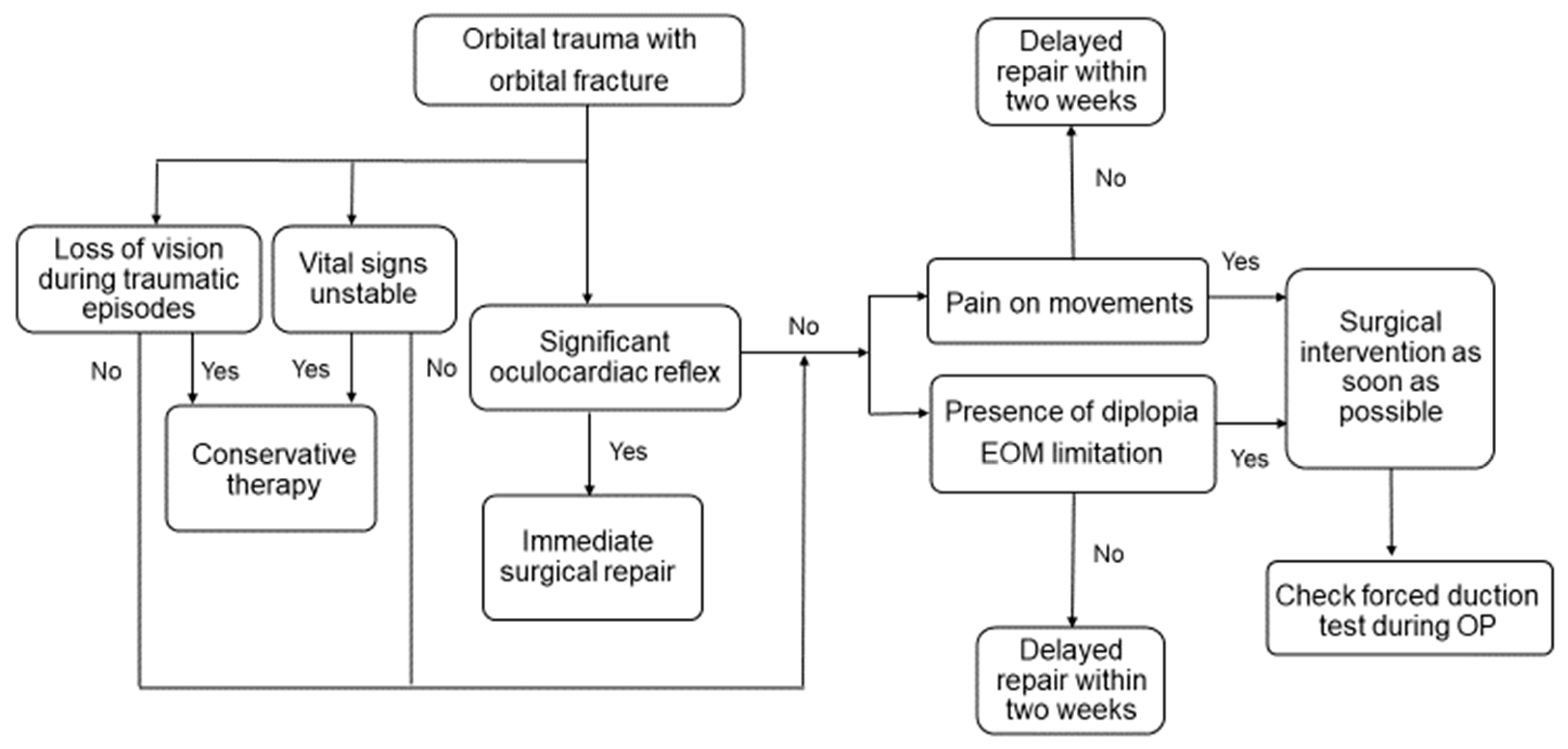
2. Methods
2.1. Surgical Procedures
2.2. Statistical Analysis
3. Results
3.1. Patient Characteristics and Demographic Data
3.2. Surgical Intervention Time and Postoperative Motility and Diplopia
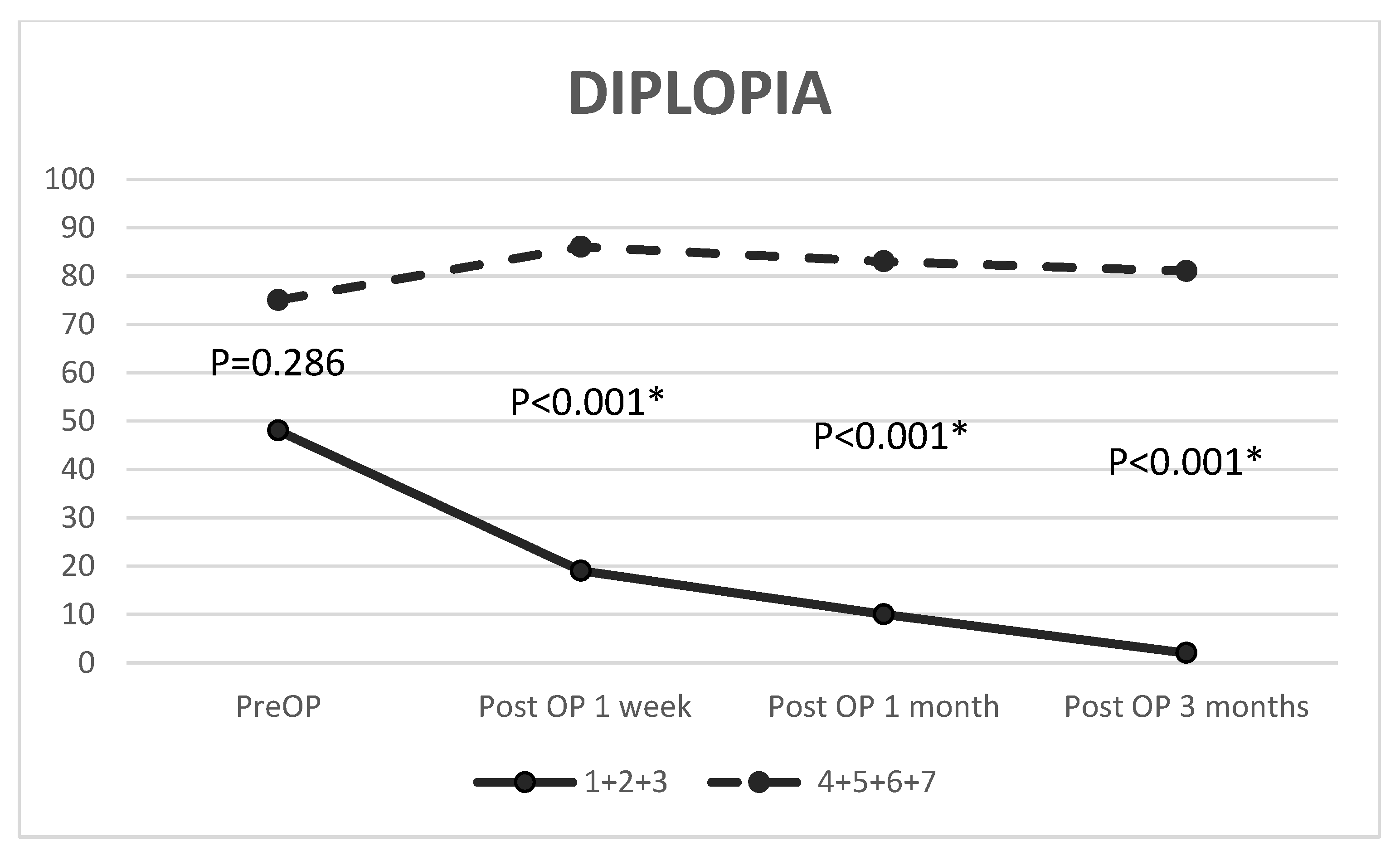
3.3. Ocular Motility and Diplopia before and after Repair
3.4. Variables Associated with Postoperative Limitation and Diplopia
4. Discussion
Supplementary Materials
Author Contributions
Funding
Institutional Review Board Statement
Informed Consent Statement
Data Availability Statement
Conflicts of Interest
References
- Lozada, K.; Kadakia, S.; Abraham, M.T.; Ducic, Y. Complications of Midface Fractures. Facial Plast. Surg. 2017, 33, 557–561. [Google Scholar] [CrossRef] [PubMed] [Green Version]
- Yamanaka, Y.; Watanabe, A.; Rajak, S.N.; Nakayama, T.; Sotozono, C. Correlation between surgical timing and postoperative ocular motility in orbital blowout fractures. Graefe’s Arch. Clin. Exp. Ophthalmol. 2021, 260, 319–325. [Google Scholar] [CrossRef] [PubMed]
- McRae, M.; Augustine, H.F.M.; Budning, A.; Antonyshyn, O. Functional Outcomes of Late Posttraumatic Enophthalmos Correction. Plast. Reconstr. Surg. 2018, 142, 169e–178e. [Google Scholar] [CrossRef] [PubMed]
- Young, S.M.; Kim, Y.D.; Kim, S.W.; Jo, H.B.; Lang, S.S.; Cho, K.; Woo, K.I. Conservatively treated orbital blowout fractures: Spontaneous radiologic improvement. Ophthalmology 2018, 125, 938–944. [Google Scholar] [CrossRef] [PubMed] [Green Version]
- Egbert, J.E.; May, K.; Kersten, R.C.; Kulwin, D.R. Pediatric orbital floor fracture: Direct extraocular muscle involvement. Ophthalmology 2000, 107, 1875–1879. [Google Scholar] [CrossRef]
- Mehta, V.; Chelnis, J.G.; Chen, Q.; Mawn, L.A. Effect of Time to Operative Intervention on Motility Outcomes Following Orbital Floor Fracture Repair in Children. Ophthalmic Plast. Reconstr. Surg. 2018, 34, 351–354. [Google Scholar] [CrossRef] [PubMed]
- Boyette, J.R.; Pemberton, J.D.; Bonilla-Velez, J. Management of orbital fractures: Challenges and solutions. Clin. Ophthalmol. 2015, 9, 2127–2137. [Google Scholar] [CrossRef] [PubMed] [Green Version]
- Burnstine, M.A. Clinical recommendations for repair of isolated orbital floor fractures: An evidence-based analysis. Ophthalmology 2002, 109, 1207–1210. [Google Scholar] [CrossRef]
- Jazayeri, H.E.; Khavanin, N.; Yu, J.W.; Lopez, J.; Ganjawalla, K.P.; Shamliyan, T.; Tannyhill, R.J.; Dorafshar, A.H. Does Early Repair of Orbital Fractures Result in Superior Patient Outcomes? A Systematic Review and Meta-Analysis. J. Oral Maxillofac. Surg. 2020, 78, 568–577. [Google Scholar] [CrossRef] [PubMed] [Green Version]
- Yu, D.Y.; Chen, C.H.; Tsay, P.K.; Leow, A.M.; Pan, C.H.; Chen, C.T. Surgical Timing and Fracture Type on the Outcome of Diplopia After Orbital Fracture Repair. Ann. Plast. Surg. 2016, 76, S91–S95. [Google Scholar] [CrossRef] [PubMed]
- Yamanaka, Y.; Watanabe, A.; Sotozono, C.; Kinoshita, S. Impact of surgical timing of postoperative ocular motility in orbital blowout fractures. Br. J. Ophthalmol. 2017, 102, 398–403. [Google Scholar] [CrossRef] [PubMed]
- Kasaee, A.; Mirmohammadsadeghi, A.; Kazemnezhad, F.; Eshraghi, B.; Akbari, M.R. The predictive factors of diplopia and extraocular movement limitations in isolated pure blow-out fracture. J. Curr. Ophthalmol. 2016, 29, 54–58. [Google Scholar] [CrossRef] [PubMed] [Green Version]
- Matteini, C.; Renzi, G.; Becelli, R.; Belli, E.; Iannetti, G. Surgical Timing in Orbital Fracture Treatment: Experience with 108 Consecutive Cases. J. Craniofacial Surg. 2004, 15, 145–150. [Google Scholar] [CrossRef] [PubMed]
- Ramphul, A.; Hoffman, G. Does Preoperative Diplopia Determine the Incidence of Postoperative Diplopia After Repair of Orbital Floor Fracture? An Institutional Review. J. Oral Maxillofac. Surg. 2017, 75, 565–575. [Google Scholar] [CrossRef] [PubMed] [Green Version]
- Scawn, R.L.; Lim, L.H.; Whipple, K.M.; Dolmetsch, A.; Priel, A.; Korn, B.; Kikkawa, D.O. Outcomes of Orbital Blow-Out Fracture Repair Performed Beyond 6 Weeks After Injury. Ophthalmic Plast. Reconstr. Surg. 2016, 32, 296–301. [Google Scholar] [CrossRef] [PubMed]
- Tahiri, Y.; Lee, J.; Tahiri, M.; Sinno, H.; Williams, B.H.; Lessard, L.; Gilardino, M.S. Preoperative diplopia: The most important prognostic factor for diplopia after surgical repair of pure orbital blowout fracture. J. Craniofacial Surg. 2010, 21, 1038–1041. [Google Scholar] [CrossRef] [PubMed]


| Group | 1 | 2 | 3 | 4 | 5 | 6 | 7 | Total | p Value |
|---|---|---|---|---|---|---|---|---|---|
| Duration from injury to surgical intervention (days) | 0–1 | 2–3 | 4–7 | 8–14 | 15–21 | 22–30 | 31~ | ||
| Number of patients (N, %) | 45 (16.7%) | 37 (13.7%) | 31 (11.5%) | 39 (14.4%) | 33 (12.2%) | 38 (14.1%) | 47 (17.4%) | 270 | <0.001 # |
| Gender (M, %) | 30 (66.7%) | 30 (81.1%) | 15 (48.4%) | 26 (66.7%) | 24 (72.7%) | 23 (60.5%) | 26 (55.3%) | 174 (64.4%) | 0.086 $ |
| Age (mean) (SD) | 33.69 (13.87) | 40.66 (15.01) | 45.24 (19.96) | 44.73 (16.79) | 41.65 (18.67) | 40.79 (19.10) | 41.25 (16.05) | 40.67 (17.15) | 0.034 #,‡,† |
| Laterality (R, %) | 30 | 22 | 19 | 19 | 13 | 17 | 18 | 138 | 0.056 $ |
| With medial wall fracture (N, %) | 21 (46.7%) | 18 (48.6%) | 16 (51.6%) | 17 (43.6%) | 24 (72.7%) | 26 (68.4%) | 31 (66.0%) | 153 (56.7%) | 0.043 $ |
| EOM limitation before OP (N, %) | 43 (95.6%) | 22 (59.5%) | 16 (51.6%) | 14 (35.9%) | 14 (42.4%) | 15 (39.5%) | 16 (34.0%) | 140 (51.9%) | <0.001 $ |
| Diplopia before OP (N, %) | 27 (60.0%) | 12 (32.43%) | 9 (29.03%) | 14 (35.90%) | 18 (54.55%) | 17 (44.74%) | 26 (55.32%) | 123 (45.56%) | 0.034 $ |
| OP time(min) (SD) | 65.36 (30.90) | 68.89 (54.10) | 64.06 (36.19) | 66.69 (29.41) | 60.27 (40.81) | 81.45 (35.30) | 82.32 (46.37) | 70.06 (40.44) | 0.001 #,*,§ |
| Group | Limitation First Week (N, %) | Limitation 1 Month (N, %) | Limitation 3 Months (N, %) | Diplopia First Week (N, %) | Diplopia 1 Month (N, %) | Diplopia 3 Months (N, %) |
|---|---|---|---|---|---|---|
| 1 | 4 (8.89%) | 0 (0.00%) | 0 (0.00%) | 5 (11.11%) | 2 (4.44%) | 0 (0.00%) |
| 2 | 7 (18.92%) | 2 (5.41%) | 1 (2.70%) | 8 (21.62%) | 4 (10.81%) | 1 (2.70%) |
| 3 | 6 (19.35%) | 4 (12.90%) | 2 (6.45%) | 6 (19.35%) | 4 (12.90%) | 1 (3.23%) |
| 4 | 12 (30.77%) | 7 (17.95%) | 4 (10.26%) | 17 (43.59%) | 16 (41.03%) | 14 (35.90%) |
| 5 | 13 (39.39%) | 12 (36.36%) | 11 (33.33%) | 18 (54.55%) | 16 (48.48%) | 16 (48.48%) |
| 6 | 13 (34.21%) | 13 (34.21%) | 13 (34.21%) | 20 (52.63%) | 20 (52.63%) | 20 (52.63%) |
| 7 | 15 (31.91%) | 15 (31.91%) | 15 (31.91%) | 31 (65.96%) | 31 (65.96%) | 31 (65.96%) |
| p value | 0.027 * | <0.001 * | <0.001 * | <0.001 * | <0.001 * | <0.001 * |
| Group | Limitation | |||||
|---|---|---|---|---|---|---|
| Before OP vs. Post-OP 1 Wk | Before OP vs. Post-OP 1 M | Before OP vs. Post-OP 3 M | ||||
| Improvement Rate (%) | p Value | Improvement Rate (%) | p Value | Improvement Rate (%) | p Value | |
| 1 + 2 +3 | 79.01% | <0.001 * | 92.59% | <0.001 * | 96.30% | <0.001 * |
| 4 + 5 + 6+ 7 | 10.17% | 0.031 * | 20.34% | 0.001 * | 27.12% | 0.001 * |
| Group | Diplopia | |||||
|---|---|---|---|---|---|---|
| Before OP vs. Post-OP 1 wk | Before OP vs. Post-OP 1 m | Before OP vs. Post-OP 3 m | ||||
| Improvement Rate (%) | p Value | Improvement Rate (%) | p Value | Improvement Rate (%) | p Value | |
| 1 + 2 + 3 | 60.42% | <0.001 * | 79.17% | <0.001 * | 95.83% | <0.001 * |
| 4 + 5 + 6 + 7 | NA | 0.281 | NA | 0.496 | NA | 0.677 |
| Post-OP 3 M Limitation | ||||
| Univariate | Multivariate | |||
| Characteristic | OR (95%) | p Value | OR (95% CI) | p Value |
| Duration from injury to OP (days) | 1.023 (1.010–1.037) | <0.001 * | 1.030 (1.015–1.048) | 0.001 * |
| OP time (minutes) | 1.010 (1.000–1.021) | 0.059 | 1.007 (0.994–1.018) | 0.267 |
| Age (years) | 0.999 (0.980–1.019) | 0.928 | ||
| Gender (M/F) | 2.611 (1.201–5.675) | 0.015 * | 2.720 (1.138–6.498) | 0.024 * |
| Combined medial wall fracture | 0.014 (0.578–1.011) | 0.996 | ||
| Pre-OP limitation | 2.412 (1.221–4.762) | 0.011 * | 2.883 (1.344–6.182) | 0.007 * |
| Post-OP 3 M Diplopia | ||||
| Univariate | Multivariate | |||
| Characteristic | OR (95%) | p Value | OR (95% CI) | p Value |
| Duration from injury to OP (days) | 1.047 (1.030–1.064) | <0.001 * | 1.091(1.063–1.119) | <0.001 * |
| OP time (minutes) | 1.011 (1.002–1.020) | 0.018 * | 1.001(0.989–1.013) | 0.269 |
| Age (years) | 0.997 (0.982–1.013) | 0.745 | ||
| Gender (M/F) | 2.840 (1.548–5.211) | 0.001 * | 3.246 (1.368–7.703) | 0.008 * |
| Combined medial wall fracture | 0.022 (0.348–1.047) | 0.995 | ||
| Pre-OP diplopia | 3.042 (1.774–5.217) | 0.001 * | 2.774 (1.496–5.145) | 0.001 * |
Publisher’s Note: MDPI stays neutral with regard to jurisdictional claims in published maps and institutional affiliations. |
© 2022 by the authors. Licensee MDPI, Basel, Switzerland. This article is an open access article distributed under the terms and conditions of the Creative Commons Attribution (CC BY) license (https://creativecommons.org/licenses/by/4.0/).
Share and Cite
Hsu, C.-R.; Lee, L.-C.; Chen, Y.-H.; Chien, K.-H. Early Intervention in Orbital Floor Fractures: Postoperative Ocular Motility and Diplopia Outcomes. J. Pers. Med. 2022, 12, 671. https://doi.org/10.3390/jpm12050671
Hsu C-R, Lee L-C, Chen Y-H, Chien K-H. Early Intervention in Orbital Floor Fractures: Postoperative Ocular Motility and Diplopia Outcomes. Journal of Personalized Medicine. 2022; 12(5):671. https://doi.org/10.3390/jpm12050671
Chicago/Turabian StyleHsu, Cherng-Ru, Lung-Chi Lee, Yi-Hao Chen, and Ke-Hung Chien. 2022. "Early Intervention in Orbital Floor Fractures: Postoperative Ocular Motility and Diplopia Outcomes" Journal of Personalized Medicine 12, no. 5: 671. https://doi.org/10.3390/jpm12050671
APA StyleHsu, C.-R., Lee, L.-C., Chen, Y.-H., & Chien, K.-H. (2022). Early Intervention in Orbital Floor Fractures: Postoperative Ocular Motility and Diplopia Outcomes. Journal of Personalized Medicine, 12(5), 671. https://doi.org/10.3390/jpm12050671