Article

Intraoperative Blood Flow Analysis of DIEP vs. ms-TRAM Flap Breast Reconstruction Combining Transit-Time Flowmetry and Microvascular Indocyanine Green Angiography

Department of Plastic and Hand Surgery and Laboratory for Tissue Engineering and Regenerative Medicine, University Hospital Erlangen, Friedrich Alexander University Erlangen-Nürnberg (FAU), 91054 Erlangen, Germany
*
Author to whom correspondence should be addressed.
J. Pers. Med. 2022, 12(3), 482; https://doi.org/10.3390/jpm12030482
Submission received: 27 January 2022 / Revised: 9 March 2022 / Accepted: 14 March 2022 / Published: 16 March 2022
(This article belongs to the Section Methodology, Drug and Device Discovery)

Abstract

:
Background: Vascular patency is the key element for high flap survival rates. The purpose of this study was to assess and compare the blood flow characteristics of deep inferior epigastric perforator (DIEP) and muscle-sparing transverse rectus abdominis musculocutaneous (ms-TRAM) flaps for autologous breast reconstruction. Methods: This prospective clinical study combined Transit-Time Flowmetry and microvascular Indocyanine Green Angiography for the measurement of blood flow volume, vascular resistance, and intrinsic transit time. Results: Twenty female patients (mean age, 52 years) received 24 free flaps (14 DIEP and 10 ms-TRAM flaps). The mean arterial blood flow of the flap in situ was 7.2 ± 1.9 mL/min in DIEP flaps and 11.5 ± 4.8 mL/min in ms-TRAM flaps (p < 0.05). After anastomosis, the mean arterial blood flow was 9.7 ± 5.6 mL/min in DIEP flaps and 13.5 ± 4.2 mL/min in ms-TRAM flaps (p = 0.07). The arterial vascular resistance of DIEP flaps was significantly higher than that of ms-TRAM flaps. The intrinsic transit time of DIEP flaps was 52 ± 18 s, and that of ms-TRAM flaps was 33 ± 11 s (p < 0.05). The flap survival rate was 100%. One DIEP flap with the highest intrinsic transit time (77 s) required surgical revision due to arterial thrombosis. Conclusion: In this study, we established the blood flow characteristics of free DIEP and ms-TRAM flaps showing different blood flow rates, vascular resistances, and intrinsic transit times. These standard values will help to determine the predictive values for vascular compromise, hence improving the safety of autologous breast reconstruction procedures.

1. Introduction

Nowadays, abdominal tissue as the main source for breast reconstruction is preferably harvested either as a complete muscle-preserving deep inferior epigastric perforator (DIEP) flap or as a muscle-sparing transverse rectus abdominis musculocutaneous (ms-TRAM) flap [1,2]. The ability to reconstruct the female breast in a like-with-like fashion with low donor site morbidity has led to its widespread use [3]. Some of the latest tissue engineering and regenerative medicine methods aiming to overcome donor site sequelae are promising but not yet clinically feasible [4,5]. Although both DIEP and ms-TRAM flaps have overall low complication rates, partial flap necrosis and total flap loss due to vascular compromise remain imminent postoperative risks [6]. Sufficient vascular perfusion remains a key aspect for the overall outcome and flap survival. In the last few years, numerous clinical studies assessed the intra- and postoperative perfusion of free flaps using different technologies [7,8,9,10,11,12,13,14]. However, several studies aiming to understand the hemodynamics of free flaps showed methodological flaws such as a heterogeneous study population, small sample sizes of the included flap types in terms of tissue composition, the sole assessment of arterial flow characteristics, or the use of nowadays outdated technologies [15,16,17,18,19]. This study measures and compares intraoperative arterial and venous blood flow and perfusion characteristics of DIEP and ms-TRAM flaps for breast reconstruction combining Transit-Time Flowmetry (TTFM) and Indocyanine Green Angiography at a microscopic level (mICG-A). TTFM, an ultrasound-based technology for the assessment of vascular blood flow, was originally introduced into clinical practice for cardiac surgery [20,21]. Validation studies showed highly accurate and reproducible measurements which enabled its extension towards other surgical specialties such as vascular surgery and microsurgery [22,23,24]. The intravenous application of Indocyanine Green in combination with microscope-integrated fluorescence-based video angiography (IR800) and the analysis tool FLOW800 (Carl Zeiss, Oberkochen, Germany) enables the recording, measurement, and assessment of the microvascular patency and blood flow characteristics of vessels just a few millimeters in diameter [25,26]. The assembly of these two state-of-the art technologies is considered a novel approach. We believe that an advanced understanding of their hemodynamic properties will improve the safety of the two most commonly used free flaps for autologous breast reconstruction. This study further aimed to establish normative blood flow and perfusion values as groundwork for the determination of predictive values for postoperative thrombotic events.

2. Materials and Methods

Patients receiving DIEP or ms-TRAM flaps for breast reconstruction were enrolled in this prospective mono-centered clinical study. The study was approved by the Ethical Committee in accordance with the Declaration of Helsinki. Prior to study inclusion, written consent was given by each patient.

2.1. Surgical Technique

Autologous breast reconstructions were performed by three experienced surgeons. All patients received a standardized computed tomography angiography (CTA) of the abdomen for perforator mapping prior to surgery. Depending on the anatomy of the selected perforators (size, course, and number), the patients included in this study received DIEP or ms-TRAM free flaps for autologous breast reconstruction. Our postoperative anticoagulation regimen usually consists of low-molecular-weight heparin application subcutaneously until full mobilization is achieved. If contraindications for low-molecular-weight heparin exist, patients usually receive weight-adjusted unfractionated heparin. Patients suffering from hyperthyroidism, thyroid adenoma or autonomy, and known allergies/hypersensitivity to Indocyanine green or sodium iodide were excluded. The patient body temperature was kept stable by using a warming mattress (37 °C) and maintaining the ambient temperature between 20 °C and 22 °C. All patients received a balanced intraoperative crystalloid volume substitution of, on average, 53 mL/kg (mean: 3780 ± 1710 mL) in order to maintain stable hemodynamic conditions. The average intraoperative blood loss was 140 ± 100 mL. The internal mammary artery (IMA) and vein (IMV) were used as recipient vessels in all cases. Each flap was harvested in a standardized fashion with the inferior epigastric artery and the inferior epigastric vein as vascular pedicle dissected towards their origin at the external iliac artery and vein. Each vascular pedicle consisted of one artery and one vein. DIEP and ms-TRAM flaps with more than one venous anastomosis were excluded from this study. All arterial and venous anastomoses were performed end to end. Arterial anastomoses were hand-sewn with interrupted nylon sutures, whereas all venous anastomoses were performed using a venous coupler device (Synovis Micro Companies Alliance, Inc., Birmingham, AL, USA).

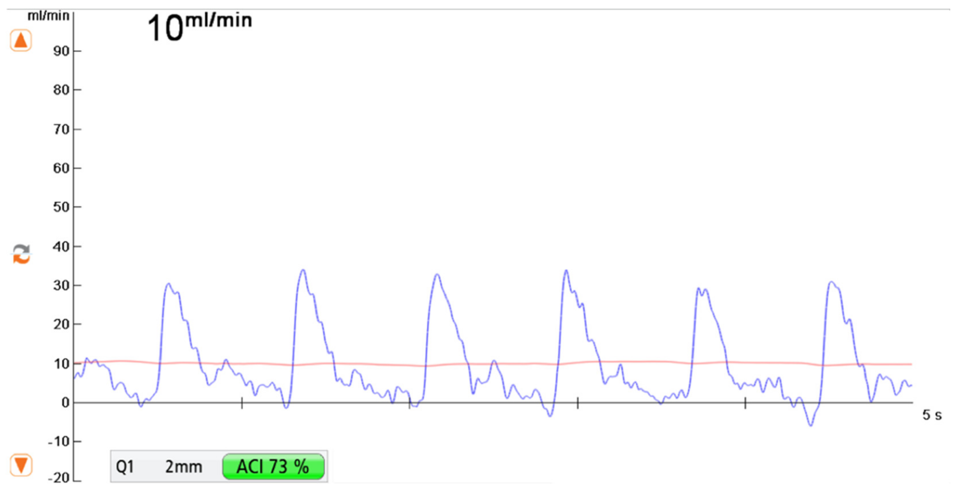
2.2. Transit-Time Flow Measurement (TTFM)

MiraQ™ Vascular (Medistim ASA, Oslo, Norway) was used for intraoperative blood flow measurements. The probe diameter ranged from 1.5 to 4 mm depending on the vessel size. Blood flow values were recorded for several minutes until a steady curve of blood flow occurred (Figure 1). Arterial and venous blood flow volume measurements were performed at three predefined time points during surgery:
-
Measurement F was taken after flap elevation and isolation on its vascular pedicle prior to free flap transfer
-
Measurement R was performed at the recipient vessel prior to anastomosis.
-
Measurement AA was taken at the vascular pedicle after anastomosis and flap reperfusion.
The mean arterial pressure was measured and documented at each measurement time point.

2.3. Microvascular Indocyanine Green Angiography (mICG-A)

In this clinical study, Indocyanine Green (ICG) was administered as an intravenous bolus (3 mL VERDYE 5 mg/mL) after arterial and venous anastomosis and flap reperfusion. The anastomosed flap pedicle was placed below the microscope (KINEVO 900, Carl Zeiss, Oberkochen, Germany). Recordings of the supplying artery and draining vein started immediately after intravenous ICG application and were continued until the intensity of the ICG markedly decreased in the artery and vein. Intraoperative fluorescence analysis requires the selection of certain regions of interest (ROI). Two ROIs were placed at the flap pedicle, one at the supplying artery, and the other at the draining vein close to the anastomosis, uncovered from any surrounding tissue (Figure 2). FLOW800 measures the intensity of ICG in the regions of interest for a time period and enables the instant visualization of blood flow variations within small vessels. The time between the maximum ICG intensity of arterial inflow and venous outflow is defined as Intrinsic Transit Time (ITT), which is considered as a parameter of blood flow velocity within the flap (Figure 2) [27].

2.4. Vascular Resistance

Arterial vascular Resistance (aVR) was calculated as millimeters of mercury per milliliter per minute (mmHg/mL/min) based on a previously reported method in adherence to the Poiseuille’s Law, using the following formula: [28,29]
R = Δ P / Q
R = aVR = arterial vascular resistance, ΔP = blood pressure gradient = mean arterial blood pressure (MAP)–mean venous blood pressure (MVP). The mean venous blood pressure (MVP) is estimated to be close to 0 and, as a result, disregarded in the calculation of vascular resistances. This results in the following formula: [28,29,30,31]
a V R = M A P / a B F

2.5. Data Analysis and Synthesis

Descriptive analysis was performed for patient demographics. Data are shown as mean ± standard deviation. Changes in blood flow and vascular resistance between the different time points within one group were calculated using the paired Student’s t-test. Blood flow and vascular resistance between DIEP and ms-TRAM flaps at different time points was analyzed using an unpaired Student’s t-test. Nonparametric data were analyzed with the Wilcoxon matched-pairs rank test within one group, whereas the Mann–Whitney U test was used for analyses of nonparametric data between DIEP and ms-TRAM flaps. The correlation of data assuming Gaussian distribution was calculated using the Pearson correlation coefficient. The Spearman’s rank correlation coefficient was used for data not passing a test for normality. The significance level was set at p < 0.05. Three outliers (one arterial and two venous blood flow values at the recipient site (Measurement R) before anastomosis) were identified using the ROUT method (Q = 1%) and appropriately excluded from statistical analysis. Statistical analyses and graphic illustrations were performed using GraphPad Prism (GraphPad Software, Inc., San Diego, CA, USA).

3. Results

A total of 20 female patients receiving 24 DIEP or ms-TRAM flaps for breast reconstruction were included in this prospective study. Patients’ average age was 52 years, ranging from 39 to 68 years. Fourteen flaps were harvested as DIEP (57%), and 10 as ms-TRAM (43%). Seven ms-TRAM flaps were classified as ms1-TRAM flap, and three as ms2-TRAM flap, according to the classification by Nahabedian et al. (Table 1) [32]. The median flap weight was 435 g, ranging from 299 to 1169 g. The median weight of DIEP flaps (390 g) was not significantly different from that of ms-TRAM flaps (491 g). The average flap ischemia time was 46 min. The venous coupler size ranged from 2.5 to 3.5 mm.

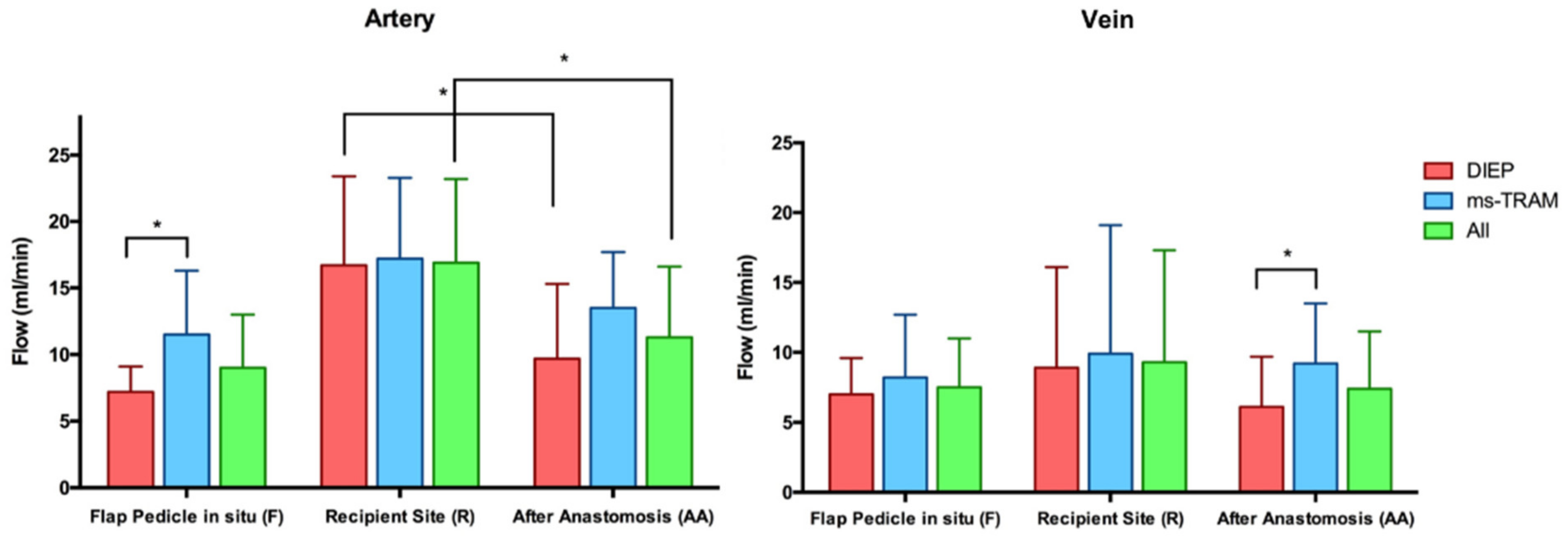
3.1. Blood Flow Volume (mL/min)

The average blood flow of the flap artery isolated as pedicle prior to free tissue transfer (F) was 9 ± 4 mL/min (mean ± SD). Its venous outflow was lower (7.5 ± 3.5 mL/min), resulting in an artery-to-vein (A/V) flow ratio of 1.4 ± 0.7. The mean blood flow of the recipient internal mammary artery and vein prior to flap anastomosis (R) was 16.9 ± 6.3 and 9.4 ± 8 mL/min, respectively. After anastomosis (AA), the arterial and venous blood flow volume was 11.3 ± 5.3 and 7.4 ± 4.1 mL/min, respectively, with an A/V flow ratio of 1.8 ± 1.3. The arterial blood flow of the intact recipient artery (R) significantly decreased after anastomosis with the flap artery (AA) (p = 0.002). However, the arterial and venous blood flow of the included flaps did not significantly change after flap transfer. (Figure 3) The blood flow of the intact recipient artery (R) did not alter the blood flow of the flaps after anastomosis. There was a significant positive correlation between the arterial inflow and the venous outflow both before (F) and after anastomosis (AA) (p < 0.05). The arterial and venous blood flow rates before and after anastomosis in DIEP flaps were lower than in ms-TRAM flaps (Table 2). There was no correlation between the arterial blood flow volume and the flap weight. The flap ischemia time did not change the blood flow rates of the examined flaps.

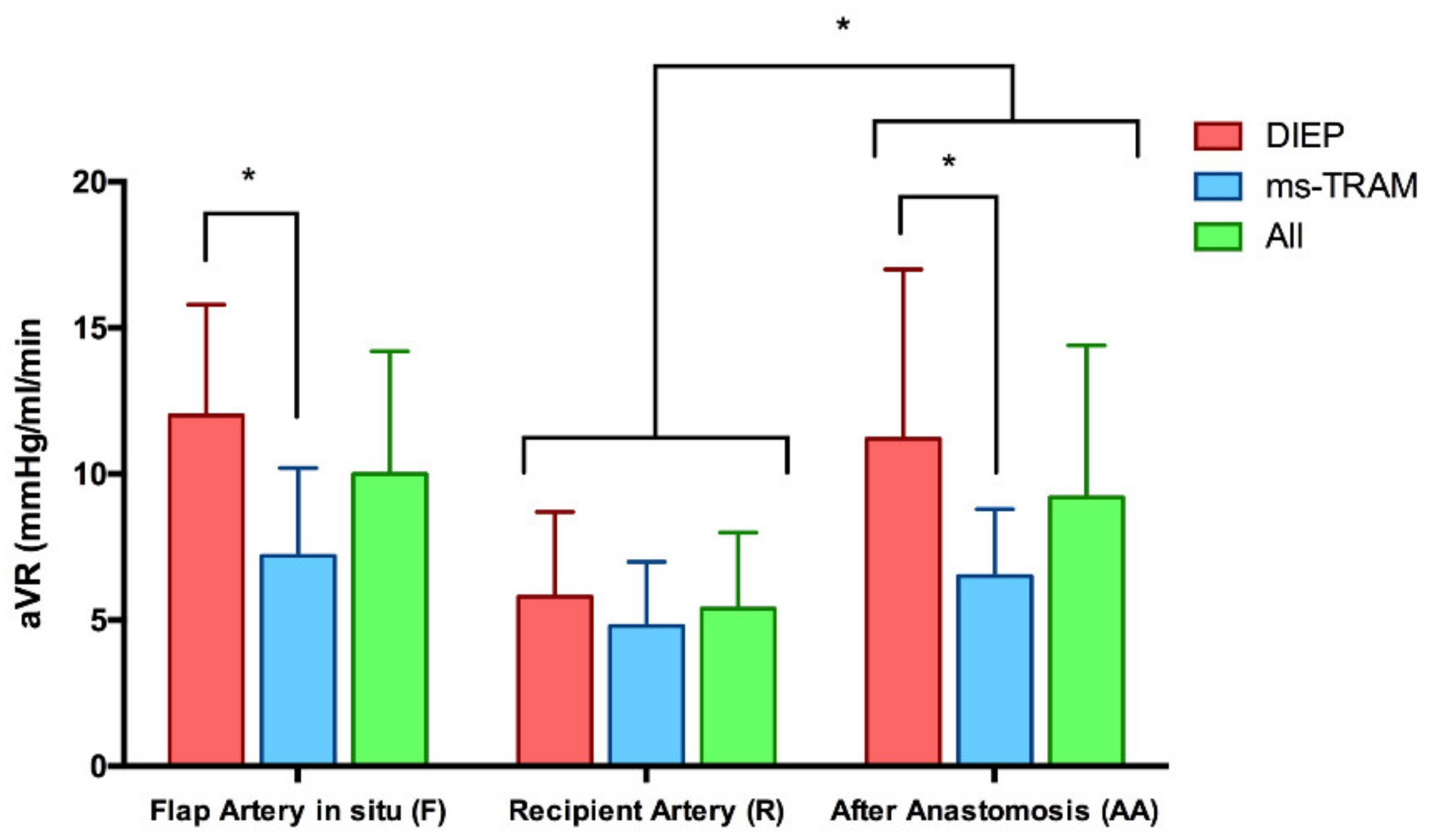
3.2. Vascular Resistance (mmHg/mL/min)

The mean arterial vascular resistance (aVR) of the included flaps prior to tissue transfer (10 ± 4.2 mmHg/mL/min) did not significantly change after anastomosis (9.2 ± 5.2 mmHg/mL/min). The vascular resistance of the recipient artery, however, significantly increased from 5.4 ± 2.6 to 9.2 ± 5.2 mmHg/mL/min after anastomosis to the flap (p < 0.001). The average arterial vascular resistance (aVR) of DIEP flaps prior to (F) and after flap transfer (AA) was 12 ± 3.8 and 11.2 ± 5.8 mmHg/mL/min, respectively. By contrast, ms-TRAM flaps had significantly lower arterial vascular resistance values prior to (7.2 ± 3 mmHg/mL/min; p = 0.004) and after flap reperfusion (6.5 ± 2.3 mmHg/mL/min; p = 0.02) (Figure 4 and Table 3). There was no correlation between the arterial vascular resistance and the weight of the included flaps before or after flap transfer.

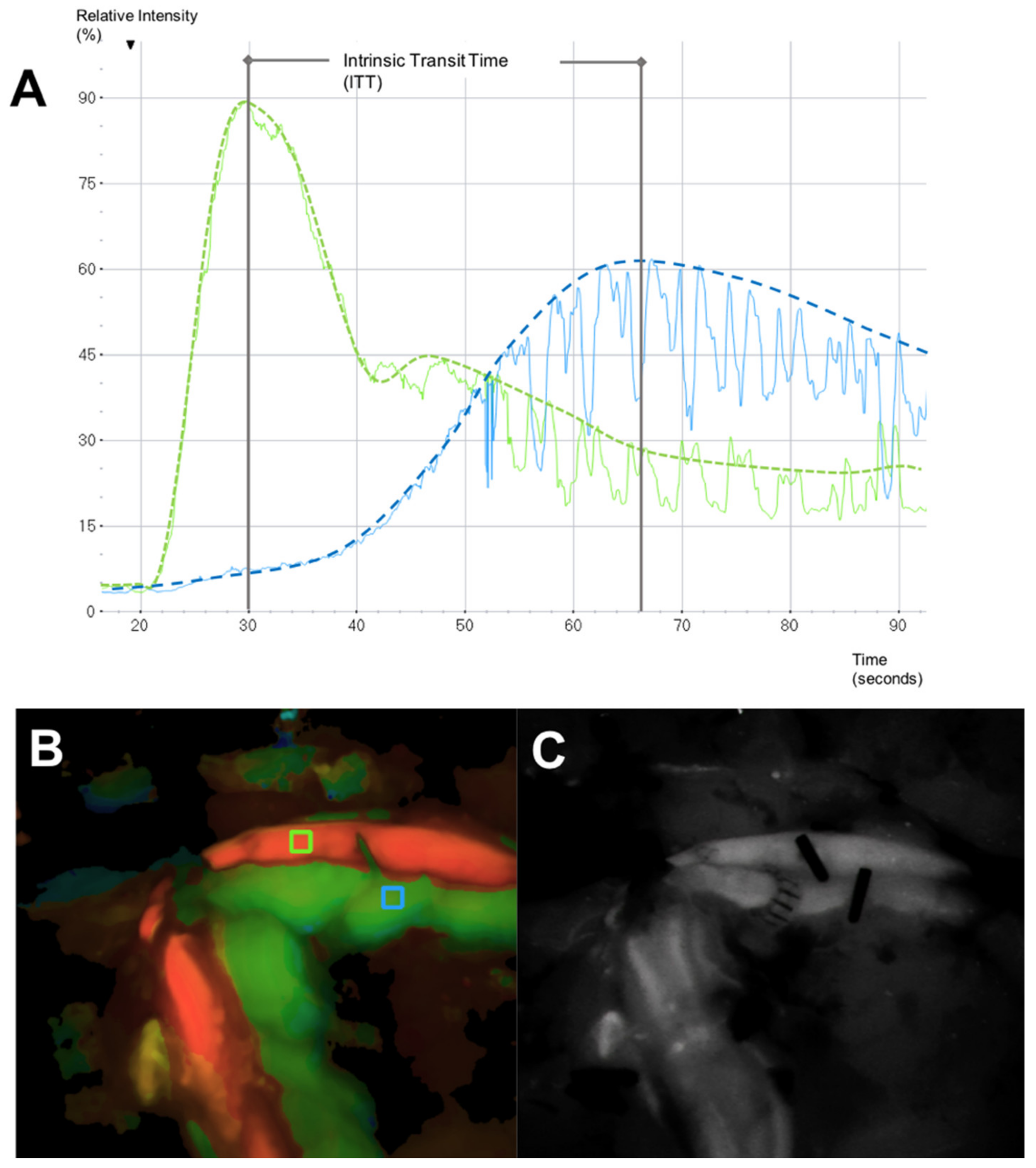
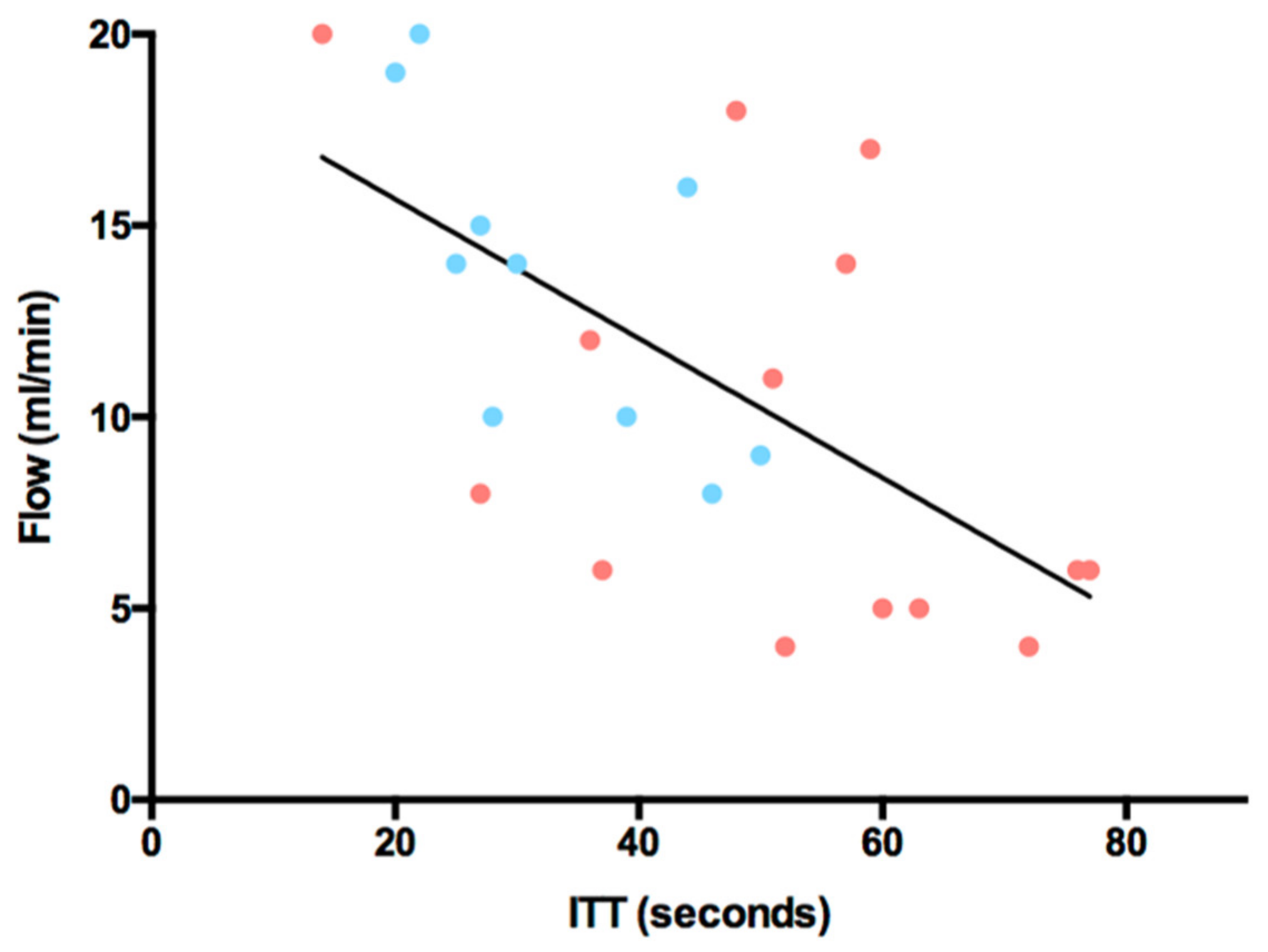
3.3. Intrinsic Transit Time

The mean Intrinsic Transit Time (ITT) after flap reperfusion was 44 ± 18 s (s), ranging from 14 to 77 s. The average ITT of DIEP flaps (52 ± 18 s) was significantly higher than the average ITT of ms-TRAM flaps (33 ± 11 s) (p = 0.005). The average vascular resistance at the time of ITT measurements was 9 ± 4.7 mmHg/mL/min. There was a significant negative correlation between the arterial blood flow and the ITT after anastomosis (p = 0.001) (Figure 5). By contrast, a significant positive correlation was seen between the arterial vascular resistance (aVR) and the ITT after anastomosis (p = 0.0006) (Figure 6). There was no correlation between the ITT and flap ischemia time, flap weight, or mean arterial pressure (MAP).
Of all included free flaps, one DIEP flap required surgical revision due to a thrombotic event occurring on the fourth day after autologous breast reconstruction. The ITT of this flap was 77 s. After emergency thrombectomy, no further complication occurred. The overall flap survival rate was 100%.

4. Discussion

Numerous recently developed technologies enable the illustration and measurement of vascularity and perfusion in free flaps at a pre-, intra, or postoperative stage, with the ultimate goal to increase their safety and efficacy [7,11,12,18,33,34,35,36]. The combination of Transit-Time Flowmetry (TTFM) and microvascular Indocyanine Green Angiography (mICG-A) is considered a unique approach aiming to meticulously evaluate and compare the intraoperative blood flow characteristics of DIEP and ms-TRAM flaps. A recent study successfully established this combination for the detection of early venous congestion in an animal flap model [37]. However, no study so far assessed the combined potential of these techniques in a clinical setting for autologous breast reconstructions. Our results show that the overall arterial blood flow of both DIEP and ms-TRAM flaps did not significantly increase after anastomosis with the recipient internal mammary vessel. The blood flow of the intact recipient artery did not influence the arterial blood flow of the included flaps. In fact, it seemed to be the opposite. In this study, both DIEP and ms-TRAM flaps downregulated the recipient artery flow towards blood flow values of the in situ flap prior to tissue transfer. These observations are supported by other studies showing that the flow of the recipient artery can either be down- or upregulated after flap anastomosis, approximating blood flow values of the flap isolated on its pedicle before tissue transfer [19,38,39,40]. Lorenzetti et al. measured the blood flow of the thoracodorsal artery before and after anastomosis with ms-TRAM flaps and reported an upregulation of the recipient artery. Before anastomosis, the thoracodorsal artery had relatively low blood flow values (4.9 ± 3 mL/min) in situ. However, after anastomosis with the ms-TRAM flap, the blood flow increased (13.7 ± 5 mL/min) towards the original blood flow rate of the isolated flap pedicle in situ before tissue transfer [38]. This phenomenon was observed not only in fasciocutaneous but also in musculocutaneous and muscle free flaps and therefore seems to be irrespective of the tissue composition [40]. Previous studies reported generally different blood flow rates and vascular resistances in fasciocutaneous, musculocutaneous, muscle, and intraperitoneal flaps [31,38]. These findings support the notion that both blood flow and vascular resistance depend on the type of tissue and its relative proportion. The tissue composition determines the vascularity of each flap, which at the same time, reflects the vascular resistance. Free flaps mainly composed of muscle tissue contain a rich vascular network connected by resistance vessels, resulting in a lower vascular resistance than fasciocutaneous flaps with a rather sparse network of much smaller vessels [30,31]. In our study, ms-TRAM flaps had an average arterial blood flow of 13.5 mL/min and a vascular resistance of 6.4 mmHg/mL/min after anastomosis. By contrast, DIEP flaps showed significantly lower blood flow values and consequently a higher vascular resistance. Although both flaps were, apart from a small segment of the rectus abdominis muscle in ms-TRAM flaps, grossly composed of the same tissue, the difference in vascular resistance seemed to be a matter of vascularity. We theorize that a larger number of perforators in ms-TRAM flaps was the main reason for a lower vascular resistance, hence providing a higher overall and weight-adjusted arterial blood flow in comparison to DIEP flaps. The overall arterial inflow of the included flaps was about 1.4 to 1.8 times greater than the venous outflow. Selber et al. reported similar results in ms-TRAM and other fasciocutaneous flaps [41]. We theorize that the peripheral leakage of small blood vessels at the flap edges caused the disparity between arterial inflow and venous outflow. An A/V ratio of more than 1 seems to a certain level inevitable and needs to be considered by surgeons during free flap flow measurements.
Microvascular Indocyanine Green Angiography (mICG-A) combined with the microscope-integrated software FLOW800 provides valuable information on vascular patency and enables the real-time visualization of arterial in- and venous outflow in free flaps [42,43,44]. It is a matter of common pathophysiological knowledge that the alteration in blood flow, as part of the Virchow’s triad, is a main contributor to thrombus formation in blood vessels [45,46]. Previous studies have already theorized that a prolonged ITT might be an indicator of low blood flow velocities, hence accounting for increased vascular resistances [27]. The combination of TTFM with mICG-A enabled to measure and detect a positive correlation of ITT with vascular resistance in free flaps. ITT, similar to blood flow, seems to depend on the flap tissue composition. In our study, the DIEP flap, which was composed of fasciocutaneous tissue, had a significantly higher average ITT (52 s) than the ms-TRAM flap (33 s), classified as musculocutaneous flap. These observations are supported by a previous study measuring a shorter ITT in muscle flaps (27.7 s) than in fasciocutaneous flaps (47.5 s) [26]. Holm et al. reported that an ITT of more than 50 s was associated with an increased risk for vascular compromise and surgical revision in free tissue transfer procedures [27]. The study, however, showed essential methodological flaws such as a heterogenous study population with varying free flap entities. In our study, eight flaps surpassed the threshold of 50 s without any hemodynamic postoperative complication. Only one DIEP flap with an ITT of 77 s required surgical revision due to a thrombotic event several days after flap transplantation. Although this was the highest ITT of all included flaps, the scarce occurrence of just a single hemodynamic complication several days after autologous breast reconstruction did not allow drawing any correlation between a prolonged ITT and the increased risk of postoperative hemodynamic complications. To the best of our knowledge, this is the first study that measures, compares, and detects hemodynamic differences between DIEP and ms-TRAM flaps. The clinical relevance of this study is the establishment of standard values of intraoperative hemodynamic and perfusion properties of DIEP and ms-TRAM flaps. We could detect significant differences in hemodynamics properties between DIEP and ms-TRAM flaps. Flaps with abnormally high or low blood flow values, according to our newly established standard hemodynamic characteristics, made a closer intraoperative assessment of anastomotic patency necessary. We are aware that these techniques do not replace a clinical examination but rather help to improve our intraoperative decision-making process. The results of this study, however, do not provide any recommendation in terms of favoring one or the other free flap type for autologous breast reconstruction. Historically, the choice between DIEP and ms-TRAM flap has been a far more extensive topic that needs to take numerous other variables into account. Our meticulous assessment of arterial and venous blood flow, arterial vascular resistance, and ITT at crucial intraoperative time points enables the establishment of normative values. This should help to assess vascular patency especially in cases where the surgeon or devices such as a regular hand-held Doppler fail to detect a more subtle vascular compromise.

5. Conclusions

In this study, we evaluated the hemodynamic characteristics of free DIEP and ms-TRAM flaps. The combination of Transit-Time Flowmetry and microvascular Indocyanine Green Angiography enabled the qualitative and quantitative intraoperative assessment of anastomotic patency. Our study serves as fundamental work for the determination of predictive values for postoperative thrombotic events and of cut-off values that will ease intraoperative decision making in the future.

Author Contributions

Conceptualization: A.G., R.E.H. and A.A.; Investigation: A.G., R.E.H., I.L. and A.A.; Methodology: A.G., I.L. and A.A.; Project administration: A.G., R.E.H. and A.A.; Resources: R.E.H.; Supervision: R.E.H. and A.A.; Validation: A.G. and A.A.; Visualization: A.G.; Writing—original draft: A.G.; Writing—review & editing: A.G., R.E.H., I.L. and A.A. All authors have read and agreed to the published version of the manuscript.

Funding

We acknowledge financial support by Deutsche Forschungsgemeinschaft and Friedrich-Alexander-Universität Erlangen-Nürnberg within the funding programme “Open Access Publication Funding”.

Institutional Review Board Statement

The study was conducted according to the guidelines of the Declaration of Helsinki and approved by the Ethics Committee of Friedrich-Alexander-University of Erlangen-Nuremberg, Germany (registration number: 447_19B).

Informed Consent Statement

Informed consent was obtained from all subjects involved in the study.

Data Availability Statement

The datasets generated during the current study are available from the corresponding author on reasonable request.

Acknowledgments

The present work was performed in fulfillment of the requirements for obtaining the degree “Dr. med.” for the author A.G.

Conflicts of Interest

The authors declare no conflict of interest.

References

  1. Eisenhardt, S.U.; Momeni, A.; von Fritschen, U.; Horch, R.E.; Stark, G.B.; Bannasch, H.; Harder, Y.; Heitmann, C.; Kremer, T.; Rieger, U.M.; et al. Breast reconstruction with the free TRAM or DIEP flap—What is the current standard? Consensus Statement of the German Speaking Working Group for Microsurgery of the Peripheral Nerves and Vessels. Handchirurgie Mikrochirurgie Plastische Chirurgie 2018, 50, 248–255. [Google Scholar] [CrossRef] [Green Version]
  2. Cai, A.; Suckau, J.; Arkudas, A.; Beier, J.P.; Momeni, A.; Horch, R.E. Autologous Breast Reconstruction with Transverse Rectus Abdominis Musculocutaneous (TRAM) or Deep Inferior Epigastric Perforator (DIEP) Flaps: An Analysis of the 100 Most Cited Articles. Med. Sci. Monit. 2019, 25, 3520–3536. [Google Scholar] [CrossRef] [PubMed]
  3. Fritschen, U.V.; Grill, B.; Wagner, J.; Schuster, H.; Sukhova, I.; Giunta, R.E.; Heitmann, C.; Andree, C.; Horch, R.E.; Kneser, U.; et al. Quality assurance in breast reconstruction—Establishment of a prospective national online registry for microsurgical breast reconstructions. Handchirurgie Mikrochirurgie Plastische Chirurgie 2020, 52, 58–66. [Google Scholar] [CrossRef]
  4. Kengelbach-Weigand, A.; Tasbihi, K.; Strissel, P.L.; Schmid, R.; Marques, J.M.; Beier, J.P.; Beckmann, M.W.; Strick, R.; Horch, R.E.; Boos, A.M. Plasticity of patient-matched normal mammary epithelial cells is dependent on autologous adipose-derived stem cells. Sci. Rep. 2019, 9, 10722. [Google Scholar] [CrossRef] [Green Version]
  5. Tong, J.; Mou, S.; Xiong, L.; Wang, Z.; Wang, R.; Weigand, A.; Yuan, Q.; Horch, R.E.; Sun, J.; Yang, J. Adipose-derived mesenchymal stem cells formed acinar-like structure when stimulated with breast epithelial cells in three-dimensional culture. PLoS ONE 2018, 13, e0204077. [Google Scholar] [CrossRef] [PubMed]
  6. Jeong, W.; Lee, S.; Kim, J. Meta-analysis of flap perfusion and donor site complications for breast reconstruction using pedicled versus free TRAM and DIEP flaps. Breast 2018, 38, 45–51. [Google Scholar] [CrossRef]
  7. Beier, J.P.; Horch, R.E.; Arkudas, A.; Dragu, A.; Schmitz, M.; Kneser, U. Decision-making in DIEP and ms-TRAM flaps: The potential role for a combined laser Doppler spectrophotometry system. J. Plast. Reconstr. Aesthetic Surg. JPRAS 2013, 66, 73–79. [Google Scholar] [CrossRef]
  8. Wechselberger, G.; Rumer, A.; Schoeller, T.; Schwabegger, A.; Ninkovic, M.; Anderl, H. Free-flap monitoring with tissue-oxygen measurement. J. Reconstr. Microsurg. 1997, 13, 125–130. [Google Scholar] [CrossRef]
  9. Machens, H.G.; Mailänder, P.; Pasel, J.; Lutz, B.S.; Funke, M.; Siemers, F.; Berger, A.C. Flap perfusion after free musculocutaneous tissue transfer: The impact of postoperative complications. Plast. Reconstr. Surg. 2000, 105, 2395–2399. [Google Scholar] [CrossRef]
  10. Udesen, A.; Løntoft, E.; Kristensen, S.R. Monitoring of free TRAM flaps with microdialysis. J. Reconstr. Microsurg. 2000, 16, 101–106. [Google Scholar] [CrossRef]
  11. Ludolph, I.; Arkudas, A.; Schmitz, M.; Boos, A.M.; Taeger, C.D.; Rother, U.; Horch, R.E.; Beier, J.P. Cracking the perfusion code?: Laser-assisted Indocyanine Green angiography and combined laser Doppler spectrophotometry for intraoperative evaluation of tissue perfusion in autologous breast reconstruction with DIEP or ms-TRAM flaps. J. Plast. Reconstr. Aesthetic Surg. JPRAS 2016, 69, 1382–1388. [Google Scholar] [CrossRef]
  12. Ludolph, I.; Horch, R.E.; Arkudas, A.; Schmitz, M. Enhancing Safety in Reconstructive Microsurgery Using Intraoperative Indocyanine Green Angiography. Front. Surg. 2019, 6, 39. [Google Scholar] [CrossRef] [Green Version]
  13. Yuen, J.C.; Feng, Z. Monitoring free flaps using the laser Doppler flowmeter: Five-year experience. Plast. Reconstr. Surg. 2000, 105, 55–61. [Google Scholar] [CrossRef]
  14. Salmi, A.M.; Tierala, E.K.; Tukiainen, E.J.; Asko-Seljavaara, S.L. Blood flow in free muscle flaps measured by color Doppler ultrasonography. Microsurgery 1995, 16, 666–672. [Google Scholar] [CrossRef]
  15. Figus, A.; Mosahebi, A.; Ramakrishnan, V. Microcirculation in DIEP flaps: A study of the haemodynamics using laser Doppler flowmetry and lightguide reflectance spectrophotometry. J. Plast. Reconstr. Aesthetic Surg. JPRAS 2006, 59, 604–612, discussion 613. [Google Scholar] [CrossRef]
  16. Heitland, A.S.; Markowicz, M.; Koellensperger, E.; Schoth, F.; Feller, A.M.; Pallua, N. Duplex ultrasound imaging in free transverse rectus abdominis muscle, deep inferior epigastric artery perforator, and superior gluteal artery perforator flaps: Early and long-term comparison of perfusion changes in free flaps following breast reconstruction. Ann. Plast. Surg. 2005, 55, 117–121. [Google Scholar] [CrossRef]
  17. Sekido, M.; Yamamoto, Y.; Sugihara, T. Arterial blood flow changes after free tissue transfer in head and neck reconstruction. Plast. Reconstr. Surg. 2005, 115, 1547–1552. [Google Scholar] [CrossRef]
  18. Nasir, S.; Baykal, B.; Altuntas, S.; Aydin, M.A. Hemodynamic differences in blood flow between free skin and muscles flaps: Prospective study. J. Reconstr. Microsurg. 2009, 25, 355–360. [Google Scholar] [CrossRef]
  19. Malagon, P.; Carrasco, C.; Vila, J.; Priego, D.; Higueras, C. Intraoperative hemodynamic changes in the arterial blood flow measured by transit time flowmetry (TTFM) during breast reconstruction with free diep flap. J. Plast. Reconstr. Aesthetic Surg. JPRAS 2020, 73, 1775–1784. [Google Scholar] [CrossRef]
  20. Groom, R.; Tryzelaar, J.; Forest, R.; Niimi, K.; Cecere, G.; Donegan, D.; Katz, S.; Weldner, P.; Quinn, R.; Braxton, J.; et al. Intra-operative quality assessment of coronary artery bypass grafts. Perfusion 2001, 16, 511–518. [Google Scholar] [CrossRef]
  21. D’Ancona, G.; Karamanoukian, H.L.; Ricci, M.; Schmid, S.; Bergsland, J.; Salerno, T.A. Graft revision after transit time flow measurement in off-pump coronary artery bypass grafting. Eur. J. Cardio-Thorac. Surg. Off. J. Eur. Assoc. Cardio-Thorac. Surg. 2000, 17, 287–293. [Google Scholar] [CrossRef] [Green Version]
  22. Albäck, A.; Mäkisalo, H.; Nordin, A.; Lepäntalo, M. Validity and reproducibility of transit time flowmetry. Ann. Chir. Gynaecol. 1996, 85, 325–331. [Google Scholar]
  23. Laustsen, J.; Pedersen, E.M.; Terp, K.; Steinbrüchel, D.; Kure, H.H.; Paulsen, P.K.; Jørgensen, H.; Paaske, W.P. Validation of a new transit time ultrasound flowmeter in man. Eur. J. Vasc. Endovasc. Surg. Off. J. Eur. Soc. Vasc. Surg. 1996, 12, 91–96. [Google Scholar] [CrossRef] [Green Version]
  24. Beldi, G.; Bosshard, A.; Hess, O.M.; Althaus, U.; Walpoth, B.H. Transit time flow measurement: Experimental validation and comparison of three different systems. Ann. Thorac. Surg. 2000, 70, 212–217. [Google Scholar] [CrossRef]
  25. Holm, C.; Mayr, M.; Höfter, E.; Dornseifer, U.; Ninkovic, M. Assessment of the patency of microvascular anastomoses using microscope-integrated near-infrared angiography: A preliminary study. Microsurgery 2009, 29, 509–514. [Google Scholar] [CrossRef]
  26. Holzbach, T.; Artunian, N.; Spanholtz, T.A.; Volkmer, E.; Engelhardt, T.O.; Giunta, R.E. Microscope-integrated intraoperative indocyanine green angiography in plastic surgery. Handchirurgie Mikrochirurgie Plastische Chirurgie 2012, 44, 84–88. [Google Scholar] [CrossRef]
  27. Holm, C.; Dornseifer, U.; Sturtz, G.; Basso, G.; Schuster, T.; Ninkovic, M. The intrinsic transit time of free microvascular flaps: Clinical and prognostic implications. Microsurgery 2010, 30, 91–96. [Google Scholar] [CrossRef]
  28. Mahabir, R.C.; Williamson, J.S.; Carr, N.J.; Courtemanche, D.J. Vascular resistance in human muscle flaps. Ann. Plast. Surg. 2001, 47, 148–152. [Google Scholar] [CrossRef]
  29. Warren, J.V.; Gorlin, R. Calculation of vascular resistance. Methods Med. Res. 1958, 7, 98–99. [Google Scholar] [PubMed]
  30. Sasmor, M.T.; Reus, W.F.; Straker, D.J.; Colen, L.B. Vascular resistance considerations in free-tissue transfer. J. Reconstr. Microsurg. 1992, 8, 195–200. [Google Scholar] [CrossRef] [PubMed]
  31. Takanari, K.; Kamei, Y.; Toriyama, K.; Yagi, S.; Torii, S. Differences in blood flow volume and vascular resistance between free flaps: Assessment in 58 cases. J. Reconstr. Microsurg. 2009, 25, 39–45. [Google Scholar] [CrossRef] [PubMed] [Green Version]
  32. Nahabedian, M.Y.; Momen, B.; Galdino, G.; Manson, P.N. Breast Reconstruction with the free TRAM or DIEP flap: Patient selection, choice of flap, and outcome. Plast. Reconstr. Surg. 2002, 110, 466–475, discussion 467–476. [Google Scholar] [CrossRef] [PubMed] [Green Version]
  33. Steiner, D.; Horch, R.E.; Ludolph, I.; Schmitz, M.; Beier, J.P.; Arkudas, A. Interdisciplinary Treatment of Breast Cancer After Mastectomy with Autologous Breast Reconstruction Using Abdominal Free Flaps in a University Teaching Hospital—A Standardized and Safe Procedure. Front. Oncol. 2020, 10, 177. [Google Scholar] [CrossRef]
  34. Hembd, A.S.; Yan, J.; Zhu, H.; Haddock, N.T.; Teotia, S.S. Intraoperative Assessment of DIEP Flap Breast Reconstruction Using Indocyanine Green Angiography: Reduction of Fat Necrosis, Resection Volumes, and Postoperative Surveillance. Plast. Reconstr. Surg. 2020, 146, 1e–10e. [Google Scholar] [CrossRef]
  35. Akita, S.; Mitsukawa, N.; Tokumoto, H.; Kubota, Y.; Kuriyama, M.; Sasahara, Y.; Yamaji, Y.; Satoh, K. Regional Oxygen Saturation Index: A Novel Criterion for Free Flap Assessment Using Tissue Oximetry. Plast. Reconstr. Surg. 2016, 138, 510e–518e. [Google Scholar] [CrossRef]
  36. Rother, U.; Muller-Mohnssen, H.; Lang, W.; Ludolph, I.; Arkudas, A.; Horch, R.E.; Regus, S.; Meyer, A. Wound closure by means of free flap and arteriovenous loop: Development of flap autonomy in the long-term follow-up. Int. Wound J. 2020, 17, 107–116. [Google Scholar] [CrossRef] [Green Version]
  37. Ritschl, L.M.; Schmidt, L.H.; Fichter, A.M.; Hapfelmeier, A.; Wolff, K.D.; Mucke, T. Multimodal analysis using flowmeter analysis, laser-Doppler spectrophotometry, and indocyanine green videoangiography for the detection of venous compromise in flaps in rats. J. Cranio-Maxillo-Fac. Surg. Off. Publ. Eur. Assoc. Cranio-Maxillo-Fac. Surg. 2018, 46, 905–915. [Google Scholar] [CrossRef]
  38. Lorenzetti, F.; Suominen, S.; Tukiainen, E.; Kuokkanen, H.; Suominen, E.; Vuola, J.; Asko-Seljavaara, S. Evaluation of blood flow in free microvascular flaps. J. Reconstr. Microsurg. 2001, 17, 163–167. [Google Scholar] [CrossRef]
  39. Lorenzetti, F.; Kuokkanen, H.; von Smitten, K.; Asko-Seljavaara, S. Intraoperative evaluation of blood flow in the internal mammary or thoracodorsal artery as a recipient vessel for a free TRAM flap. Ann. Plast. Surg. 2001, 46, 590–593. [Google Scholar] [CrossRef]
  40. Lorenzetti, F.; Giordano, S.; Tukiainen, E. Intraoperative hemodynamic evaluation of the latissimus dorsi muscle flap: A prospective study. J. Reconstr. Microsurg. 2012, 28, 273–278. [Google Scholar] [CrossRef]
  41. Selber, J.C.; Garvey, P.B.; Clemens, M.W.; Chang, E.I.; Zhang, H.; Hanasono, M.M. A prospective study of transit-time flow volume measurement for intraoperative evaluation and optimization of free flaps. Plast. Reconstr. Surg. 2013, 131, 270–281. [Google Scholar] [CrossRef]
  42. Preidl, R.H.; Schlittenbauer, T.; Weber, M.; Neukam, F.W.; Wehrhan, F. Assessment of free microvascular flap perfusion by intraoperative fluorescence angiography in craniomaxillofacial surgery. J. Cranio-Maxillo-Fac. Surg. Off. Publ. Eur. Assoc. Cranio-Maxillo-Fac. Surg. 2015, 43, 643–648. [Google Scholar] [CrossRef]
  43. Eguchi, T.; Kawaguchi, K.; Basugi, A.; Kanai, I.; Hamada, Y. Intraoperative real-time assessment of blood flow using indocyanine green angiography after anastomoses in free-flap reconstructions. Br. J. Oral Maxillofac. Surg. 2017, 55, 628–630. [Google Scholar] [CrossRef]
  44. Hitier, M.; Cracowski, J.L.; Hamou, C.; Righini, C.; Bettega, G. Indocyanine green fluorescence angiography for free flap monitoring: A pilot study. J. Cranio-Maxillo-Fac. Surg. Off. Publ. Eur. Assoc. Cranio-Maxillo-Fac. Surg. 2016, 44, 1833–1841. [Google Scholar] [CrossRef]
  45. Aschoff, L. Thrombosis. Lectures on Pathology; Paul, B., Ed.; Hoeber, Inc.: New York, NY, USA, 1924; pp. 253–278. [Google Scholar]
  46. Bagot, C.N.; Arya, R. Virchow and his triad: A question of attribution. Br. J. Haematol. 2008, 143, 180–190. [Google Scholar] [CrossRef]
Figure 1. Transit-Time Flow Volume Measurement (TTFM) showing a flow volume of 10 mL/min with an Acoustic Coupling Index (ACI) of 73% using a 2 mm probe.
Jpm 12 00482 g001
Figure 2. (A) Microvascular Indocyanine Green Angiography (mICG-A) flow curves in two selected regions of interest (ROI) (green curve: arterial flow, blue curve: venous flow). The spikes are artefacts caused by respiratory movements. (B) Delay Map obtained with FLOW800 illustrating both ROIs (green ROI placed at the artery, blue ROI placed at the vein) and picturing the two flow curves. (C) Gray-scale map of fluorescence intensity (Intensity Map) illustrating both artery and vein after anastomosis.
Jpm 12 00482 g002
Figure 3. Arterial and venous blood flows (mL/min) at three predefined time points (F, R, AA). The bars represent means ± standard error (* indicates significant differences).
Jpm 12 00482 g003
Figure 4. Arterial Vascular Resistance (aVR) at three predefined time points (F, R, AA). The bars represent means ± standard error (* indicates significant differences).
Jpm 12 00482 g004
Figure 5. Arterial blood flow (ml/min) versus Intrinsic Transit Time (ITT, seconds); y = −0.182x + 19.33; p = 0.001; r2 = 0.3875; red dots = DIEP flaps; blue dots = ms-TRAM flaps.
Jpm 12 00482 g005
Figure 6. Arterial Vascular Resistance (aVR, mmHg/mL/min) versus Intrinsic Transit Time (ITT, seconds); y = 0.1744x + 1.308; p = 0.0006; r2 = 0.4196; red dots = DIEP flaps; blue dots = ms-TRAM flaps.
Jpm 12 00482 g006
Table 1. Characteristics of the included patients. Abbreviations: DIEP: deep inferior epigastric perforator; ms-TRAM: muscle-sparing transverse rectus abdominis musculocutaneous.
Patient NumberAgeType of FlapFlap WeightIschemia TimeIntrinsic Transit TimeHemodynamic Postoperative Complications
(Years) (g)(min)(s)
146DIEP4597037-
255ms-TRAM3404946-
DIEP3145336-
357DIEP10364527-
446ms-TRAM3444650-
539ms-TRAM11693522-
662DIEP3093760-
749DIEP3705614-
ms-TRAM3106425-
847DIEP5144276-
957ms-TRAM3285630-
1055DIEP5915459-
1151DIEP3525077Arterial Thrombosis
1255ms-TRAM4644228-
ms-TRAM5173227-
1344DIEP3654472-
1456DIEP3104848-
1549DIEP6884463-
1643DIEP2994151-
1764ms-TRAM6914120-
ms-TRAM8104044-
1868DIEP7134357-
1942ms-TRAM6313839-
2051DIEP4104252-
Table 2. Comparison of the Blood flow (mL/min) of the Flap Pedicle in situ (F), of the Recipient Artery (R), and After Anastomosis (AA).
Flow in mL/in (Mean ± SD)p-Value
Flap Pedicle In Situ (F)Recipient Vessel (R)After Anastomosis (AA)
Type of FlapNo. of FlapsArteryVeinRatio A/VArteryVeinArteryVeinRatio A/VArtery vs. VeinF vs. AAR vs. AA
All249 ± 47.5 ± 3.51.4 ± 0.716.9 ± 6.39.4 ± 811.3 ± 5.37.4 ± 4.11.8 ± 1.30.07 (F); 0.0001 (AA)0.1 (A); 0.9 (V)0.002 (A); 0.4 (V)
DIEP147.2 ± 1.97 ± 2.61.1 ± 0.316.7 ± 6.78.9 ± 7.29.7 ± 5.66.1 ± 3.61.9 ± 1.60.5 (F); 0.002 (AA)0.2 (A); 0.5 (V)0.01 (A); 0.2 (V)
ms-TRAM1011.5 ± 4.88.2 ± 4.51.8 ± 117.2 ± 6.19.9 ± 9.213.5 ± 4.29.2 ± 4.31.7 ± 0.80.04 (F); 0.02 (AA)0.1 (A); 0.5 (V)0.1 (A); 0.9 (V)
p-value (DIEP vs.
ms-TRAM)
0.020.50.060.850.960.070.040.8
Bold numbers indicate significant differences.
Table 3. Comparison of Arterial Vascular Resistances (mmHg/mL/min) of the flap pedicle in situ (F), of the Recipient Artery (R) and After Anastomosis (AA).
Arterial Vascular Resistance (aVR) in mmHg/mL/min (Mean ± SD)p-Value
Type of FlapFlap Artery (FA)Recipient Artery (RA)After Anastomosis (AA)FA vs. AARA vs. AA
All10 ± 4.25.4 ± 2.69.2 ± 5.20.50.0002
DIEP12 ± 3.85.8 ± 2.911.2 ± 5.80.70.005
ms-TRAM7.2 ± 34.8 ± 2.26.5 ± 2.30.50.02
p-value (DIEP vs. ms-TRAM)0.0040.40.02
The bold numbers indicate significant differences.
Publisher’s Note: MDPI stays neutral with regard to jurisdictional claims in published maps and institutional affiliations.

