Article

Resting-State Isolated Effective Connectivity of the Cingulate Cortex as a Neurophysiological Biomarker in Patients with Severe Treatment-Resistant Schizophrenia

1
Department of Neuropsychiatry, Keio University School of Medicine, Tokyo 160-8582, Japan
2
Department of Psychiatry, Komagino Hospital, Tokyo 193-8505, Japan
3
Graduate School of Media and Governance, Keio University, Kanagawa, Tokyo 252-0882, Japan
4
Faculty of Environment and Information Studies, Keio University, Kanagawa, Tokyo 252-0882, Japan
5
Temerty Centre for Therapeutic Brain Intervention, Centre for Addiction and Mental Health, Department of Psychiatry, University of Toronto, Toronto, ON M6J 1H4, Canada
*
Authors to whom correspondence should be addressed.
J. Pers. Med. 2020, 10(3), 89; https://doi.org/10.3390/jpm10030089
Submission received: 21 June 2020 / Revised: 9 August 2020 / Accepted: 12 August 2020 / Published: 14 August 2020

Abstract

:
Background: The neural basis of treatment-resistant schizophrenia (TRS) remains unclear. Previous neuroimaging studies suggest that aberrant connectivity between the anterior cingulate cortex (ACC) and default mode network (DMN) may play a key role in the pathophysiology of TRS. Thus, we aimed to examine the connectivity between the ACC and posterior cingulate cortex (PCC), a hub of the DMN, computing isolated effective coherence (iCoh), which represents causal effective connectivity. Methods: Resting-state electroencephalogram with 19 channels was acquired from seventeen patients with TRS and thirty patients with non-TRS (nTRS). The iCoh values between the PCC and ACC were calculated using sLORETA software. We conducted four-way analyses of variance (ANOVAs) for iCoh values with group as a between-subject factor and frequency, directionality, and laterality as within-subject factors and post-hoc independent t-tests. Results: The ANOVA and post-hoc t-tests for the iCoh ratio of directionality from PCC to ACC showed significant findings in delta (t45 = 7.659, p = 0.008) and theta (t45 = 8.066, p = 0.007) bands in the left side (TRS < nTRS). Conclusion: Left delta and theta PCC and ACC iCoh ratio may represent a neurophysiological basis of TRS. Given the preliminary nature of this study, these results warrant further study to confirm the importance of iCoh as a clinical indicator for treatment-resistance.

1. Introduction

Approximately one-third of patients with schizophrenia do not respond to antipsychotic treatment [1,2], which is considered as treatment-resistant schizophrenia (TRS). As the quality of life in patients with TRS is remarkably disturbed through their lifespan, understanding the pathophysiology of TRS is a priority for mental health research. However, the neural basis of TRS, especially the difference from non-treatment resistant schizophrenia (nTRS), remains unclear [3].
One brain region commonly reported to show abnormal structural and functional findings in patients with schizophrenia is the anterior cingulate cortex (ACC) [4,5,6]. The ACC is an area crucial for integrating emotional, cognitive/attentional, and nociceptive functioning, as well as motor processing [7]. Additionally, previous proton magnetic resonance spectroscopy studies demonstrated that patients with TRS showed increased levels of glutamatergic neurometabolites in the ACC compared with patients with nTRS [8] or healthy controls [9,10,11]. Thus, while dysfunction of the ACC is among pathological neural bases for schizophrenia, it may also be related to that for TRS.
Several resting-state functional magnetic resonance imaging (fMRI) studies have shown that connectivity within the default mode network (DMN) is increased in patients with schizophrenia compared with healthy controls [12,13,14]. The DMN correlates closely with the resting-state human brain activity and is thought to be involved in the monitoring of internal processes as well as internal and external cognition [15]. A number of studies suggested that impaired DMN may be related to various types of symptoms such as cognitive impairment and psychotic symptoms and be associated with long-term clinical outcomes in patients with schizophrenia [16,17,18]. In addition, previous fMRI studies have indicated that connectivity between the posterior cingulate cortex (PCC), one of the core nodes of the DMN, and ACC is associated with both positive and negative symptoms in patients with schizophrenia [19,20,21]. Notably, Alonso-Solís et al. reported that patients with TRS demonstrated decreased functional connectivity between the PCC and ACC compared with patients with nTRS [22]. Moreover, patients with schizophrenia who had higher severity of hallucination or delusions demonstrated reduced fractional anisotropy values of the cingulum bundle, as measured by diffusion tensor tractography [23], as well as a reduced magnetization transfer ratio, as measured by MRI [24]. These findings suggest that both functional and structural aberrant connectivity between the ACC and DMN may play a key role in the pathophysiology of TRS.
A recent development in computational techniques has enabled non-invasive measurements of scalp electroencephalography (EEG) to estimate not only local activities at arbitrary brain regions, but also functional connectivities between any two brain regions. Recently, in particular, a new method has been developed to calculate effective directional connectivities called “isolated effective coherence (iCoh)” [25]. The iCoh is considered to represent one of the causal effective connectivities that can specifically estimate the directionality of information flow along a specific path. Most of the brain nodes not only directly, but also indirectly affect one another. Distinguishing between them leads to more precise information. Although it is difficult to do so, the partial directed coherence (PDC) can be used to quantify direct connections that are not confounded by indirect paths, their directionality, and their spectral characteristics. However, this method is influenced by the sender nodes of interest and may decrease in the presence of many nodes, even if the relationship between a sender and receiver node of a particular interest remains unchanged [26]. Here, the iCoh is a novel method that overcomes the abovementioned limitations by estimating the partial coherence under a multivariate autoregressive model. Of note, the better accuracy of the iCoh method has been confirmed by several studies compared with the PDC [25,26,27].
For further investigation of the pathophysiology of TRS, it is indispensable to detect the direction of abnormality between the ACC and DMN. In this study, we hypothesized that the aberrant effective connectivity between the PCC and ACC may be associated with the pathophysiology of TRS. Therefore, we aimed to investigate the causal effective connectivities as indexed by iCoh of resting-state EEG, focusing on the path between the PCC and ACC between patients with TRS and nTRS.

2. Materials and Methods

2.1. Participants

This study was approved by the ethics committees at Komagino Hospital (IRB code: 20160504) on 22 October 2016 and Keio University School of Medicine (IRB code: 20160320) on 23 July 2018. All participants were included following the completion of an informed consent procedure. All patients were recruited from Komagino Hospital, Tokyo, Japan and had a diagnosis of schizophrenia or schizoaffective disorder based on the Diagnostic and Statistical Manual of Mental Disorders IV. Seventeen patients with TRS and thirty patients with nTRS were enrolled in this study. Treatment-resistance to antipsychotics was defined by the modified treatment response and resistance in psychosis (TRRIP) working group consensus criteria [28]. Specifically, TRS criteria included a history of treatment failure to optimal treatment with at least two previous non-clozapine antipsychotics, while nTRS criteria included the following: (i) current intake of a non-clozapine single antipsychotic and (ii) treatment response: every positive and negative syndrome scale (PANSS) [29] positive score less than 3 points, and clinical global impression score less than 3 points. We excluded patients who had (i) substance abuse/dependence within the past 6 months; (ii) history of head trauma resulting in loss of consciousness for more than 30 min; (iii) serious or unstable physical illness; or (iv) current administration of lamotrigine, topiramate, or memantine.

2.2. Clinical Assessments

The severity of clinical symptoms was assessed with the PANSS by experienced qualified psychiatrists (R.T. and S.N.).

2.3. Measurement and Preprocessing of Resting-State EEG

Resting-state EEG was acquired for approximately 5 min with a 19-channel EEG system (Neurofax EEG-1214, Nihon Kohden, Inc., Tokyo, Japan) according to the 10–20 international system using a linked earlobes reference. Subjects were instructed to keep their eyes closed while staying awake during the EEG recording. EEG data were recorded at the sampling rate of 500 Hz and electrode impedances were kept below 5 kΩ during the recording. EEG data were band-pass filtered off-line at 0.1–100 Hz. Blink and eye-movement related artifacts were removed using independent component analysis. After removing the periods contaminated with noise with a visual inspection, EEG data were concatenated and preprocessed with R software (2018). Subsequently, preprocessed EEG data was processed using standardized low-resolution brain electromagnetic tomography, which is implemented within sLORETA software [25,30].

2.4. iCoh Analysis

In the present study, we calculated the causal effective connectivity as indexed with the iCoh using sLORETA software [25] among the various functional connectivity indices. The iCoh is defined by the formula based on a multivariate autoregressive model, calculating the corresponding partial coherences after setting all irrelevant connections to zero other than the particular directional paths of interest. Here, a multivariate autoregressive model is a mathematical model of two-time series data that can be estimated using a linear sum of the history of the two-time series data. The partial coherence is a measure of connection between two complex-valued random variables after removing the effect of other measured variables. Again, technical details are described in a previously published article [25]. Information on effective connectivity provided by the iCoh method is supposed to represent “direct” paths of connections between the pairs of regions, excluding the influence of indirect connection paths [25]. Furthermore, iCoh provides two-directional estimators for the strength of oscillatory information flow between each pair of regions such as from region “A” to “B” and from region “B” to “A” [31].
The primary analysis of causal effective connectivity as indexed by iCoh was performed for region of interest (ROI) pairs between the PCC and ACC individually for each group. Subsequently, connectivity for each frequency band (i.e., delta: 1.5–3 Hz, theta: 4–7 Hz, alpha: 8–13 Hz, beta: 14–30 Hz, low-gamma: 30–45 Hz, and high-gamma: 55–70 Hz) was calculated. The ROI names, abbreviations, and the Montreal Neurological Institute (MNI)-coordinates are listed in Supplemental Table S1.

2.5. Statistical Analysis

Statistical analyses were performed using the SPSS software (version 25, SPSS Inc., Chicago, IL, USA). Clinico-demographic characteristics, including age, sex, years of education, age of onset, treatment duration, chlorpromazine (CPZ), and PANSS total scores were compared between the groups by χ2-tests or independent t-tests for categorical or continuous variables, respectively. In this study, normal distributions of the iCoh data were confirmed with Shapiro–Wilk tests before performing the parametric statistical testing. The iCoh values were statistically analyzed by four-way repeated-measures analysis of variance (rm-ANOVA) using “group” (i.e., two groups: TRS and nTRS) as a between-subject factor and “frequency” (i.e., six frequency bands: delta, theta, alpha, beta, low-gamma, and high-gamma), “directionality” (two directions: e.g., PCC to ACC and ACC to PCC), and “laterality” (two lateralities: right and left) as within-subject factors. When significant differences were found in any interactions, subsequent post-hoc rm-ANOVAs (i.e., three-way and two-way ANOVAs) were conducted. Finally, we performed post-hoc independent t-tests for the ratio of bidirectionality of iCoh values. The ratio was calculated as follows: [(PCC to ACC) − (ACC to PCC)]/[(PCC to ACC) + (ACC to PCC)]. Here, the significance level of alpha was set as 0.05, however, only for post hoc analyses of four-way ANOVA, the alpha level was set as 0.01 depending on the number of frequency bands (0.05/5 = 0.01). Pearson’s correlation coefficients between chlorpromazine (CPZ) equivalent daily doses and iCoh values were calculated in order to check the effect of antipsychotics on the iCoh as a confounding factor.
In addition, Pearson’s correlation coefficients were calculated for the results showing significant findings in the above ANOVA model to examine the correlations among iCoh values within the ROIs and clinical symptoms as assessed with the PANSS total scores.
Moreover, we conducted a receiver operating characteristic (ROC) analysis to investigate the sensitivity and specificity of the iCoh index in discriminating between TRS and nTRS.

3. Results

3.1. Clinico-Demographics Data

Clinico-demographic data are summarized in Table 1. There were no significant group differences in age, sex, years of education, age of onset, and treatment duration other than CPZ equivalent daily doses and PANSS total scores, suggesting the nature of differences between TRS and nTRS.

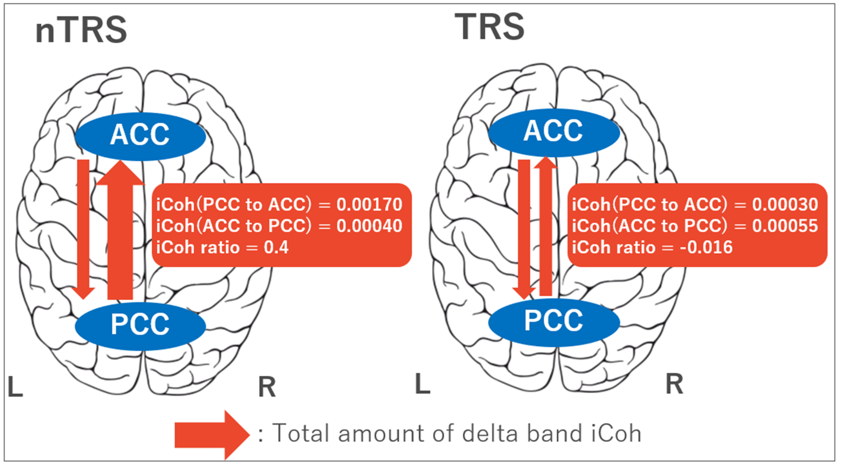
3.2. Four-Way ANOVA for iCoh Values

Four-way ANOVAs for iCoh values between the PCC and ACC indicated the following results. There was a significant group × frequency × directionality × laterality interaction for iCoh values between the PCC and ACC connectivity among the four-way ANOVAs. Consequently, post-hoc independent t-tests for the iCoh ratio of directionality from PCC to ACC showed significant findings that the ratio was decreased in TRS compared with nTRS in delta (t45 = 7.659, p = 0.008; alpha = 0.01) and theta (t45 = 8.066, p = 0.007; alpha = 0.01) frequency bands in the left side (Figure 1). The results of ANOVAs and post-hoc independent t-tests are summarized in Supplemental Table S2.
Of note, there were no significant correlations between CPZ equivalent daily doses and iCoh values in either TRS group (r = −0.196, p = 0.225) or nTRS group (r = 0.064, p = 0.368).

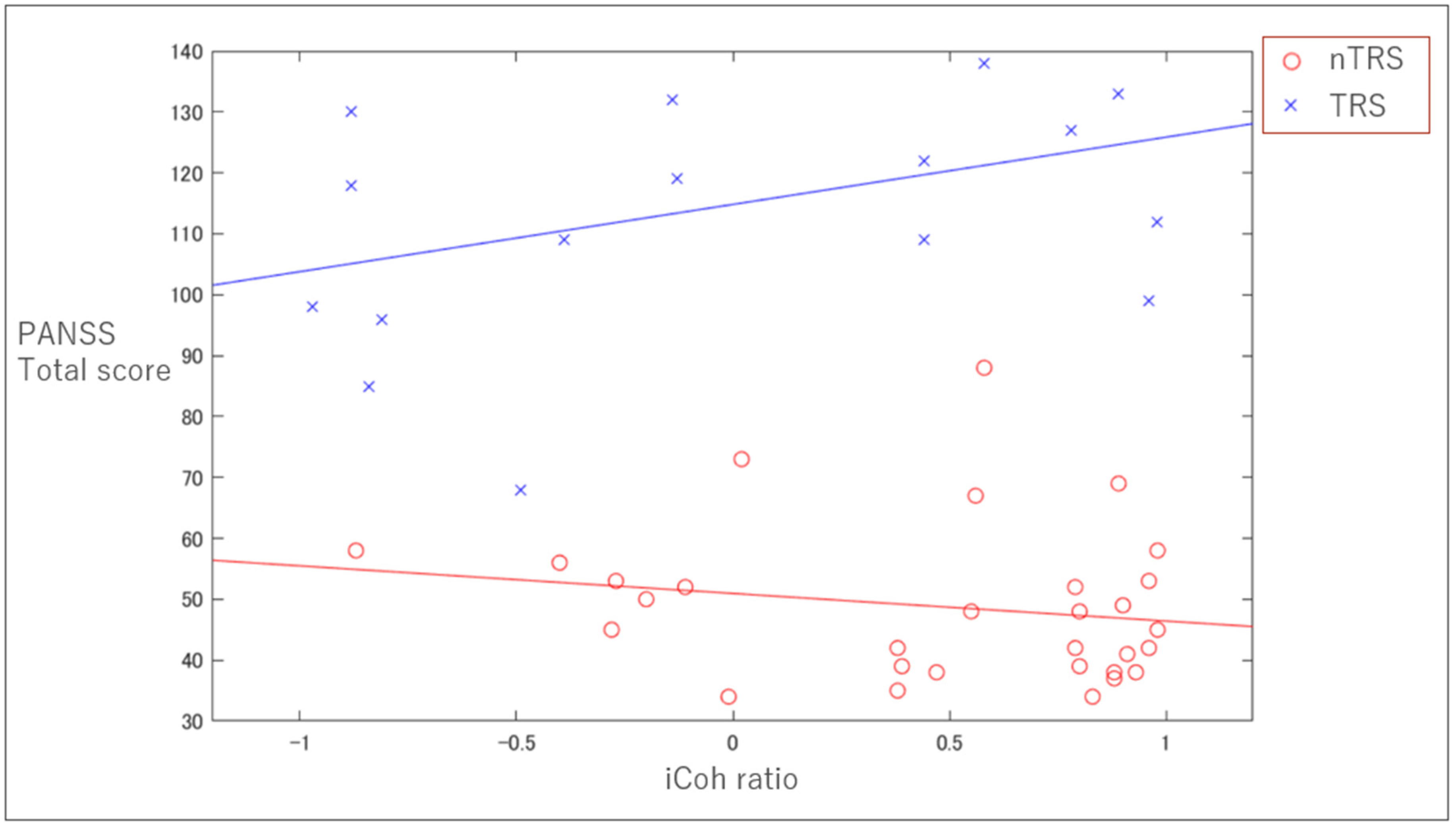
3.3. Clinical Correlation with iCoh

Pearson’s correlational analyses indicated a trend toward a significant correlation between the iCoh ratio for the left delta PCC–ACC connectivity and PANSS total score only for TRS group (r = 0.38, p = 0.069), but not for nTRS group (r = −0.18, p = 0.17) (Figure 2).

3.4. ROC Analysis of the iCoh Ratio between TRS and nTRS

Regarding the discrimination between TRS and nTRS groups, the ROC analysis that employed the iCoh ratio for the left delta PCC–ACC connectivity showed a significant asymptotic p-value (p = 0.023; confidence interval: 0.536–0.868) with an area under the curve of 0.70. Further, the sensitivity and specificity at the optimum point of the ROC curve were 0.64 and 0.70, respectively.

4. Discussion

In the present study, we found that patients with TRS showed a significantly lower iCoh ratio between the PCC and ACC in delta and theta frequency bands over the left side than that of patients with nTRS. Furthermore, there was a trend toward a positive correlation between the PCC and ACC iCoh ratio in delta band over the left side and PANSS total scores in patients with TRS, but not in nTRS, suggesting that the higher iCoh ratio between the PCC and ACC in delta band over the left side was associated with greater psychotic symptoms severity in TRS group. These findings suggest that the absolute flow of information from the DMN to ACC was significantly attenuated in patients with TRS compared with patients with nTRS, while patients with TRS who had more severe psychiatric symptoms showed an increasing trend in relative information flow from the DMN to ACC.
Counterintuitively, we found a positive relationship between symptom severity and PCC–ACC iCoh ratio in the TRS group, while there was no association between them in the non-TRS group. These findings suggest that TRS may be accounted for by the hybrid model of categorical and continuous characteristics [32]. Although previous studies have shown consistent findings of the ACC abnormalities in patients with schizophrenia, no studies so far have examined the information flow in the cingulate bundle between the ACC and PCC in detail in this disorder. In addition, our result of a positive relationship between the PCC–ACC iCoh ratio and clinical severity may be related to impaired function of the ACC in patients with TRS.
The PCC is considered to play a crucial role in mediating spontaneous activity [33,34]. In addition, the PCC is thought to contribute to the essential functions such as emotional salience [35] and autobiographical memory [36]. Thus, dysfunction of the PCC may be related to clinical symptoms of schizophrenia such as hallucinations [37], delusions [38], or disorganized thinking [39]. Additionally, the PCC is also one of the core nodes of the theory of mind (ToM) network, which represents the cognitive ability to understand others as intentional agents by inferring their mental states [40,41]. Some studies showed that patients with schizophrenia had decreased activity of the ToM network including the PCC during the ToM task [5,42]. Thus, the PCC may be crucial as a pathological basis for this disorder. On the other hand, the ACC plays a role in mediating awareness and attention [43,44]. The ACC is thought to be a core region affected by schizophrenia [44] and dysfunction of the ACC may induce severity of symptoms or global impairment of cognitive function [45,46,47]. For example, a smaller volume of the ACC is significantly correlated with more severe positive symptoms of schizophrenia [45] and related to global cognitive impairment measured by the Brief Assessment of Cognition in Schizophrenia [47]. Notably, previous studies noted that patients with TRS had elevated levels of glutamatergic neurometabolites in the ACC [9,10,11]. These findings suggest that our results may explain the features of TRS including more severe positive symptoms, poorer cognitive function, and social function compared with nTRS. Additionally, impaired functional communication between the two regions might make them worse reciprocally. However, the ROC analysis using the iCoh ratio demonstrated a moderate accuracy to differentiate between patients with TRS and nTRS. While the iCoh ratio between the PCC and ACC may be a potential biomarker to distinguish between the two groups, future work is needed to disentangle the pathophysiology of TRS with a combination of multimodal biological measures.
Several studies have shown that the left cingulum is more related to positive symptoms of schizophrenia compared with the right cingulum. Reduced extracellular free-water as measured by diffusion MRI in the left cingulum was associated with delusions in patients with schizophrenia [48]. In addition, Palaniyappan et al. demonstrated that a reduced magnetization transfer ratio in the left cingulum was associated with a higher severity of delusions, while no such relationship was observed in the right cingulum [24]. Collectively, both functional and structural connectivities between the left PCC and ACC may be related to the severity of symptoms as represented by delusions. Additionally, Yuan et al. revealed that patients with schizophrenia who had never been treated for a long term showed more sever white matter abnormalities in the left cingulum-hippocampus pathway compared with patients with schizophrenia who had been treated [49]. This finding supports our hypothesis that the persistent symptoms observed in patients with TRS may be associated with functional abnormalities in the left cingulate cortex. Thus, these findings are in line with our result that the reduction of iCoh ratio was present only in the left side.
Unlike the neuroimaging studies, EEG enables the assessment of cortical network dynamics because of the high temporal resolution. Delta band oscillations are linked with learning, memory encoding and retrieval, and motivation and reward processes [50,51]. The activity of theta band oscillations has been linked to working memory, emotional arousal, and fear conditioning [51]. For example, Hlinka et al. reported that, in an inter-subject experimental design, a strong relationship was established between functional connectivity in delta band oscillations and the DMN [52]. Furthermore, Neuner et al. demonstrated a highly significant correlation between delta band oscillation and spontaneous blood-oxygen-level dependent (BOLD) signal within the DMN using simultaneous fMRI–EEG study [53]. Thus, delta band oscillations may represent the normal functioning of the DMN.
Our findings also suggest a new treatment option for TRS such as neuromodulation. Specifically, non-invasive novel neurostimulation techniques including transcranial magnetic stimulation (TMS) and deep brain stimulation (DBS) enable us to modulate the local neural connectivity [54,55]. Thus, the abnormal neural connectivity in the cingulate bundle can be one of the therapeutic targets. Given the limited treatment options for TRS, neurostimulation targeting the pathological neural basis as described above may be a promising therapeutic strategy.
There are several limitations to the present study. First, we did not include a healthy control group. Comparison between patients with schizophrenia and health control group may reveal comprehensive dysfunction in patients with schizophrenia, which will help to clarify the position of our current findings. Second, we did not include the potential covariates in the statistical analyses such as the dose of antipsychotics as indexed with CPZ equivalent daily doses. However, we did not see a correlation between the dose of antipsychotics and iCoh values or clinical severity. Third, the present study included relatively small sample sizes in both subdiagnostic groups (i.e., TRS and nTRS). Owing to Coronavirus disease 2019 (COVID-19), we could not continue to enroll subjects at this stage. Therefore, our findings warrant further studies with larger sample sizes in this illness using the TRRIP working group consensus criteria [28]. Forth, we focused only on the effective connectivity between the ACC and PCC as a hypothesis-based manner; however, there may be other potential network abnormalities in patients with schizophrenia. Further research is needed using multimodal imaging based on multifaceted perspectives. Fifth, we have only assessed psychiatric symptoms using the PANSS. Thus, there were no clinical measures for other symptoms like depression or anxiety. As schizophrenia is a multifaceted disorder, future research needs to include a variety of clinical measures. Lastly, in this study, we used the standard 10–20 EEG system using 19 electrodes, which could lead to incorrect localization. Because the number of source-level electrodes in the present study was small, we may not have been able to accurately estimate the source of the deep brain signals. Therefore, the present preliminary analyses warrant further studies to confirm the reproducibility and reliability of these results by, for example, using a higher resolution EEG system with 64 channel electrodes and combining them with more sophisticated signal source analysis techniques. However, several previous studies have performed the sLORETA analysis on 19-electrode EEGs [56,57,58,59].

5. Conclusions

In conclusion, we found significant differences in the iCoh ratio between the left PCC and ACC in delta and theta bands between patients with TRS and nTRS. Taken together, these findings may represent part of the underlying neural basis of TRS. The present findings warrant further research in larger sample sizes with multimodal examinations to elucidate underlying mechanisms of treatment-resistance in this illness.

Supplementary Materials

The following are available online at https://www.mdpi.com/2075-4426/10/3/89/s1. Table S1: MNI coordinates, Table S2: Summary of significant results of 4-way ANOVA.

Author Contributions

Conceptualization, Y.N.; methodology, Y.N. and T.M.; software, F.M.; validation, M.W.; formal analysis, M.W. and Y.N.; data curation, T.M., S.H., K.M., and Y.K.; writing: original draft preparation, M.W., F.M., and Y.N.; manuscript editing, Y.N., S.T., K.O., S.F., D.M.B., and Z.J.D.; visualization, M.W.; supervision, Y.N. and S.N.; project administration, R.T., S.H., K.M., Y.K., and S.N.; funding acquisition, S.N., M.M., and Y.N. All authors have read and agreed to the published version of the manuscript.

Funding

This work was supported by the Japan Society for the Promotion of Science and AMED to Y.N., S.N., and M.M. The funding agency did not contribute to the study design; in the data collection, analyses, and interpretation; in the writing of the manuscript; and in the decision to submit the manuscript for publication.

Acknowledgments

We thank Nishikata for his technical support. Furthermore, we appreciate all the patients who have participated in this study.

Conflicts of Interest

M.W., R.T., F.M., T.M., S.T., K.O., S.H., K.M., and Y.K. report no biomedical interests. S.N. has received fellowship grants from CIHR, Japan Research Foundation for Clinical Pharmacology, Naito Foundation, Takeda Science Foundation, Uehara Memorial Foundation, and Daiichi Sankyo Scholarship Donation Program within the past three years. S.N. has also received research supports, manuscript fees, or speaker’s honoraria from Dainippon Sumitomo Pharma, Meiji-Seika Pharma, Otsuka Pharmaceutical, Shionogi, and Yoshitomi Yakuhin within the past three years. Z.J.D. has received research and equipment in-kind support for an investigator-initiated study through Brainsway Inc and Magventure Inc. His work was supported by the Ontario Mental Health Foundation (OMHF), the Canadian Institutes of Health Research (CIHR), the National Institutes of Mental Health (NIMH), and the Temerty Family and Grant Family and through the Centre for Addiction and Mental Health (CAMH) Foundation and the Campbell Institute. M.M. has received research support from Japan Society for the Promotion of Science and grants or speaker’s honoraria from Daiichi Sankyo, Dainippon-Sumitomo Pharma, Eisai, Eli Lilly, Fuji Film RI Pharma, Janssen Pharmaceutical, Mochida Pharmaceutical, M.S.D., Nippon Chemipher, Novartis Pharma, Ono Yakuhin, Otsuka Pharmaceutical, Pfizer, Takeda Yakuhin, Tsumura, and Yoshitomi Yakuhin within the past three years. D.M.B. receives research support from the Canadian Institutes of Health Research (CIHR), National Institutes of Health—US (NIH), Weston Brain Institute, Brain Canada, and the Temerty Family through the CAMH Foundation and the Campbell Research Institute. He received research support and in-kind equipment support for an investigator-initiated study from Brainsway Ltd. and he is the site principal investigator for three sponsor-initiated studies for Brainsway Ltd. He received in-kind equipment support from Magventure for an investigator-initiated study. He received medication supplies for an investigator-initiated trial from Indivior. He has participated in an advisory board for Janssen. Y.N. has received a Grant-in-Aid for Young Scientists (KAKENHI), a research grant from Japan Agency for Medical Research and development (AMED), an investigator-initiated clinical study grant from TEIJIN PHARMA LIMITED. Y.N. also received research grants from Japan Health Foundation, Meiji Yasuda Mental Health Foundation, Mitsui Life Social Welfare Foundation, Takeda Science Foundation, SENSHIN Medical Research Foundation, Health Science Center Foundation, Mochida Memorial Foundation for Medical and Pharmaceutical Research, and Daiichi Sankyo Scholarship Donation Program. He has received research supports from Otsuka Pharmaceutical, Shionogi, and Meiji Seika Pharma. Y.N. also received equipment-in-kind supports for an investigator-initiated study from Magventure Inc., Inter Reha Co., Ltd., Rogue Resolutions Ltd., and Miyuki Giken Co., Ltd.

Abbreviations

ACCanterior cingulate cortex
CPZchlorpromazine
DMNdefault mode network
DBSdeep brain stimulation
EEGelectroencephalography
fMRIfunctional magnetic resonance imaging
iCohisolated effective coherence
IPLinferior parietal lobe
ITLinferior temporal lobe
mPFCmedial prefrontal cortex
MNIMontreal Neurological Institute
PANSSpositive and negative syndrome scale
PCCposterior cingulate cortex
rm-ANOVArepeated-measures analysis of variance
ROCreceiver operating characteristic
ROIsregions of interest
sLORETAstandardized low-resolution brain electromagnetic tomography
TRRIPtreatment response and resistance in psychosis
TMStranscranial magnetic stimulation
TRStreatment-resistant schizophrenia
nTRSnon treatment-resistant schizophrenia

References

  1. Hasan, A.; Falkai, P.; Wobrock, T.; Lieberman, J.; Glenthoj, B.; Gattaz, W.F.; Thibaut, F.; Möller, H.-J. World Federation of Societies of Biological Psychiatry (WFSBP) Guidelines for Biological Treatment of Schizophrenia, Part 1: Update 2012 on the acute treatment of schizophrenia and the management of treatment resistance. World J. Biol. Psychiatry 2012, 13, 318–378. [Google Scholar] [CrossRef] [PubMed]
  2. Samara, M.T.; Dold, M.; Gianatsi, M.; Nikolakopoulou, A.; Helfer, B.; Salanti, G.; Leucht, S. Efficacy, Acceptability, and Tolerability of Antipsychotics in Treatment-Resistant Schizophrenia: A network meta-analysis. JAMA Psychiatry 2016, 73, 199. [Google Scholar] [CrossRef] [Green Version]
  3. Nakajima, S.; Takeuchi, H.; Plitman, E.; Fervaha, G.; Gerretsen, P.; Caravaggio, F.; Chung, J.K.; Iwata, Y.; Remington, G.; Graff-Guerrero, A. Neuroimaging findings in treatment-resistant schizophrenia: A systematic review: Lack of neuroimaging correlates of treatment-resistant schizophrenia. Schizophr. Res. 2015, 164, 164–175. [Google Scholar] [CrossRef] [PubMed] [Green Version]
  4. Carter, C.S.; Macdonald, A.; Ross, L.L.; Stenger, V.A. Anterior Cingulate Cortex Activity and Impaired Self-Monitoring of Performance in Patients with Schizophrenia: An Event-Related fMRI Study. Am. J. Psychiatry 2001, 158, 1423–1428. [Google Scholar] [CrossRef]
  5. Walter, H.; Ciaramidaro, A.; Adenzato, M.; Vasic, N.; Ardito, R.B.; Erk, S.; Bara, B.G. Dysfunction of the social brain in schizophrenia is modulated by intention type: An fMRI study. Soc. Cogn. Affect. Neurosci. 2009, 4, 166–176. [Google Scholar] [CrossRef] [PubMed] [Green Version]
  6. Yan, H.; Tian, L.; Yan, J.; Sun, W.; Liu, Q.; Zhang, Y.-B.; Li, X.-M.; Zang, Y.-F.; Zhang, D. Functional and Anatomical Connectivity Abnormalities in Cognitive Division of Anterior Cingulate Cortex in Schizophrenia. PLoS ONE 2012, 7, e45659. [Google Scholar] [CrossRef]
  7. Salgado-Pineda, P.; Landin-Romero, R.; Fakra, E.; Delaveau, P.; Amann, B.L.; Blin, O. Structural Abnormalities in Schizophrenia: Further Evidence on the Key Role of the Anterior Cingulate Cortex. Neuropsychobiology 2014, 69, 52–58. [Google Scholar] [CrossRef]
  8. Mouchlianitis, E.; Bloomfield, M.A.P.; Law, V.; Beck, K.; Selvaraj, S.; Rasquinha, N.; Waldman, A.D.; Turkheimer, F.E.; Egerton, A.; Stone, J.; et al. Treatment-Resistant Schizophrenia Patients Show Elevated Anterior Cingulate Cortex Glutamate Compared to Treatment-Responsive. Schizophr. Bull. 2015, 42, 744–752. [Google Scholar] [CrossRef]
  9. Iwata, Y.; Nakajima, S.; Plitman, E.; Caravaggio, F.; Kim, J.; Shah, P.; Mar, W.; Chavez, S.; De Luca, V.; Mimura, M.; et al. Glutamatergic Neurometabolite Levels in Patients With Ultra-Treatment-Resistant Schizophrenia: A Cross-Sectional 3T Proton Magnetic Resonance Spectroscopy Study. Biol. Psychiatry 2019, 85, 596–605. [Google Scholar] [CrossRef]
  10. Demjaha, A.; Egerton, A.; Murray, R.M.; Kapur, S.; Howes, O.D.; Stone, J.; McGuire, P. Antipsychotic Treatment Resistance in Schizophrenia Associated with Elevated Glutamate Levels but Normal Dopamine Function. Boil. Psychiatry 2014, 75, e11–e13. [Google Scholar] [CrossRef]
  11. Tarumi, R.; Tsugawa, S.; Noda, Y.; Plitman, E.; Honda, S.; Matshusita, K.; Chavez, S.; Sawada, K.; Wada, M.; Matsui, M.; et al. Levels of glutamatergic neurometabolites in patients with severe treatment-resistant schizophrenia: A proton magnetic resonance spectroscopy study. Neuropsychopharmacology 2020, 46, S313. [Google Scholar] [CrossRef] [PubMed]
  12. Jafri, M.J.; Pearlson, G.D.; Stevens, M.C.; Calhoun, V.D. A method for functional network connectivity among spatially independent resting-state components in schizophrenia. NeuroImage 2008, 39, 1666–1681. [Google Scholar] [CrossRef] [PubMed] [Green Version]
  13. Skudlarski, P.; Jagannathan, K.; Anderson, K.; Stevens, M.C.; Calhoun, V.D.; Skudlarska, B.A.; Pearlson, G. Brain Connectivity Is Not Only Lower but Different in Schizophrenia: A Combined Anatomical and Functional Approach. Biol. Psychiatry 2010, 68, 61–69. [Google Scholar] [CrossRef] [PubMed] [Green Version]
  14. Zhou, C.; Yu, M.; Tang, X.; Wang, X.; Zhang, X.; Zhang, X.R.; Chen, J. Convergent and divergent altered patterns of default mode network in deficit and non-deficit schizophrenia. Prog. Neuro Psychopharmacol. Biol. Psychiatry 2019, 89, 427–434. [Google Scholar] [CrossRef] [PubMed]
  15. Buckner, R.L.; Schacter, D.L.; Andrews-Hanna, J.R. The Brain’s Default Network. Ann. N. Y. Acad. Sci. 2008, 1124, 1–38. [Google Scholar] [CrossRef] [PubMed] [Green Version]
  16. Krukow, P.; Jonak, K.; Grochowski, C.; Plechawska-Wójcik, M.; Karakuła-Juchnowicz, H. Resting-state hyperconnectivity within the default mode network impedes the ability to initiate cognitive performance in first-episode schizophrenia patients. Prog. Neuro Psychopharmacol. Biol. Psychiatry 2020, 102, 109959. [Google Scholar] [CrossRef]
  17. Lee, H.; Lee, D.-K.; Park, K.; Kim, C.-E.; Ryu, S. Default mode network connectivity is associated with long-term clinical outcome in patients with schizophrenia. NeuroImage Clin. 2019, 22, 101805. [Google Scholar] [CrossRef] [PubMed]
  18. Fox, J.M.; Abram, S.V.; Reilly, J.L.; Eack, S.; Goldman, M.B.; Csernansky, J.G.; Wang, L.; Smith, M.J. Default mode functional connectivity is associated with social functioning in schizophrenia. J. Abnorm. Psychol. 2017, 126, 392–405. [Google Scholar] [CrossRef] [PubMed]
  19. Bluhm, R.L.; Miller, J.; Lanius, R.A.; Osuch, E.A.; Boksman, K.; Neufeld, R.; Théberge, J.; Schaefer, B.; Williamson, P. Spontaneous Low-Frequency Fluctuations in the BOLD Signal in Schizophrenic Patients: Anomalies in the Default Network. Schizophr. Bull. 2007, 33, 1004–1012. [Google Scholar] [CrossRef]
  20. Garrity, A.G.; Pearlson, G.D.; McKiernan, K.; Lloyd, D.; Kiehl, K.A.; Calhoun, V.D. Aberrant “default mode” functional connectivity in schizophrenia. Am. J. Psychiatry 2007, 164, 450–457. [Google Scholar] [CrossRef]
  21. Hare, S.M.; Ford, J.M.; Mathalon, D.H.; Damaraju, E.; Bustillo, J.; Belger, A.; Lee, H.J.; A Mueller, B.; O Lim, K.; Brown, G.G.; et al. Salience–Default Mode Functional Network Connectivity Linked to Positive and Negative Symptoms of Schizophrenia. Schizophr. Bull. 2018, 45, 892–901. [Google Scholar] [CrossRef] [PubMed]
  22. Alonso-Solís, A.; Vives-Gilabert, Y.; Grasa, E.; Portella, M.J.; Rabella, M.; Sauras, R.B.; Roldán, A.; Núñez-Marín, F.; Gomez-Anson, B.; Pérez, V.; et al. Resting-state functional connectivity alterations in the default network of schizophrenia patients with persistent auditory verbal hallucinations. Schizophr. Res. 2015, 161, 261–268. [Google Scholar] [CrossRef] [PubMed]
  23. Whitford, T.J.; Lee, S.W.; Oh, J.S.; De Luis-García, R.; Savadjiev, P.; Alvarado, J.L.; Westin, C.-F.; Niznikiewicz, M.; Nestor, P.G.; McCarley, R.W.; et al. Localized abnormalities in the cingulum bundle in patients with schizophrenia: A Diffusion Tensor tractography study. NeuroImage Clin. 2014, 5, 93–99. [Google Scholar] [CrossRef] [PubMed] [Green Version]
  24. Palaniyappan, L.; Al-Radaideh, A.; Mougin, O.; Das, T.; Gowland, P.A.; Liddle, P.F. Aberrant myelination of the cingulum and Schneiderian delusions in schizophrenia: A 7T magnetization transfer study. Psychol. Med. 2018, 49, 1890–1896. [Google Scholar] [CrossRef]
  25. Pascual-Marqui, R.D.; Biscay, R.J.; Bosch-Bayard, J.; Lehmann, D.; Kochi, K.; Kinoshita, T.; Yamada, N.; Sadato, N. Assessing direct paths of intracortical causal information flow of oscillatory activity with the isolated effective coherence (iCoh). Front. Hum. Neurosci. 2014, 8, 448. [Google Scholar] [CrossRef]
  26. Schelter, B.; Timmer, J.; Eichler, M. Assessing the strength of directed influences among neural signals using renormalized partial directed coherence. J. Neurosci. Methods 2009, 179, 121–130. [Google Scholar] [CrossRef] [PubMed]
  27. Plomp, G.; Quairiaux, C.; Michel, C.M.; Astolfi, L. The physiological plausibility of time-varying Granger-causal modeling: Normalization and weighting by spectral power. NeuroImage 2014, 97, 206–216. [Google Scholar] [CrossRef]
  28. Howes, O.D.; McCutcheon, R.A.; Agid, O.; De Bartolomeis, A.; Van Beveren, N.J.; Birnbaum, M.L.; Bloomfield, M.A.P.; Bressan, R.A.; Buchanan, R.W.; Carpenter, W.T.; et al. Treatment-Resistant Schizophrenia: Treatment Response and Resistance in Psychosis (TRRIP) Working Group Consensus Guidelines on Diagnosis and Terminology. Am. J. Psychiatry 2017, 174, 216–229. [Google Scholar] [CrossRef]
  29. Kay, S.R.; Fiszbein, A.; Opler, L.A. The Positive and Negative Syndrome Scale (PANSS) for Schizophrenia. Schizophr. Bull. 1987, 13, 261–276. [Google Scholar] [CrossRef]
  30. Pascual-Marqui, R.D.; Michel, C.; Lehmann, D. Low resolution electromagnetic tomography: A new method for localizing electrical activity in the brain. Int. J. Psychophysiol. 1994, 18, 49–65. [Google Scholar] [CrossRef]
  31. Kitaura, Y.; Nishida, K.; Yoshimura, M.; Mii, H.; Katsura, K.; Ueda, S.; Ikeda, S.; Pascual-Marqui, R.D.; Ishii, R.; Kinoshita, T. Functional localization and effective connectivity of cortical theta and alpha oscillatory activity during an attention task. Clin. Neurophysiol. Pr. 2017, 2, 193–200. [Google Scholar] [CrossRef] [PubMed]
  32. Mouchlianitis, E.; McCutcheon, R.A.; Howes, O.D. Brain-imaging studies of treatment-resistant schizophrenia: A systematic review. Lancet Psychiatry 2016, 3, 451–463. [Google Scholar] [CrossRef] [Green Version]
  33. Coito, A.; Michel, C.M.; Vulliemoz, S.; Plomp, G. Directed functional connections underlying spontaneous brain activity. Hum. Brain Mapp. 2019, 40, 879–888. [Google Scholar] [CrossRef] [PubMed] [Green Version]
  34. Fransson, P.; Marrelec, G. The precuneus/posterior cingulate cortex plays a pivotal role in the default mode network: Evidence from a partial correlation network analysis. NeuroImage 2008, 42, 1178–1184. [Google Scholar] [CrossRef]
  35. Tao, Y.; Liu, B.; Zhang, X.; Li, J.; Qin, W.; Yu, C.; Jiang, T. The Structural Connectivity Pattern of the Default Mode Network and Its Association with Memory and Anxiety. Front. Neuroanat. 2015, 9, 152. [Google Scholar] [CrossRef] [Green Version]
  36. Maddock, R.J.; Garrett, A.S.; Buonocore, M.H. Remembering familiar people: The posterior cingulate cortex and autobiographical memory retrieval. Neuroscience 2001, 104, 667–676. [Google Scholar] [CrossRef]
  37. Lefebvre, S.; Demeulemeester, M.; Leroy, A.; Delmaire, C.; Lopes, R.; Pins, D.; Thomas, P.; Jardri, R. Network dynamics during the different stages of hallucinations in schizophrenia. Hum. Brain Mapp. 2016, 37, 2571–2586. [Google Scholar] [CrossRef]
  38. Manoliu, A.; Riedl, V.; Zherdin, A.; Mühlau, M.; Schwerthöffer, D.; Scherr, M.; Peters, H.; Zimmer, C.; Förstl, H.; Bäuml, J.; et al. Aberrant dependence of default mode/central executive network interactions on anterior insular salience network activity in schizophrenia. Schizophr. Bull. 2014, 40, 428–437. [Google Scholar] [CrossRef] [Green Version]
  39. Lagioia, A.; Van De Ville, D.; Debbané, M.; Lazeyras, F.; Eliez, S. Adolescent resting state networks and their associations with schizotypal trait expression. Front. Syst. Neurosci. 2010, 4, 4. [Google Scholar] [CrossRef] [Green Version]
  40. Tettamanti, M.; Vaghi, M.M.; Bara, B.G.; Cappa, S.F.; Enrici, I.; Adenzato, M. Effective connectivity gateways to the Theory of Mind network in processing communicative intention. NeuroImage 2017, 155, 169–176. [Google Scholar] [CrossRef] [Green Version]
  41. Quidé, Y.; Wilhelmi, C.; Green, M.J. Structural brain morphometry associated with theory of mind in bipolar disorder and schizophrenia. PsyCh J. 2020, 9, 234–246. [Google Scholar] [CrossRef]
  42. Corcoran, R.; Mercer, G.; Frith, C.D. Schizophrenia, symptomatology and social inference: Investigating “theory of mind” in people with schizophrenia. Schizophr. Res. 1995, 17, 5–13. [Google Scholar] [CrossRef]
  43. Hakamata, Y.; Iwase, M.; Kato, T.; Senda, K.; Inada, T. The Neural Correlates of Mindful Awareness: A Possible Buffering Effect on Anxiety-Related Reduction in Subgenual Anterior Cingulate Cortex Activity. PLoS ONE 2013, 8, e75526. [Google Scholar] [CrossRef] [PubMed] [Green Version]
  44. Wu, D.; Deng, H.; Xiao, X.; Zuo, Y.; Sun, J.; Wang, Z. Persistent Neuronal Activity in Anterior Cingulate Cortex Correlates with Sustained Attention in Rats Regardless of Sensory Modality. Sci. Rep. 2017, 7, 43101. [Google Scholar] [CrossRef] [PubMed] [Green Version]
  45. Choi, J.-S.; Kang, D.-H.; Kim, J.-J.; Ha, T.-H.; Roh, K.S.; Youn, T.; Kwon, J.S. Decreased caudal anterior cingulate gyrus volume and positive symptoms in schizophrenia. Psychiatry Res. Neuroimaging 2005, 139, 239–247. [Google Scholar] [CrossRef] [PubMed]
  46. Nelson, B.D.; Bjorkquist, O.A.; Olsen, E.K.; Herbener, E.S. Schizophrenia symptom and functional correlates of anterior cingulate cortex activation to emotion stimuli: An fMRI investigation. Psychiatry Res. 2015, 234, 285–291. [Google Scholar] [CrossRef] [Green Version]
  47. Ohi, K.; Shimada, T.; Nemoto, K.; Kataoka, Y.; Yasuyama, T.; Kimura, K.; Okubo, H.; Uehara, T.; Kawasaki, Y. Cognitive clustering in schizophrenia patients, their first-degree relatives and healthy subjects is associated with anterior cingulate cortex volume. NeuroImage Clin. 2017, 16, 248–256. [Google Scholar] [CrossRef] [Green Version]
  48. Oestreich, L.K.; Pasternak, O.; Shenton, M.E.; Kubicki, M.; Gong, X.; Whitford, T.J.; Australian Schizophrenia Research Bank; McCarthy-Jones, S.; Whitford, T.J. Abnormal white matter microstructure and increased extracellular free-water in the cingulum bundle associated with delusions in chronic schizophrenia. NeuroImage Clin. 2016, 12, 405–414. [Google Scholar] [CrossRef] [Green Version]
  49. Xiao, Y.; Sun, H.; Shi, S.; Jiang, D.; Tao, B.; Zhao, Y.; Zhang, W.; Gong, Q.; Sweeney, J.A.; Lui, S. White Matter Abnormalities in Never-Treated Patients With Long-Term Schizophrenia. Am. J. Psychiatry 2018, 175, 1129–1136. [Google Scholar] [CrossRef] [Green Version]
  50. Ekstrom, A.; Watrous, A.J. Multifaceted roles for low-frequency oscillations in bottom-up and top-down processing during navigation and memory. NeuroImage 2014, 85, 667–677. [Google Scholar] [CrossRef] [Green Version]
  51. Knyazev, G.G. Motivation, emotion, and their inhibitory control mirrored in brain oscillations. Neurosci. Biobehav. Rev. 2007, 31, 377–395. [Google Scholar] [CrossRef] [PubMed]
  52. Hlinka, J.; Alexakis, C.; Diukova, A.; Liddle, P.F.; Auer, D.P. Slow EEG pattern predicts reduced intrinsic functional connectivity in the default mode network: An inter-subject analysis. NeuroImage 2010, 53, 239–246. [Google Scholar] [CrossRef] [PubMed]
  53. Neuner, I.; Arrubla, J.; Werner, C.J.; Hitz, K.; Boers, F.; Kawohl, W.; Shah, N.J. The Default Mode Network and EEG Regional Spectral Power: A Simultaneous fMRI-EEG Study. PLoS ONE 2014, 9, e88214. [Google Scholar] [CrossRef] [PubMed] [Green Version]
  54. Tik, M.; Hoffmann, A.; Sladky, R.; Tomova, L.; Hummer, A.; De Lara, L.I.N.; Bukowski, H.; Pripfl, J.; Biswal, B.B.; Lamm, C.; et al. Towards understanding rTMS mechanism of action: Stimulation of the DLPFC causes network-specific increase in functional connectivity. Neuroimage 2017, 162, 289–296. [Google Scholar] [CrossRef]
  55. Chiken, S.; Nambu, A. Mechanism of Deep Brain Stimulation: Inhibition, Excitation, or Disruption? Neuroscientist 2016, 22, 313–322. [Google Scholar] [CrossRef]
  56. Lehmann, D.; Faber, P.L.; Tei, S.; Pascual-Marqui, R.D.; Milz, P.; Kochi, K. Reduced functional connectivity between cortical sources in five meditation traditions detected with lagged coherence using EEG tomography. NeuroImage 2012, 60, 1574–1586. [Google Scholar] [CrossRef] [Green Version]
  57. A Ponomarev, V.; Kropotov, I.D. Improving source localization of ERPs in the GO/NOGO task by modeling of their cross-covariance structure. Fiziol. Cheloveka 2013, 39, 36–50. [Google Scholar]
  58. Coben, R.; Hammond, D.C.; Arns, M. 19 Channel Z-Score and LORETA Neurofeedback: Does the Evidence Support the Hype? Appl. Psychophysiol. Biofeedback 2019, 44, 1–8. [Google Scholar] [CrossRef] [Green Version]
  59. Eugene, A.R.; Masiak, J. Identifying Treatment Response of Sertraline in a Teenager with Selective Mutism using Electrophysiological Neuroimaging. Int. J. Clin. Pharmacol. Toxicol. 2016, 5, 216–219. [Google Scholar] [CrossRef]
Figure 1. Schematics of the causal effective connectivity between the posterior cingulate cortex (PCC) and anterior cingulate cortex (ACC). In patients with treatment-resistant schizophrenia (TRS), the isolated effective coherence (iCoh) ratios [(PCC to ACC) − (ACC to PCC)]/[(PCC to ACC) + (ACC to PCC)] in delta and theta bands over the left side were significantly decreased compared with patients with non-TRS (nTRS).
Jpm 10 00089 g001
Figure 2. The relationship between the iCho ratio for the left delta PCC–ACC connectivity and positive and negative syndrome scale (PANSS) total score. In patients with TRS, there is trend toward a relationship (r = 0.38, p = 0.069), but not for the nTRS group (r = −0.18, p = 0.17).
Jpm 10 00089 g002
Table 1. Clinico-demographics data.
nTRS
(n = 30)
TRS
(n = 17)
t-Value (Chi-Squared Value for Sex),
p-Value
Age, mean (SD), years41.2 (12.6)42.4 (13.4)t45 = 0.29, p = 0.78
Sex (number of male) (%)13 (43)5 (29)χ245 = 0.89, p = 0.34
Education, mean (SD), years13.3 (1.81)13.3 (2.42)t45 = 0.06, p = 0.95
Age of onset, mean (SD), years26.0 (9.47)26.6 (7.64)t45 = 0.23, p = 0.82
Treatment duration, mean (SD), years14.5 (12.0)15.5 (11.3)t45 = 0.27, p = 0.79
Chlorpromazine equivalents, mean (SD), mg406 (233.5)748 (319.0)t45 = 4.22, p < 0.001 *
PANSS total, mean (SD)48.8 (12.7)114.6 (21.2)t45 = 13.4, p < 0.001 *
TRS: treatment-resistant schizophrenia; nTRS: non treatment-resistant schizophrenia; SD: standard deviation; PANSS: positive and negative syndrome scale, * = p < 0.05.

