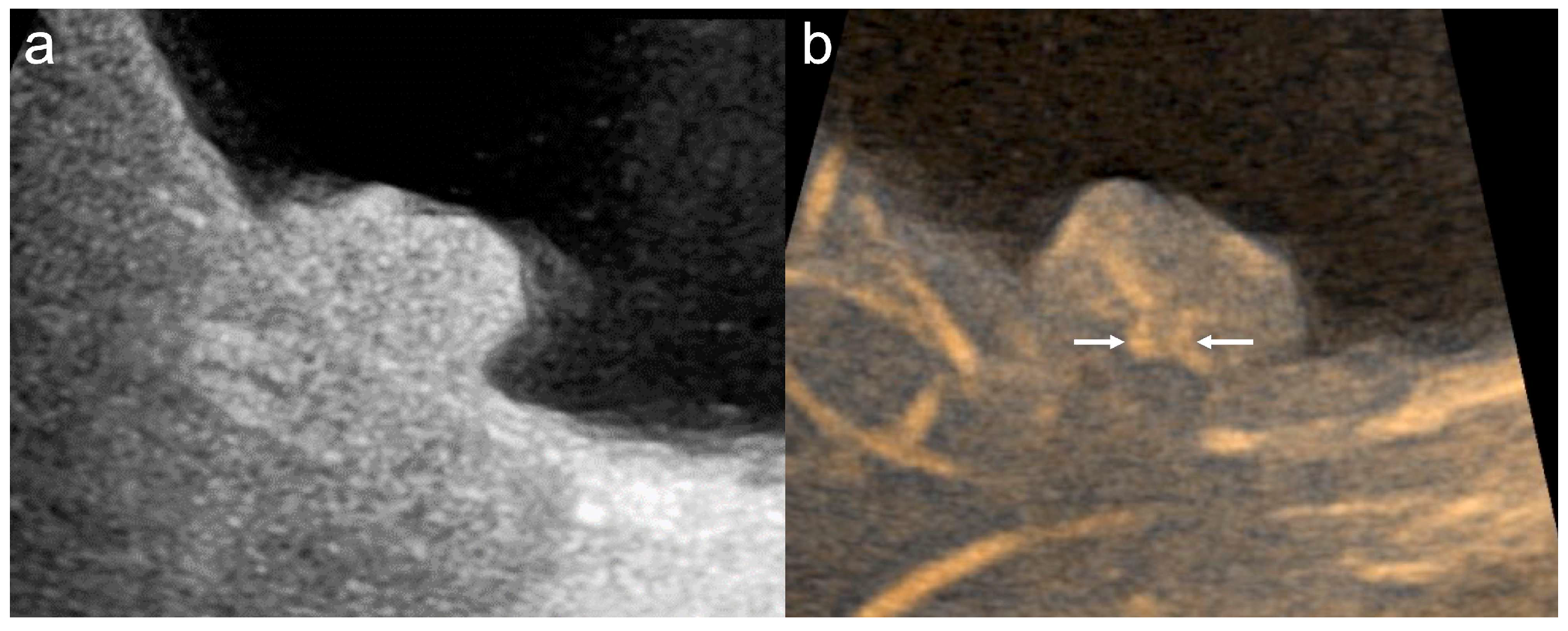
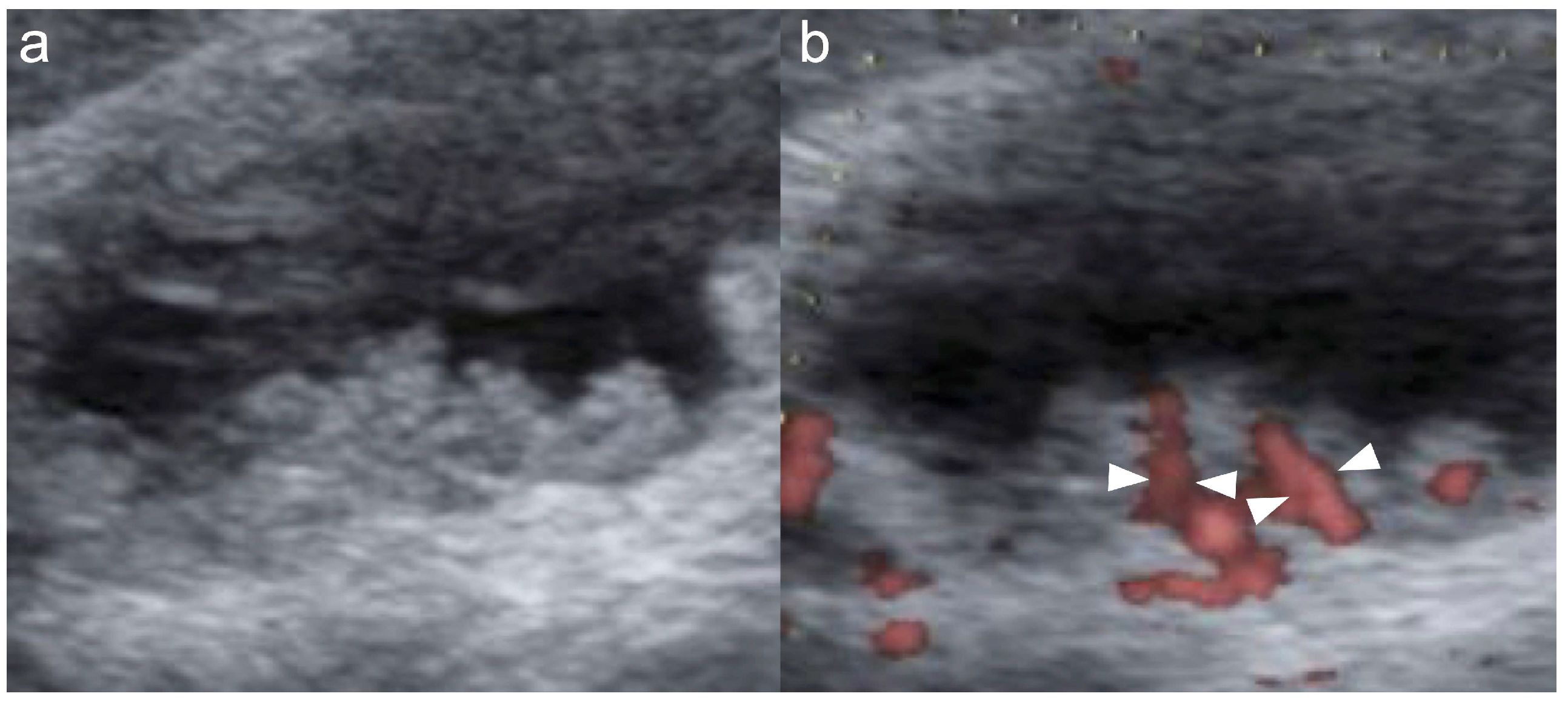
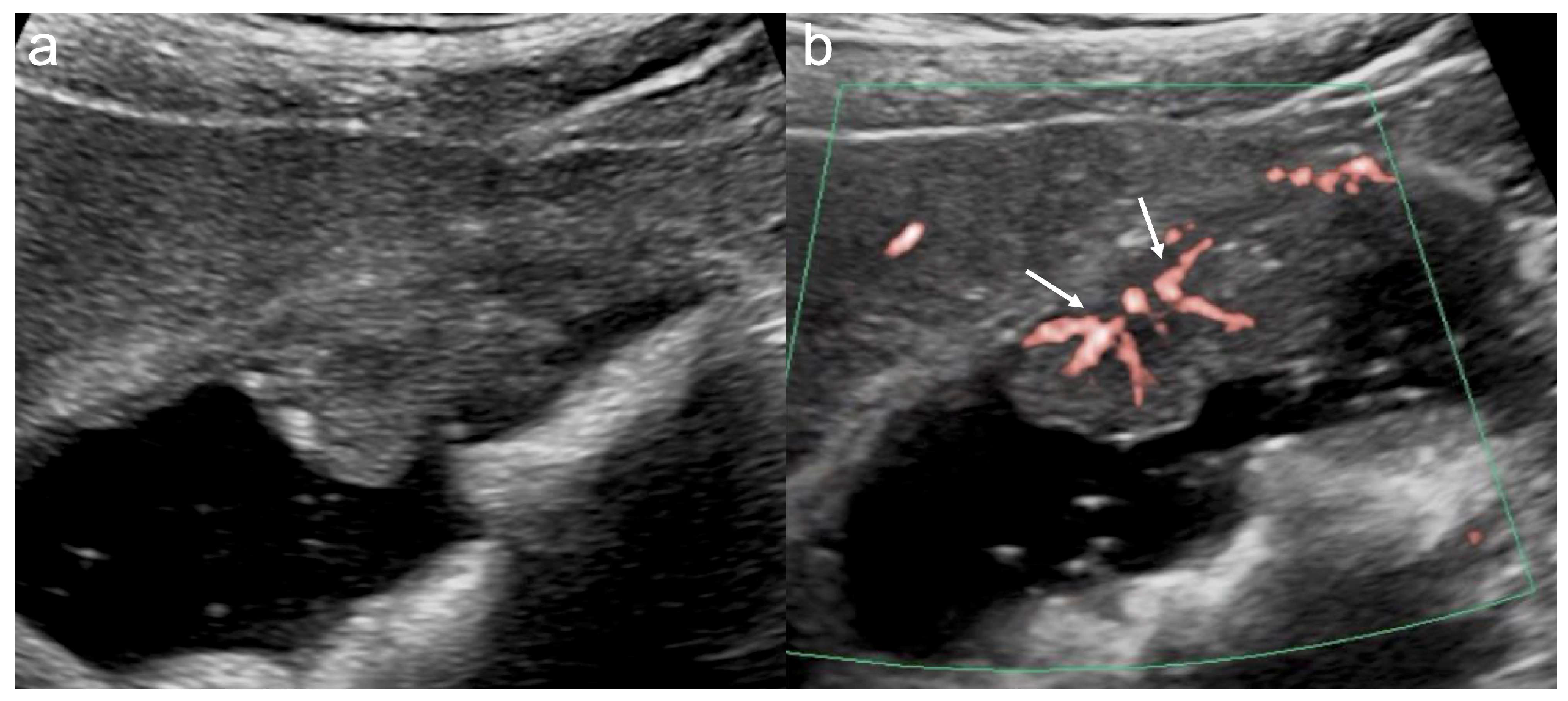
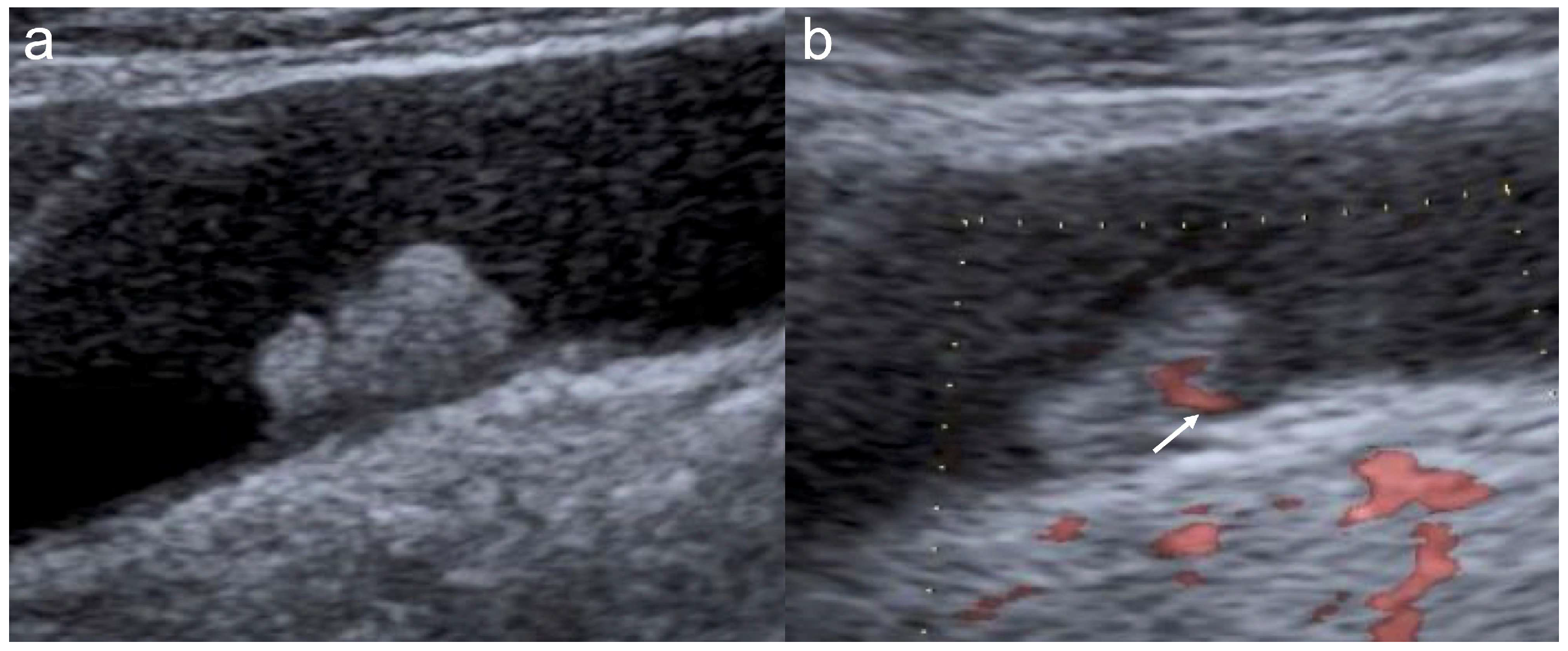
Vascular Features of Gallbladder Lesions Using Microvascular Flow Imaging on Transabdominal Ultrasonography: A Retrospective Study
 , , , ,
Abstract
1. Introduction
2. Materials and Methods
2.1. Patients
2.2. TUS Examinations
2.3. Microvascular Flow Imaging
2.4. Image Evaluation
2.5. Study Design
2.6. Statistical Analysis
2.7. Ethics
2.8. Use of Generative AI
3. Results
3.1. Patient Characteristics
3.2. Interobserver Agreement
3.3. Imaging Findings After Consensus Review
4. Discussion
5. Conclusions
Author Contributions
Funding
Institutional Review Board Statement
Informed Consent Statement
Data Availability Statement
Acknowledgments
Conflicts of Interest
References
- Goetze, T.O. Gallbladder carcinoma: Prognostic factors and therapeutic options. World J. Gastroenterol. 2015, 21, 12211–12217. [Google Scholar] [CrossRef] [Scilit]
- Roa, J.C.; García, P.; Kapoor, V.K.; Maithel, S.K.; Javle, M.; Koshiol, J. Gallbladder cancer. Nat. Rev. Dis. Primers 2022, 8, 69. [Google Scholar] [CrossRef] [Scilit] [PubMed]
- Wennmacker, S.Z.; Lamberts, M.P.; Di Martino, M.; Drenth, J.P.; Gurusamy, K.S.; van Laarhoven, C.J. Transabdominal ultrasound and endoscopic ultrasound for diagnosis of gallbladder polyps. Cochrane Database Syst. Rev. 2018, 8, CD012233. [Google Scholar] [CrossRef] [Scilit] [PubMed]
- Pickering, O.; Pucher, P.H.; Toale, C.; Hand, F.; Anand, E.; Cassidy, S.; McEntee, G.; Toh, S.K. Prevalence and sonographic detection of gallbladder polyps in a western European population. J. Surg. Res. 2020, 250, 226–231. [Google Scholar] [CrossRef] [Scilit] [PubMed]
- Runner, G.J.; Corwin, M.T.; Siewert, B.; Eisenberg, R.L. Gallbladder wall thickening. AJR Am. J. Roentgenol. 2014, 202, W1–W12. [Google Scholar] [CrossRef] [Scilit]
- Bhatt, N.R.; Gillis, A.; Smoothey, C.O.; Awan, F.N.; Ridgway, P.F. Evidence based management of polyps of the gall bladder: A systematic review of the risk factors of malignancy. Surgeon 2016, 14, 278–286. [Google Scholar] [CrossRef] [Scilit]
- Okaniwa, S. How Can We Manage Gallbladder Lesions by Transabdominal Ultrasound? Diagnostics 2021, 11, 784. [Google Scholar] [CrossRef] [Scilit]
- Foley, K.G.; Lahaye, M.J.; Thoeni, R.F.; Soltes, M.; Dewhurst, C.; Barbu, S.T.; Vashist, Y.K.; Rafaelsen, S.R.; Arvanitakis, M.; Perinel, J.; et al. Management and follow-up of gallbladder polyps: Updated joint guidelines between the ESGAR, EAES, EFISDS and ESGE. Eur. Radiol. 2022, 32, 3358–3368. [Google Scholar] [CrossRef] [Scilit]
- Badea, R.; Zaro, R.; Opincariu, I.; Chiorean, L. Ultrasound in the examination of the gallbladder—A holistic approach: Grey scale, Doppler, CEUS, elastography, and 3D. Med. Ultrason. 2014, 16, 345–355. [Google Scholar] [CrossRef] [Scilit]
- Miyoshi, H.; Inui, K.; Katano, Y.; Tachi, Y.; Yamamoto, S. B-mode ultrasonographic diagnosis in gallbladder wall thickening. J. Med. Ultrason. 2021, 48, 175–186. [Google Scholar] [CrossRef] [Scilit]
- Xu, J.-M.; Guo, L.-H.; Xu, H.-X.; Zheng, S.-G.; Liu, L.-N.; Sun, L.-P.; Lu, M.-D.; Xie, X.-Y.; Wang, W.-P.; Hu, B.; et al. Differential diagnosis of gallbladder wall thickening: The usefulness of contrast-enhanced ultrasound. Ultrasound Med. Biol. 2014, 40, 2794–2804. [Google Scholar] [CrossRef] [Scilit]
- Sun, L.-P.; Guo, L.-H.; Xu, H.-X.; Liu, L.-N.; Xu, J.-M.; Zhang, Y.-F.; Liu, C.; Bo, X.-W.; Xu, X.-H. Value of contrast-enhanced ultrasound in the differential diagnosis between gallbladder adenoma and gallbladder adenoma canceration. Int. J. Clin. Exp. Med. 2015, 8, 1115–1121. [Google Scholar]
- Miwa, H.; Numata, K.; Sugimori, K.; Sanga, K.; Hirotani, A.; Tezuka, S.; Goda, Y.; Irie, K.; Ishii, T.; Kaneko, T.; et al. Differential diagnosis of gallbladder polypoid lesions using contrast-enhanced ultrasound. Abdom. Radiol. 2019, 44, 1367–1378. [Google Scholar] [CrossRef] [Scilit]
- Matsumoto, N.; Ogawa, M.; Miura, T.; Shiozawa, K.; Abe, M.; Nakagawara, H.; Moriyama, M. B-flow imaging of vascular structure for the diagnosis of liver tumor. J. Med. Ultrason. 2013, 40, 409–415. [Google Scholar] [CrossRef] [Scilit] [PubMed]
- Lu, R.; Meng, Y.; Zhang, Y.; Zhao, W.; Wang, X.; Jin, M.; Guo, R. Superb microvascular imaging (SMI) compared with conventional ultrasound for evaluating thyroid nodules. BMC Med. Imaging 2017, 17, 65. [Google Scholar] [CrossRef] [Scilit]
- Kin, T.; Nagai, K.; Hayashi, T.; Takahashi, K.; Katanuma, A. Efficacy of superb microvascular imaging of ultrasound for diagnosis of gallbladder lesion. J. Hepatobiliary Pancreat. Sci. 2020, 27, 977–983. [Google Scholar] [CrossRef] [Scilit]
- Miwa, H.; Numata, K.; Sugimori, K.; Kaneko, T.; Maeda, S. Vascular evaluation using transabdominal ultrasound for gallbladder polyps. J. Med. Ultrason. 2021, 48, 159–173. [Google Scholar] [CrossRef] [Scilit]
- Matsui, M.; Jikuzono, T.; Kure, S.; Ishibashi, O.; Akasu, H.; Sugitani, I. Usefulness of ultrasonographic detective flow imaging to diagnose parathyroid tumors: Two case reports. J. Nippon. Med. Sch. 2022, 90, 460–464. [Google Scholar] [CrossRef] [Scilit] [PubMed]
- Miwa, H.; Sugimori, K.; Maeda, S. Vessel images of gallbladder polypoid lesions on detective flow imaging endoscopic ultrasonography. Dig. Endosc. 2023, 35, e61–e62. [Google Scholar] [CrossRef] [Scilit] [PubMed]
- Yamashita, Y.; Ashida, R.; Tamura, T.; Shimokawa, T.; Yamazaki, H.; Kawaji, Y.; Tamura, T.; Hatamaru, K.; Itonaga, M.; Kitano, M. Novel Technique of Endoscopic Ultrasonography for the Differential Diagnosis of Gallbladder Lesions and Intraductal Papillary Mucinous Neoplasms: A Single-Center Prospective Study. Diagnostics 2023, 13, 2132. [Google Scholar] [CrossRef] [Scilit]
- Chen, S.-H.; Xiang, X.-Z.; Che, P.-F.; Hu, B.; Shui, D.; Zhao, Y.; Wang, L. Superb Microvascular Imaging for the Differentiation of Benign and Malignant Breast Lesions: A System Review and Meta-Analysis. J. Ultrasound Med. 2023, 42, 1385–1399. [Google Scholar] [CrossRef] [Scilit]
- Hofmann, A.G.; Mlekusch, I.; Wickenhauser, G.; Assadian, A.; Taher, F. Clinical Applications of B-Flow Ultrasound: A Scoping Review of the Literature. Diagnostics 2023, 13, 397. [Google Scholar] [CrossRef] [Scilit] [PubMed]
- Chantarojanasiri, T.; Hirooka, Y.; Kawashima, H.; Ohno, E.; Kongkam, P.; Goto, H. The role of endoscopic ultrasound in the diagnosis of gallbladder diseases. J. Med. Ultrason. 2017, 44, 63–70. [Google Scholar] [CrossRef] [Scilit] [PubMed]
- Hirooka, Y.; Naitoh, Y.; Goto, H.; Furukawa, T.; Ito, A.; Hayakawa, T. Differential diagnosis of gall-bladder masses using colour Doppler ultrasonography. J. Gastroenterol. Hepatol. 1996, 11, 840–846. [Google Scholar] [CrossRef] [Scilit] [PubMed]
- Komatsuda, T.; Ishida, H.; Konno, K.; Hamashima, Y.; Naganuma, H.; Sato, M.; Watanabe, S. Gallbladder carcinoma: Color Doppler sonography. Abdom. Imaging 2000, 25, 194–197. [Google Scholar] [CrossRef] [Scilit]
- Wang, W.; Yang, Z.-L.; Liu, J.-Q.; Jiang, S.; Miao, X.Y. Identification of CD146 expression, angiogenesis, and lymphangiogenesis as progression, metastasis, and poor-prognosis related markers for gallbladder adenocarcinoma. Tumor Biol. 2012, 33, 173–182. [Google Scholar] [CrossRef] [Scilit]
- Liu, L.-N.; Xu, H.-X.; Lu, M.-D.; Xie, X.-Y.; Wang, W.-P.; Hu, B.; Yan, K.; Ding, H.; Tang, S.-S.; Qian, L.-X.; et al. Contrast-enhanced ultrasound in the diagnosis of gallbladder diseases: A multi-center experience. PLoS ONE 2012, 7, e48371. [Google Scholar] [CrossRef] [Scilit]
- Yuan, H.-X.; Cao, J.-Y.; Kong, W.-T.; Xia, H.-S.; Wang, X.; Wang, W.-P. Contrast-enhanced ultrasound in diagnosis of gallbladder adenoma. Hepatobiliary Pancreat. Dis. Int. 2015, 14, 201–207. [Google Scholar] [CrossRef] [Scilit]
- Fei, X.; Lu, W.-P.; Luo, Y.-K.; Xu, J.-H.; Li, Y.-M.; Shi, H.-Y.; Jiao, Z.-Y.; Li, H.-T. Contrast-enhanced ultrasound may distinguish gallbladder adenoma from cholesterol polyps: A prospective case-control study. Abdom. Imaging 2015, 40, 2355–2363. [Google Scholar] [CrossRef] [Scilit]
- Kong, W.-T.; Shen, H.-Y.; Qiu, Y.-D.; Han, H.; Wen, B.-J.; Wu, M. Application of contrast enhanced ultrasound in gallbladder lesion: Is it helpful to improve the diagnostic capabilities? Med. Ultrason. 2018, 20, 420–426. [Google Scholar] [CrossRef] [Scilit]
- Zhang, H.-P.; Bai, M.; Gu, J.-Y.; He, Y.-Q.; Qiao, X.-H.; Du, L.-F. Value of contrast-enhanced ultrasound in the differential diagnosis of gallbladder lesion. World J. Gastroenterol. 2018, 24, 744–751. [Google Scholar] [CrossRef] [Scilit]
- Zhuang, B.; Li, W.; Wang, W.; Lin, M.; Xu, M.; Xie, X.; Lu, M.; Xie, X. Contrast-enhanced ultrasonography improves the diagnostic specificity for gallbladder-confined focal tumors. Abdom. Radiol. 2018, 43, 1134–1142. [Google Scholar] [CrossRef] [Scilit]
- Gupta, P.; Dutta, U.; Rana, P.; Singhal, M.; Gulati, A.; Kalra, N.; Soundararajan, R.; Kalage, D.; Chhabra, M.; Sharma, V.; et al. Gallbladder reporting and data system (GB-RADS) for risk stratification of gallbladder wall thickening on ultrasonography: An international expert consensus. Abdom. Radiol. 2022, 47, 554–565. [Google Scholar] [CrossRef] [Scilit]



| Invasive Malignant (N = 10) | Non-Malignant (N = 32) | p Value | |
|---|---|---|---|
| Age, years (median, range) | 70.5 (52–83) | 60.5 (23–83) | 0.030 † |
| <65, n (%) | 4 (40%) | 21 (66%) | 0.268 * |
| ≥65, n (%) | 6 (60%) | 11 (34%) | |
| Sex | |||
| Male | 3 (30%) | 18 (56%) | 0.465 * |
| Female | 7 (70%) | 14 (44%) | |
| Lesion size, mm (median, range) | 16.5 (6–30) | 8 (5–20) | <0.001 † |
| <15 mm, n (%) | 3 (30%) | 30 (94%) | <0.001 * |
| ≥15 mm, n (%) | 7 (70%) | 2 (6.3%) | |
| Probes | 0.451 * | ||
| Convex type | 5 (50%) | 10 (31%) | |
| Linear type | 5 (50%) | 22 (69%) | |
| MVFI modality | 0.102 * | ||
| DFI, n (%) | 4 (40%) | 22 (69%) | |
| SMI, n (%) | 4 (40%) | 9 (28%) | |
| B-flow, n (%) | 2 (20%) | 1 (3.1%) | |
| Diagnosis confirmed by | |||
| Surgery | 10 (100%) | 12 (38%) | <0.001 * |
| Follow-up | 0 (0%) | 20 (62%) |
| Reader 1 | Reader 2 | Kappa Value | ||
|---|---|---|---|---|
| B-mode | Echogenicity | 0.54 | ||
| Hyper-echoic | 24% (10/42) | 14% (6/42) | ||
| Hypo-echoic | 76% (32/42) | 86% (36/42) | ||
| Morphology * | 0.53 | |||
| Sessile | 36% (12/33) | 33% (11/33) | ||
| Pedunculated | 64% (21/33) | 67% (22/33) | ||
| Heterogeneity | 0.35 | |||
| Heterogeneous | 26% (11/42) | 14% (6/42) | ||
| Homogeneous | 74% (31/42) | 86% (36/42) | ||
| MVFI | Vascular flow detection | 0.79 | ||
| Present | 81% (34/42) | 74% (31/42) | ||
| Absent | 19% (8/42) | 26% (11/42) | ||
| Number of basal vessels * | 0.91 | |||
| Single or none | 76% (25/33) | 79% (26/33) | ||
| Multiple | 24% (8/33) | 21% (7/33) | ||
| Shape of vessels | 0.66 | |||
| Dot or none | 45% (19/42) | 48% (20/42) | ||
| Linear | 55% (23/42) | 52% (22/42) | ||
| Thickness of vessels | 0.72 | |||
| Thin or none | 81% (34/42) | 76% (32/42) | ||
| Dilated | 19% (8/42) | 24% (10/42) | ||
| Irregularity of vessels | 0.48 | |||
| Regular or none | 86% (36/42) | 88% (37/42) | ||
| Irregular | 14% (6/42) | 12% (5/42) |
| Malignant (N = 10) | Non-Malignant (N = 32) | p Value | ||
|---|---|---|---|---|
| B-mode | Echogenicity | 0.022 | ||
| Hyper-echoic | 50% (5/10) | 13% (4/32) | ||
| Hypo-echoic | 50% (5/10) | 87% (28/32) | ||
| Morphology * | 0.044 | |||
| Sessile | 67% (6/9) | 25% (6/24) | ||
| Pedunculated | 33% (3/9) | 75% (18/24) | ||
| Heterogeneity | 0.040 | |||
| Heterogeneous | 50% (5/10) | 16% (5/32) | ||
| Homogeneous | 50% (5/10) | 84% (27/32) | ||
| MVFI | Vascular flow detection | 0.041 | ||
| Present | 100% (10/10) | 66% (21/32) | ||
| Absent | 0% (0/10) | 34% (11/32) | ||
| Number of basal vessels * | <0.001 | |||
| Single or none | 22% (2/9) | 100% (24/24) | ||
| Multiple | 78% (7/9) | 0% (0/24) | ||
| Shape of vessels | <0.001 | |||
| Dot or none | 0% (0/10) | 59% (19/32) | ||
| Linear | 100% (10/10) | 41% (13/32) | ||
| Thickness of vessels | <0.001 | |||
| Thin or none | 20% (2/10) | 94% (30/32) | ||
| Dilated | 80% (8/10) | 6.3% (2/32) | ||
| Irregularity of vessels | <0.001 | |||
| Regular or none | 30% (3/10) | 100% (32/32) | ||
| Irregular | 70% (7/10) | 0% (0/32) |
Disclaimer/Publisher’s Note: The statements, opinions and data contained in all publications are solely those of the individual author(s) and contributor(s) and not of MDPI and/or the editor(s). MDPI and/or the editor(s) disclaim responsibility for any injury to people or property resulting from any ideas, methods, instructions or products referred to in the content. |
© 2026 by the authors. Licensee MDPI, Basel, Switzerland. This article is an open access article distributed under the terms and conditions of the Creative Commons Attribution (CC BY) license.
Share and Cite
Miwa, H.; Oishi, R.; Sakai, N.; Soma, R.; Shibasaki, K.; Ishino, Y.; Tsunoda, S.; Endo, K.; Funaoka, A.; Suzuki, Y.; et al. Vascular Features of Gallbladder Lesions Using Microvascular Flow Imaging on Transabdominal Ultrasonography: A Retrospective Study. Diagnostics 2026, 16, 1393. https://doi.org/10.3390/diagnostics16091393
Miwa H, Oishi R, Sakai N, Soma R, Shibasaki K, Ishino Y, Tsunoda S, Endo K, Funaoka A, Suzuki Y, et al. Vascular Features of Gallbladder Lesions Using Microvascular Flow Imaging on Transabdominal Ultrasonography: A Retrospective Study. Diagnostics. 2026; 16(9):1393. https://doi.org/10.3390/diagnostics16091393
Chicago/Turabian StyleMiwa, Haruo, Ritsuko Oishi, Nene Sakai, Ryo Soma, Kozue Shibasaki, Yugo Ishino, Shotaro Tsunoda, Kazuki Endo, Akihiro Funaoka, Yuichi Suzuki, and et al. 2026. "Vascular Features of Gallbladder Lesions Using Microvascular Flow Imaging on Transabdominal Ultrasonography: A Retrospective Study" Diagnostics 16, no. 9: 1393. https://doi.org/10.3390/diagnostics16091393
APA StyleMiwa, H., Oishi, R., Sakai, N., Soma, R., Shibasaki, K., Ishino, Y., Tsunoda, S., Endo, K., Funaoka, A., Suzuki, Y., Tsuchiya, H., Komiyama, S., Morimoto, M., & Maeda, S. (2026). Vascular Features of Gallbladder Lesions Using Microvascular Flow Imaging on Transabdominal Ultrasonography: A Retrospective Study. Diagnostics, 16(9), 1393. https://doi.org/10.3390/diagnostics16091393