


Review

Advances in the Diagnosis of Invasive Pulmonary Mold Infections: Focus on Diagnostic Performance and Cost-Effectiveness of Diagnostic Tests

by
Spyridon Papadimatos
1,†,
Andreas Tziotis
2,†,
Panos Arvanitis
3,
Audrey Le-Mahajan
4 and
Dimitrios Farmakiotis
4,*
1
Division of Colon and Rectal Surgery, Department of Surgery, Beth Israel Deaconess Medical Center, 330 Brookline Ave, Boston, MA 02215, USA
2
Department of Obstetrics and Gynecology, Beth Israel Deaconess Medical Center, 330 Brookline Ave, Boston, MA 02215, USA
3
Department of Neurology, Washington University in St. Louis, 1 Barnes Jewish Hospital Plaza, St. Louis, MO 63110, USA
4
Division of Infectious Diseases, Beth Israel Deaconess Medical Center, 330 Brookline Ave, Boston, MA 02215, USA
*
Author to whom correspondence should be addressed.
These authors contributed equally to this work.
Diagnostics 2026, 16(9), 1384; https://doi.org/10.3390/diagnostics16091384
Submission received: 16 March 2026 / Revised: 18 April 2026 / Accepted: 27 April 2026 / Published: 2 May 2026

Abstract

Invasive pulmonary mold infections (IPMIs) are critical complications in immunocompromised patients, contributing significantly to morbidity and mortality. Diagnosing pathogens like Aspergillus species (spp.) and the Mucorales remains challenging due to non-specific clinical presentations and the limitations of traditional culture methods. This review provides an up-to-date synopsis of IPMI diagnostic tools, focusing on their diagnostic performance, turnaround time (TAT), and cost-effectiveness. We conducted a narrative review of the current literature regarding clinical evaluation, radiographic findings, invasive diagnostics, and non-invasive assays, including next-generation sequencing (NGS) and volatile organic compounds (VOCs). Chest computerized tomography (CT) remains a vital first step, though classic signs like the “halo” or “reverse halo” are neither sensitive nor specific. Traditional diagnostics are limited by low sensitivity and delayed results. While plasma microbial cell-free DNA (mcfDNA) NGS offers rapid TAT (24–48 h) and high specificity, its suboptimal sensitivity for Aspergillus spp. (<50%) and high cost remain significant barriers. Investigational VOC “breath tests” show promising sensitivity (77–96%) but lack standardization. Future research must prioritize the standardization of non-invasive microbiologic testing modalities, particularly those with rapid TAT such as bedside “breath tests” and high-throughput mcfDNA NGS. Development of clinical algorithms that balance cost-effectiveness with timely pathogen diagnosis based on the patient’s degree of immunosuppression is essential to improve survival in high-risk populations.

1. Introduction

Despite advances in antifungal therapy, Invasive pulmonary mold infections (IPMIs) remain among the most severe infectious complications in hematopoietic cell transplant (HCT), and organ transplant (OT) recipients, patients with hematologic malignancies (HM), and patients receiving immunosuppressive agents, contributing substantially to morbidity and mortality [1,2]. Aspergillus spp. and the Mucorales are the leading culprit pathogens, while Fusarium spp. and Scedosporium spp. are less common but commonly associated with antifungal resistance and poorer outcomes [3,4,5]. Mortality rates remain high across all IPMIs, reaching up to 90% in certain clinical scenarios [3].
Defects in neutrophil function and, less so, cell-mediated immunity are the major risk factors for IPMIs [2,4,6]. In immunocompromised patients, the absence of typical signs and symptoms such as fever, cough, and dyspnea can delay diagnosis [2,7]. Certain radiographic findings, including the “halo” and “air-crescent” signs suggestive of invasive pulmonary aspergillosis (IPA) and the “reverse halo” sign or pleural effusions suggestive of mucormycosis, may assist in diagnosis [6,8,9] (Figure 1). However, these findings are not always present (thus, not sensitive) and can overlap with other IPMIs and opportunistic pulmonary syndromes (thus, not specific). Traditional microbiologic diagnostic methods, including fungal stains, cytology, and bronchoalveolar lavage (BAL) or tissue culture, although widely available, are limited by low sensitivity, need for invasive sampling, dependence on organism burden, and delayed turnaround times (TATs) [5,7].
Non-culture-based fungal diagnostics, like serum galactomannan (GM, positive if >0.5 [optical density, OD]) for IPA, β-D-glucan (BDG) for invasive candidiasis and other fungal infections, and mold-specific polymerase chain reaction (PCR), have improved early detection of certain IPMIs, but need to be interpreted carefully due to variable specificity and potential cross-reactivity [10,11,12]. Next-generation sequencing (NGS) of microbial cell-free DNA (mcfDNA)-based assays in plasma and BAL allow for rapid, non-invasive and culture-independent identification of invasive molds. However, clinical validation remains limited, and cost and availability pose important barriers to widespread use [1,13,14,15].
In this narrative review, we present an up-to-date synopsis of diagnostic tools for IPMIs in immunocompromised hosts, focused on diagnostic performance, TAT, and cost considerations for conventional and novel tests.

2. Clinical Evaluation

Diagnosing IPMIs is almost impossible by clinical exam alone. Identifying host risk factors is the first, and often the most important step. Prolonged neutropenia has been most strongly associated as an independent risk factor. Several other “immunomodulating” agents, such as Bruton’s tyrosine kinase and JAK inhibitors have emerged as predisposing factors to IPMIs through poorly understood off-target effects [9,15]. Patients with prolonged intensive care unit (ICU) stay are also at risk for IPMIs, especially IPA, even without any other classic risk factors. Early symptoms and signs of IPMIs are usually mild and nearly always non-specific, overlapping with other infections and underlying HM, such as cough (usually dry), fever and malaise [1,2,4,7,13]. Chest pain can occur with tissue infarction. The physical exam is often unrevealing, although sometimes can reveal skin [16] or oral (hard palate eschar from invasive sinusitis in the setting of sino-orbital or rhinocerebral mucormycosis) [17] lesions, suggesting an invasive mold infection. It should be noted that all details of medical history are important because in many patients underlying medical problems can confound the differential diagnosis and management decisions. For example, frequent thrombocytopenia makes invasive testing risky or not feasible.

3. Radiographic Findings

Computerized tomography (CT) of the chest is far more sensitive than chest X-ray (CXR), gives results fast (within hours), and, although costly (up to >$4000 depending on the type and protocol used), is widely available, even outside referral hospitals, and indicated as the first step in all patients with suspected IPMIs, because it can identify pulmonary lesions as the cause of the patient symptoms and can guide the areas to undergo BAL or even biopsy.
The term “halo sign” describes a pulmonary nodule surrounded by ground glass attenuation, whereas the “vessel occlusion sign” refers to the interruption of a pulmonary artery branch within a lesion. Both signs reflect angioinvasion with surrounding hemorrhage, and can be observed in IPMIs, especially IPA (Figure 1). Cavities, and especially the “air crescent” sign are also considered suggestive of IPA but can be present in other fungal and bacterial infections. The reverse halo sign, a rounded area of “ground-glass” opacities surrounded by a ring of consolidation, and the “bird’s sign”, a reverse halo with intersecting internal strands, are caused by central necrosis and are considered diagnostic of angioinvasive mucormycosis, especially in neutropenic patients (Figure 1) [8].
In one study, multiple nodules and pleural effusion(s) differentiated IPA from pulmonary mucormycosis (PM) [18]. In another, more recent report, large consolidative lesions and absence of airway invasion, as well as the reverse halo sign differentiated PM from IPA [19] (Figure 1). Nonetheless, in patients with uncontrolled diabetes, PM can also present with endobronchial, exophytic lesions [17]. Notably, chest CT with angiography can reliably rule out IPMIs by absence of vessel infarction [20,21,22].
It should be again noted that such signs are non-specific, and may also occur in other lung diseases such as bacterial infections, organizing pneumonia or tuberculosis. Furthermore, in non-neutropenic immunocompromised patients, the classic signs seen on chest CT are frequently absent, and diagnostic imaging can be more challenging; in such cases, chest CT may be supplemented by other imaging techniques, such as CT angiogram or positron emission tomography (PET) CT, when clinically indicated [23]. In ICU patients, diffuse infiltrates or acute respiratory distress syndrome (ARDS)-like changes can mask the underlying diagnosis, making CT interpretation more challenging [9]. PET CT is increasingly used for staging and monitoring the response of IPMIs to treatment, but its use for infection surveillance is not routinely recommended.

4. Invasive Diagnostics

The “gold standard” for diagnosis of IPMIs requires invasive bronchoscopy with tissue biopsy, though this cannot be performed safely in patients who are at high risk for respiratory decompensation or major bleeding [1,2]. If unable to obtain tissue specimens, the next highest quality microbiologic evidence for IPMIs would be acquired via bronchoscopy with bronchoalveolar lavage (BAL). Importantly, traditional culture positivity in addition to diagnosis, allows for susceptibility testing (Figure 1F). A “probable” diagnosis of IPMIs can be made if BAL fungal tests are positive in the right host with the right radiographic findings [23]. Use of respiratory specimens other than BAL, like sputum, or endotracheal aspirate, may be pursued, but obtaining adequate quantity and quality specimens are essential, given high risk of detecting colonization and overall lower diagnostic yield [9,23]. Molecular methods (PCR or NGS) from respiratory samples usually have better sensitivity than cultures and faster TAT [5,10,14]; however, lack of PCR platform standardization still leads to variable accuracy across laboratories. It is also difficult to know if a positive result represents true infection or non-pathogenic airway colonization, especially for Aspergillus spp. [9]. A multiplex PCR panel for Aspergillus spp., the Mucorales, Nocardia spp. and Pneumocystis jirovecii is commercially available in the US (Viracor-Eurofins, Table 1). One study showed poor sensitivity (31%) but high specificity (97%) for aspergillosis [24]. In the same study, specificity for the Mucorales and Nocardia spp. was 100% (no confirmed false positives), but neither were identified by the standard of care, despite compatible clinical syndromes; therefore, the sensitivity could not be calculated. Another study [9] looked at panfungal and Mucorales PCR assays in several biological samples, including tissue from biopsy, and BAL was 100% sensitive and 91% specific. Further studies are needed to determine the diagnostic yield and negative predictive value of such assays.
Assays based on mcfDNA-NGS can detect a wide range of organisms directly from plasma or BAL [1,13]. These tests are very promising, but expensive, not widely available, and can yield false positives from contamination, colonization, or transient DNAemia [13,14]. The utility of BAL NGS is, therefore, still largely under development. The greatest advantage of NGS is rapid TAT (Table 1). Traditional methods (such as culture, histopathology, and antigen detection) still play a critical role, but are slow and often lack sensitivity.
Serum GM, in the right clinical setting, has excellent specificity and sensitivity for invasive aspergillosis. Likewise, detection of GM in BAL is often indicative of an IPMI, especially from Aspergillus spp., but test specificity is dependent on the host (with highest specificity in HM patients). Furthermore, GM can be false positive, especially in the BAL, even with very high values, from other reasons (cross-reactivity with other mannans) including heavy Candida colonization [40] or food aspiration [41]. It should be noted that, unlike BAL GM, there is no role in testing BDG levels in the BAL, a test that is largely non-specific for fungal infections and also has very poor reproducibility [42].
Broad-range PCR for bacteria, viruses and fungi in BAL or tissue has been better studied, and is most helpful when stains are positive, but growth is absent (for example, in the setting of antimicrobial treatment [43]). It takes longer than NGS (5–10 vs. 2–3 business days, Table 1) but is less costly, and depending on the libraries built for NGS, can be potentially more sensitive given targeted DNA primers. For tissue samples with visualized hyphae but without corresponding microbiological studies or culture growth, another option for mold identification is immunohistochemistry with mold-specific antibodies at the Centers for Disease Control (CDC) [44].
Cytology testing is another important yet often overlooked component of BAL diagnostics. In addition to helping diagnose malignancy, which is sometimes on the differential (especially with lymphangitic spread that can mimic atypical pneumonia or cavitated tumors), it can also provide a diagnosis of mold infections, since the Gomori Methenamine Silver stain used in histopathology is more sensitive than the Calcofluor stain in the microbiology laboratory [7]. In addition, cytology can supplement direct fluorescent antibody testing for Pneumocystis, identify viral cytopathic changes (HSV, CMV) or highlight larvae in Strongyloides hyperinfection, which are often important considerations on the differential diagnosis for atypical pulmonary infection in immunocompromised hosts [7,45,46].

5. Non-Invasive Diagnostics

Culture provides a definitive diagnosis and allows for antifungal susceptibility testing with potential resistance profiles [9,23]. However, conventional non-invasive sampling for organism growth is limited to blood cultures, which rarely yield mold. The majority of molds do not grow from blood, as most filamentous fungi do not circulate in a viable form nor can they survive in standard broth media [47]. Exceptions are Fusarium spp. and Scedosporium/Lomentospora spp., which are capable of producing conidia in the bloodstream [9,23]. Detection of these molds in blood cultures generally reflects disseminated disease [48].
With regards to non-culture diagnostics, serum GM and BDG are helpful but largely imperfect. GM, a polysaccharide released from the cell wall of Aspergillus spp. during hyphal growth, can be used as a genus-specific biomarker for IPA. The assay has been validated for use in serum and BAL, although detection in other tissues (e.g., plasma, CSF) may also support the diagnosis of IPA when compatible with clinical and radiologic findings [9,23]. Diagnostic thresholds consistent with probable infection include a GM index of ≥1.0 in serum, plasma, BAL fluid, cerebrospinal fluid (CSF), or a serum or plasma index ≥ 0.7 combined with a BAL index ≥ 0.8 [23]. The test’s sensitivity varies widely, ranging from 20% to 90%, depending on host factors, fungal burden, and specimen type. Exposure to mold active antifungal agents substantially reduces assay sensitivity [9,23].
GM is quite specific for Aspergillus, but false positives can occur with certain antibiotics or foods, and results are less reliable in non-neutropenic patients [11]. It should be noted that with recent modifications in antibiotic manufacturing, especially piperacillin/tazobactam, the risk for false positive cross-reactivity from antibiotics is considered minimal [49]. In the largest meta-analysis of GM diagnostic performance today, its sensitivity and specificity among patients with hematologic malignancies were 92 and 90%, respectively. Sensitivity increased to 99% with either positive GM or non-invasive (serum) PCR for Aspergillus, and specificity was 95% and 98% with two positive GM or positive GM and PCR, respectively, indicating potential utility for repeat or combined blood testing [50].
The BDG assay is a panfungal antigen test that detects a cell wall polysaccharide present in most pathogenic fungi (including Pneumocystis), with the exceptions of Cryptococcus, Blastomyces, and the Mucorales [9,11,12,23]. As such, it is not specific for diagnosing IPMIs, nor for any specific fungal pathogen. As another caveat, BDG is almost always (falsely) positive after IVIg administration, and can also be falsely elevated in hemodialysis or bacterial sepsis [12].
Aspergillus spp. PCR assays, although not yet as standardized or widely used as GM, constitute a robust diagnostic tool for both screening and confirmation of IPA as they are both sensitive and specific [9]. Current evidence supports their use on serum, plasma, whole blood, and BAL fluid, with the strongest data derived from studies in patients with hematologic malignancies and those undergoing HCT [9,23]. Diagnostic criteria consistent with probable infection include two or more consecutive positive PCR tests from plasma, serum, or whole blood, two or more positive replicate tests from BAL fluid, or at least one positive blood-based PCR in combination with one positive BAL PCR [23]. As previously mentioned, the combined use of PCR and GM can substantially increase the diagnostic yield of non-invasive testing when IPA is suspected. Importantly, PCR assays have the unique ability to detect Aspergillus at both the genus and species levels. Furthermore, some platforms may also offer the potential to identify mutations associated with triazole resistance making them potentially valuable in settings where azole resistance is prevalent [23].
Beyond aspergillosis, blood PCR can be used to diagnose mucormycosis. In a prospective study including several susceptible hosts (mostly with HM, but also OT and diabetes) with suspected mucormycosis, the Mucorales quantitative PCR assay, demonstrated 85.2% sensitivity and 89.9% specificity for the diagnosis of proven or probable mucormycosis [9,35].
One promising and rapidly evolving methodology to diagnose IMI and other elusive infectious syndromes is non-invasive NGS in plasma. Two tests are commercially available in the US: The NexGen Assay for the detection of fungi/mycobacteria/Nocardia spp. (Viracor-Eurofins), and the “Karius” microbial cell-free DNA metagenomic NGS assay (Karius Inc., Redwood City, CA, USA) that can detect any of >1000 DNA pathogens in blood (Karius Spectrum—KS) or BAL (Karius Focus—KF), including almost all medically important molds. Whereas clinical data on NexGen are limited to an ongoing open-label, investigator-initiated study, several studies recently summarized have highlighted the utility of KS in diagnosing IMI, but also the assay’s notable shortcomings [51].
The specificity for KS in IPMI is excellent (nearly 100% across studies) [51,52,53]; however, its sensitivity for the most common IPMI, IPA has been consistently reported <50% [51,52,53]. In the aforementioned study, the denominators of proven/probable IPA (host + clinical criteria but also +GM) could include cases with false +GM; however, mcfDNA NGS in plasma likely has suboptimal sensitivity for the detection of Aspergillus spp., and potentially other less angioinvasive than the Mucorales. The reasons for these observations are still unclear, but likely related to the structure of mold DNA, variable angioinvasion and burden of disease, and the need for computational algorithms to remove human (=other eukaryotic) DNA and exclude lab contamination of the specimen with Aspergillus spp.
These limitations do not apply to the same extent to the more angioinvasive Mucorales, and the sensitivity of KS is higher for mucormycoses, as high as 100% in a recent case series [53], although sensitivity of KS for diagnosis of early or more localized mucormycoses may be lower in real-life. Notably, mcfDNA NGS testing in BAL is expected to have higher sensitivity for pulmonary IMI [52], pending publication of additional high-quality, peer-reviewed data.
The main advantages of mcfDNA NGS, especially in plasma as a non-invasive diagnostic modality are rapid TAT (2–3 business days), high specificity, and persistent detection in the setting of antimicrobial use. An important limitation is the cost.
Several novel technologies are under investigation for the non-invasive diagnosis of fungal pneumonia. One of the most promising is gas chromatography mass spectrometry (GC MS)-analysis of volatile organic compounds (VOCs) produced by the metabolic activity of infecting fungi in exhaled breath (“breath test”), allowing for rapid, non-invasive detection of respiratory fungal and bacterial pathogens [1,9]. In observational studies, GC MS analysis of exhaled metabolites demonstrated sensitivity ranging from 77% to 96% and specificity of 78% to 97% for IPA, particularly in transplant recipients and other immunocompromised hosts [38,54]. The methodology is under development for the detection of other molds, too [1,55,56,57]. As of now, the technology is investigational and requires specialized laboratory equipment, but the ultimate goal is implementation of a portable device that can be used at the bedside. For the time being, its specific diagnostic yield, TAT, and cost-effectiveness remain unknown.

6. Conclusions and Future Directions

Major diagnostic advances have been made in the last few years in the care of immunocompromised patients at risk for IPMIs, focusing on two main areas: 1. Advanced, faster diagnostics with higher sensitivity than traditional cultures, especially fungal biomarkers and molecular testing; 2. Implementation of sensitive imaging, mainly high-resolution CT, with or without angiogram protocols to detect angioinvasion, and PET scan. Nevertheless, IPMIs remain an area of major concern in infectious diseases, mainly associated with their high mortality rates, and challenges in timely and accurate diagnosis. Unfortunately, IPMIs are still often surprisingly diagnosed at autopsy [40,58,59], highlighting the need to better understand host risk factors and enhance our capacity for early, accurate, non-invasive diagnostic modalities with short TAT.
Therefore, future research is needed to focus on (a) the development of such novel diagnostic methods, including “breath tests”; (b) further characterization of the specificity, sensitivity, predictive values and cost-effectiveness of costly, high throughput NGS assays with rapid TAT; (c) development and validation of specific diagnostic algorithms that factor into clinical decision-making the cost of different (serial) tests and the ideal time-to-pathogen diagnosis, across different hosts and urgency to diagnose (e.g., plan for imminent added immunosuppression as in allo-HCT candidates) clinical scenarios.

Author Contributions

Conceptualization, A.L.-M. and D.F.; methodology, S.P., A.T., P.A., A.L.-M. and D.F.; validation, S.P., A.T., A.L.-M. and D.F.; data curation, S.P. and A.T.; visualization, S.P., A.T. and P.A.; project administration, S.P. and A.T.; writing—original draft preparation, S.P., A.T. and P.A.; writing—review and editing, S.P., A.T., P.A., A.L.-M. and D.F.; supervision, A.L.-M. and D.F. All authors have read and agreed to the published version of the manuscript.

Funding

This work was funded by the National Cancer Institute/National Institutes of Health, grant number U01CA287008.

Institutional Review Board Statement

Not applicable.

Informed Consent Statement

Not applicable.

Data Availability Statement

No new data were created or analyzed in this study.

Conflicts of Interest

Dimitrios Farmakiotis has received research support from Astellas, Viracor-Eurofins, Merck, and Astrazeneca and honoraria from Viracor; Audrey Le-Mahajan has received research support from Astrazeneca, Viracor and Karius and honoraria from Karius. The remaining authors declare that the research was conducted in the absence of any commercial or financial relationships that could be construed as a potential conflict of interest.

Abbreviations

The following abbreviations are used in this manuscript:
IPMIInvasive pulmonary mold infection
HCTHematopoietic cell transplant
OTOrgan transplant
HMHematologic malignancies
IPAInvasive pulmonary aspergillosis
BALBronchoalveolar lavage
TATTurnaround time
GMGalactomannan
BDGBeta-D-glucan
PCRPolymerase chain reaction
NGSNext-generation sequencing
mcfDNAMicrobial Cell-free DNA
ICUIntensive care unit
CTComputed tomography
CXRChest X-ray
PETPositron emission tomography
GGOGround-glass opacity
GMSGomori methenamine silver
HSVHerpes simplex virus
CMVCytomegalovirus
CDCCenters for Disease Control and Prevention
VOCsVolatile organic compounds
GC-MSGas chromatography–mass spectrometry
IVIgIntravenous immunoglobulin
ARDSAcute respiratory distress syndrome
PMPulmonary mucormycosis

References

  1. Azar, M.M.; Turbett, S.; Gaston, D.; Gitman, M.; Razonable, R.; Koo, S.; Hanson, K.; Kotton, C.; Silveira, F.; Banach, D.B.; et al. A consensus conference to define the utility of advanced infectious disease diagnostics in solid organ transplant recipients. Am. J. Transplant. 2022, 22, 3150–3169. [Google Scholar] [CrossRef] [Scilit] [PubMed]
  2. Murali, S.; Marks, A.; Heeger, A.; Dako, F.; Febbo, J. Pneumonia in the Immunocompromised Host. Semin. Roentgenol. 2022, 57, 90–104. [Google Scholar] [CrossRef] [Scilit]
  3. Tong, X.; Liu, T.; Jiang, K.; Wang, D.; Liu, S.; Wang, Y.; Fan, H. Clinical Characteristics and Prognostic Risk Factors of Patients with Proven Invasive Pulmonary Aspergillosis: A Single-Institution Retrospective Study. Front. Med. 2021, 8, 756237. [Google Scholar] [CrossRef] [Scilit]
  4. Jung, J.; Kim, M.; Lee, H.; Park, Y.; Lee, S.-O.; Choi, S.-H.; Kim, Y.; Woo, J.; Kim, S.-H. Comparison of computed tomographic findings in pulmonary mucormycosis and invasive pulmonary aspergillosis. Clin. Microbiol. Infect. 2015, 21, 684.e11–684.e18. [Google Scholar] [CrossRef] [Scilit] [PubMed]
  5. Scherer, E.; Iriart, X.; Bellanger, A.P.; Dupont, D.; Guitard, J.; Gabriel, F.; Cassaing, S.; Charpentier, E.; Guenounou, S.; Cornet, M.; et al. Quantitative PCR (qPCR) Detection of Mucorales DNA in Bronchoalveolar Lavage Fluid To Diagnose Pulmonary Mucormycosis. J. Clin. Microbiol. 2018, 56, e00289-18. [Google Scholar] [CrossRef] [Scilit]
  6. Azoulay, E.; Russell, L.; Van de Louw, A.; Metaxa, V.; Bauer, P.; Povoa, P.; Montero, J.G.; Loeches, I.M.; Mehta, S.; Puxty, K.; et al. Diagnosis of severe respiratory infections in immunocompromised patients. Intensive Care Med. 2020, 46, 298–314. [Google Scholar] [CrossRef] [Scilit]
  7. Fernández-Cruz, A.; Magira, E.; Heo, S.T.; Evans, S.; Tarrand, J.; Kontoyiannis, D.P. Bronchoalveolar Lavage Fluid Cytology in Culture-Documented Invasive Pulmonary Aspergillosis in Patients with Hematologic Diseases: Analysis of 67 Episodes. J. Clin. Microbiol. 2018, 56, e00962-18. [Google Scholar] [CrossRef] [Scilit] [PubMed]
  8. Georgiadou, S.P.; Sipsas, N.V.; Marom, E.M.; Kontoyiannis, D.P. The diagnostic value of halo and reversed halo signs for invasive mold infections in compromised hosts. Clin. Infect. Dis. 2011, 52, 1144–1155. [Google Scholar] [CrossRef] [Scilit]
  9. Azar, M.M. A Diagnostic Approach to Fungal Pneumonia: An Infectious Diseases Perspective. Chest 2024, 165, 559–572. [Google Scholar] [CrossRef] [Scilit]
  10. Gali, V.; Al-Ghanamah, R.; Finnigan, K.; Kalchiem-Dekel, O.; Kamboj, M.; Hohl, T.M.; Babady, N.E.; Papanicolaou, G.A.; Lee, Y.J. Evaluating the clinical utility of Aspergillus, Mucorales, and Nocardia bronchoalveolar PCRs for the diagnosis of invasive pulmonary infections in patients with hematological malignancies. J. Clin. Microbiol. 2025, 63, e0135524. [Google Scholar] [CrossRef] [Scilit]
  11. Szvalb, A.D.; Malek, A.E.; Jiang, Y.; Bhatti, M.M.; Wurster, S.; Kontoyiannis, D.P. Serum (1,3)-Beta-d-Glucan has suboptimal performance for the diagnosis of Pneumocystis jirovecii pneumonia in cancer patients and correlates poorly with respiratory burden as measured by quantitative PCR. J. Infect. 2020, 81, 443–451. [Google Scholar] [CrossRef] [Scilit]
  12. Morjaria, S.; Frame, J.; Franco-Garcia, A.; Geyer, A.; Kamboj, M.; Babady, N.E. Clinical Performance of (1,3) Beta-D Glucan for the Diagnosis of Pneumocystis Pneumonia (PCP) in Cancer Patients Tested with PCP Polymerase Chain Reaction. Clin. Infect. Dis. 2019, 69, 1303–1309. [Google Scholar] [CrossRef] [Scilit]
  13. Bergin, S.P.; Chemaly, R.F.; Dadwal, S.S.; A Hill, J.; Lee, Y.J.; Haidar, G.; Luk, A.; Drelick, A.; Chin-Hong, P.V.; Benamu, E.; et al. Plasma Microbial Cell-Free DNA Sequencing in Immunocompromised Patients with Pneumonia: A Prospective Observational Study. Clin. Infect. Dis. 2024, 78, 775–784. [Google Scholar] [CrossRef] [Scilit] [PubMed]
  14. Arvanitis, M.; Ziakas, P.D.; Zacharioudakis, I.M.; Zervou, F.N.; Caliendo, A.M.; Mylonakis, E. PCR in diagnosis of invasive aspergillosis: A meta-analysis of diagnostic performance. J. Clin. Microbiol. 2014, 52, 3731–3742. [Google Scholar] [CrossRef] [Scilit] [PubMed]
  15. Pechacek, J.; Lionakis, M.S. Invasive Fungal Infections as a Complication of New Therapies. Annu. Rev. Med. 2025, 77, 509–523. [Google Scholar] [CrossRef] [Scilit]
  16. Farmakiotis, D.; Ciurea, A.M.; Cahuayme-Zuniga, L.; Kontoyiannis, D.P. The diagnostic yield of skin biopsy in patients with leukemia and suspected infection. J. Infect. 2013, 67, 265–272. [Google Scholar] [CrossRef] [Scilit]
  17. Farmakiotis, D.; Kontoyiannis, D.P. Mucormycoses. Infect. Dis. Clin. N. Am. 2016, 30, 143–163. [Google Scholar] [CrossRef] [Scilit]
  18. Chamilos, G.; Marom, E.M.; Lewis, R.E.; Lionakis, M.S.; Kontoyiannis, D.P. Predictors of pulmonary zygomycosis versus invasive pulmonary aspergillosis in patients with cancer. Clin. Infect. Dis. 2005, 41, 60–66. [Google Scholar] [CrossRef] [Scilit] [PubMed]
  19. Jang, H.M.; Kim, M.Y.; Lim, S.Y.; Chang, E.; Bae, S.; Jung, J.; Kim, M.J.; Chong, Y.P.; Choi, S.; Lee, S.; et al. CT Findings for Differentiating Pulmonary Mucormycosis from Invasive Pulmonary Aspergillosis, Prior to Invasive Procedure Such as a Biopsy or Surgery: A 22-Year Single-Center Experience. Mycoses 2025, 68, e70115. [Google Scholar] [CrossRef] [Scilit]
  20. Stanzani, M.; Sassi, C.; Lewis, R.E.; Tolomelli, G.; Bazzocchi, A.; Cavo, M.; Vianelli, N.; Battista, G. High resolution computed tomography angiography improves the radiographic diagnosis of invasive mold disease in patients with hematological malignancies. Clin. Infect. Dis. 2015, 60, 1603–1610. [Google Scholar] [CrossRef] [Scilit]
  21. Stanzani, M.; Sassi, C.; Lewis, R.; Sartor, C.; Rasetto, G.; Cavo, M.; Battista, G. Early low-dose computed tomography with pulmonary angiography to improve the early diagnosis of invasive mould disease in patients with haematological malignancies: A pilot study. J. Infect. 2021, 83, 371–380. [Google Scholar] [CrossRef] [Scilit]
  22. Lewis, R.E.; Stanzani, M.; Morana, G.; Sassi, C. Radiology-based diagnosis of fungal pulmonary infections in high-risk hematology patients: Are we making progress? Curr. Opin. Infect. Dis. 2023, 36, 250–256. [Google Scholar] [CrossRef] [Scilit]
  23. Donnelly, J.P.; Chen, S.C.; Kauffman, C.A.; Steinbach, W.J.; Baddley, J.W.; Verweij, P.E.; Clancy, C.J.; Wingard, J.R.; Lockhart, S.R.; Groll, A.H.; et al. Revision and Update of the Consensus Definitions of Invasive Fungal Disease from the European Organization for Research and Treatment of Cancer and the Mycoses Study Group Education and Research Consortium. Clin. Infect. Dis. 2020, 71, 1367–1376. [Google Scholar] [CrossRef] [Scilit] [PubMed]
  24. Smith, C.B.; Shi, X.; Liesman, R.M.; A Thomas, L.; Bahr, N.C.; Brownback, K.R. Evaluation of the Diagnostic Accuracy and Clinical Utility of Fungal Profile Plus Polymerase Chain Reaction Assay in Pulmonary Infections. Open. Forum Infect. Dis. 2022, 9, ofac646. [Google Scholar] [CrossRef] [Scilit] [PubMed]
  25. Clinical Laboratory Fee Schedule|CMS. Available online: https://www.cms.gov/medicare/payment/fee-schedules/clinical-laboratory-fee-schedule-clfs (accessed on 10 April 2026).
  26. Guarner, J.; Brandt, M.E. Histopathologic diagnosis of fungal infections in the 21st century. Clin. Microbiol. Rev. 2011, 24, 247–280. [Google Scholar] [CrossRef] [Scilit]
  27. Patterson, T.F.; Thompson, G.R.; Denning, D.W.; Fishman, J.A.; Hadley, S.; Herbrecht, R.; Kontoyiannis, D.P.; Marr, K.A.; Morrison, V.A.; Nguyen, M.H.; et al. Practice Guidelines for the Diagnosis and Management of Aspergillosis: 2016 Update by the Infectious Diseases Society of America. Clin. Infect. Dis. 2016, 63, e1–e60. [Google Scholar] [CrossRef] [Scilit]
  28. Hage, C.A.; Carmona, E.M.; Epelbaum, O.; Evans, S.E.; Gabe, L.M.; Haydour, Q.; Knox, K.S.; Kolls, J.K.; Murad, M.H.; Wengenack, N.L.; et al. Microbiological Laboratory Testing in the Diagnosis of Fungal Infections in Pulmonary and Critical Care Practice. An Official American Thoracic Society Clinical Practice Guideline. Am. J. Respir. Crit. Care Med. 2019, 200, 535–550. [Google Scholar] [CrossRef] [Scilit]
  29. Croxatto, A.; Prod’hom, G.; Greub, G. Applications of MALDI-TOF mass spectrometry in clinical diagnostic microbiology. FEMS Microbiol. Rev. 2012, 36, 380–407. [Google Scholar] [CrossRef] [Scilit]
  30. Lass-Flörl, C.; Mutschlechner, W.; Aigner, M.; Grif, K.; Marth, C.; Girschikofsky, M.; Grander, W.; Greil, R.; Russ, G.; Cerkl, P.; et al. Utility of PCR in diagnosis of invasive fungal infections: Real-life data from a multicenter study. J. Clin. Microbiol. 2013, 51, 863–868. [Google Scholar] [CrossRef] [Scilit] [PubMed]
  31. Leeflang, M.M.G.; Debets-Ossenkopp, Y.J.; Wang, J.; E Visser, C.; Scholten, R.J.P.M.; Hooft, L.; A Bijlmer, H.; Reitsma, J.B.; Zhang, M.; Bossuyt, P.M.M.; et al. Galactomannan detection for invasive aspergillosis in immunocompromised patients. Cochrane Database Syst. Rev. 2015, 2015, CD007394. [Google Scholar] [CrossRef] [Scilit]
  32. Guo, Y.-L.; Chen, Y.-Q.; Wang, K.; Qin, S.-M.; Wu, C.; Kong, J.-L. Accuracy of BAL galactomannan in diagnosing invasive aspergillosis: A bivariate metaanalysis and systematic review. Chest 2010, 138, 817–824. [Google Scholar] [CrossRef] [Scilit]
  33. Karageorgopoulos, D.E.; Vouloumanou, E.K.; Ntziora, F.; Michalopoulos, A.; Rafailidis, P.I.; Falagas, M.E. β-D-glucan assay for the diagnosis of invasive fungal infections: A meta-analysis. Clin. Infect. Dis. 2011, 52, 750–770. [Google Scholar] [CrossRef] [Scilit]
  34. Mengoli, C.; Cruciani, M.; A Barnes, R.; Loeffler, J.; Donnelly, J.P. Use of PCR for diagnosis of invasive aspergillosis: Systematic review and meta-analysis. Lancet Infect. Dis. 2009, 9, 89–96. [Google Scholar] [CrossRef] [Scilit]
  35. Millon, L.; Caillot, D.; Berceanu, A.; Bretagne, S.; Lanternier, F.; Morio, F.; Letscher-Bru, V.; Dalle, F.; Denis, B.; Alanio, A.; et al. Evaluation of Serum Mucorales Polymerase Chain Reaction (PCR) for the Diagnosis of Mucormycoses: The MODIMUCOR Prospective Trial. Clin. Infect. Dis. 2022, 75, 777–785. [Google Scholar] [CrossRef] [Scilit]
  36. Blauwkamp, T.A.; Thair, S.; Rosen, M.J.; Blair, L.; Lindner, M.S.; Vilfan, I.D.; Kawli, T.; Christians, F.C.; Venkatasubrahmanyam, S.; Wall, G.D.; et al. Analytical and clinical validation of a microbial cell-free DNA sequencing test for infectious disease. Nat. Microbiol. 2019, 4, 663–674. [Google Scholar] [CrossRef] [Scilit]
  37. MacIntyre, A.T.; Hirst, A.; Duttagupta, R.; Hollemon, D.; Hong, D.K.; Blauwkamp, T.A. Budget Impact of Microbial Cell-Free DNA Testing Using the Karius® Test as an Alternative to Invasive Procedures in Immunocompromised Patients with Suspected Invasive Fungal Infections. Appl. Health Econ. Health Policy 2021, 19, 231–241. [Google Scholar] [CrossRef] [Scilit]
  38. Koo, S.; Thomas, H.R.; Daniels, S.D.; Lynch, R.C.; Fortier, S.M.; Shea, M.M.; Rearden, P.; Comolli, J.C.; Baden, L.R.; Marty, F.M. A breath fungal secondary metabolite signature to diagnose invasive aspergillosis. Clin. Infect. Dis. 2014, 59, 1733–1740. [Google Scholar] [CrossRef] [Scilit] [PubMed]
  39. Caillot, D.; Couaillier, J.-F.; Bernard, A.; Casasnovas, O.; Denning, D.W.; Mannone, L.; Lopez, J.; Couillault, G.; Piard, F.; Vagner, O.; et al. Increasing volume and changing characteristics of invasive pulmonary aspergillosis on sequential thoracic computed tomography scans in patients with neutropenia. J. Clin. Oncol. 2001, 19, 253–259. [Google Scholar] [CrossRef] [Scilit] [PubMed]
  40. Kitmiridou, D.; Aung, S.N.; Farmakiotis, D. Disseminated Mucormycosis with Positive Aspergillus Galactomannan. Case Rep. Infect. Dis. 2018, 2018, 4294013. [Google Scholar] [CrossRef] [Scilit] [PubMed]
  41. Knipe, R.S.; Alba, G.A.; Barnes, J.M.H.; Hariri, L.P. Case 15-2022: A 57-Year-Old Man with Persistent Cough and Pulmonary Opacities. N. Engl. J. Med. 2022, 386, 1933–1944. [Google Scholar] [CrossRef] [Scilit]
  42. Rose, S.R.; Vallabhajosyula, S.; Velez, M.G.; Fedorko, D.P.; VanRaden, M.J.; Gea-Banacloche, J.C.; Lionakis, M.S. The utility of bronchoalveolar lavage beta-D-glucan testing for the diagnosis of invasive fungal infections. J. Infect. 2014, 69, 278–283. [Google Scholar] [CrossRef] [Scilit]
  43. Kubiak, J.; Morgan, A.; Kirmaier, A.; Arnaout, R.; Riedel, S. Universal PCR for bacteria, mycobacteria, and fungi: A 10-year retrospective review of clinical indications and patient outcomes. J. Clin. Microbiol. 2023, 61, e0095223. [Google Scholar] [CrossRef] [Scilit]
  44. Eyzaguirre, E.J.; Walker, D.H.; Zaki, S.R. Immunohistology of Infectious Diseases. Diagn. Immunohistochem. 2006, 58, 43–64. [Google Scholar] [CrossRef] [Scilit]
  45. Pandita, A.; Dannheim, K.; Madhuripan, N.; Ehsan, A.; Farmakiotis, D. Acute Necrotizing Pneumonia from Herpes Simplex Virus. Am. J. Med. 2021, 134, e121–e122. [Google Scholar] [CrossRef] [Scilit]
  46. Stephens, M.L.; Mathew, A.; Banerjee, D.; Farmakiotis, D. COVID-associated pulmonary aspergillosis and herpes simplex virus pneumonia in a renal transplant recipient. Transpl. Infect. Dis. 2022, 24, e13978. [Google Scholar] [CrossRef] [Scilit]
  47. Kirn, T.J.; Weinstein, M.P. Update on blood cultures: How to obtain, process, report, and interpret. Clin. Microbiol. Infect. 2013, 19, 513–520. [Google Scholar] [CrossRef] [Scilit]
  48. Amaya, S.; Giovannini-Sanguineti, G.; Lopez, C.; Alonso, C.D.; Riedel, S. Disseminated Lomentospora prolificans infection in a neutropenic patient with acute monocytic leukemia: A clinical and diagnostic challenge. ASM Case Rep. 2025, 1, e00093-25. [Google Scholar] [CrossRef] [Scilit] [PubMed]
  49. Vergidis, P.; Razonable, R.R.; Wheat, L.J.; Estes, L.; Caliendo, A.M.; Baden, L.R.; Wingard, J.R.; Baddley, J.; Assi, M.; Norris, S.; et al. Reduction in false-positive Aspergillus serum galactomannan enzyme immunoassay results associated with use of piperacillin-tazobactam in the United States. J. Clin. Microbiol. 2014, 52, 2199–2201. [Google Scholar] [CrossRef] [Scilit]
  50. Arvanitis, M.; Anagnostou, T.; Mylonakis, E. Galactomannan and Polymerase Chain Reaction-Based Screening for Invasive Aspergillosis Among High-Risk Hematology Patients: A Diagnostic Meta-analysis. Clin. Infect. Dis. 2015, 61, 1263–1272. [Google Scholar] [CrossRef] [Scilit] [PubMed]
  51. Allos, H.; John, T.M.; Stewart, A.G. Microbial cell-free DNA for diagnosis of bacterial and fungal infection in the immunocompromised host—What do we know? Curr. Opin. Infect. Dis. 2025, 38, 535–543. [Google Scholar] [CrossRef] [Scilit] [PubMed]
  52. Huygens, S.; Schauwvlieghe, A.; Wlazlo, N.; Moors, I.; Boelens, J.; Reynders, M.; Chong, G.-L.; Klaassen, C.H.W.; A Rijnders, B.J. Diagnostic Value of Microbial Cell-free DNA Sequencing for Suspected Invasive Fungal Infections: A Retrospective Multicenter Cohort Study. Open. Forum Infect. Dis. 2024, 11, ofae252. [Google Scholar] [CrossRef] [Scilit]
  53. Sim, B.Z.; Mah, J.K.; Heldman, M.R.; Stanly, K.L.; E Hanson, K.; Caliendo, A.M.; Andes, D.; Ostrosky-Zeichner, L.; Wingard, J.R.; Alexander, B.D. Plasma Microbial Cell-free DNA Metagenomic Sequencing for Diagnosis of Invasive Fungal Diseases Among High-risk Outpatient and Inpatient Immunocompromised Hosts. Clin. Infect. Dis. 2025, 81, 1008–1014. [Google Scholar] [CrossRef] [Scilit] [PubMed]
  54. Ghosh, C.; Leon, A.; Koshy, S.; Aloum, O.; Al-Jabawi, Y.; Ismail, N.; Weiss, Z.F.; Koo, S. Breath-Based Diagnosis of Infectious Diseases: A Review of the Current Landscape. Clin. Lab. Med. 2021, 41, 185–202. [Google Scholar] [CrossRef] [Scilit] [PubMed]
  55. Chambers, S.T.; Syhre, M.; Murdoch, D.R.; McCartin, F.; Epton, M.J. Detection of 2-pentylfuran in the breath of patients with Aspergillus fumigatus. Sabouraudia 2009, 47, 468–476. [Google Scholar] [CrossRef] [Scilit] [PubMed]
  56. Li, Z.-T.; Zeng, P.-Y.; Chen, Z.-M.; Guan, W.-J.; Wang, T.; Lin, Y.; Li, S.-Q.; Zhang, Z.-J.; Zhan, Y.-Q.; Wang, M.-D.; et al. Exhaled Volatile Organic Compounds for Identifying Patients with Chronic Pulmonary Aspergillosis. Front. Med. 2021, 8, 720119. [Google Scholar] [CrossRef] [Scilit]
  57. Acharige, M.J.T.; Koshy, S.; Ismail, N.; Aloum, O.; Jazaerly, M.; Astudillo, C.L.; Koo, S. Breath-based diagnosis of fungal infections. J. Breath. Res. 2018, 12, 027108. [Google Scholar] [CrossRef] [Scilit]
  58. Lewis, R.E.; Cahyame-Zuniga, L.; Leventakos, K.; Chamilos, G.; Ben-Ami, R.; Tamboli, P.; Tarrand, J.; Bodey, G.P.; Luna, M.; Kontoyiannis, D.P. Epidemiology and sites of involvement of invasive fungal infections in patients with haematological malignancies: A 20-year autopsy study. Mycoses 2013, 56, 638–645. [Google Scholar] [CrossRef] [Scilit]
  59. Tsikala-Vafea, M.; Cao, W.; Olszewski, A.J.; Donahue, J.E.; Farmakiotis, D. Fatal Mucormycosis and Aspergillosis in an Atypical Host: What Do We Know about Mixed Invasive Mold Infections? Case Rep. Infect. Dis. 2020, 2020, 8812528. [Google Scholar] [CrossRef] [Scilit]
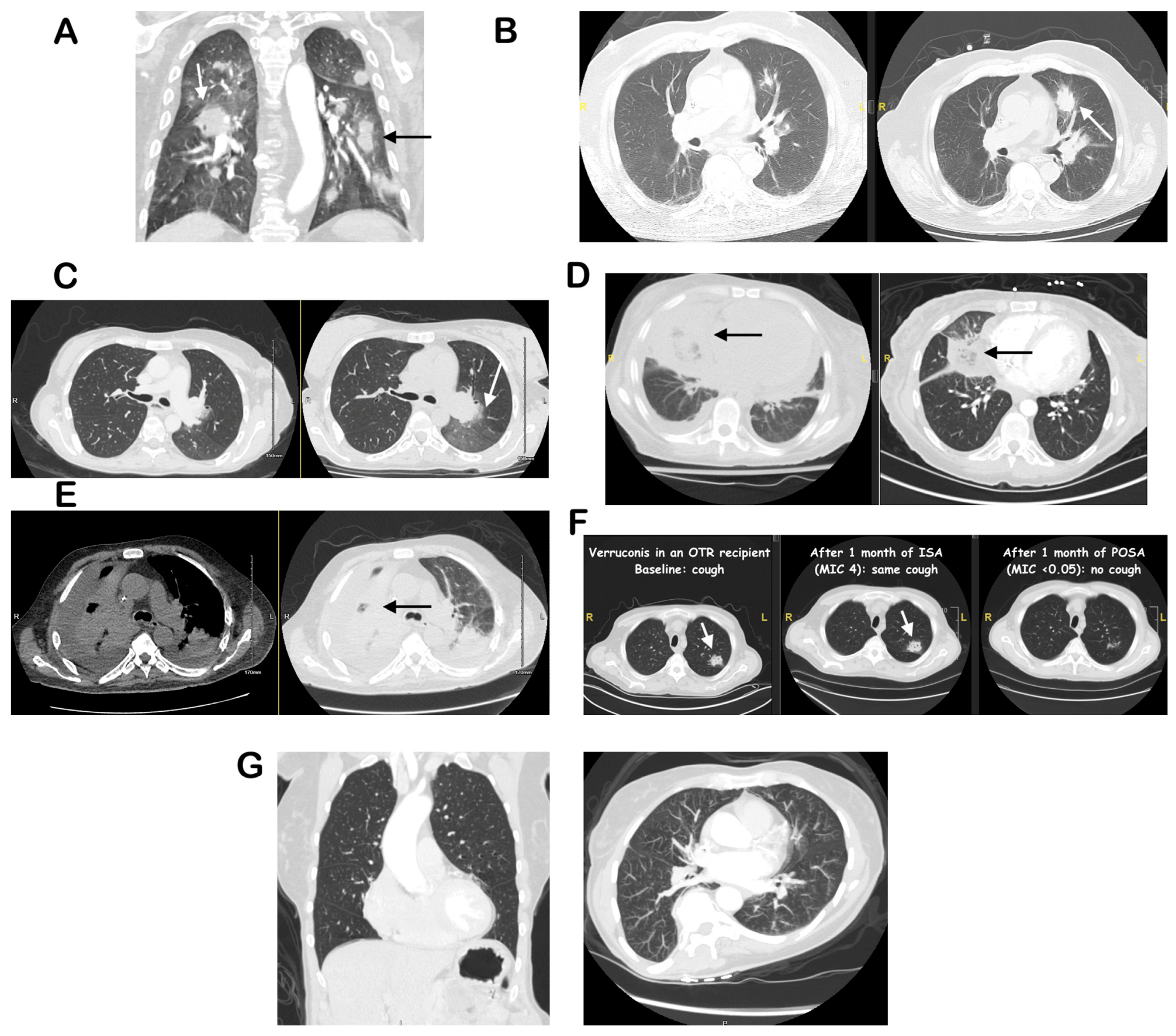
Figure 1. Arrows indicate characteristic abnormal findings. (A): Multifocal consolidations in the setting of high-dose steroids, serum GM > assay cut-off (OD 8), autopsy showed angioinvasive septate narrow-angled hyphae, consistent with IPA. (B): Enlarging nodule with “halo sign” in a neutropenic patient with acute myeloid leukemia, positive serum bDG, BAL PCR and plasma mcfDNA positive for Aspergillus fumigatus. (C): Nodule with “halo sign” in a neutropenic patient with aplastic anemia, positive BAL Galactomannan and plasma mcfDNA positive for Aspergillus flavus/oryzae. (D): “Reverse Halo” sign with rapid progression off treatment, negative BAL and tissue biopsies. Plasma mcfDNA-NGS was positive for Rhizopus delemar and autopsy showed angioinvasive aseptate broad-angled hyphae consistent with mucormycosis. (E): Large consolidation with BAL growth of a Syncephalastrum species (not further speciated). (F): Multiple pulmonary nodules with BAL growth of Verruconis galopava (“black” mold), and response to different anti-mold agents (OTR: Organ transplant recipient, POSA: Posaconazole, ISA: Isavuconazole), correlating with its minimal inhibitory concentrations (MIC). (G): Normal chest CT.
Diagnostics 16 01384 g001
Table 1. Diagnostic modalities for invasive pulmonary mold infections in immunocompromised patients.
CategoryTest/ModalitySpecimenTypical TATSensitivity (%)Specificity (%)Approximate Cost (USD, 2024–2025) *
Microscopy & HistopathologyDirect stains (GMS, PAS, calcofluor)BAL, sputum, tissueMinutes–hours<50High when tissue invasion present~$10–30 per specimen[25,26,27]
Microscopy & HistopathologyHistopathology (FFPE or fresh tissue)BiopsyHours–1 dayVariable (sampling dependent)High for tissue invasion; demonstrates angioinvasion~$100–200 per biopsy (pathology technical fee)[25,26,27]
Culture-Based DiagnosticsFungal cultureBAL, sputum, tissue, sterile fluids1–7 days (up to 21 days for slow growers)~50 overall; <30 in respiratory IPA~100 (species-level identification)~$30–60 per culture set[25,27,28]
Culture-Based DiagnosticsBlood culture for moldsBlood1–7 daysLow overall; higher for Fusarium/Scedosporium spp.High; usually indicates disseminated disease~$60–120 per set (aerobic/anaerobic bottles)[25,27,28]
Culture-Based DiagnosticsMALDI-TOF MS (from culture)Culture isolateMinutes once colony availableSame as culture yield~100 (species-level identification)Incremental ~$5–15 per isolate (reagent cost)[25,29]
Targeted Molecular/Tissue-Based TestsPCR + sequencing (species-level)FFPE or fresh tissue1–3 daysHigh when fungal elements seen on histologyHigh; useful for non-distinct morphology or culture-negative disease~$250–400 per assay[25,30]
Non-Culture BiomarkersSerum galactomannan (GM)Serum, plasma1–3 days20–90 (host- and specimen-dependent)~80–90~$100–200 per test[25,27,31]
Non-Culture BiomarkersBAL galactomannanBAL fluid1–3 daysUp to ~90~90~$120–220 per test[25,32]
Non–Culture Biomarkers(1→3)-β-D-glucan (BDG)SerumSame day–1 day~60–80 (highest in hematologic/HSCT)~70–90; reduced in ICU due to false positives~$400–450 per test[25,33]
Fungal PCR (Pathogen-Specific)Aspergillus PCRSerum, plasma, whole blood, BAL1–2 days70–10085–95~$200–350 per assay[25,34]
Fungal PCR (Pathogen-Specific)Mucorales PCRSerum, BAL, tissue1–2 days~85~89–90~$200–350 per assay[35]
Broad Molecular & Advanced DiagnosticsBroad-range fungal PCRTissue, sterile fluids1–3 days50–70High; adjunct when microscopy positive but cultures/PCR negative~$250–450 per assay[25,30]
Broad Molecular & Advanced DiagnosticsPlasma microbial cell-free DNA sequencing/mNGSPlasma (±BAL, tissue)24–48 h40–7080–90~$1800–2500 per test[36,37]
Emerging Non-invasive TechnologiesExhaled VOCs by GC–MSExhaled breathMinutes–hours77–96 (IPA and CPA)78–97Research only; estimated reagent cost ~$200–400 per run[38]
ImagingHigh-resolution CT (HRCT) chestImagingImmediateHigh for typical angioinvasive patternsLimited; findings often non-specific in non-neutropenic hosts~$130–200 (Medicare technical payment) to >$500 list price[27,39]
* Approximate direct test costs in US dollars based on 2024–2025 cash prices, manufacturer information, and fee-schedule estimates where available; values illustrate relative order of magnitude rather than provide exact charges. Actual costs vary between institutions, health systems, countries, and payers and may differ substantially from the amounts billed to or paid by patients and insurers.
Disclaimer/Publisher’s Note: The statements, opinions and data contained in all publications are solely those of the individual author(s) and contributor(s) and not of MDPI and/or the editor(s). MDPI and/or the editor(s) disclaim responsibility for any injury to people or property resulting from any ideas, methods, instructions or products referred to in the content.


