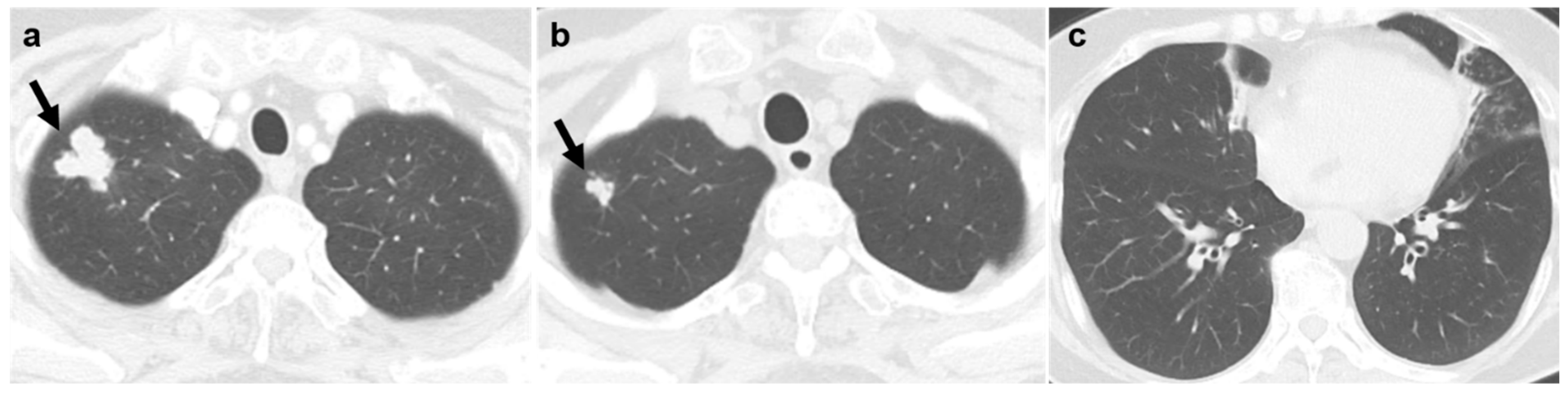
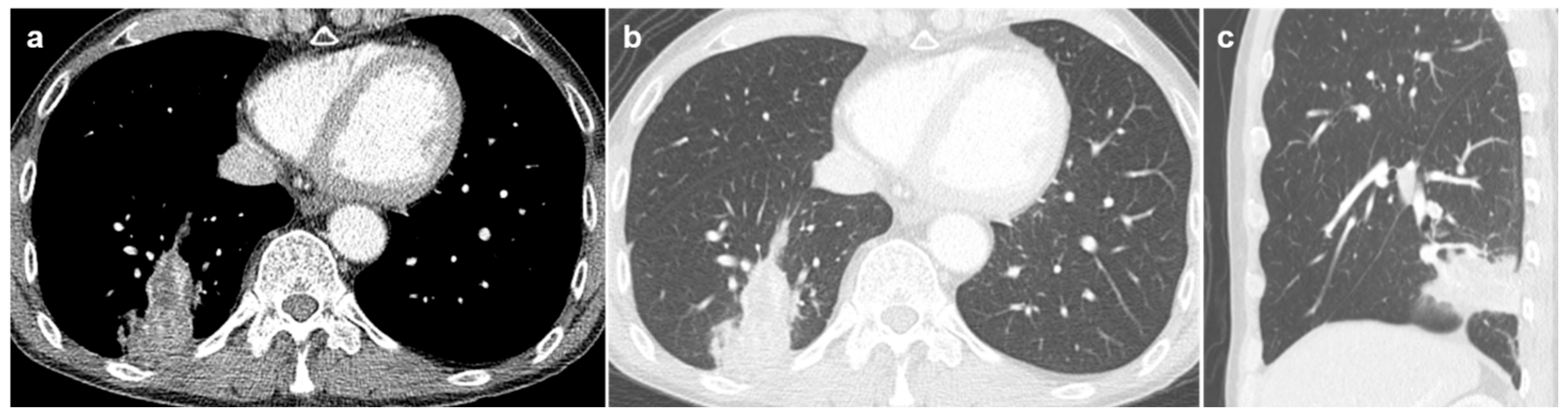
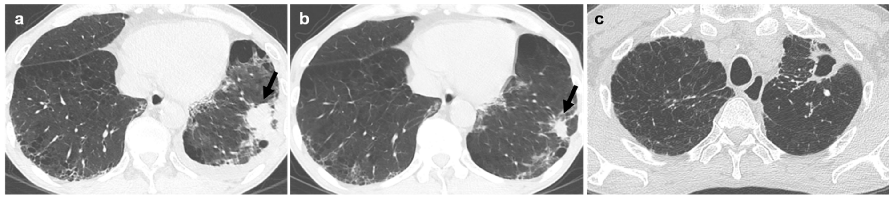
Differentiation of Lung Malignancy from Benign Lesions in Patients with Nontuberculous Mycobacterial Infection: A Retrospective Analysis of Biopsy-Proven Cases


Abstract
1. Introduction
2. Materials and Methods
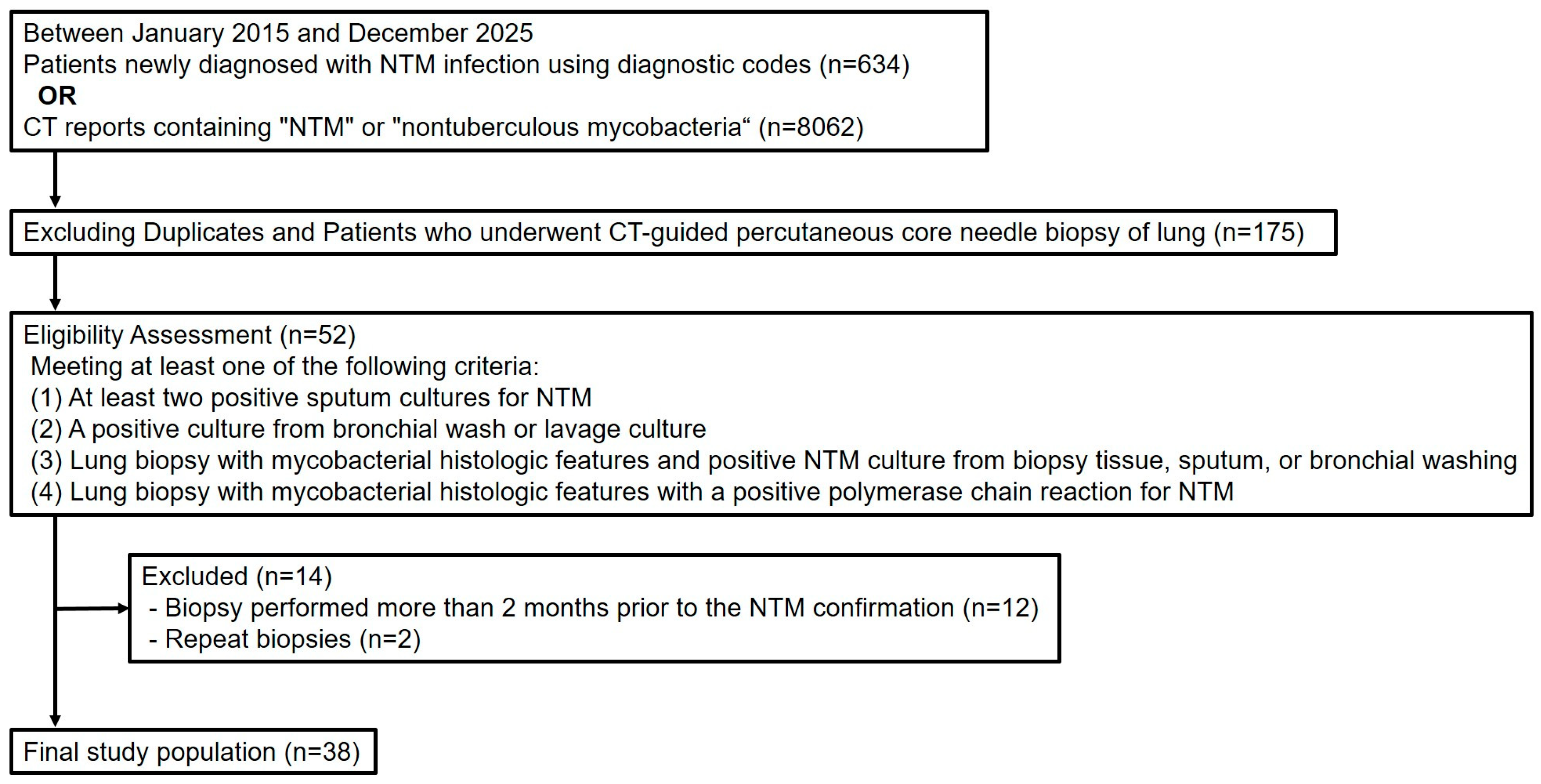
2.1. Study Population
2.2. Microbiologic Studies
2.3. Histopathology
2.4. Imaging Techniques and Analyses of CT Features
2.5. Procedure of Percutaneous Core Needle Biopsy
2.6. Statistical Analyses
3. Results
3.1. Clinical Characteristics, CT Features, and Pathologic Results of PCNB
3.2. Clinical and Radiologic Characteristics of Benign and Malignant PCNB Lesions
4. Discussion
Supplementary Materials
Author Contributions
Funding
Institutional Review Board Statement
Informed Consent Statement
Data Availability Statement
Conflicts of Interest
References
- Kim, J.-Y. Increasing burden of nontuberculous mycobacterial pulmonary disease. J. Korean Med. Assoc. 2024, 67, 4–10. [Google Scholar] [CrossRef]
- Johnson, M.M.; Odell, J.A. Nontuberculous mycobacterial pulmonary infections. J. Thorac. Dis. 2014, 6, 210–220. [Google Scholar]
- Park, J.H.; Shin, S.; Kim, T.S.; Park, H. Clinically refined epidemiology of nontuberculous mycobacterial pulmonary disease in South Korea: Overestimation when relying only on diagnostic codes. BMC Pulm. Med. 2022, 22, 195. [Google Scholar] [CrossRef]
- Dahl, V.N.; Mølhave, M.; Fløe, A.; van Ingen, J.; Schön, T.; Lillebaek, T.; Andersen, A.B.; Wejse, C. Global trends of pulmonary infections with nontuberculous mycobacteria: A systematic review. Int. J. Infect. Dis. 2022, 125, 120–131. [Google Scholar] [CrossRef] [PubMed]
- Daley, C.L.; Iaccarino, J.M.; Lange, C.; Cambau, E.; Wallace, R.J., Jr.; Andrejak, C.; Böttger, E.C.; Brozek, J.; Griffith, D.E.; Guglielmetti, L.; et al. Treatment of nontuberculous mycobacterial pulmonary disease: An official ATS/ERS/ESCMID/IDSA clinical practice guideline. Clin. Infect. Dis. 2020, 71, e1–e36. [Google Scholar] [CrossRef] [PubMed]
- Dettmer, S.; Ringshausen, F.C.; Fuge, J.; Maske, H.L.; Welte, T.; Wacker, F.; Rademacher, J. Computed tomography in adults with bronchiectasis and nontuberculous mycobacterial pulmonary disease: Typical imaging findings. J. Clin. Med. 2021, 10, 2736. [Google Scholar] [CrossRef]
- Hong, S.J.; Kim, T.J.; Lee, J.-H.; Park, J.-S. Nontuberculous mycobacterial pulmonary disease mimicking lung cancer: Clinicoradiologic features and diagnostic implications. Medicine 2016, 95, e3978. [Google Scholar] [CrossRef] [PubMed]
- Beck, K.S.; Han, D.H.; Lee, K.Y.; Kim, S.J. Role of CT-guided transthoracic biopsy in the diagnosis of mycobacterial infection. J. Investig. Med. 2019, 67, 850–855. [Google Scholar] [CrossRef]
- Ose, N.; Takeuchi, Y.; Kitahara, N.; Matumura, A.; Kodama, K.; Shiono, H.; Susaki, Y.; Funakoshi, Y.; Takabatake, H.; Shintani, Y. Analysis of pulmonary nodules caused by nontuberculous mycobacteriosis in 101 resected cases: Multi-center retrospective study. J. Thorac. Dis. 2021, 13, 977–989. [Google Scholar] [CrossRef]
- Kusumoto, T.; Asakura, T.; Suzuki, S.; Okamori, S.; Namkoong, H.; Fujiwara, H.; Yagi, K.; Kamata, H.; Ishii, M.; Betsuyaku, T.; et al. Development of lung cancer in patients with nontuberculous mycobacterial lung disease. Respir. Investig. 2019, 57, 157–164. [Google Scholar] [CrossRef]
- Dettmer, S.; Heiß-Neumann, M.; Wege, S.; Maske, H.; Ringshausen, F.C.; Joean, O.; Theissig, N.; Ewen, R.; Wacker, F.; Rademacher, J. Evaluation of treatment response with serial CT in patients with non-tuberculous mycobacterial pulmonary disease. Eur. Radiol. 2025, 35, 798–805. [Google Scholar] [CrossRef] [PubMed]
- Griffith, D.E.; Aksamit, T.; Brown-Elliott, B.A.; Catanzaro, A.; Daley, C.; Gordin, F.; Holland, S.M.; Horsburgh, R.; Huitt, G.; Iademarco, M.F.; et al. An official ATS/IDSA statement: Diagnosis, treatment, and prevention of nontuberculous mycobacterial diseases. Am. J. Respir. Crit. Care Med. 2007, 175, 367–416. [Google Scholar] [CrossRef] [PubMed]
- Kobashi, Y.; Kittaka, M.; Mouri, K.; Kato, S.; Oka, M. Clinical analysis of pulmonary nontuberculous mycobacterial disease complicated by lung cancer during the follow-up periods. Open J. Respir. Dis. 2016, 6, 59. [Google Scholar] [CrossRef][Green Version]
- Lande, L.; Peterson, D.D.; Gogoi, R.; Daum, G.; Stampler, K.; Kwait, R.; Yankowski, C.; Hauler, K.; Danley, J.; Sawicki, K.; et al. Association between pulmonary Mycobacterium avium complex infection and lung cancer. J. Thorac. Oncol. 2012, 7, 1345–1351. [Google Scholar] [CrossRef]
- Bercier, J. Non-tuberculous mycobacterial disease and lung adenocarcinoma: Birds of a feather flock together. A case report of increasing lung mass despite non-tuberculous mycobacterial treatment. In American Thoracic Society International Conference Abstracts; American Thoracic Society: New York, NY, USA, 2023; p. A2142. [Google Scholar]
- Agarwal, S.; Nair, G.; Patel, S.M.; Deshpande, P.; Sarangdhar, N. A rare case of co-existing non-small cell lung carcinoma and non-tuberculous mycobacteria. Cureus 2023, 15, e50456. [Google Scholar] [CrossRef]
- Taira, N.; Kawasaki, H.; Takahara, S.; Chibana, K.; Atsumi, E.; Kawabata, T. The presence of coexisting lung cancer and non-tuberculous mycobacterium in a solitary mass. Am. J. Case Rep. 2018, 19, 748–752. [Google Scholar] [CrossRef]
- Conic, J.; Lapinel, N.; Ali, J.; Boulmay, B. Association between non-tuberculous mycobacterial infection and aerodigestive cancers: A case series highlighting different features, sequence and association. Respir. Med. Case Rep. 2022, 40, 101751. [Google Scholar] [CrossRef]
- Yang, X.; Wisselink, H.J.; Vliegenthart, R.; Heuvelmans, M.A.; Groen, H.J.M.; Vonder, M.; Dorrius, M.D.; de Bock, G.H. Association between chest CT-defined emphysema and lung cancer: A systematic review and meta-analysis. Radiology 2022, 304, 322–330. [Google Scholar] [CrossRef]
- Parris, B.A.; O’Farrell, H.E.; Fong, K.M.; Yang, I.A. Chronic obstructive pulmonary disease (COPD) and lung cancer: Common pathways for pathogenesis. J. Thorac. Dis. 2019, 11, S2155–S2172. [Google Scholar] [CrossRef]
- Lee, H.; Jang, J.G.; Kim, Y.; Min, K.H.; Ahn, J.H.; Yoo, K.H.; Kang, M.G.; Kim, J.S.; Moon, J.Y. Prevalence of chronic obstructive pulmonary disease in patients with nontuberculous mycobacterial pulmonary disease: A systematic review and meta-analysis. J. Pers. Med. 2024, 14, 1089. [Google Scholar] [CrossRef]
- Wu, F.Z.; Huang, Y.L.; Wu, C.C.; Tang, E.K.; Chen, C.S.; Mar, G.Y.; Yen, Y.; Wu, M.T. Assessment of selection criteria for low-dose lung screening CT among Asian ethnic groups in Taiwan: From mass screening to specific risk-based screening for non-smoker lung cancer. Clin. Lung Cancer 2016, 17, e45–e56. [Google Scholar] [CrossRef]



| Group 1 (n = 22) | Group 2 (n = 16) | |||
|---|---|---|---|---|
| Clinical characteristics | ||||
| Age (year, mean ± SD) | 70.3 ± 7.4 | 67.4 ± 11.2 | ||
| Sex | Male | 16 (72.7) | 7 (43.8) | |
| Female | 6 (27.3) | 9 (56.3) | ||
| Median time interval between diagnosis of NTM-PD and PCNB (days, median [IQR]) | 551.5 [372.5–1021.0] | 6.5 [4.0–8.8] | ||
| NTM species | ||||
| Mycobacterium avium | 13 (59.1) | 6 (37.5) | ||
| Mycobacterium intracellulare | 4 (18.2) | 6 (37.5) | ||
| Mycobacterium kansasii | 0 (0.0) | 1 (6.3) | ||
| Mycobacterium fortuitum complex | 2 (9.1) | 1 (6.3) | ||
| Unknown | 3 (13.6) | 2 (12.5) | ||
| Underlying malignancy | ||||
| Yes | 8 (36.4) | 2 (12.5) | ||
| None | 14 (63.6) | 14 (87.5) | ||
| Pathologic results of PCNB | ||||
| Malignancy | 7 (31.8) | 2 (12.5) | ||
| Poorly differentiated malignancy | 1 (4.5) | 0 (0.0) | ||
| Squamous cell carcinoma | 4 (18.2) | 1 (6.3) | ||
| Adenocarcinoma | 1 (4.5) | 1 (6.3) | ||
| Metastatic infiltrating urothelial carcinoma from urinary bladder | 1 (4.5) | 0 (0.0) | ||
| Benign | 15 (68.2) | 14 (87.5) | ||
| Chronic inflammation with necrosis | 6 (27.3) | 3 (18.8) | ||
| Chronic inflammation | 5 (22.7) | 6 (37.5) | ||
| Necrosis | 0 (0.0) | 5 (31.3) | ||
| Anthracofibrosis | 3 (13.6) | 0 (0.0) | ||
| Fungal ball, consistent with Aspergillus | 1 (4.5) | 0 (0.0) | ||
| Chest CT Findings | Group 1 (n = 22) | Group 2 (n = 16) | ||
|---|---|---|---|---|
| Location | ||||
| Right upper lobe | 6 (27.3) | 1 (6.3) | ||
| Right middle lobe | 1 (4.5) | 2 (12.5) | ||
| Right lower lobe | 6 (27.3) | 7 (43.8) | ||
| Left upper lobe | 4 (18.2) | 3 (18.8) | ||
| Left lower lobe | 5 (22.7) | 3 (18.8) | ||
| Morphologic appearance | ||||
| Lobulated irregular nodule | 11 (50.0) | 8 (50.0) | ||
| Smooth round nodule | 5 (22.7) | 3 (18.8) | ||
| Patchy consolidation | 4 (18.2) | 5 (31.3) | ||
| Subpleural thickening | 2 (9.1) | 0 (0.0) | ||
| Size (mm, mean ± SD) | 34.9 ± 17.8 | 33.6 ± 13.9 | ||
| Cavity | 5 (22.7) | 4 (25.0) | ||
| Necrosis | 7 (31.8) | 8 (50.0) | ||
| Associated multiple nodules | ||||
| Yes | 16 (72.7) | 14 (87.5) | ||
| No | 6 (27.3) | 2 (12.5) | ||
| Underlying two typical forms of NTM-PD | ||||
| Nodular bronchiectatic | 12 (54.5) | 5 (31.3) | ||
| Upper lobe fibrocavitary | 3 (13.6) | 0 (0.0) | ||
| None | 7 (31.8) | 11 (68.7) | ||
| Interval change of PCNB lesion on previous follow-up CT | ||||
| Newly appeared | 5 (22.7) | 3 (18.8) | ||
| Gradual growth | 14 (63.6) | 6 (37.5) | ||
| Fluctuation | 2 (9.1) | 0 (0.0) | ||
| No change | 0 (0.0) | 3 (18.8) | ||
| N/A | 1 (4.5) | 4 (25.0) | ||
| Interval change of other lesions | ||||
| Yes | 8 (36.4) | 6 (37.5) | ||
| No | 12 (54.5) | 5 (31.3) | ||
| N/A | 2 (9.1) | 5 (31.3) | ||
| Emphysema | 11 (50.0) | 4 (25.0) | ||
| Lymphadenopathy | 6 (27.3) | 5 (31.3) | ||
| Pleural effusion | 1 (4.5) | 3 (18.8) | ||
| Benign (n = 29) | Malignant (n = 9) | p-Value | Univariable Logistic Regression | Multivariable Logistic Regression | ||||
|---|---|---|---|---|---|---|---|---|
| p-Value | Odds Ratio | p-Value | Odds Ratio | |||||
| Age (year, mean ± SD) | 67.3 ± 9.1 | 74.7 ± 8.6 | 0.045 | 0.049 | 1.106 [1.000–1.222] | |||
| Sex | 0.061 | 0.074 | 7.467 [0.825–67.573] | |||||
| Male | 15 (51.7) | 8 (88.9) | ||||||
| Female | 14 (48.3) | 1 (11.1) | ||||||
| Location | 0.321 | 0.53 | ||||||
| Right upper lobe | 6 (20.7) | 1 (11.1) | ||||||
| Right middle lobe | 3 (10.3) | 0 | ||||||
| Right lower lobe | 10 (34.5) | 3 (33.3) | ||||||
| Left upper lobe | 6 (20.7) | 1 (11.1) | ||||||
| Left lower lobe | 4 (13.8) | 4 (44.4) | ||||||
| Size (mm, mean ± SD) | 33.5 ± 17.3 | 39.7 ± 14.8 | 0.306 | 0.329 | 1.022 [0.978–1.069] | |||
| Cavity or necrosis | 19 (65.5) | 4 (44.4) | 0.436 | 0.265 | 0.421 [0.092–1.928] | |||
| Shape * | 0.057 | 0.023 | 13.091 [1.436–119.338] | 0.023 | 19.856 [1.516–260.089] | |||
| Lobulated irregular nodule | 11 (37.9) | 8 (88.9) | ||||||
| Smooth round nodule | 7 (24.1) | 1 (11.1) | ||||||
| Patchy consolidation | 9 (31.0) | 0 | ||||||
| Subpleural thickening | 2 (6.9) | 0 | ||||||
| Associated multiple nodules | 0.071 | 0.061 | 0.200 [0.037–1.080] | |||||
| Yes | 25 (86.2) | 5 (55.6) | ||||||
| No | 4 (13.8) | 4 (44.4) | ||||||
| Underlying two typical forms of NTM-PD | 0.717 | 0.722 | ||||||
| Nodular bronchiectatic | 14 (48.3) | 3 (33.3) | ||||||
| Upper lobe fibrocavitary | 2 (6.9) | 1 (11.1) | ||||||
| None | 13 (44.8) | 5 (55.6) | ||||||
| Interval change of PCNB lesion on previous follow-up CT | 0.356 | 0.986 | 0.714 [0.128–3.995] | |||||
| Newly appeared | 5 (17.2) | 3 (33.3) | ||||||
| Gradual growth | 14 (48.3) | 6 (66.7) | ||||||
| Fluctuation | 2 (6.9) | 0 | ||||||
| No change | 3 (10.3) | 0 | ||||||
| N/A | 5 (17.2) | 0 | ||||||
| Interval change of other lesions | 0.094 | 0.09 | 7.091 [0.737–68.236] | |||||
| Yes | 13 (44.8) | 1 (11.1) | ||||||
| No | 11 (37.9) | 6 (66.7) | ||||||
| N/A | 5 (17.2) | 2 (22.2) | ||||||
| Emphysema | 7 (24.1) | 8 (88.9) | <0.001 | 0.005 | 25.143 [2.660–237.624] | 0.005 | 35.531 [2.857–441.824] | |
| Lymphadenopathy | 7 (24.1) | 4 (44.4) | 0.241 | 0.248 | 2.514 [0.525–12.036] | |||
| Pleural effusion | 2 (6.9) | 2 (22.2) | 0.233 | 0.214 | 3.857 [0.459–32.424] | |||
Disclaimer/Publisher’s Note: The statements, opinions and data contained in all publications are solely those of the individual author(s) and contributor(s) and not of MDPI and/or the editor(s). MDPI and/or the editor(s) disclaim responsibility for any injury to people or property resulting from any ideas, methods, instructions or products referred to in the content. |
© 2026 by the authors. Licensee MDPI, Basel, Switzerland. This article is an open access article distributed under the terms and conditions of the Creative Commons Attribution (CC BY) license.
Share and Cite
Hong, W.; Lee, I.J.; Kwon, L.M.; Kim, M.-J.; Nam, H. Differentiation of Lung Malignancy from Benign Lesions in Patients with Nontuberculous Mycobacterial Infection: A Retrospective Analysis of Biopsy-Proven Cases. Diagnostics 2026, 16, 1321. https://doi.org/10.3390/diagnostics16091321
Hong W, Lee IJ, Kwon LM, Kim M-J, Nam H. Differentiation of Lung Malignancy from Benign Lesions in Patients with Nontuberculous Mycobacterial Infection: A Retrospective Analysis of Biopsy-Proven Cases. Diagnostics. 2026; 16(9):1321. https://doi.org/10.3390/diagnostics16091321
Chicago/Turabian StyleHong, Wonju, In Jae Lee, Lyo Min Kwon, Min-Jeong Kim, and Hyunseung Nam. 2026. "Differentiation of Lung Malignancy from Benign Lesions in Patients with Nontuberculous Mycobacterial Infection: A Retrospective Analysis of Biopsy-Proven Cases" Diagnostics 16, no. 9: 1321. https://doi.org/10.3390/diagnostics16091321
APA StyleHong, W., Lee, I. J., Kwon, L. M., Kim, M.-J., & Nam, H. (2026). Differentiation of Lung Malignancy from Benign Lesions in Patients with Nontuberculous Mycobacterial Infection: A Retrospective Analysis of Biopsy-Proven Cases. Diagnostics, 16(9), 1321. https://doi.org/10.3390/diagnostics16091321