Artificial Intelligence in Gastrointestinal Wireless Capsule Endoscopy: A Systematic Literature Review and Meta-Analysis



Abstract
1. Introduction
1.1. The Global Burden of Gastrointestinal Disorders
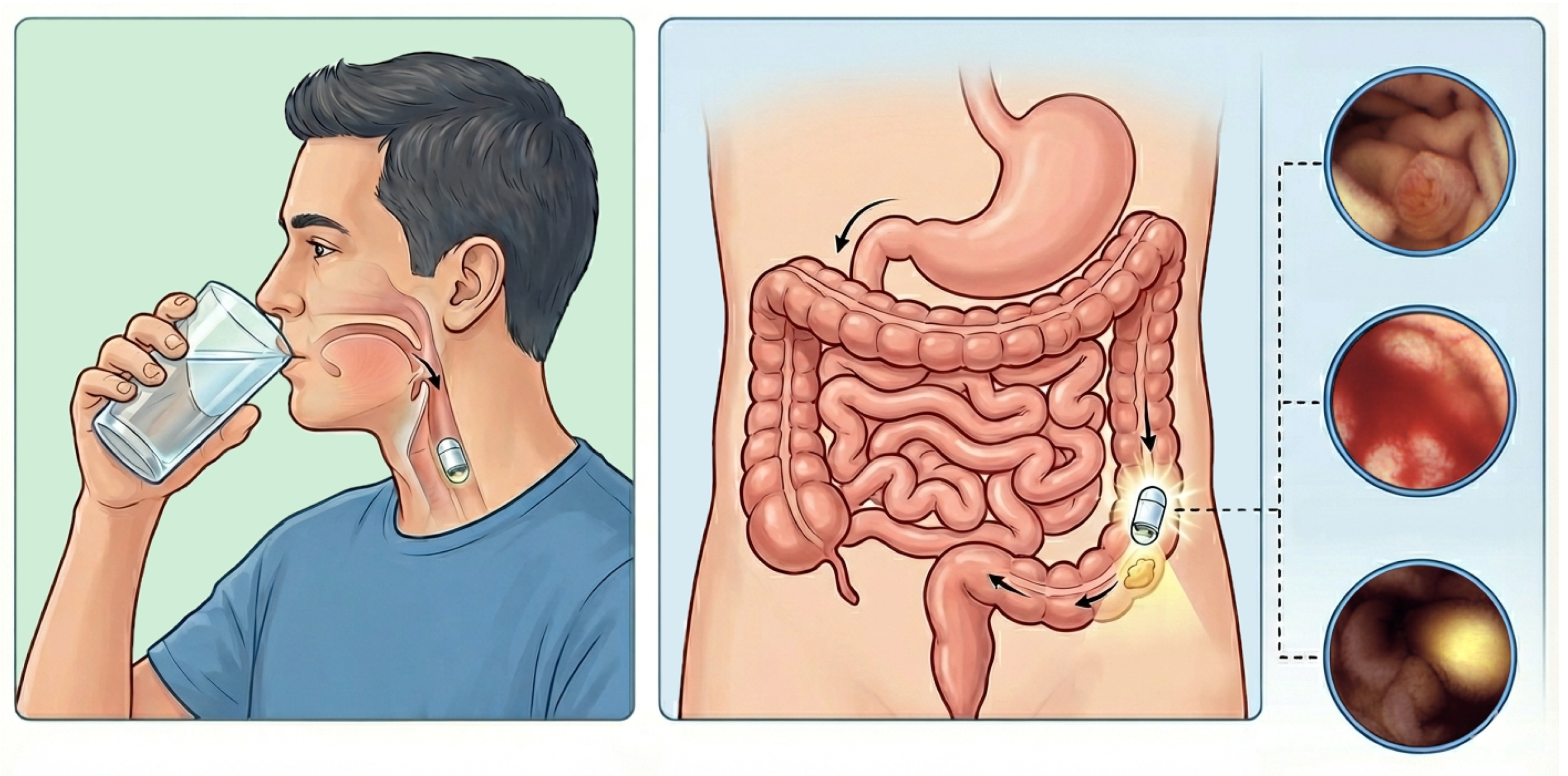
1.2. Wireless Capsule Endoscopy
1.3. The Limitations of Human Interpretation
1.4. The Promise of Artificial Intelligence
2. Research Gaps and Objectives
- (1)
- Almost all review articles on the use of AI in WCE applications are restricted to specific settings, such as particular diseases, GI locations, or predefined applications. To address this limitation, in the current study we extracted all original journal research articles that applied AI for the analysis of WCE outputs. In other words, our search strategy was not restricted to specific diseases, applications, or GI locations.
- (2)
- Moreover, almost all studies in this domain have largely focused on analyzing and comparing the performance metric values. However, several critical technical and clinical barriers must be considered to improve the trustworthiness of AI results and to increase the likelihood of successful deployment of AI systems in clinical practice. Therefore, in this study, we go beyond performance metrics and highlight these barriers and provide recommendations for future research.
- (3)
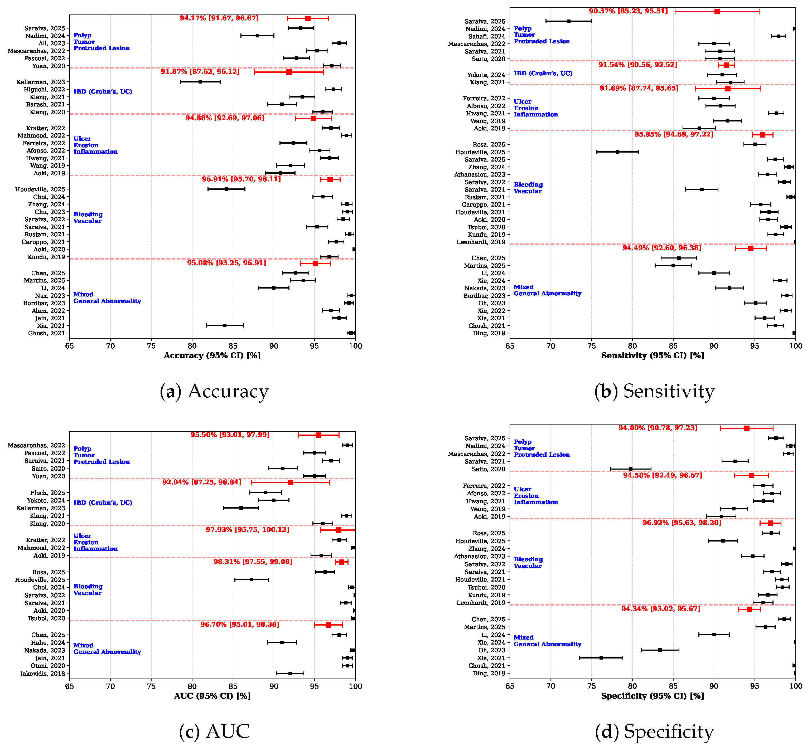
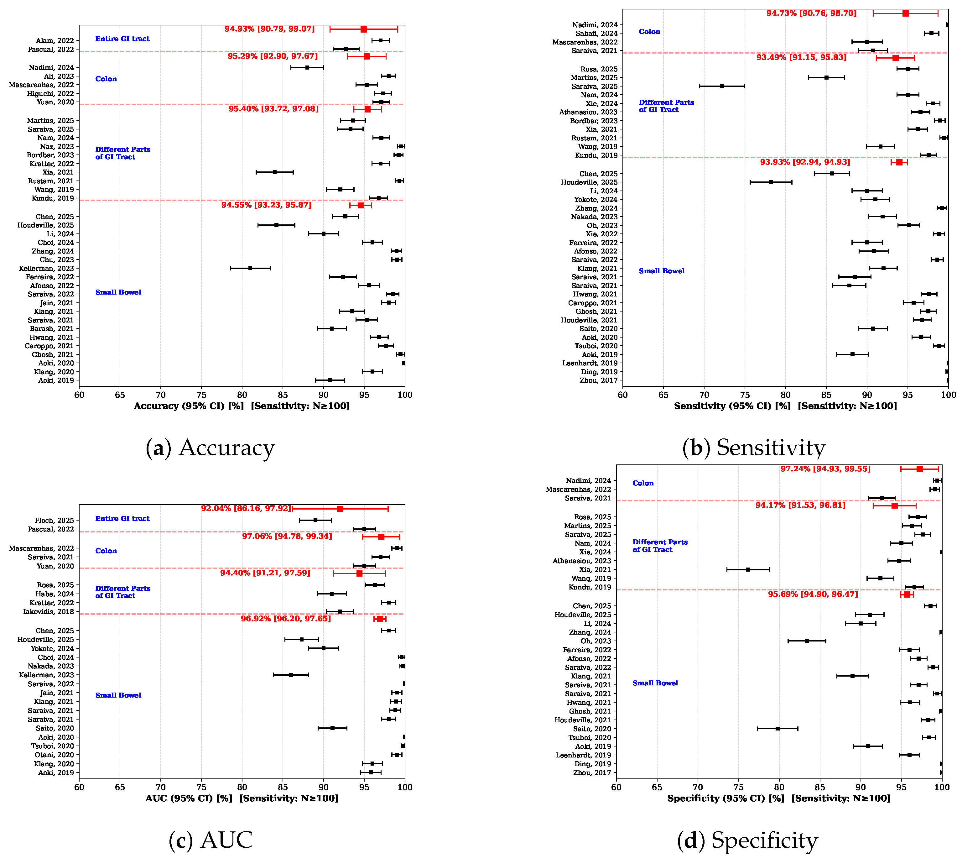
- Finally, this study presents a more comprehensive meta-analysis by systematically structuring the results across two complementary dimensions including clinical indication–based domains and GI tract locations. The included studies were first grouped into five categories based on their clinical indications, and subsequently reclassified into four categories according to the GI tract locations.
3. Method
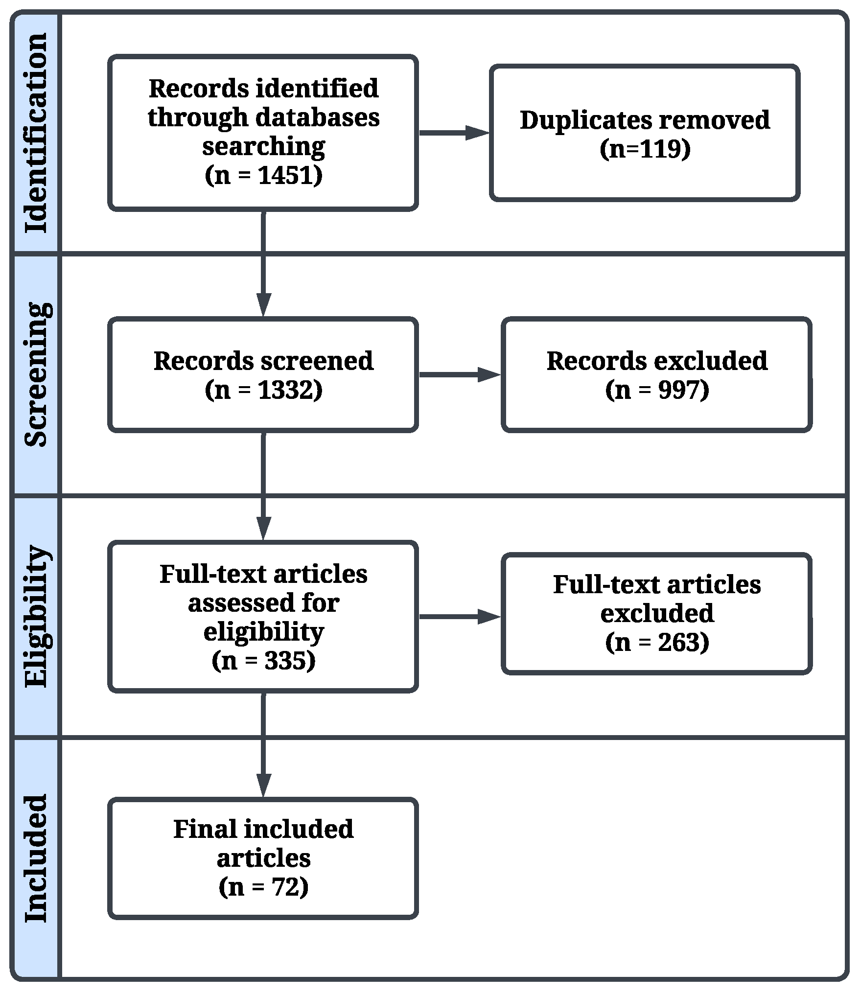
3.1. Search Strategy and Study Selection
3.2. Data Extraction
3.3. Risk of Bias Assessment
3.4. Meta-Analysis
4. Results
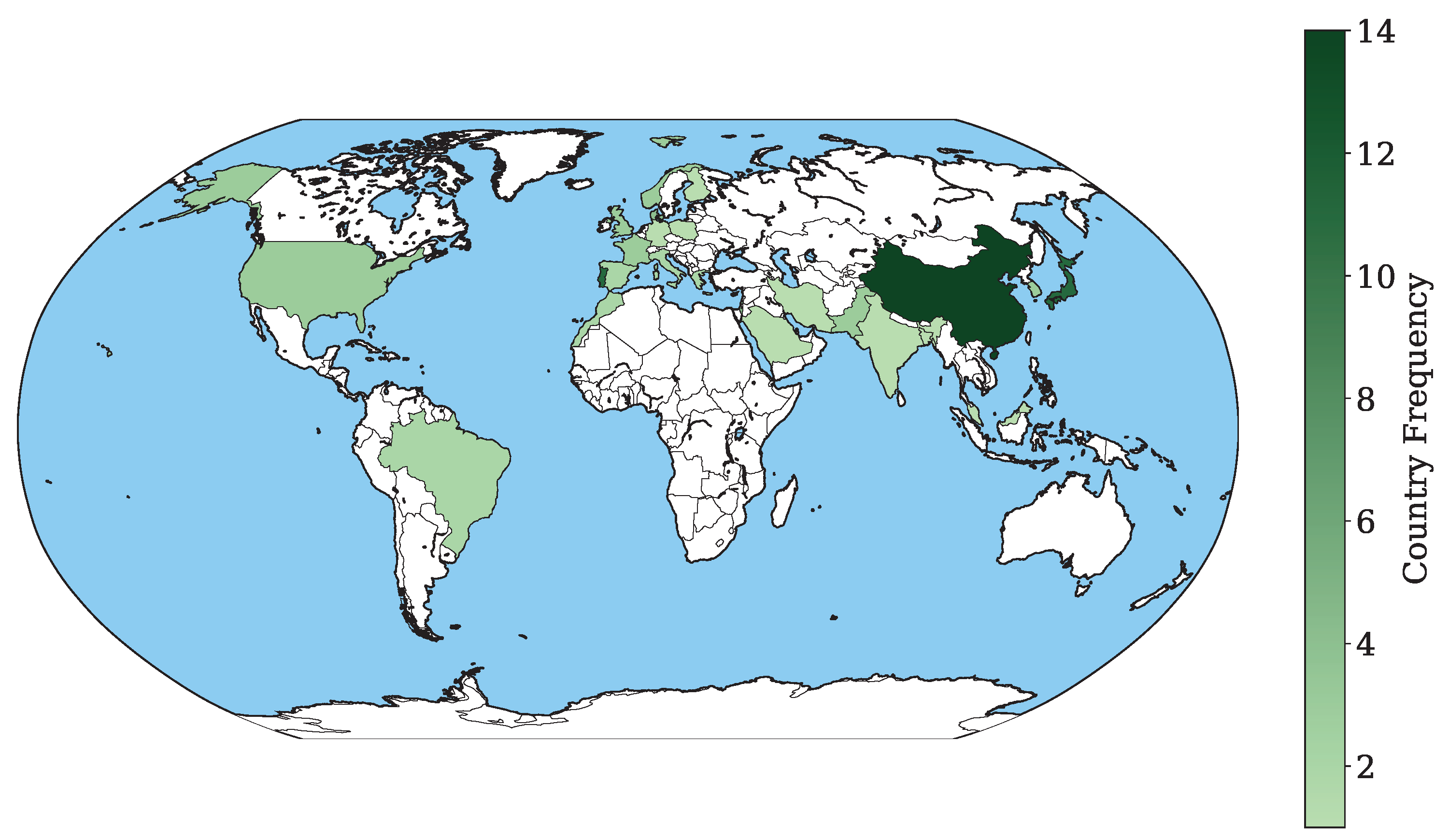
4.1. Geographical Distribution
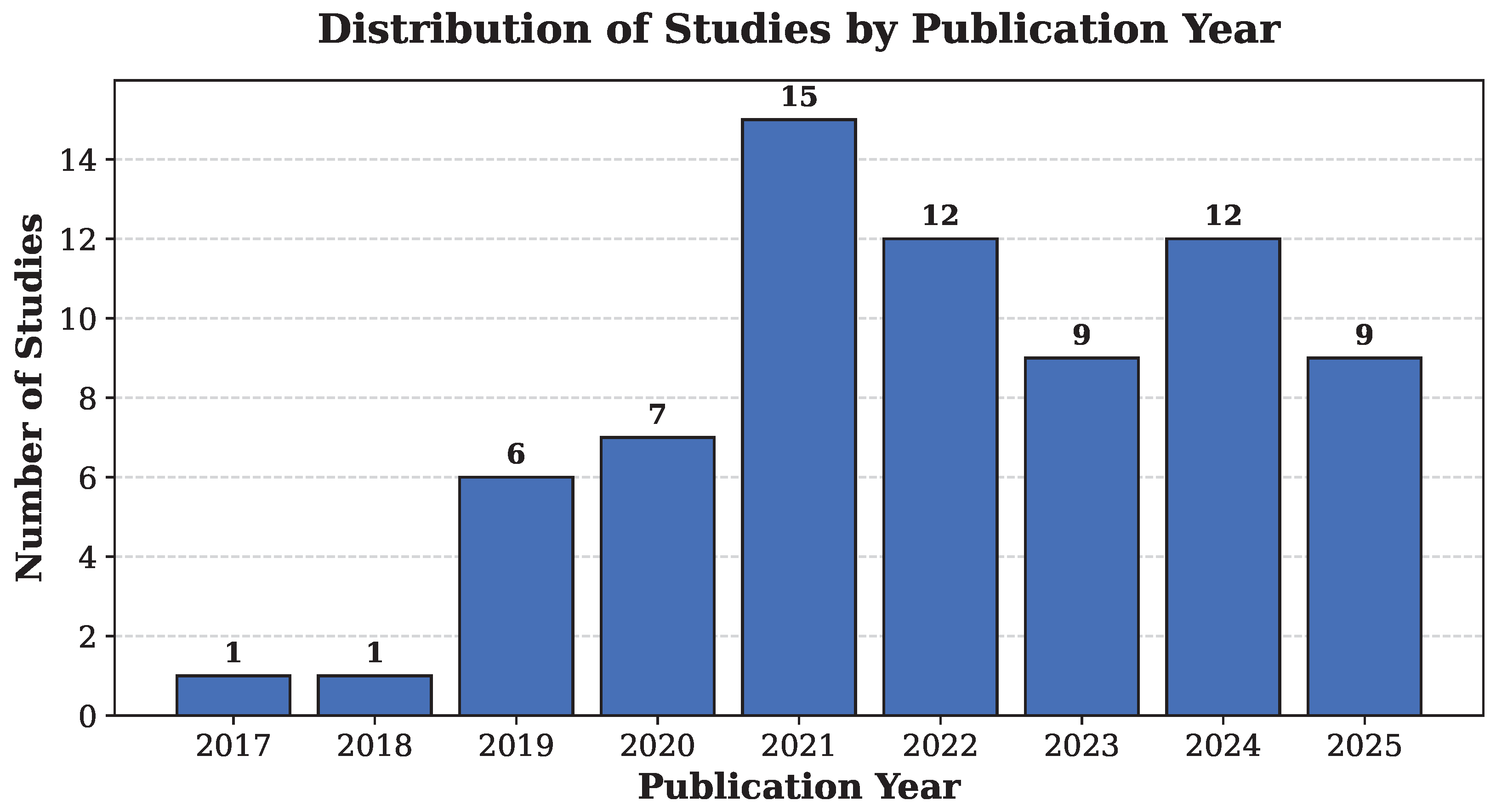
4.2. Publication Trend
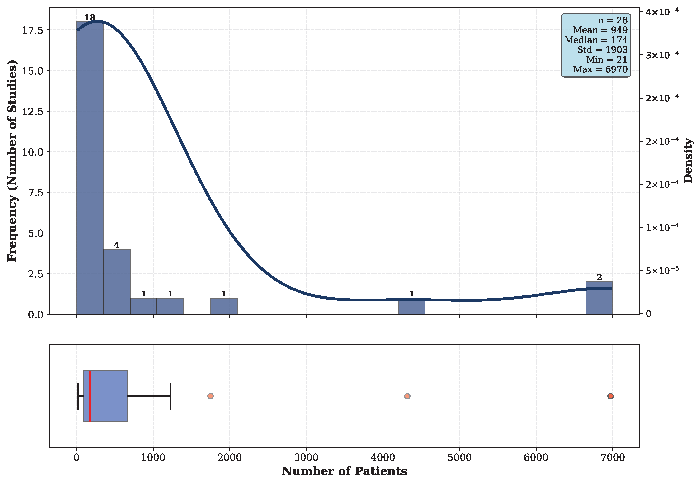
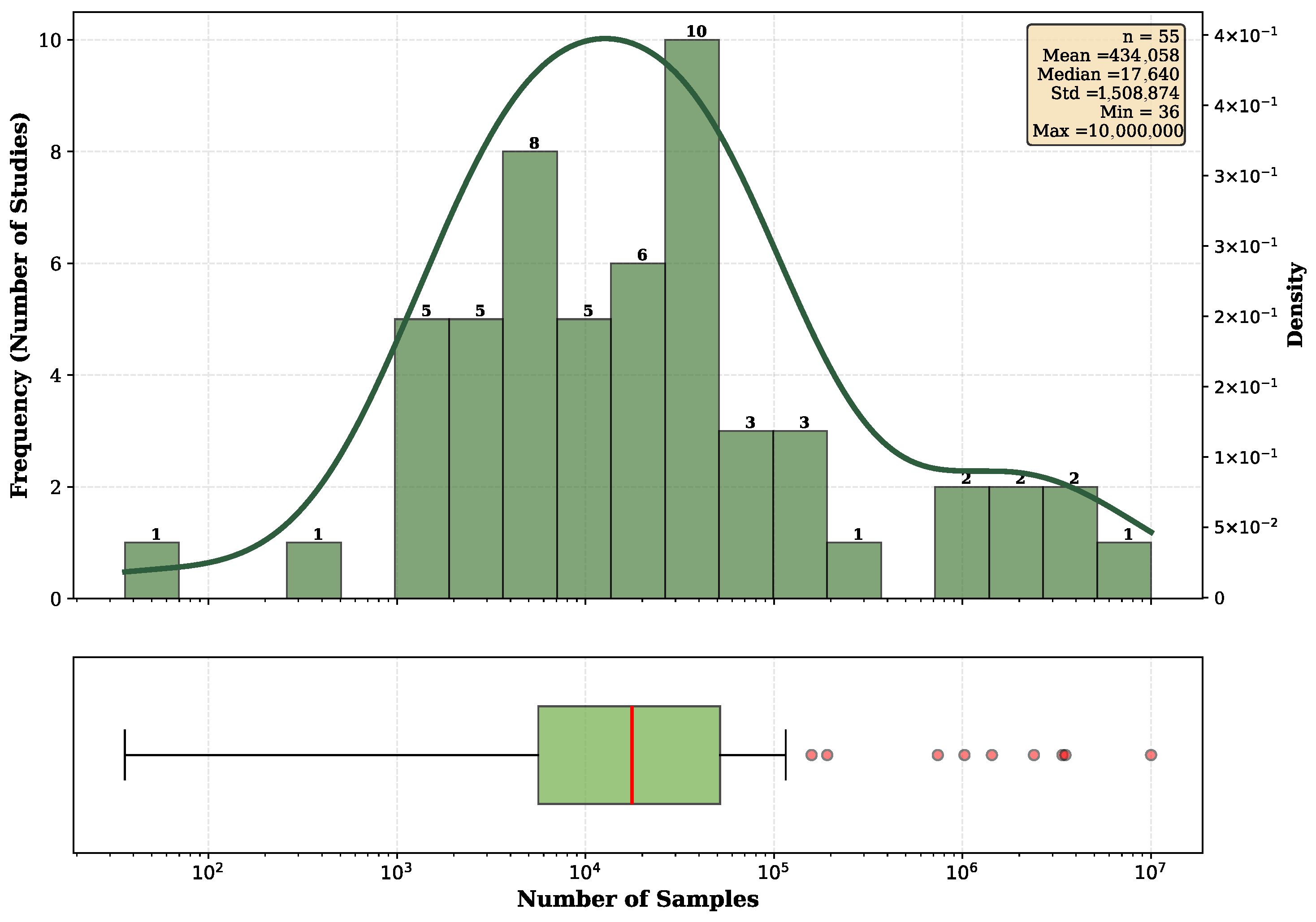
4.3. Cohort Presentation
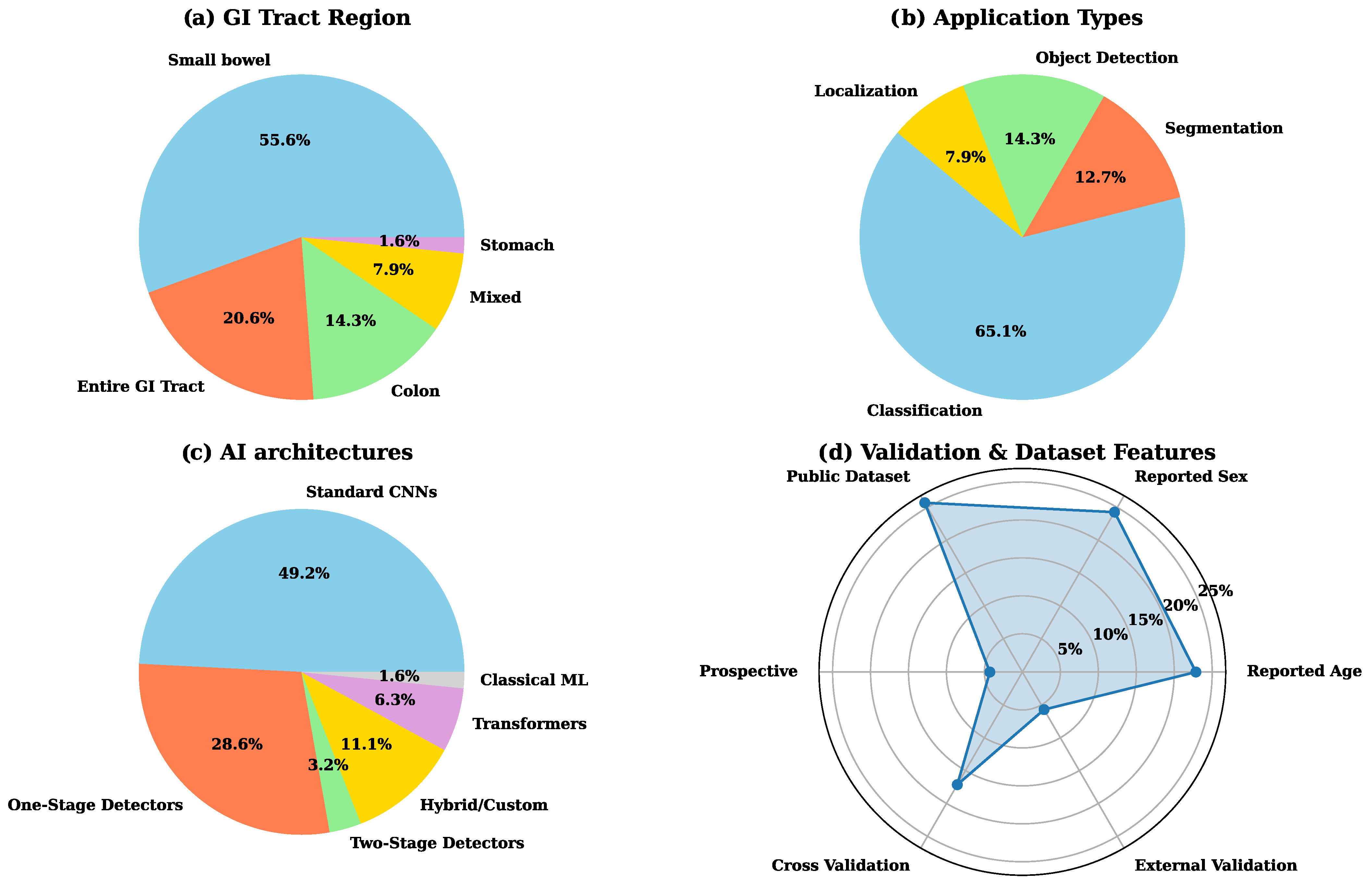
4.4. GI Tract Locations
4.5. Applications
4.6. AI Models
4.7. Study Design and Validation Strategies
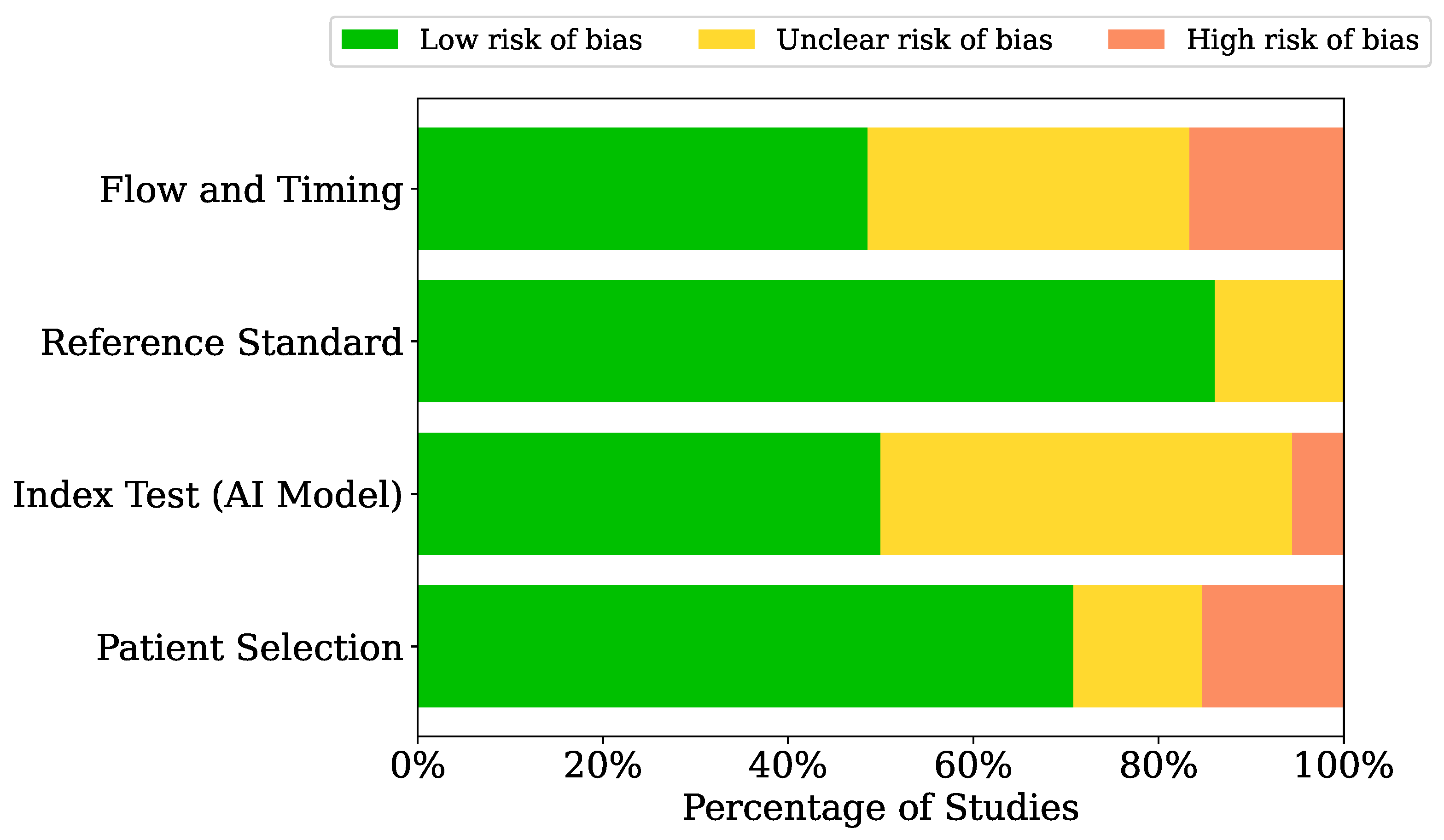
4.8. ROB Assessment
4.9. Meta-Analysis
5. Discussion
5.1. Datasets
5.2. AI Analysis
5.3. Linking WCE and Conventional Endoscopy
5.4. Comparison with Expert Interpretation
5.5. Strengths and Limitations
6. Conclusions
Supplementary Materials
Author Contributions
Funding
Institutional Review Board Statement
Informed Consent Statement
Data Availability Statement
Conflicts of Interest
Appendix A. Characteristics of Included Studies

| First Author | Year | Country | Region of GI Tract | Time Period | Samples | Patients Type | Age | Sex | Data Source | Study Design | Setting |
|---|---|---|---|---|---|---|---|---|---|---|---|
| Teng Zhou [86] | 2017 | China | Small bowel | 2008–2009 | 11 celiac disease patients and 10 controls, analyzed via capsule endoscopy | Patients with celiac disease and controls | 50.5 (females mean), 44 (males mean) | 45.45% male | Columbia University Medical Center | Retrospective | Clinical practice |
| Dimitris K. Iakovidis [80] | 2018 | Greece, UK | Entire GI tract | 2015–2018 | 10,698 images | Patients undergoing endoscopy | - | - | Two public datasets (MICCAI and KID) | Retrospective | Analysis |
| Zhen Ding [77] | 2019 | China | Small bowel | 2016–2018 | 6970 patients | Patients with small-bowel diseases and normal variants | - | - | Collected by authors from 77 medical centers | Retrospective | Clinical practice |
| Romain Leenhardt [30] | 2019 | France | Small bowel | 2011–2018 | 2946 images with lesions, 600 gastrointestinal angiectasia frames | Patients with gastrointestinal bleeding | 72 (mean) | 61% male | CAD-CAP database | Retrospective | Analysis |
| Amit Kumar Kundu [61] | 2019 | Bangladesh | Entire GI tract | 2019 | 2350 images (450 bleeding, 1900 non-bleeding) | Patients with gastrointestinal abnormalities | - | - | Public dataset | Retrospective | Analysis |
| Tomonori Aoki [51] | 2019 | Japan | Small bowel | 2009–2018 | 115 patients (training), 65 patients (validation), 5360 training images, 10,440 validation images | Patients with gastrointestinal abnormalities | 63 (training set mean) | 54% male (training dataset) | University of Tokyo Hospital | Retrospective | Clinical practice |
| Sen Wang [31] | 2019 | China | Entire GI tract | - | 1416 videos | Ulcer patients | - | 73% male | 30 hospitals and 100 medical examination centers, China | Retrospective | Clinical practice |
| Libin Lan [108] | 2019 | China | Small bowel | - | 7381 wireless capsule endoscopy images | Patients with gastrointestinal abnormalities | - | - | Datasets from Jinshan Science & Technology and Given Imaging | Retrospective | Analysis |
| U Deding [109] | 2020 | Denmark | Colon | 2016–2018 | 97 patients | Patients with incomplete optical colonoscopy | 64.5 (median) | 25 % male | Odense University Hospital (Denmark) | Prospective | Clinical practice |
| Eyal Klang [45] | 2020 | Israel | Small bowel | - | 17,640 images from 49 patients | Patients with known Crohn’s disease and control subjects | 28 (mean) | 50% male | Department of Gastroenterology at Sheba Medical Center | Retrospective | Clinical practice |
| Keita Otani [79] | 2020 | Japan | Small bowel | 2009–2019 | 167 patients; 398 erosions/ulcers, 538 vascular lesions, 4590 tumors, 34,437 normal images | Patients with gastrointestinal abnormalities | 63.6 (mean) | 55% male | Tokyo Hospital | Retrospective | Clinical practice |
| Yixuan Yuan [40] | 2020 | Hong Kong | Colon | 2019 | 7200 images from 80 patients | Patients with gastrointestinal abnormalities | - | - | Not mentioned | Retrospective | Analysis |
| Akiyoshi Tsuboi [73] | 2020 | Japan | Small bowel | - | 141 patients (training), 28 patients (validation) | Patients with diagnosed small-bowel angioectasia using capsule endoscopy | 68.5 (validation set mean) | 54% male (validation set) | Hiroshima University Hospital, University of Tokyo Hospital, Sendai Kousei Hospital | Retrospective | Clinical practice |
| Tomonori Aoki [60] | 2020 | Japan | Small bowel | 2009–2015 | 27,847 images (training), 10,208 images (test) | Patients with gastrointestinal abnormalities | 53.4 (mean) | 56% male | The University of Tokyo Hospital (Japan) | Retrospective | Clinical practice |
| Hiroaki Saito [32] | 2020 | Japan | Small bowel | 2009–2018 | 30,584 images for training; 17,507 for testing | Patients with and without small bowel protruding lesions | 60.1 (mean) | 65.8% male | Sendai Kousei Hospital (Japan), The University of Tokyo (Japan), Hiroshima University Hospital (Japan), Given Imaging (Israel) | Retrospective | Clinical practice |
| Charles Houdeville [82] | 2021 | France | Small bowel | 2021 | 1200 frames | Patients with gastrointestinal bleeding | - | - | CAD-CAP | Retrospective | Analysis |
| Tonmoy Ghosh [69] | 2021 | USA | Small bowel | - | 2350 images (450 bleeding, 1900 non-bleeding) | Patients with suspected intestinal bleeding | - | - | endoscopy.org and KID public datasets | Retrospective | Analysis |
| Chao Liao [110] | 2021 | China | Small bowel | - | 50,000 frames across 9 videos | Patients undergoing gastrointestinal motility assessment | - | - | Jinshan Science & Technology Company | Retrospective | Clinical practice |
| Jürgen Herp [111] | 2021 | Denmark | Colon | - | 42 patients, 84 videos | Patients with colonoscopy follow-up need | - | - | Odense University Hospital | Retrospective | Clinical practice |
| Andrea Caroppo [59] | 2021 | Italy | Small bowel | - | 2352 images (303 with bleeding) | Patients with gastrointestinal bleeding | - | - | Public KID Dataset | Retrospective | Analysis |
| Furqan Rustam [58] | 2021 | Pakistan, South Korea | Entire GI tract | - | 5000 images from 33 patients | Patients with gastrointestinal tract infections | - | - | Sheikh Zayed Hospital, Rahim Yar Khan | Retrospective | Clinical practice |
| Ji Xia [68] | 2021 | China | Stomach | 2014–2018 | 697 patients, 1,023,955 capsule endoscopy images | Patients with gastrointestinal abnormalities | - | - | Changhai Hospital, Shanghai | Retrospective | Clinical practice |
| Yunseob Hwang [50] | 2021 | Republic of Korea | Small bowel | 2007–2019 | 526 mall bowel capsule endoscopy videos (training), 5760 independent images (validation) | Patients undergoing mall bowel capsule endoscopy | - | - | Multiple hospitals in Korea | Retrospective | Clinical practice |
| Miguel José Mascarenhas Saraiva [85] | 2021 | Portugal | Small bowel | 2015–2020 | 4319 patients, 53,555 capsule endoscopy images | Patients with gastrointestinal abnormalities | - | - | São João University Hospital and ManopH Gastroenterology Clinic | Retrospective | Clinical practice |
| Tomonori Aoki [112] | 2021 | Japan | Small bowel | 2009–2018 | 379 patients, 66,028 capsule endoscopy images for training | Patients undergoing small-bowel capsule endoscopy | 62.3 (test set mean) | 57% male (test set) | University of Tokyo, Hiroshima University Hospital, Sendai Kosei Hospital | Retrospective | Clinical practice |
| Miguel José Mascarenhas Saraiva [84] | 2021 | Portugal | Colon | 2010–2020 | 3,387,259 frames from 24 colon capsule endoscopy exams, 3640 images for training/validation | Patients undergoing colon capsule endoscopy for detection of colonic lesions | - | - | São João University Hospital, Porto, Portugal | Retrospective | Clinical practice |
| Yiftach Barash [44] | 2021 | Israel | Small bowel | - | 17,640 images (7391 ulcers, 10,249 normal) | Patients with Crohn’s disease | - | - | Sheba Medical Center, Tel Hashomer (Israel), Medtronic Dublin (Ireland) | Retrospective | Clinical practice |
| Miguel Mascarenhas Saraiva [57] | 2021 | Portugal | Small bowel | 2020–2021 | 6740 images (training and validation) | Patients undergoing DAE for suspected mid-gastrointestinal bleeding | - | - | São João University Hospital, Porto, Portugal | Retrospective | Clinical practice |
| Eyal Klang [43] | 2021 | Israel | Small bowel | 2009–2018 | 27,892 images (1942 strictures; 14,266 normal mucosa; 11,684 ulcers) | Crohn’s Disease patients | - | - | Sheba Medical Center, Tel Hashomer (Israel), Medtronic Dublin (Ireland) | Retrospective | Clinical practice |
| Samir Jain [67] | 2021 | India, Norway, Czech Republic, Malaysia | Small bowel | 2018–2021 | Combined KID dataset; images with augmentation | Patients with gastrointestinal anomalies | - | - | KID dataset | Retrospective | Analysis |
| Miguel Mascarenhas Saraiva [56] | 2022 | Portugal | Small bowel | 2015–2020 | 1229 patients; 22,095 frames | Patients with suspected gastrointestinal bleeding | - | - | São João University Hospital, Porto, Portugal | Retrospective | Clinical practice |
| Guillem Pascual [39] | 2022 | Spain, Denmark | Small intestine, colon | 2016–2021 | Generic: 1,185,033 frames, Polyp dataset: 248,136 frames, CAD-CAP dataset: 1800 images | Mixed patients | - | - | CAD-CAP, Internal dataset | Retrospective | Analysis |
| João Afonso [49] | 2022 | Portugal | Small bowel | 2015–2020 | 1483 wireless capsule endoscopy exams, 6130 frames (4233 containing ulcers and erosions) | Patients with inflammatory bowel disease | - | - | São João University Hospital, Porto, Portugal | Retrospective | Clinical practice |
| Jun-Xiao Zhou [113] | 2022 | China | Small bowel | 2016–2019 | 277 polyp images from 480 patients | Patients with gastrointestinal disorders | - | - | Guangzhou First People’s Hospital, CVC-Colon, CVC-Clinic | Retrospective | Clinical practice |
| Md. Jahin Alam [66] | 2022 | Bangladesh, USA | Entire GI tract | 2016–2018 | 47,238 images | Patients with gastrointestinal abnormalities | - | - | Kvasir-Capsule public dataset | Retrospective | Analysis |
| Naoki Higuchi [42] | 2022 | Japan | Colon | 2018–2020 | 739,021 images | Patients with moderate or mild ulcerative colitis | - | - | Hirosaki University Hospital, Japan | Prospective | Clinical practice |
| João Pedro Sousa Ferreira [48] | 2022 | Portugal | Small bowel and Colon | 2017–2020 | 8085 images | Patients with Crohn’s Disease | - | - | São João University Hospital and ManopH Gastroenterology Clinic, Portugal | Retrospective | Clinical practice |
| Saqib Mahmood [47] | 2022 | Pakistan | Entire GI tract | 2016–2018 | 47,238 labeled frames from 117 videos | Patients with GI tract abnormalities | - | - | Kvasir-Capsule dataset | Retrospective | Analysis |
| Meryem Souaidi [114] | 2022 | Morocco | Entire GI tract | - | WCE, PillCam-COLON, CVC-ClinicDB, ETIS-Larib, PASCAL VOC, COCO | Patients with gastrointestinal polyp indications | - | - | PillCam-COLON, CVC-ClinicDB, ETIS-Larib, PASCAL VOC, COCO | Retrospective | Analysis |
| Miguel Mascarenhas [38] | 2022 | Portugal | Colon | 2010–2020 | 124 patients, 5715 colon capsule endoscopy frames | Patients with protruding lesions detected via colon capsule endoscopy | - | - | São João University Hospital and ManopH Gastroenterology Clinic | Retrospective | Clinical practice |
| Tom Kratter [46] | 2022 | Israel | Small bowel, Colon | - | 33,100 capsule endoscopy images | Patients with Crohn’s disease and normal mucosa | - | - | Sheba Medical Center, Tel Hashomer (Israel), Medtronic Dublin (Ireland) | Retrospective | Clinical practice |
| Xia Xie [33] | 2022 | China | Small bowel | 2012–2021 | 5825 small bowel capsule endoscopy examinations | Patients undergoing small bowel capsule endoscopy | 49.8 (mean) | 60.9% male | 51 medical centers, China | Retrospective | Clinical practice |
| SangYup Oh [75] | 2023 | South Korea | Small bowel | 2002–2022 | 1,431,344 images (across 260 cases) | Mixed gastrointestinal patients | - | - | Dongguk University Ilsan Hospital, South Korea | Retrospective | Clinical practice |
| Raizy Kellerman [41] | 2023 | Israel | Small bowel | 2011–2021 | 101 patients | Newly diagnosed Crohn’s patients | 27 (median) | 46.5% male | Sheba Medical Center, Israel | Retrospective | Clinical practice |
| Mehrdokht Bordbar [34] | 2023 | Iran | Entire GI tract | - | 29 patients, 14,691 frames | Mixed gastrointestinal patients | 53.5 (mean) | 66% male | Namazi Hospital, Shiraz, Iran | Retrospective | Clinical practice |
| Sharib Ali [37] | 2023 | UK, Norway, France, Italy, Egypt | Colon | - | 300 patients, 8037 frames | Patients undergoing colonoscopy for colorectal cancer screening | - | - | Public PolypGen dataset | Retrospective | Analysis |
| Ye Chu [55] | 2023 | China | Small bowel | 2014–2020 | 12,403 images | Patients with angiodysplasias undergoing capsule endoscopy | - | - | Ruijin Hospital, China | Retrospective | Clinical practice |
| Sofia A. Athanasiou [72] | 2023 | Greece | Entire GI tract | 2019–2020 | 6016 frames | Patients with bleeding, angiodysplasias, hemangiomas, or lesions predisposing to bleeding | - | - | Kapodistrian University of Athens, Attikon University Hospital, Laikon University Hospital, Aristotle University of Thessaloniki | Prospective | Clinical practice |
| Akihiko Sumioka [115] | 2023 | Japan | Small bowel | 2011–2021 | 26 patients | Primary small-bowel follicular lymphoma patients | 60.9 (mean) | 50% male | Hiroshima University Hospital, Japan | Retrospective | Clinical practice |
| Javeria Naz [65] | 2023 | Pakistan, Norway, UK, Saudi Arabia | Entire GI tract | 2016–2018 | >30,000 images | Patients with gastrointestinal abnormalities | - | - | Kvasir-V1 and other pulic sources | Retrospective | Analysis |
| Ayako Nakada [74] | 2023 | Japan | Small bowel | 2009–2019 | 651 patients, >10 million wireless capsule endoscopy images | Patients with obscure gastrointestinal bleeding, small intestine tumors, abdominal symptoms | - | - | Nine hospitals across Japan | Retrospective | Analysis |
| Tomonori Aoki [116] | 2024 | Japan | Small bowel | 2009–2019 | 36 wireless capsule endoscopy videos, 43 lesions | Patients with abnormal lesions in wireless capsule endoscopy | - | - | University of Tokyo, Hiroshima University Hospital, Sendai Kosei Hospital, and Japan Medical Association | Retrospective | Clinical practice |
| Ali Sahafi [22] | 2024 | Denmark, Poland | Colon | - | 2500 images | Patients with gastrointestinal abnormalities | - | - | KID Dataset | Retrospective | Analysis |
| Rui-Ya Zhang [54] | 2024 | China | Small bowel | 2013–2023 | 111,861 images | Patients with gastrointestinal issues | - | - | Shanxi Provincial Hospital, China | Retrospective | Clinical practice |
| Tsedeke Temesgen Habe [81] | 2024 | Finland | Entire GI tract | 2016–2018 | 47,238 images (class-annotated) | Patients with gastrointestinal abnormalities | - | - | Kvasir-Capsule public dataset | Retrospective | Analysis |
| Mohamed Achraf Belabbes [100] | 2024 | Morocco | Entire GI tract | 2023–2024 | Several datasets: PillCam COLON, Kvasir-SEG, ETIS-Larib, CVC-ClinicDB | Patients with suspected polyps | - | - | PillCam COLON, Kvasir-SEG, ETIS-Larib, CVC-ClinicDB | Retrospective | Analysis |
| Kyung Seok Choi [53] | 2024 | South Korea | Small bowel | 2018–2020 | 103 paitient (1,037,286 images) | Patients with suspected small-bowel bleeding (SSBB) | 64.7 (mean) | 62.2% male | Consecutive patient data from Yeouido St. Mary’s and Seoul St. Mary’s Hospitals | Retrospective | Clinical practice |
| Akihito Yokote [70] | 2024 | Japan | Small bowel | 2014–2021 | 954 patients, 18,481 images | Patients undergoing small bowel capsule endoscopy | - | - | Kyushu University Hospital | Retrospective | Clinical practice |
| Dong Liu [98] | 2024 | China | Colon | 2016–2018 | 3 public datasets: 1000 polyp images (Kvasir-SEG), 590 images (Kvasir-Instrument), 55 images (KvasirCapsule-SEG) | Patients with gastrointestinal abnormalities | - | - | Kvasir-Capsule public dataset | Retrospective | Analysis |
| Xia Xie [76] | 2024 | China | Stomach and Small bowel | 2021–2022 | 1069 training, 342 validation | Patients with gastrointestinal abnormalities | 42 (median) | 45.61% male | Three hospitals in Chongqing (China), University of Southern Denmark, Jinshan Science & Technology (China) | Retrospective | Clinical practice |
| Seung-Joo Nam [83] | 2024 | Republic of Korea | Stomach, Small bowel, Colon | 2018–2021 | 126 paitient (2,392,462 images) | Patients with gastrointestinal abnormalities | - | - | Kangwon National University Hospital (South Korea), Dongguk University Ilsan Hospital (South Korea) | Retrospective | Clinical practice |
| Lan Li [64] | 2024 | China | Small bowel | 2016–2022 | 1452 patients for training; 298 for testing | Patients with small bowel lesions | - | - | Zhejiang University | Retrospective | Clinical practice |
| Esmaeil S. Nadimi [36] | 2024 | Denmark | Large Intestine (Colon) | 2021 | 5838 polyp & 5573 normal images (augmented) for recognition; 5838 for size; 144 (49 neoplastic, 95 non-neoplastic) for characterization | FIT-positive screening participants undergoing colon capsule endoscopy | - | - | Odense University Hospital and University of Southern Denmark | Retrospective | Analysis |
| Yeong Seok Kwon [117] | 2025 | Korea | Small Bowel | 2011–2022 | 28,279 small-bowel images, 32 full-length SBCE videos for external validation | Patients undergoing small-bowel capsule endoscopy for suspected bleeding | - | - | Chuncheon & Dongtan Sacred Heart (training); Yeungnam & Ewha Univ. Hospitals (external) | Prospective | Clinical validation |
| Miguel José Mascarenhas Saraiva [35] | 2025 | Portugal | Small and Large Intestine | 2021–2023 | 191,455 frames extracted from 1245 CE/CCE exams | Patients undergoing capsule or colon capsule endoscopy for diagnostic evaluation | - | - | CHU São João & ManopH Clinic (Portugal) | Retrospective | Analysis |
| Patrícia Andrade [118] | 2025 | Multiple (Europe & USA) | Small Intestine | 2021–2024 | 259 SBCE exams (PillCam SB3 and Olympus EC-10) | Crohn’s disease patients undergoing small-bowel capsule endoscopy | - | - | Multiple hospitals across Europe and the USA | Retrospective | Clinical practice |
| Miguel Mascarenhas Saraiva [119] | 2025 | Portugal, Spain, Brazil, USA | Entire GI tract | - | 330 capsule endoscopy videos | Patients undergoing capsule endoscopy across Portugal, Spain, Brazil and the USA | - | - | Multi-centre dataset using PillCam, Olympus and OMOM systems | Prospective | Clinical practice |
| Maxime Le Floch [78] | 2025 | Germany | Small Intestine | 2011–2023 | 80 VCE recordings; 3,513,539 frames annotated | Patients undergoing video capsule endoscopy | - | - | University Hospital Carl Gustav Carus, Germany | Retrospective | Analysis |
| Miguel Martins [63] | 2025 | Portugal, Spain | Esophagus and Stomach | 2021–2023 | 59,482 frames extracted from 774 capsule endoscopy procedures | Patients undergoing capsule endoscopy of the oesophagus and stomach | - | - | São João University Hospital, Portugal & La Princesa University Hospitals, Spain | Retrospective | Analysis |
| Charles Houdeville [52] | 2025 | France, Netherlands | Small Intestine | 2019–2021 | 148 patients; 1525 images | Patients with suspected small-bowel bleeding undergoing SBCE | Training: 72 (mean); Validation: 78 (mean) | 52 % male | Sorbonne University Hospital, Paris & Radboud University Medical Center, Nijmegen | Retrospective | Analysis |
| Jian Chen [62] | 2025 | China | Small Intestine | - | 34,799 images from multiple public datasets | Patients undergoing small-bowel capsule endoscopy with various lesions | - | - | Multi institutional datasets from Chinese hospitals and public sources | Retrospective | Analysis |
| Bernardo Rosa [71] | 2025 | Portugal | Entire GI tract | 2024 | 100 patients | Patients undergoing panenteric capsule endoscopy for suspected mid-to-lower GI bleeding | 66.5 (median) | 35 % male | Hospital da Senhora da Oliveira & São João University Hospital, Portugal | Prospective | Clinical practice |
| First Author | Gastrointestinal Problem | Application | AI Models | Performance Metrics | Cross Validation | External Validation |
|---|---|---|---|---|---|---|
| Teng Zhou [86] | Detection of celiac disease | Image classification | GoogLeNet | Acc: 100%, Sens: 100%, Spec: 100% | 7-fold cross-validation performed | No |
| Dimitris K. Iakovidis [80] | Detecting and localizing gastrointestinal anomalies | Image classification and anomaly localization | Weakly supervised CNN with saliency detection | Classification AUC (mean): 88.85%, localization (mean): AUC: 0.86 | 10-fold cross-validation | Yes |
| Zhen Ding [77] | Detection of inflammation, ulcer, polyps, lymphangiectasia, bleeding, vascular disease, protruding lesion | Image classification | ResNet | Per-patient (Sens: 99.88%, NPV: 99.77%), Per-lesion (Sens: 99.90%, NPV: 99.77%) | No | No |
| Romain Leenhardt [30] | Detection of GI angiectasia | Image classification and segmentation | CNN | Sens: 100%, Spec: 96%, Prec: 96.15%, NPV: 100% | No | No |
| Amit Kumar Kundu [61] | Detection and localization of bleeding regions | Image classification and bleeding zone segmentation | Support vector machine | Acc: 96.77%, Sens: 97.55%, Spec: 96.59% | 10-fold cross-validation | No |
| Tomonori Aoki [51] | Detection of erosions and ulcerations in the small bowel | Image classification | SSD | Acc: 90.8%, Sens: 88.2%, Spec: 90.9%, AUC: 0.958 | No | No |
| Sen Wang [31] | Ulcers detection | Image classification | ResNet34 | Acc: 92.05%, Sens: 91.64%, Spec: 92.42%, prec: 92.37%, F1 score 91.99%, AUC: 0.97 | 5-fold cross-validation | No |
| Libin Lan [108] | Detection of bleeding, polyps, and tumors | Image classification | CascadeProposal (based on Fast R-CNN) | Performance average: overall mAP: 72.17%, per-Class mAP: 70.54%, region proposal methods mAP: 69.40% | No | No |
| U Deding [109] | Polyp detection | Image localization and tracking | AI-based tracking algorithm | Acc (mean): 77% | No | No |
| Eyal Klang [45] | Ulcers detection in Crohn’s disease | Image classification | Xception | Acc: 96%, Sens: 94.8%, Spec: 97%, Prec: 95.8%, NPV: 96.4%, AUC: 0.99% | 5-fold cross-validation | No |
| Keita Otani [79] | Detection of small bowel lesions | Image classification and object detection | RetinaNet and SSD | RetinaNet (best model) performance average: AUC: 0.93 | 5-fold cross-validation | Yes |
| Yixuan Yuan [40] | Polyp detection | Image classification | DenseNet-UDCS | Acc: 93.19%, Sens: 95.12%, Prec: 94.56%, F1 score: 94.84% | 10-fold cross-validation | No |
| Akiyoshi Tsuboi [73] | Detection of small-bowel angioectasia | Image classification | SSD | Sens: 98.8%, Spec: 98.4%, AUC: 0.99 | No | Yes |
| Tomonori Aoki [60] | Blood content detection | Image classification | ResNet50 | Acc: 99.89%, Sens: 96.63%, Spec: 99.96%, Prec: 98.06%, NPV: 99.93%, AUC: 0.99, Sens: 96.63% | No | No |
| Hiroaki Saito [32] | Detection of polyps, nodules, epithelial tumors, submucosal tumors, and venous structures | Image classification | SSD | Sens: 90.7%, Spec: 79.8%, AUC: 0.91 | No | No |
| Charles Houdeville [82] | Angiectasias detection | Image classification | Axaro | Performance average: Acc: 99.7%, Prec: 98.2%, NPV: 96.9% | No | Yes |
| Tonmoy Ghosh [69] | Detection of bleeding | Image classification, bleeding zone segmentation | AlexNet, SegNet | Bleeding classification (F1 score: 98.49%, Sens: 97.51%, Spec: 99.88%, Prec: 99.50%, Acc: 99.44 | No | No |
| Chao Liao [110] | GI motility assessment | Region-based in consecutive WCE frames | ResNet-50) | Percentage of correct key-points: 66.49% | No | No |
| Jürgen Herp [111] | Screening for polyps, inflammatory diseases, and tumors | Capsule localization and path reconstruction | Feature Point Tracking with filtering | Path difference: 4 ± 0.7 cm, Classification Acc: 86%, Section Acc: 92% | No | Yes |
| Andrea Caroppo [59] | Bleeding and lesion classification | Image classification | Fusion of VGG19, InceptionV3, and ResNet50 | Performance average: Acc: 96.95%, Sens: 97.26%, Spec: 96.41%, Prec: 97.79%, F1 score: 97.52% | 10-fold cross-validation | No |
| Furqan Rustam [58] | Bleeding detection | Image classification | MobileNet+custom CNN | Acc: 99%, Prec: 100%, Recall: 99.4%, F1 score: 99.7% | 10-fold cross-validation | No |
| Ji Xia [68] | Detection of various gastric lesions | Image classification | ResNet-34 and Faster-RCNN | Performance average: Acc: 81.17%, Sens: 94.89%, Spec: 68.77%, Prec: 55.57%, NPV: 99.90%, AUC: 0.84 | No | No |
| Yunseob Hwang [50] | Classification and localization of small-bowel lesions | Image classification and lesion localization | VGGNet | Performance average: Acc: 96.73%, Sens: 96.34%, Spec: 96.04%, Prec: 96.11%, NPV: 96.41%, AUC: 0.99 | 10-fold cross-validation | Yes |
| Miguel José Mascarenhas Saraiva [85] | Detection small bowel lesions | Image classification | Xception | Acc: 98%, Sens: 87.8%, Spec: 99.4%, NPV: 99.4% | No | No |
| Tomonori Aoki [112] | Detection of mucosal breaks, angioectasia, Protruding lesions, and bleeding | Image classification | SSD and ResNet50 | Performance average: Acc: 92.60%, Sens: 99.36%, Spec: 81.18% | No | No |
| Miguel José Mascarenhas Saraiva [84] | Protruding polyps, epithelial tumors, submucosal tumors, and nodes | Image classification | Xception | Acc: 92.2%, Sens: 90.7%, Spec: 92.6%, Prec: 79.2%, NPV: 96.9%, AUC: 0.97 | No | No |
| Yiftach Barash [44] | Ulcer severity in Crohn’s disease | Image classification and ulcer severity grading | Ordinal CNN (ResNet based) | Performance average: Acc: 77.13%, Sens: 66.20%, Spec: 84.57%, F1 score: 67.5%, AUC: 0.82 | 5-fold cross-validation | No |
| Miguel Mascarenhas Saraiva [57] | Detection of GI angioectasia | Image classification | Xception | Acc: 95.3%, Sens: 88.5%, Spec: 97.1%, Prec: 88.8%, NPV: 97%, AUC: 0.98, | No | No |
| Eyal Klang [43] | Detection of intestinal strictures in Crohn’s Disease | Image classifcation | EfficientNetB5 | Performance average: Acc: 86.2%, Sens: 92%, Spec: 89%, Prec: 55%, NPV: 99%, F1 score: 69%, AUC: 0.93 | 10-fold cross-validation | No |
| Samir Jain [67] | Localization and detection of inflammatory, polyp, vascular, and normal mucosa | Image classification | WCENet (Custom CNN, Grad-CAM++, SegNet) | Performance average: Acc: 98%, Sens: 98%, Spec: 98%, Prec: 98%, F1 score: 0.98, AUC: 0.99, localization IoU: 0.60 | 5-fold cross-validation | No |
| Miguel Mascarenhas Saraiva [56] | Obscure gastrointestinal bleeding | Image classification | Xception | Sens: 98.6%, Spec: 98.9%, Acc: 98.5%, Prec: 98.7%, AUC: 1.0 | No | No |
| Guillem Pascual [39] | Polyp detection | Image classification | ResNet with SimCLR, TLBA | Acc: 92.77%, AUC: 0.95 | 5-fold cross-validation | No |
| João Afonso [49] | Detection of ulcers and erosions | Image classification with bleeding risk | Xception | Acc: 95.6%, Sens: 90.8%, Spec: 97.1%, Prec: 93.4%, NPV: 97.1% | No | No |
| Jun-Xiao Zhou [113] | Polyp segmentation | Image segmentation | Ensemble with SegNet, U-Net, Attention-UNet, ResNet-UNet, HarDMSEG | Guangzhou First People’s Hospital dataset (IoU: 0.55, Dice: 0.65), CVC datasets (IoU: 0.84, Dice: 0.90) | No | No |
| Md. Jahin Alam [66] | Detection of ulcer, erosion, bleeding, lymphangiectasia, and angiectasia | Image classification | RAt-CapsNet | Binary (Acc: 98.51%, Prec: 99.02%, Sens: 98.45%, F1 score: 98.73%), 3-class Acc: 97.92%, 4-class: 95.65% | No | No |
| Naoki Higuchi [42] | Ulcerative colitis | Image classification for severity scoring | ResNet50 | Acc: 97.3% | No | No |
| João Pedro Sousa Ferreira [48] | Detection of ulcers and erosions in Crohn’s disease | Image classification | Xception | Acc: 92.4%, Sens: 90.0%, Spec: 96.0%, Prec: 96.6%, NPV: 99.5%, AUC: 1 | No | No |
| Saqib Mahmood [47] | Detection of peptic Ulcer and other GI disorders | Image classification | GI Disease-Detection Network with BL-SMOTE | Acc: 98.9%, Prec: 98.9%, Sens: 98.8% AUC: 0.99, F1 score: 98.9% | No | No |
| Meryem Souaidi [114] | Polyp detection | Object detection | MP-FSSD | Average mAP: 93.4% | 5-fold cross-validation | Yes |
| Miguel Mascarenhas [38] | Protruding polyps, epithelial tumors, and subepithelial lesions | Image classification | Xception | Acc: 95.3%, Sens: 90.0%, Spec: 99.1%, AUC: 0.99 | 3-fold cross-validation | No |
| Tom Kratter [46] | Detection and grading of ulcer | Image classification and grading | EfficientNetB4 | Performance average: Acc: 87.87%, F1 score: 97.8%, AUC: 0.97 | 5-fold cross-validation | No |
| Xia Xie [33] | Detection of small bowel abnormalities | Image classification | EfficientNet and YOLO | Sens: 93.45% | No | Yes |
| SangYup Oh [75] | Detection of small bowel lesions | Video classification | Transformer-based VWCE-Net | Sens: 95.1%, Spec: 83.4% | No | No |
| Raizy Kellerman [41] | Predicting the need for biological therapy in Crohn’s Disease | Video classification | TimeSformer | Acc: 81%, Prec: 81%, Sens: 75%, Spec: 84% AUC: 0.86% | 5-fold cross-validation | No |
| Mehrdokht Bordbar [34] | Detection of ulcers, bleeding, polyps, and erosions, and vascular abnormalities | Image classification | 3D-CNN | Acc: 99.20%, Sens: 98.92%, Prec: 99.51%, NPV: 98.92%, F1 score: 99.21% | No | Yes |
| Sharib Ali [37] | Polyp detection and segmentation | Image classification and segmentation | FCN-8s, U-Net, PSPNet, DeepLabV3+, ResNet-UNet | Single frame: (Acc: 98%, Dice score: 0.82, Prec: 92%, Sens: 81%), Sequence-based (Acc: 97%, Dice score: 0.71, Prec: 90%, Sens: 73 | No | Yes |
| Ye Chu [55] | Segmentation angiodysplasias | Image segmentation | ResNet50 with feature fusion | Acc: 99%, mIOU: 0.69, PPV: 94.27%, NPV: 98.74% | No | No |
| Sofia A. Athanasiou [72] | Organ boundary detection | Image classification | CNN | Acc: 95.56%, Sens: 91.82%, | No | Yes |
| Akihiko Sumioka [115] | Disease surveillance evaluation of primary small-bowel follicular lymphoma | Image classification and disease surveillance | EfficientDet | Acc: 85.6%, Sens: 81.2%, Spec: 88.6%, AUC: 0.91 | No | No |
| Javeria Naz [65] | Detection of GI abnormalities | Image classification | XcepNet23 and ResNet18 | Average performance (Acc: 99.62%, Sens: 99.34%, Spec: 99.94%, Prec: 99.34%, F1 score: 100%) | 5-fold cross-validation | No |
| Ayako Nakada [74] | Detection of ulcerations, vascular lesions, and tumors | Object detection | Revised RetinaNet | Performance average: Acc: 93.87%, Sens: 89.10%, Spec: 94.73%, IoU: 82.33%, AUC: 0.99 | 5-fold cross-validation | No |
| Tomonori Aoki [116] | Detection of mucosal breaks, angioectasia, protruding lesions | Image classification | SSD, EfficientDet | SSD Sens: 67%, EfficientDet Sens: 79% | No | No |
| Ali Sahafi [22] | Polyps segmentation | Image segmentation | YOLO-V8 | Prec: 98%, Recall: 97.9%, mAP: 97.4%, Dice score: 97.94% | No | No |
| Rui-Ya Zhang [54] | Various small bowel lesions with bleeding risks | Image classification and lesion detection | ResNet-50 and YOLO-V5 | Acc: 98.96%, Sens: 99.17%, Spec: 99.92%, AUC: 0.98 | No | No |
| Tsedeke Temesgen Habe [81] | Detection of polyps, angiectasia, erosions, ulcers, and bleeding | Object detection and classification | SSD300, EfficientDet, Faster R-CNN, RetinaNet, YOLOv3, RTMDet, Faster R-CNN+ResNet | Best performance: Prec: 99.67%, Sens: 99.67%, Spec: 99.97%, F1 score: 99.67% | 5-fold cross-validation | No |
| Mohamed Achraf Belabbes [100] | Polyp detection | Object detection and localization | SPDNet (SSD based) | Performance average: mAP: 91.7% | 5-fold cross-validation | Yes |
| Kyung Seok Choi [53] | Detection of small-bowel lesions | Image classification | VGGNet | Acc: 96%, Sens: 96.5%, Spec: 95.8%, AUC: 0.99 | No | No |
| Akihito Yokote [70] | Detection of small-bowel lesions | Image classification and object detection | YOLOv5 | Sens: 91%, Prec: 67.6%. F1 score: 77.6%, AUC: 0.98 | 3-fold cross-validation | No |
| Dong Liu [98] | Segmentation of colonic polyps | Image segmentation | NA-SegFormer | Performance average: Dice Score: 90.54%, Acc: 93.04%, IoU: 85.71%, Prec: 86.84%, Recall: 95.64% | 5-fold cross-validation | Yes |
| Xia Xie [76] | Detection of gastric and small bowel lesions | Image classification | EfficientNet and YOLO | Performance average: Acc: 96.93%, Sens: 97.77%, Spec: 99.80% | No | No |
| Seung-Joo Nam [83] | Real-time organ localization and transit time estimation | Localization and transit time | Hybrid CNN | Average classification performance (Acc: 97.1%, F1 score: 97.1%, Sens: 96.87%, Spec: 98.33%), transit time performance (gastric transit time error: 4.3 ± 9.7 min, small bowel transit time error: 24.7 ± 33.8 min ) | No | No |
| Lan Li [64] | Detection of small-bowel lesions | Image classification and localization | YOLOv5 | Performance average: Acc: 92.76%, Sens: 93.86%, Spec: 91.54% | No | Yes |
| Esmaeil S. Nadimi [36] | Colonic polyp detection, size estimation and neoplastic classification | Recognition and characterisation | NasNetLarge-based recognition; VGG16-based classifier | Recognition: Sensitivity 99.9%, Specificity 99.4%, NPV 99.8%; Characterisation: Sensitivity 82%, Specificity 80%, Accuracy 81% | No | No |
| Yeong Seok Kwon [117] | Obscure gastrointestinal bleeding (erosions/ulcers, angiodysplasia, bleeding) | Lesion classification | DenseNet201 ensemble with post filter | Accuracy 99.4% (erosions/ulcers), 99.8% (angiodysplasia), 99.9% (bleeding); Sensitivity 90.1% (erosions/ulcers), 100% (angiodysplasia & bleeding); Specificity 99.7%, 99.8%, 99.9% | No | Yes |
| Miguel Jose Mascarenhas Saraiva [35] | Protruding lesions (polyps, epithelial tumours, subepithelial lesions) | Image classification | Modified ResNet | Sensitivity: 79.7%, Specificity 96.5%, PPV 81.5%, NPV 96.0%, Accuracy 93.7% | Yes | No |
| Patricia Andrade [118] | Ulcers and erosions in Crohn’s disease | Detection | Convolutional neural network | AI review: Sensitivity 90.2%, Specificity 84.4%, PPV 76.1%, NPV 94.0%, Accuracy 86.5% | No | Yes |
| Miguel Mascarenhas Saraiva [119] | Pleomorphic lesions across entire GI tract | Detection | Deep neural network | Sensitivity 97.5%, Detection rate 96.1% (605/635 lesions) | No | Yes |
| Maxime Le Floch [78] | Multi-label classification of technical artefacts, view quality, anatomical segments and pathologies | Multi-task classification | ResNet-50 | Anatomical segments: Micro F1 0.89, Macro F1 0.71, Macro accuracy 0.81, Micro accuracy 0.89 | Yes | No |
| Miguel Martins [63] | Pleomorphic oesophageal and gastric lesions | Detection | CNN with cross-covariance transformer (XCiT) | Test set: Sensitivity 92.2%, Specificity 95.1%, PPV 79.3%, NPV 98.3%, Accuracy 94.6% | Yes | No |
| Charles Houdeville [52] | Small bowel capsule endoscopy lesion detection | Classification | Random forest | Specificity 91.1%, AUC 0.873, Accuracy 84.2% | No | Yes |
| Jian Chen [62] | Multiple small bowel lesions | Multi-task classification | FocalNet (transformer-based) | Precision 88.12%, Sensitivity 85.69%, F1 85.84%, Specificity 98.58%, Accuracy 85.69%, AUC 0.98 | No | Yes |
| Bernardo Rosa [71] | Potential haemorrhagic lesions across entire GI tract | Detection | AI PCE method | Sensitivity 95.2%, Specificity 97.3%, PPV 98.4%, NPV 92.3%, AUROC 0.963 | No | Yes |
References
- Zhang, Y.; Chu, X.; Wang, L.; Yang, H. Global patterns in the epidemiology, cancer risk, and surgical implications of inflammatory bowel disease. Gastroenterol. Rep. 2024, 12, goae053. [Google Scholar] [CrossRef]
- Li, M.; Cao, S.; Xu, R.H. Global trends and epidemiological shifts in gastrointestinal cancers: Insights from the past four decades. Cancer Commun. 2025, 45, 774–788. [Google Scholar] [CrossRef] [PubMed]
- Yao, L.; Nie, J.; Kong, L.; Shao, S.; Xu, X. The global epidemiology of gastrointestinal cancer burden attributable to dietary risks: A systematic analysis of the Global Burden of Disease Study 2021. Front. Nutr. 2025, 12, 1677735. [Google Scholar] [CrossRef] [PubMed]
- Caron, B.; Honap, S.; Peyrin-Biroulet, L. Epidemiology of inflammatory bowel disease across the ages in the era of advanced therapies. J. Crohn’s Colitis 2024, 18, ii3–ii15. [Google Scholar] [CrossRef] [PubMed]
- Liu, Y.; Li, J.; Yang, G.; Meng, D.; Long, X.; Wang, K. Global burden of inflammatory bowel disease in the elderly: Trends from 1990 to 2021 and projections to 2051. Front. Aging 2024, 5, 1479928. [Google Scholar] [CrossRef]
- Rose, T.C.; Pennington, A.; Kypridemos, C.; Chen, T.; Subhani, M.; Hanefeld, J.; Ricciardiello, L.; Barr, B. Analysis of the burden and economic impact of digestive diseases and investigation of research gaps and priorities in the field of digestive health in the European Region-White Book 2: Executive summary. United Eur. Gastroenterol. J. 2022, 10, 657–662. [Google Scholar] [CrossRef]
- Peery, A.F.; Dellon, E.S.; Lund, J.; Crockett, S.D.; McGowan, C.E.; Bulsiewicz, W.J.; Gangarosa, L.M.; Thiny, M.T.; Stizenberg, K.; Morgan, D.R.; et al. Burden of gastrointestinal disease in the United States: 2012 update. Gastroenterology 2012, 143, 1179–1187. [Google Scholar] [CrossRef]
- Rey, J.F. Gastric examination by guided capsule endoscopy: A new era. Lancet Gastroenterol. Hepatol. 2021, 6, 879–880. [Google Scholar] [CrossRef]
- Oka, P.; McAlindon, M.; Sidhu, R. Capsule endoscopy-a non-invasive modality to investigate the GI tract: Out with the old and in with the new? Expert Rev. Gastroenterol. Hepatol. 2022, 16, 591–599. [Google Scholar] [CrossRef]
- Habe, T.T.; Haataja, K.; Toivanen, P. Precision enhancement in wireless capsule endoscopy: A novel transformer-based approach for real-time video object detection. Front. Artif. Intell. 2025, 8, 1529814. [Google Scholar] [CrossRef]
- Rosa, B.; Cotter, J. Capsule endoscopy and panendoscopy: A journey to the future of gastrointestinal endoscopy. World J. Gastroenterol. 2024, 30, 1270. [Google Scholar] [CrossRef]
- Pennazio, M.; Rondonotti, E.; Despott, E.J.; Dray, X.; Keuchel, M.; Moreels, T.; Sanders, D.S.; Spada, C.; Carretero, C.; Valdivia, P.C.; et al. Small-bowel capsule endoscopy and device-assisted enteroscopy for diagnosis and treatment of small-bowel disorders: European Society of Gastrointestinal Endoscopy (ESGE) Guideline–Update 2022. Endoscopy 2023, 55, 58–95. [Google Scholar] [CrossRef]
- Spada, C.; Piccirelli, S.; Hassan, C.; Ferrari, C.; Toth, E.; González-Suárez, B.; Keuchel, M.; McAlindon, M.; Finta, Á; Rosztóczy, A.; et al. AI-assisted capsule endoscopy reading in suspected small bowel bleeding: A multicentre prospective study. Lancet Digit. Health 2024, 6, e345–e353. [Google Scholar] [CrossRef] [PubMed]
- Morera, H.; Warman, R.; Anudu, A.; Uche, C.; Radosavljevic, I.; Reddy, N.; Kayastha, A.; Baviriseaty, N.; Mhaskar, R.; Borkowski, A.A.; et al. Reduction of video capsule endoscopy reading times using deep learning with small data. Algorithms 2022, 15, 339. [Google Scholar] [CrossRef]
- Cortegoso Valdivia, P.; Deding, U.; Bjørsum-Meyer, T.; Baatrup, G.; Fernández-Urién, I.; Dray, X.; Boal-Carvalho, P.; Ellul, P.; Toth, E.; Rondonotti, E.; et al. Inter/intra-observer agreement in video-capsule endoscopy: Are we getting it all wrong? A systematic review and meta-analysis. Diagnostics 2022, 12, 2400. [Google Scholar] [CrossRef] [PubMed]
- Vats, A.; Mohammed, A.; Pedersen, M. From labels to priors in capsule endoscopy: A prior guided approach for improving generalization with few labels. Sci. Rep. 2022, 12, 15708. [Google Scholar] [CrossRef]
- Soffer, S.; Klang, E.; Shimon, O.; Nachmias, N.; Eliakim, R.; Ben-Horin, S.; Kopylov, U.; Barash, Y. Deep learning for wireless capsule endoscopy: A systematic review and meta-analysis. Gastrointest. Endosc. 2020, 92, 831–839. [Google Scholar] [CrossRef]
- Eker, B.U.; Solak, F.Z. Robust, explainable, and statistically validated gastrointestinal image analysis using modern deep learning architectures. Signal Image Video Process. 2025, 19, 1265. [Google Scholar] [CrossRef]
- Oğuz, F.E.; Alkan, A.; Klepaczko, A.; Strumillo, P.; İspiroğlu, M. Artificial intelligence supported colonoscopy bowel preparation assessment: A video-based approach. Signal Image Video Process. 2025, 19, 946. [Google Scholar] [CrossRef]
- Bchir, O.; Ben Ismail, M.M.; AlZahrani, N. Multiple bleeding detection in wireless capsule endoscopy. Signal Image Video Process. 2019, 13, 121–126. [Google Scholar] [CrossRef]
- Da Rio, L.; Spadaccini, M.; Parigi, T.L.; Gabbiadini, R.; Dal Buono, A.; Busacca, A.; Maselli, R.; Fugazza, A.; Colombo, M.; Carrara, S.; et al. Artificial intelligence and inflammatory bowel disease: Where are we going? World J. Gastroenterol. 2023, 29, 508. [Google Scholar] [CrossRef] [PubMed]
- Sahafi, A.; Koulaouzidis, A.; Lalinia, M. Polypoid lesion segmentation using YOLO-V8 network in wireless video capsule endoscopy images. Diagnostics 2024, 14, 474. [Google Scholar] [CrossRef] [PubMed]
- Souaidi, M.; Lafraxo, S.; Kerkaou, Z.; El Ansari, M.; Koutti, L. A multiscale polyp detection approach for gi tract images based on improved densenet and single-shot multibox detector. Diagnostics 2023, 13, 733. [Google Scholar] [CrossRef] [PubMed]
- Kusters, C.H.; Jaspers, T.J.; Boers, T.G.; Jong, M.R.; Jukema, J.B.; Fockens, K.N.; de Groof, A.J.; Bergman, J.J.; van der Sommen, F.; De With, P.H. Will Transformers change gastrointestinal endoscopic image analysis? A comparative analysis between CNNs and Transformers, in terms of performance, robustness and generalization. Med. Image Anal. 2025, 99, 103348. [Google Scholar] [CrossRef]
- Page, M.J.; McKenzie, J.E.; Bossuyt, P.M.; Boutron, I.; Hoffmann, T.C.; Mulrow, C.D.; Shamseer, L.; Tetzlaff, J.M.; Akl, E.A.; Brennan, S.E.; et al. The PRISMA 2020 statement: An updated guideline for reporting systematic reviews. BMJ 2021, 372, n71. [Google Scholar] [CrossRef]
- Moons, K.G.; de Groot, J.A.; Bouwmeester, W.; Vergouwe, Y.; Mallett, S.; Altman, D.G.; Reitsma, J.B.; Collins, G.S. Critical appraisal and data extraction for systematic reviews of prediction modelling studies: The CHARMS checklist. PLoS Med. 2014, 11, e1001744. [Google Scholar] [CrossRef]
- Whiting, P.F.; Rutjes, A.W.; Westwood, M.E.; Mallett, S.; Deeks, J.J.; Reitsma, J.B.; Leeflang, M.M.; Sterne, J.A.; Bossuyt, P.M. QUADAS-2 Group*. QUADAS-2: A revised tool for the quality assessment of diagnostic accuracy studies. Ann. Intern. Med. 2011, 155, 529–536. [Google Scholar] [CrossRef]
- DerSimonian, R.; Laird, N. Meta-analysis in clinical trials. Control. Clin. Trials 1986, 7, 177–188. [Google Scholar] [CrossRef]
- Naemi, A.; Schmidt, T.; Mansourvar, M.; Ebrahimi, A.; Wiil, U.K. Quantifying the impact of addressing data challenges in prediction of length of stay. BMC Med. Inform. Decis. Mak. 2021, 21, 298. [Google Scholar] [CrossRef]
- Leenhardt, R.; Vasseur, P.; Li, C.; Saurin, J.C.; Rahmi, G.; Cholet, F.; Becq, A.; Marteau, P.; Histace, A.; Dray, X.; et al. A neural network algorithm for detection of GI angiectasia during small-bowel capsule endoscopy. Gastrointest. Endosc. 2019, 89, 189–194. [Google Scholar] [CrossRef]
- Wang, S.; Xing, Y.; Zhang, L.; Gao, H.; Zhang, H. Deep convolutional neural network for ulcer recognition in wireless capsule endoscopy: Experimental feasibility and optimization. Comput. Math. Methods Med. 2019, 2019, 7546215. [Google Scholar] [CrossRef]
- Saito, H.; Aoki, T.; Aoyama, K.; Kato, Y.; Tsuboi, A.; Yamada, A.; Fujishiro, M.; Oka, S.; Ishihara, S.; Matsuda, T.; et al. Automatic detection and classification of protruding lesions in wireless capsule endoscopy images based on a deep convolutional neural network. Gastrointest. Endosc. 2020, 92, 144–151. [Google Scholar] [CrossRef] [PubMed]
- Xie, X.; Xiao, Y.F.; Zhao, X.Y.; Li, J.J.; Yang, Q.Q.; Peng, X.; Nie, X.B.; Zhou, J.Y.; Zhao, Y.B.; Yang, H.; et al. Development and validation of an artificial intelligence model for small bowel capsule endoscopy video review. JAMA Netw. Open 2022, 5, e2221992. [Google Scholar] [CrossRef] [PubMed]
- Bordbar, M.; Helfroush, M.S.; Danyali, H.; Ejtehadi, F. Wireless capsule endoscopy multiclass classification using three-dimensional deep convolutional neural network model. BioMed. Eng. Online 2023, 22, 124. [Google Scholar] [CrossRef] [PubMed]
- Saraiva, M.J.M.; Almeida, M.J.; Martins, M.; Afonso, J.; Ribeiro, T.; Cardoso, P.M.M.S.; Mendes, F.M.C.S.; Mota, J.; Andrade, A.P.; Cardoso, H.; et al. Deep learning and capsule endoscopy: Automatic panendoscopic detection of protruding lesions. BMJ Open Gastroenterol. 2025, 12, e001655. [Google Scholar] [CrossRef]
- Nadimi, E.S.; Braun, J.M.; Schelde-Olesen, B.; Khare, S.; Gogineni, V.C.; Blanes-Vidal, V.; Baatrup, G. Towards full integration of explainable artificial intelligence in colon capsule endoscopy’s pathway. Sci. Rep. 2025, 15, 5960. [Google Scholar] [CrossRef]
- Ali, S.; Jha, D.; Ghatwary, N.; Realdon, S.; Cannizzaro, R.; Salem, O.E.; Lamarque, D.; Daul, C.; Riegler, M.A.; Anonsen, K.V.; et al. A multi-centre polyp detection and segmentation dataset for generalisability assessment. Sci. Data 2023, 10, 75. [Google Scholar] [CrossRef]
- Mascarenhas, M.; Afonso, J.; Ribeiro, T.; Cardoso, H.; Andrade, P.; Ferreira, J.P.; Saraiva, M.M.; Macedo, G. Performance of a deep learning system for automatic diagnosis of protruding lesions in colon capsule endoscopy. Diagnostics 2022, 12, 1445. [Google Scholar] [CrossRef]
- Pascual, G.; Laiz, P.; García, A.; Wenzek, H.; Vitrià, J.; Seguí, S. Time-based self-supervised learning for Wireless Capsule Endoscopy. Comput. Biol. Med. 2022, 146, 105631. [Google Scholar] [CrossRef]
- Yuan, Y.; Qin, W.; Ibragimov, B.; Zhang, G.; Han, B.; Meng, M.Q.H.; Xing, L. Densely connected neural network with unbalanced discriminant and category sensitive constraints for polyp recognition. IEEE Trans. Autom. Sci. Eng. 2019, 17, 574–583. [Google Scholar] [CrossRef]
- Kellerman, R.; Bleiweiss, A.; Samuel, S.; Margalit-Yehuda, R.; Aflalo, E.; Barzilay, O.; Ben-Horin, S.; Eliakim, R.; Zimlichman, E.; Soffer, S.; et al. Spatiotemporal analysis of small bowel capsule endoscopy videos for outcomes prediction in Crohn’s disease. Ther. Adv. Gastroenterol. 2023, 16, 17562848231172556. [Google Scholar] [CrossRef]
- Higuchi, N.; Hiraga, H.; Sasaki, Y.; Hiraga, N.; Igarashi, S.; Hasui, K.; Ogasawara, K.; Maeda, T.; Murai, Y.; Tatsuta, T.; et al. Automated evaluation of colon capsule endoscopic severity of ulcerative colitis using ResNet50. PLoS ONE 2022, 17, e0269728. [Google Scholar] [CrossRef]
- Klang, E.; Grinman, A.; Soffer, S.; Margalit Yehuda, R.; Barzilay, O.; Amitai, M.M.; Konen, E.; Ben-Horin, S.; Eliakim, R.; Barash, Y.; et al. Automated detection of Crohn’s disease intestinal strictures on capsule endoscopy images using deep neural networks. J. Crohn’s Colitis 2021, 15, 749–756. [Google Scholar] [CrossRef] [PubMed]
- Barash, Y.; Azaria, L.; Soffer, S.; Yehuda, R.M.; Shlomi, O.; Ben-Horin, S.; Eliakim, R.; Klang, E.; Kopylov, U. Ulcer severity grading in video capsule images of patients with Crohn’s disease: An ordinal neural network solution. Gastrointest. Endosc. 2021, 93, 187–192. [Google Scholar] [CrossRef] [PubMed]
- Klang, E.; Barash, Y.; Margalit, R.Y.; Soffer, S.; Shimon, O.; Albshesh, A.; Ben-Horin, S.; Amitai, M.M.; Eliakim, R.; Kopylov, U. Deep learning algorithms for automated detection of Crohn’s disease ulcers by video capsule endoscopy. Gastrointest. Endosc. 2020, 91, 606–613. [Google Scholar] [CrossRef] [PubMed]
- Kratter, T.; Shapira, N.; Lev, Y.; Mauda, O.; Moshkovitz, Y.; Shitrit, R.; Konyo, S.; Ukashi, O.; Dar, L.; Shlomi, O.; et al. Deep learning multi-domain model provides accurate detection and grading of mucosal ulcers in different capsule endoscopy types. Diagnostics 2022, 12, 2490. [Google Scholar] [CrossRef]
- Mahmood, S.; Fareed, M.M.S.; Ahmed, G.; Dawood, F.; Zikria, S.; Mostafa, A.; Jilani, S.F.; Asad, M.; Aslam, M. A robust deep model for classification of peptic ulcer and other digestive tract disorders using endoscopic images. Biomedicines 2022, 10, 2195. [Google Scholar] [CrossRef]
- Ferreira, J.P.S.; de Mascarenhas Saraiva, M.J.d.Q.e.C.; Afonso, J.P.L.; Ribeiro, T.F.C.; Cardoso, H.M.C.; Ribeiro Andrade, A.P.; de Mascarenhas Saraiva, M.N.G.; Parente, M.P.L.; Natal Jorge, R.; Lopes, S.I.O.; et al. Identification of ulcers and erosions by the novel Pillcam™ Crohn’s capsule using a convolutional neural network: A multicentre pilot study. J. Crohn’s Colitis 2022, 16, 169–172. [Google Scholar] [CrossRef]
- Afonso, J.; Saraiva, M.M.; Ferreira, J.P.S.; Cardoso, H.; Ribeiro, T.; Andrade, P.; Parente, M.; Jorge, R.N.; Macedo, G. Automated detection of ulcers and erosions in capsule endoscopy images using a convolutional neural network. Med. Biol. Eng. Comput. 2022, 60, 719–725. [Google Scholar] [CrossRef]
- Hwang, Y.; Lee, H.H.; Park, C.; Tama, B.A.; Kim, J.S.; Cheung, D.Y.; Chung, W.C.; Cho, Y.S.; Lee, K.M.; Choi, M.G.; et al. Improved classification and localization approach to small bowel capsule endoscopy using convolutional neural network. Dig. Endosc. 2021, 33, 598–607. [Google Scholar] [CrossRef]
- Aoki, T.; Yamada, A.; Aoyama, K.; Saito, H.; Tsuboi, A.; Nakada, A.; Niikura, R.; Fujishiro, M.; Oka, S.; Ishihara, S.; et al. Automatic detection of erosions and ulcerations in wireless capsule endoscopy images based on a deep convolutional neural network. Gastrointest. Endosc. 2019, 89, 357–363. [Google Scholar] [CrossRef] [PubMed]
- Houdeville, C.; Souchaud, M.; Leenhardt, R.; Goltstein, L.C.; Velut, G.; Beaumont, H.; Dray, X.; Histace, A. Toward automated small bowel capsule endoscopy reporting using a summarizing machine learning algorithm: The SUM UP study. Clin. Res. Hepatol. Gastroenterol. 2025, 49, 102509. [Google Scholar] [CrossRef] [PubMed]
- Choi, K.S.; Park, D.; Kim, J.S.; Cheung, D.Y.; Lee, B.I.; Cho, Y.S.; Kim, J.I.; Lee, S.; Lee, H.H. Deep learning in negative small-bowel capsule endoscopy improves small-bowel lesion detection and diagnostic yield. Dig. Endosc. 2024, 36, 437–445. [Google Scholar] [CrossRef] [PubMed]
- Zhang, R.Y.; Qiang, P.P.; Cai, L.J.; Li, T.; Qin, Y.; Zhang, Y.; Zhao, Y.Q.; Wang, J.P. Automatic detection of small bowel lesions with different bleeding risks based on deep learning models. World J. Gastroenterol. 2024, 30, 170. [Google Scholar] [CrossRef]
- Chu, Y.; Huang, F.; Gao, M.; Zou, D.W.; Zhong, J.; Wu, W.; Wang, Q.; Shen, X.N.; Gong, T.T.; Li, Y.Y.; et al. Convolutional neural network-based segmentation network applied to image recognition of angiodysplasias lesion under capsule endoscopy. World J. Gastroenterol. 2023, 29, 879. [Google Scholar] [CrossRef]
- Mascarenhas Saraiva, M.; Ribeiro, T.; Afonso, J.; Ferreira, J.P.; Cardoso, H.; Andrade, P.; Parente, M.P.; Jorge, R.N.; Macedo, G. Artificial intelligence and capsule endoscopy: Automatic detection of small bowel blood content using a convolutional neural network. GE-Port. J. Gastroenterol. 2022, 29, 331–338. [Google Scholar] [CrossRef]
- Mascarenhas Saraiva, M.; Ribeiro, T.; Afonso, J.; Andrade, P.; Cardoso, P.; Ferreira, J.; Cardoso, H.; Macedo, G. Deep Learning and Device-Assisted Enteroscopy: Automatic Detection of Gastrointestinal Angioectasia. Medicina 2021, 57, 1378. [Google Scholar] [CrossRef]
- Rustam, F.; Siddique, M.A.; Siddiqui, H.U.R.; Ullah, S.; Mehmood, A.; Ashraf, I.; Choi, G.S. Wireless capsule endoscopy bleeding images classification using CNN based model. IEEE Access 2021, 9, 33675–33688. [Google Scholar] [CrossRef]
- Caroppo, A.; Leone, A.; Siciliano, P. Deep transfer learning approaches for bleeding detection in endoscopy images. Comput. Med. Imaging Graph. 2021, 88, 101852. [Google Scholar] [CrossRef]
- Aoki, T.; Yamada, A.; Kato, Y.; Saito, H.; Tsuboi, A.; Nakada, A.; Niikura, R.; Fujishiro, M.; Oka, S.; Ishihara, S.; et al. Automatic detection of blood content in capsule endoscopy images based on a deep convolutional neural network. J. Gastroenterol. Hepatol. 2020, 35, 1196–1200. [Google Scholar] [CrossRef]
- Kundu, A.K.; Fattah, S.A. Probability density function based modeling of spatial feature variation in capsule endoscopy data for automatic bleeding detection. Comput. Biol. Med. 2019, 115, 103478. [Google Scholar] [CrossRef]
- Chen, J.; Wang, H.; Zhang, Z.; Xia, K.; Gao, F.; Xu, X.; Wang, G. Development and Validation of a Multi-Task Artificial Intelligence-Assisted System for Small Bowel Capsule Endoscopy. Int. J. Gen. Med. 2025, 18, 2521–2536. [Google Scholar] [CrossRef]
- Martins, M.; Mascarenhas, M.J.; Almeida, M.J.; Afonso, J.; Ribeiro, T.; Cardoso, P.; Mendes, F.; Mota, J.; Andrade, P.; Cardoso, H.; et al. A ubiquitous and interoperable deep learning model for automatic detection of pleomorphic gastroesophageal lesions. Sci. Rep. 2025, 15, 22889. [Google Scholar] [CrossRef]
- Li, L.; Yang, L.; Zhang, B.; Yan, G.; Bao, Y.; Zhu, R.; Li, S.; Wang, H.; Chen, M.; Jin, C.; et al. Automated detection of small bowel lesions based on capsule endoscopy using deep learning algorithm. Clin. Res. Hepatol. Gastroenterol. 2024, 48, 102334. [Google Scholar] [CrossRef]
- Naz, J.; Sharif, M.I.; Sharif, M.I.; Kadry, S.; Rauf, H.T.; Ragab, A.E. A comparative analysis of optimization algorithms for gastrointestinal abnormalities recognition and classification based on ensemble XcepNet23 and ResNet18 features. Biomedicines 2023, 11, 1723. [Google Scholar] [CrossRef]
- Alam, M.J.; Rashid, R.B.; Fattah, S.A.; Saquib, M. Rat-capsnet: A deep learning network utilizing attention and regional information for abnormality detection in wireless capsule endoscopy. IEEE J. Transl. Eng. Health Med. 2022, 10, 3300108. [Google Scholar] [CrossRef] [PubMed]
- Jain, S.; Seal, A.; Ojha, A.; Yazidi, A.; Bures, J.; Tacheci, I.; Krejcar, O. A deep CNN model for anomaly detection and localization in wireless capsule endoscopy images. Comput. Biol. Med. 2021, 137, 104789. [Google Scholar] [CrossRef] [PubMed]
- Xia, J.; Xia, T.; Pan, J.; Gao, F.; Wang, S.; Qian, Y.Y.; Wang, H.; Zhao, J.; Jiang, X.; Zou, W.B.; et al. Use of artificial intelligence for detection of gastric lesions by magnetically controlled capsule endoscopy. Gastrointest. Endosc. 2021, 93, 133–139. [Google Scholar] [CrossRef] [PubMed]
- Ghosh, T.; Chakareski, J. Deep transfer learning for automated intestinal bleeding detection in capsule endoscopy imaging. J. Digit. Imaging 2021, 34, 404–417. [Google Scholar] [CrossRef]
- Yokote, A.; Umeno, J.; Kawasaki, K.; Fujioka, S.; Fuyuno, Y.; Matsuno, Y.; Yoshida, Y.; Imazu, N.; Miyazono, S.; Moriyama, T.; et al. Small bowel capsule endoscopy examination and open access database with artificial intelligence: The SEE-artificial intelligence project. DEN Open 2024, 4, e258. [Google Scholar] [CrossRef]
- Rosa, B.; Saraiva, M.J.M.; Afonso, J.; Gonçalves, T.C.; Mendes, F.; Moreira, M.J.; Martins, M.; de Castro, F.D.; Ribeiro, T.; Cardoso, P.; et al. Artificial intelligence-assisted versus conventional reading in pan-intestinal capsule endoscopy for suspected mid-lower gastrointestinal bleeding: A retrospective analysis of a prospective cohort. BMJ Open Gastroenterol. 2025, 12, e001906. [Google Scholar] [CrossRef] [PubMed]
- Athanasiou, S.A.; Sergaki, E.S.; Polydorou, A.A.; Polydorou, A.A.; Stavrakakis, G.S.; Afentakis, N.M.; Vardiambasis, I.O.; Zervakis, M.E. Revealing the Boundaries of Selected Gastro-Intestinal (GI) Organs by Implementing CNNs in Endoscopic Capsule Images. Diagnostics 2023, 13, 865. [Google Scholar] [CrossRef] [PubMed]
- Tsuboi, A.; Oka, S.; Aoyama, K.; Saito, H.; Aoki, T.; Yamada, A.; Matsuda, T.; Fujishiro, M.; Ishihara, S.; Nakahori, M.; et al. Artificial intelligence using a convolutional neural network for automatic detection of small-bowel angioectasia in capsule endoscopy images. Dig. Endosc. 2020, 32, 382–390. [Google Scholar] [CrossRef] [PubMed]
- Nakada, A.; Niikura, R.; Otani, K.; Kurose, Y.; Hayashi, Y.; Kitamura, K.; Nakanishi, H.; Kawano, S.; Honda, T.; Hasatani, K.; et al. Improved Object Detection Artificial Intelligence Using the Revised RetinaNet Model for the Automatic Detection of Ulcerations, Vascular Lesions, and Tumors in Wireless Capsule Endoscopy. Biomedicines 2023, 11, 942. [Google Scholar] [CrossRef]
- Oh, S.; Oh, D.; Kim, D.; Song, W.; Hwang, Y.; Cho, N.; Lim, Y.J. Video Analysis of Small Bowel Capsule Endoscopy Using a Transformer Network. Diagnostics 2023, 13, 3133. [Google Scholar] [CrossRef]
- Xie, X.; Xiao, Y.F.; Yang, H.; Peng, X.; Li, J.J.; Zhou, Y.Y.; Fan, C.Q.; Meng, R.P.; Huang, B.B.; Liao, X.P.; et al. A new artificial intelligence system for both stomach and small bowel capsule endoscopy. Gastrointest. Endosc. 2024, 100, 878-e1. [Google Scholar] [CrossRef]
- Ding, Z.; Shi, H.; Zhang, H.; Meng, L.; Fan, M.; Han, C.; Zhang, K.; Ming, F.; Xie, X.; Liu, H.; et al. Gastroenterologist-level identification of small-bowel diseases and normal variants by capsule endoscopy using a deep-learning model. Gastroenterology 2019, 157, 1044–1054. [Google Scholar] [CrossRef]
- Le Floch, M.; Wolf, F.; McIntyre, L.; Weinert, C.; Palm, A.; Volk, K.; Herzog, P.; Kirk, S.H.; Steinhäuser, J.L.; Stopp, C.; et al. Galar-a large multi-label video capsule endoscopy dataset. Sci. Data 2025, 12, 828. [Google Scholar] [CrossRef]
- Otani, K.; Nakada, A.; Kurose, Y.; Niikura, R.; Yamada, A.; Aoki, T.; Nakanishi, H.; Doyama, H.; Hasatani, K.; Sumiyoshi, T.; et al. Automatic detection of different types of small-bowel lesions on capsule endoscopy images using a newly developed deep convolutional neural network. Endoscopy 2020, 52, 786–791. [Google Scholar] [CrossRef]
- Iakovidis, D.K.; Georgakopoulos, S.V.; Vasilakakis, M.; Koulaouzidis, A.; Plagianakos, V.P. Detecting and locating gastrointestinal anomalies using deep learning and iterative cluster unification. IEEE Trans. Med. Imaging 2018, 37, 2196–2210. [Google Scholar] [CrossRef]
- Habe, T.T.; Haataja, K.; Toivanen, P. Efficiency meets Accuracy: Benchmarking Object Detection Models for Pathology Detection in Wireless Capsule Endoscopy. IEEE Access 2024, 12, 126793–126817. [Google Scholar] [CrossRef]
- Houdeville, C.; Souchaud, M.; Leenhardt, R.; Beaumont, H.; Benamouzig, R.; McAlindon, M.; Grimbert, S.; Lamarque, D.; Makins, R.; Saurin, J.C.; et al. A multisystem-compatible deep learning-based algorithm for detection and characterization of angiectasias in small-bowel capsule endoscopy. A proof-of-concept study. Dig. Liver Dis. 2021, 53, 1627–1631. [Google Scholar] [CrossRef]
- Nam, S.J.; Moon, G.; Park, J.H.; Kim, Y.; Lim, Y.J.; Choi, H.S. Deep Learning-Based Real-Time Organ Localization and Transit Time Estimation in Wireless Capsule Endoscopy. Biomedicines 2024, 12, 1704. [Google Scholar] [CrossRef] [PubMed]
- Saraiva, M.; Ferreira, J.; Cardoso, H.; Afonso, J.; Ribeiro, T.; Andrade, P.; Parente, M.; Jorge, R.; Macedo, G. Artificial intelligence and colon capsule endoscopy: Development of an automated diagnostic system of protruding lesions in colon capsule endoscopy. Tech. Coloproctology 2021, 25, 1243–1248. [Google Scholar] [CrossRef] [PubMed]
- Saraiva, M.J.M.; Afonso, J.; Ribeiro, T.; Ferreira, J.; Cardoso, H.; Andrade, A.P.; Parente, M.; Natal, R.; Saraiva, M.M.; Macedo, G. Deep learning and capsule endoscopy: Automatic identification and differentiation of small bowel lesions with distinct haemorrhagic potential using a convolutional neural network. BMJ Open Gastroenterol. 2021, 8, e000753. [Google Scholar] [CrossRef] [PubMed]
- Zhou, T.; Han, G.; Li, B.N.; Lin, Z.; Ciaccio, E.J.; Green, P.H.; Qin, J. Quantitative analysis of patients with celiac disease by video capsule endoscopy: A deep learning method. Comput. Biol. Med. 2017, 85, 1–6. [Google Scholar] [CrossRef]
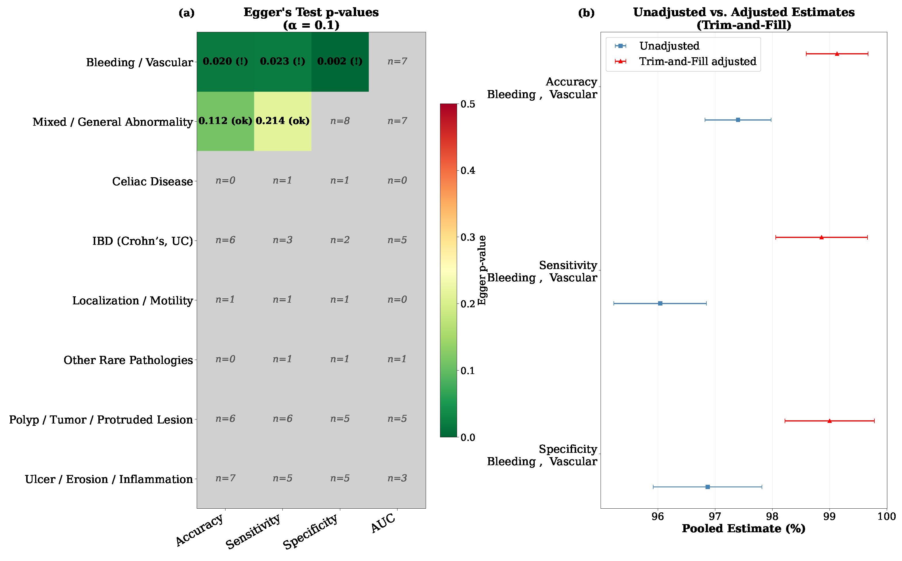
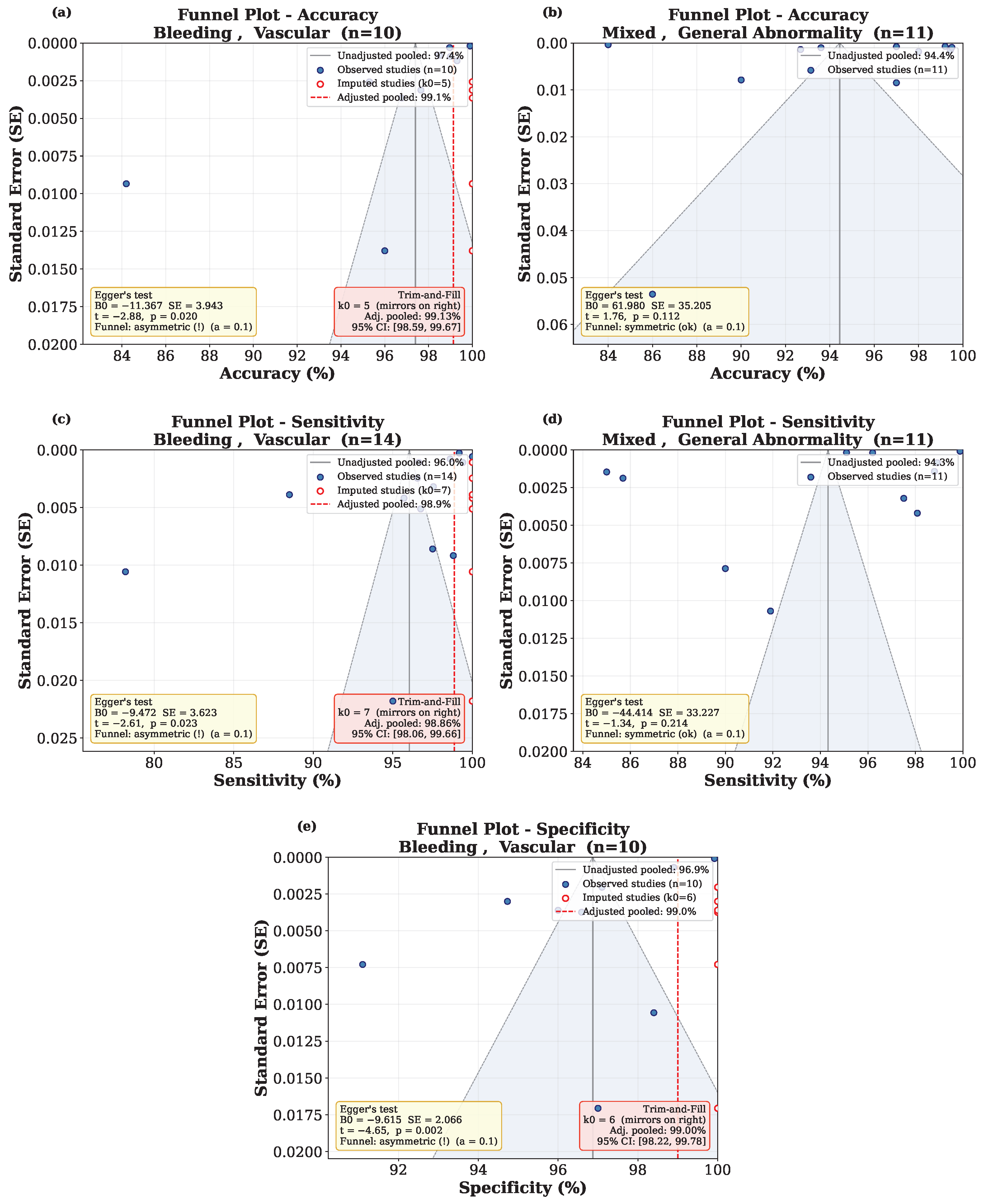
- Egger, M.; Smith, G.D.; Schneider, M.; Minder, C. Bias in meta-analysis detected by a simple, graphical test. BMJ 1997, 315, 629–634. [Google Scholar] [CrossRef]
- Duval, S.; Tweedie, R. Trim and fill: A simple funnel-plot–based method of testing and adjusting for publication bias in meta-analysis. Biometrics 2000, 56, 455–463. [Google Scholar] [CrossRef]
- Molder, A.; Balaban, D.V.; Jinga, M.; Molder, C.C. Current evidence on computer-aided diagnosis of celiac disease: Systematic review. Front. Pharmacol. 2020, 11, 341. [Google Scholar] [CrossRef]
- Pal, P.; Pooja, K.; Nabi, Z.; Gupta, R.; Tandan, M.; Rao, G.V.; Reddy, N. Artificial intelligence in endoscopy related to inflammatory bowel disease: A systematic review. Indian J. Gastroenterol. 2024, 43, 172–187. [Google Scholar] [CrossRef]
- Kim, H.J.; Gong, E.J.; Bang, C.S.; Lee, J.J.; Suk, K.T.; Baik, G.H. Computer-aided diagnosis of gastrointestinal protruded lesions using wireless capsule endoscopy: A systematic review and diagnostic test accuracy meta-analysis. J. Pers. Med. 2022, 12, 644. [Google Scholar] [CrossRef]
- Mohan, B.P.; Khan, S.R.; Kassab, L.L.; Ponnada, S.; Chandan, S.; Ali, T.; Dulai, P.S.; Adler, D.G.; Kochhar, G.S. High pooled performance of convolutional neural networks in computer-aided diagnosis of GI ulcers and/or hemorrhage on wireless capsule endoscopy images: A systematic review and meta-analysis. Gastrointest. Endosc. 2021, 93, 356–364. [Google Scholar] [CrossRef] [PubMed]
- Bang, C.S.; Lee, J.J.; Baik, G.H. Computer-aided diagnosis of gastrointestinal ulcer and hemorrhage using wireless capsule endoscopy: Systematic review and diagnostic test accuracy meta-analysis. J. Med. Internet Res. 2021, 23, e33267. [Google Scholar] [CrossRef] [PubMed]
- Qin, K.; Li, J.; Fang, Y.; Xu, Y.; Wu, J.; Zhang, H.; Li, H.; Liu, S.; Li, Q. Convolution neural network for the diagnosis of wireless capsule endoscopy: A systematic review and meta-analysis. Surg. Endosc. 2022, 36, 16–31. [Google Scholar] [CrossRef] [PubMed]
- Naemi, A.; Tashk, A.; Azar, A.S.; Samimi, T.; Tavassoli, G.; Mohasefi, A.B.; Khanshan, E.N.; Najafabad, M.H.; Tarighi, V.; Wiil, U.K.; et al. Applications of Artificial Intelligence for Metastatic Gastrointestinal Cancer: A Systematic Literature Review. Cancers 2025, 17, 558. [Google Scholar] [CrossRef]
- Naemi, A.; Schmidt, T.; Mansourvar, M.; Wiil, U.K. Personalized Predictive Models for Identifying Clinical Deterioration Using LSTM in Emergency Departments. In Proceedings of the EFMI-STC, Virtual Event, 26–27 November 2020; pp. 152–156. [Google Scholar]
- Smedsrud, P.H.; Thambawita, V.; Hicks, S.A.; Gjestang, H.; Nedrejord, O.O.; Næss, E.; Borgli, H.; Jha, D.; Berstad, T.J.D.; Eskeland, S.L.; et al. Kvasir-Capsule, a video capsule endoscopy dataset. Sci. Data 2021, 8, 142. [Google Scholar] [CrossRef]
- Liu, D.; Lu, C.; Sun, H.; Gao, S. NA-segformer: A multi-level transformer model based on neighborhood attention for colonoscopic polyp segmentation. Sci. Rep. 2024, 14, 22527. [Google Scholar] [CrossRef]
- Obayya, M.; Al-Wesabi, F.N.; Maashi, M.; Mohamed, A.; Hamza, M.A.; Drar, S.; Yaseen, I.; Alsaid, M.I. Modified salp swarm algorithm with deep learning based gastrointestinal tract disease classification on endoscopic images. IEEE Access 2023, 11, 25959–25967. [Google Scholar] [CrossRef]
- Belabbes, M.A.; Oukdach, Y.; Souaidi, M.; Koutti, L.; Charfi, S. Advancements in Polyp Detection: A Developed Single Shot Multibox Detector Approach. IEEE Access 2024, 12, 19199–19215. [Google Scholar] [CrossRef]
- Yang, J.; Chang, L.; Li, S.; He, X.; Zhu, T. WCE polyp detection based on novel feature descriptor with normalized variance locality-constrained linear coding. Int. J. Comput. Assist. Radiol. Surg. 2020, 15, 1291–1302. [Google Scholar] [CrossRef]
- Lin, T.Y.; Maire, M.; Belongie, S.; Hays, J.; Perona, P.; Ramanan, D.; Dollár, P.; Zitnick, C.L. Microsoft coco: Common objects in context. In Proceedings of the European Conference on Computer Vision; Springer: Berlin/Heidelberg, Germany, 2014; pp. 740–755. [Google Scholar]
- Moen, S.; Vuik, F.E.; Kuipers, E.J.; Spaander, M.C. Artificial intelligence in colon capsule endoscopy—A systematic review. Diagnostics 2022, 12, 1994. [Google Scholar] [CrossRef]
- Naemi, A.; Sahafi, A. Benchmarking Large Language Models for MIMIC-IV Clinical Note Summarization. J. Healthc. Inform. Res. 2026, 10, 95–115. [Google Scholar] [CrossRef]
- Zhu, J.Y.; Park, T.; Isola, P.; Efros, A.A. Unpaired image-to-image translation using cycle-consistent adversarial networks. In Proceedings of the IEEE International Conference on Computer Vision, Venice, Italy, 22–29 October 2017; pp. 2223–2232. [Google Scholar]
- Ganin, Y.; Ustinova, E.; Ajakan, H.; Germain, P.; Larochelle, H.; Laviolette, F.; March, M.; Lempitsky, V. Domain-adversarial training of neural networks. J. Mach. Learn. Res. 2016, 17, 1–35. [Google Scholar]
- Sushma, B.; Aparna, P. Summarization of wireless capsule endoscopy video using deep feature matching and motion analysis. IEEE Access 2020, 9, 13691–13703. [Google Scholar] [CrossRef]
- Lan, L.; Ye, C.; Wang, C.; Zhou, S. Deep convolutional neural networks for WCE abnormality detection: CNN architecture, region proposal and transfer learning. IEEE Access 2019, 7, 30017–30032. [Google Scholar] [CrossRef]
- Deding, U.; Herp, J.; Havshoei, A.; Kobaek-Larsen, M.; Buijs, M.; Nadimi, E.; Baatrup, G. Colon capsule endoscopy versus CT colonography after incomplete colonoscopy. Application of artificial intelligence algorithms to identify complete colonic investigations. United Eur. Gastroenterol. J. 2020, 8, 782–789. [Google Scholar] [CrossRef] [PubMed]
- Liao, C.; Wang, C.; Bai, J.; Lan, L.; Wu, X. Deep learning for registration of region of interest in consecutive wireless capsule endoscopy frames. Comput. Methods Programs Biomed. 2021, 208, 106189. [Google Scholar] [CrossRef]
- Herp, J.; Deding, U.; Buijs, M.M.; Kroijer, R.; Baatrup, G.; Nadimi, E.S. Feature point tracking-based localization of colon capsule endoscope. Diagnostics 2021, 11, 193. [Google Scholar] [CrossRef]
- Aoki, T.; Yamada, A.; Kato, Y.; Saito, H.; Tsuboi, A.; Nakada, A.; Niikura, R.; Fujishiro, M.; Oka, S.; Ishihara, S.; et al. Automatic detection of various abnormalities in capsule endoscopy videos by a deep learning-based system: A multicenter study. Gastrointest. Endosc. 2021, 93, 165–173. [Google Scholar] [CrossRef]
- Zhou, J.X.; Yang, Z.; Xi, D.H.; Dai, S.J.; Feng, Z.Q.; Li, J.Y.; Xu, W.; Wang, H. Enhanced segmentation of gastrointestinal polyps from capsule endoscopy images with artifacts using ensemble learning. World J. Gastroenterol. 2022, 28, 5931. [Google Scholar] [CrossRef]
- Souaidi, M.; El Ansari, M. A new automated polyp detection network MP-FSSD in WCE and colonoscopy images based fusion single shot multibox detector and transfer learning. IEEE Access 2022, 10, 47124–47140. [Google Scholar] [CrossRef]
- Sumioka, A.; Tsuboi, A.; Oka, S.; Kato, Y.; Matsubara, Y.; Hirata, I.; Takigawa, H.; Yuge, R.; Shimamoto, F.; Tada, T.; et al. Disease surveillance evaluation of primary small-bowel follicular lymphoma using capsule endoscopy images based on a deep convolutional neural network (with video). Gastrointest. Endosc. 2023, 98, 968–976. [Google Scholar] [CrossRef]
- Aoki, T.; Yamada, A.; Oka, S.; Tsuboi, M.; Kurokawa, K.; Togo, D.; Tanino, F.; Teshima, H.; Saito, H.; Suzuki, R.; et al. Comparison of clinical utility of deep learning-based systems for small-bowel capsule endoscopy reading. J. Gastroenterol. Hepatol. 2024, 39, 157–164. [Google Scholar] [CrossRef] [PubMed]
- Kwon, Y.S.; Park, T.Y.; Kim, S.E.; Park, Y.; Lee, J.G.; Lee, S.P.; Kim, K.O.; Jang, H.J.; Yang, Y.J.; Cho, B.J. Deep learning-based localization and lesion detection in capsule endoscopy for patients with suspected small-bowel bleeding. World J. Gastroenterol. 2025, 31, 106819. [Google Scholar] [CrossRef]
- Andrade, P.; Mascarenhas, M.; Mendes, F.; Rosa, B.; Cardoso, P.; Afonso, J.; Ribeiro, T.; Martins, M.; Mota, J.; Almeida, M.J.; et al. AI-Assisted Capsule Endoscopy for Detection of Ulcers and Erosions in Crohn’s Disease: A Multicenter Validation Study. Clin. Gastroenterol. Hepatol. 2025, in press. [Google Scholar] [CrossRef]
- Mascarenhas Saraiva, M.; Ferreira, J.; Afonso, J.; Mendes, F.; Sonnier, W.P.; Rosa, B.; Ribeiro, T.; Gonçalves, T.C.; Martins, M.; Campelo, P.; et al. Real-Life Clinical Validation of Artificial Intelligence-Assisted Detection and Differentiation of Pleomorphic Lesions in Capsule Endoscopy. Am. J. Gastroenterol. 2025, 121, 993–1000. [Google Scholar] [CrossRef]

| Question | Description |
|---|---|
| Q1 | Which AI models have been used for WCE across different diagnostic tasks? |
| Q2 | How do AI models perform across studies based on reported performance metrics? |
| Q3 | How do dataset characteristics (size, diversity, imbalance, and annotation quality) influence model performance and generalizability? |
| Q4 | What barriers currently limit the clinical adoption of AI in WCE, and how can future research address these barriers? |
| Group | Search Criteria |
|---|---|
| G1—AI keywords | Artificial intelligence, Machine learning, Learning algorithms, Deep learning, Unsupervised machine learning, Supervised learning, Image classification, Object detection, Segmentation, Localization |
| G2—Technical keywords | Capsule endoscopy, Wireless capsule endoscopy, Video capsule endoscopy |
| G3—Medical keywords | Gastrointestinal, Stomach, Small bowel, Colon, Esophagus, Polyp, Ulcer, Bleeding, Inflammation, Crohn’s disease, Lesion, Celiac, Angiectasias |
| G4—Document type | English journal articles |
| G5—Publication year | 1 January 2000–1 January 2026 |
| G6—Final result | G1 AND G2 AND G3 AND G4 AND G5 |
| Inclusion Criteria | Exclusion Criteria |
|---|---|
| - AI studies on WCE images. | - Studies using traditional statistical models. |
| - Cohorts comprising relevant patient groups for WCE. | - The primary focus is not AI applications on WCE images. |
| - Journal articles written in English. | - Non-English and non-journal articles. |
| - Review studies. |
Disclaimer/Publisher’s Note: The statements, opinions and data contained in all publications are solely those of the individual author(s) and contributor(s) and not of MDPI and/or the editor(s). MDPI and/or the editor(s) disclaim responsibility for any injury to people or property resulting from any ideas, methods, instructions or products referred to in the content. |
© 2026 by the authors. Licensee MDPI, Basel, Switzerland. This article is an open access article distributed under the terms and conditions of the Creative Commons Attribution (CC BY) license.
Share and Cite
Sahafi, A.; Koulaouzidis, A.; Naemi, A. Artificial Intelligence in Gastrointestinal Wireless Capsule Endoscopy: A Systematic Literature Review and Meta-Analysis. Diagnostics 2026, 16, 1269. https://doi.org/10.3390/diagnostics16091269
Sahafi A, Koulaouzidis A, Naemi A. Artificial Intelligence in Gastrointestinal Wireless Capsule Endoscopy: A Systematic Literature Review and Meta-Analysis. Diagnostics. 2026; 16(9):1269. https://doi.org/10.3390/diagnostics16091269
Chicago/Turabian StyleSahafi, Ali, Anastasios Koulaouzidis, and Amin Naemi. 2026. "Artificial Intelligence in Gastrointestinal Wireless Capsule Endoscopy: A Systematic Literature Review and Meta-Analysis" Diagnostics 16, no. 9: 1269. https://doi.org/10.3390/diagnostics16091269
APA StyleSahafi, A., Koulaouzidis, A., & Naemi, A. (2026). Artificial Intelligence in Gastrointestinal Wireless Capsule Endoscopy: A Systematic Literature Review and Meta-Analysis. Diagnostics, 16(9), 1269. https://doi.org/10.3390/diagnostics16091269