Article

Three-Dimensional Analysis of Facial Skeleton Textures in CBCT as an Early Warning Sign of Osteoporosis—A Pilot Study


1
Department of Maxillofacial Surgery, Medical University of Lodz, 251 Pomorska Str., 92-213 Lodz, Poland
2
Department of Maxillofacial Surgery, Medical University of Gdansk, 80-210 Gdańsk, Poland
3
Institute of Electronics, Lodz University of Technology, Al. Politechniki 10, 93-590 Łódź, Poland
4
Department of Otolaryngology, Hospital in Sosnowiec, Zegadłowicza 3, 41-200 Sosnowiec, Poland
5
Head and Neck Surgery Clinic for Children and Young Adults, Department of Clinical Pediatrics, University of Warmia and Mazury, ul. Oczapowskiego 2, 10-719 Olsztyn, Poland
6
III Department of Radiology and Diagnostic Imaging, Medical University of Lodz, 90-419 Lodz, Poland
7
Department of Oral and Maxillofacial Surgery, Cliniques Universitaires Saint Luc, UCLouvain, 1348 Brussels, Belgium
8
Oral and Maxillofacial Surgery Research Lab (OMFSLab), NMSK, Institut de Recherche Expérimentale et Clinique (IREC), UCLouvain, 1348 Brussels, Belgium
9
Department of Perioperative Dentistry, Ludwika Rydygiera Collegium Medicum in Bydgoszcz, Nicolaus Copernicus University in Torun, 85-067 Torun, Poland
*
Author to whom correspondence should be addressed.
Diagnostics 2026, 16(8), 1217; https://doi.org/10.3390/diagnostics16081217

Submission received: 8 March 2026 / Revised: 13 April 2026 / Accepted: 15 April 2026 / Published: 19 April 2026
(This article belongs to the Section Medical Imaging and Theranostics)

Abstract

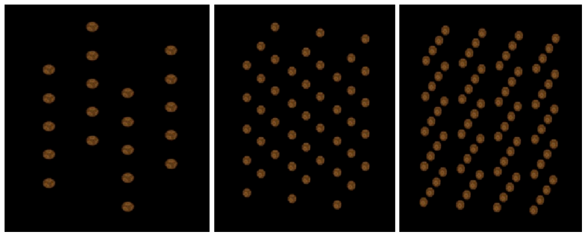
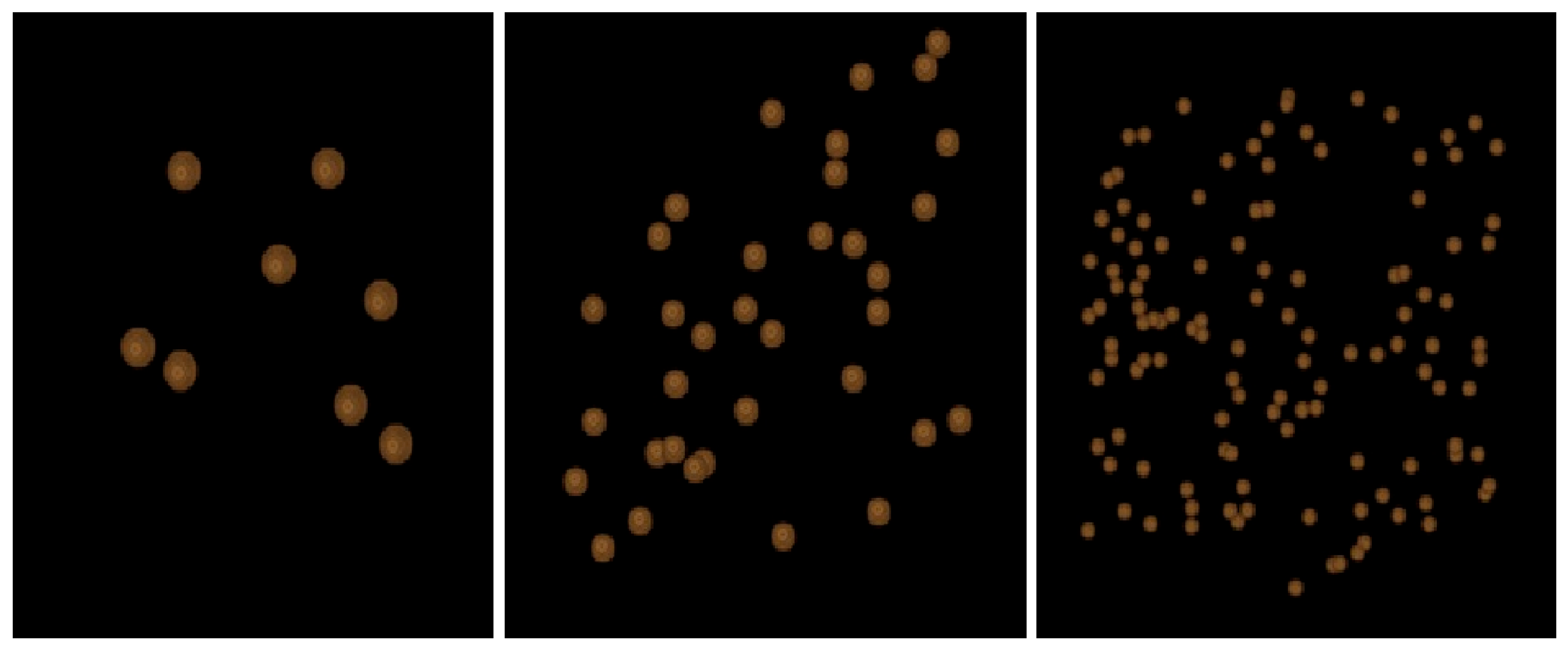
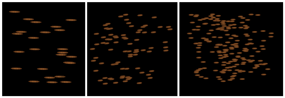
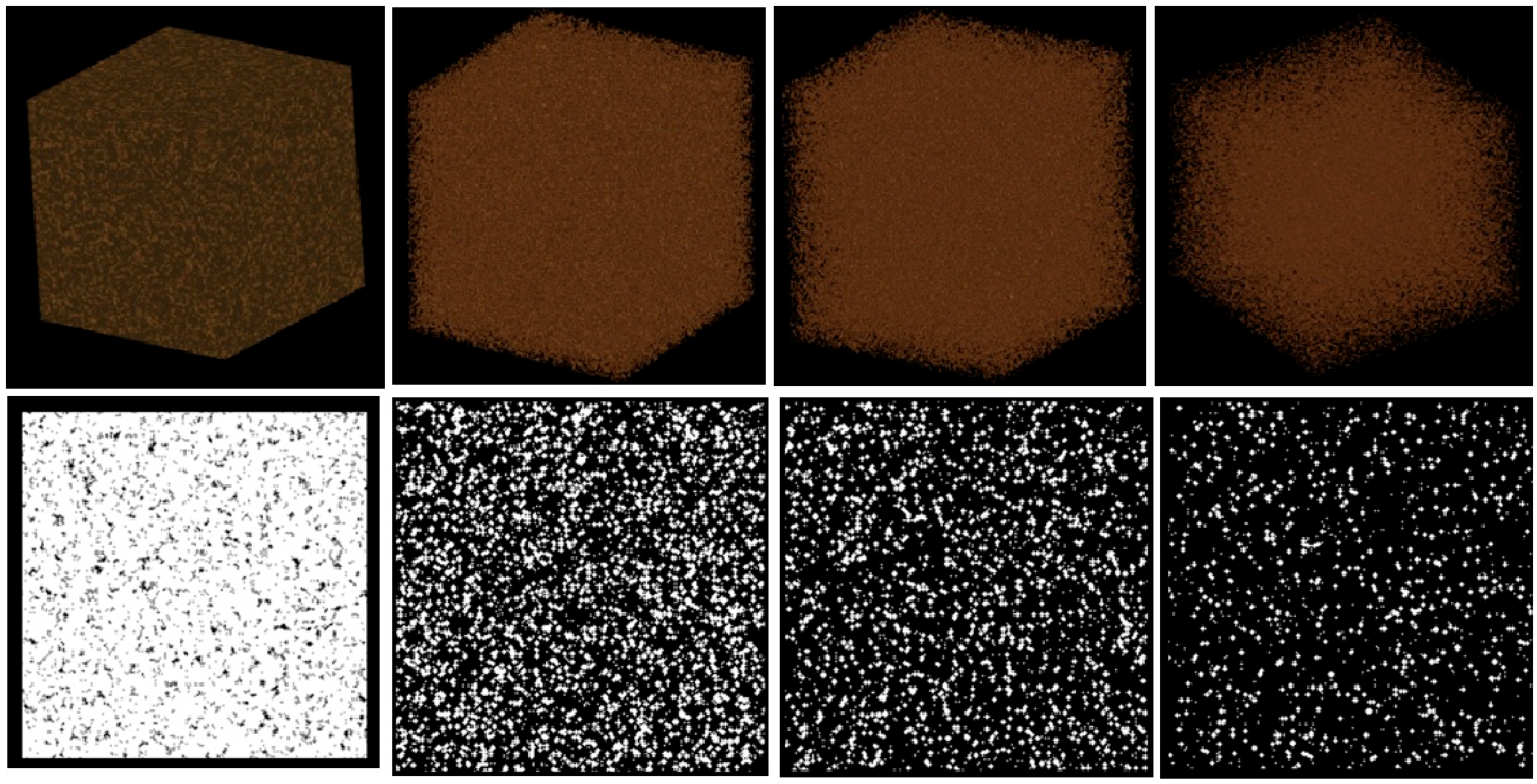
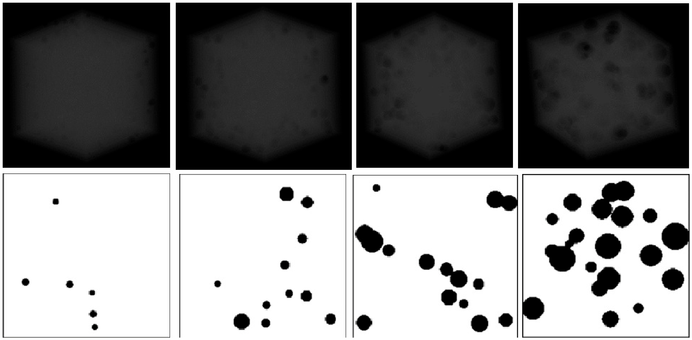
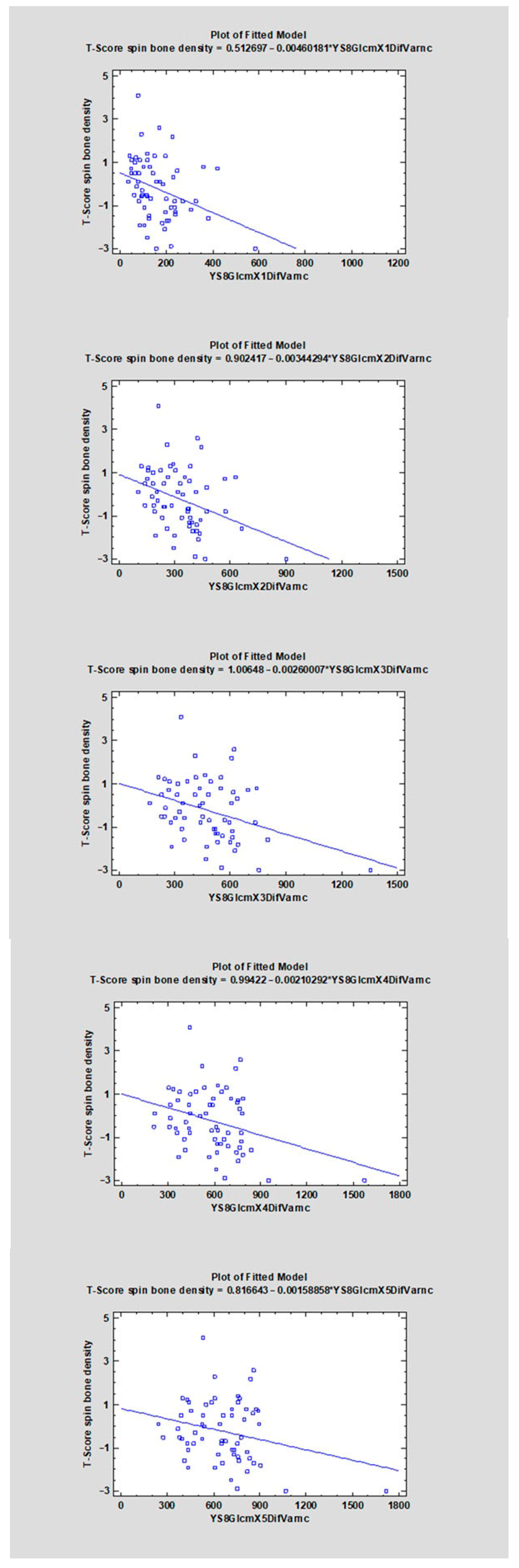
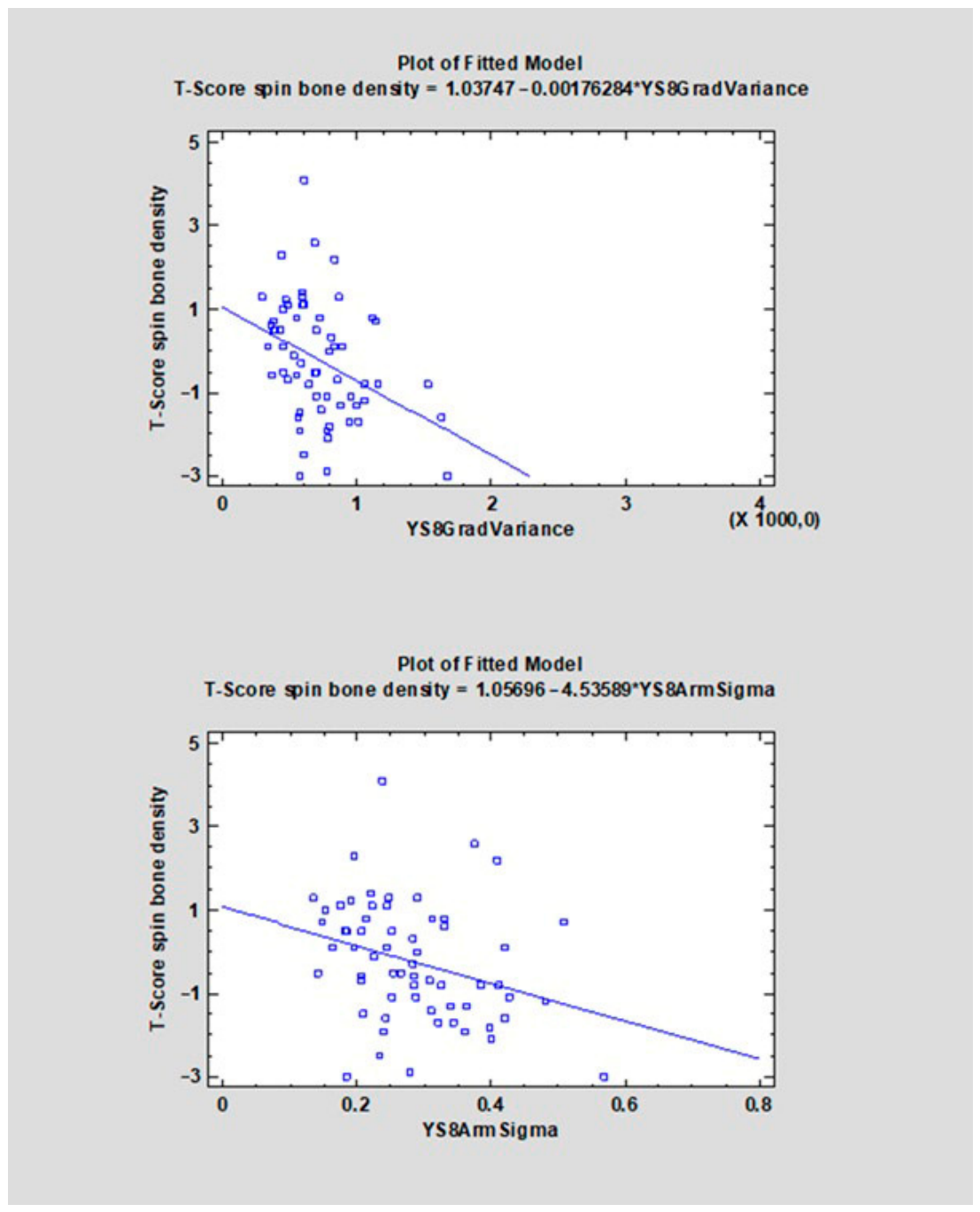
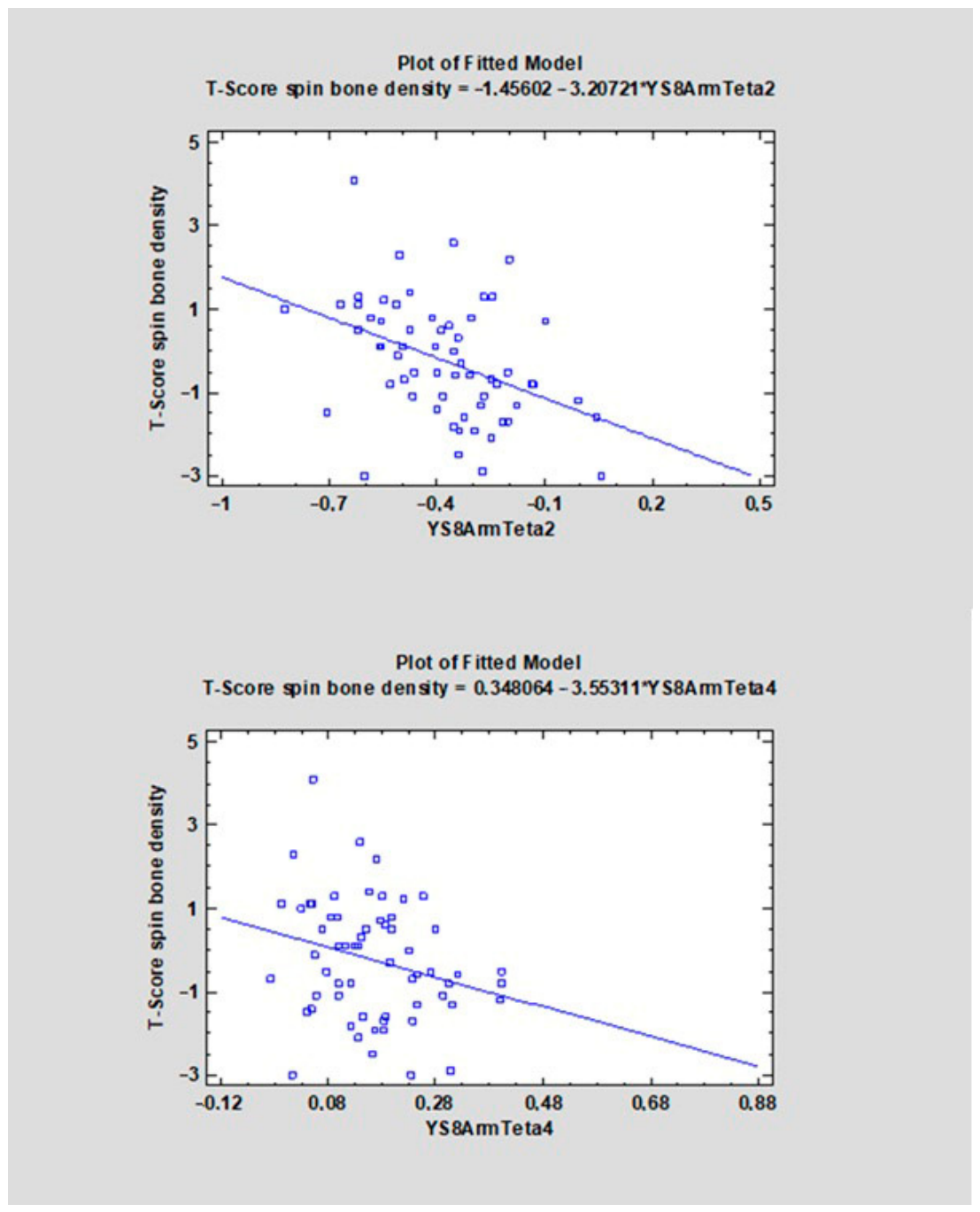
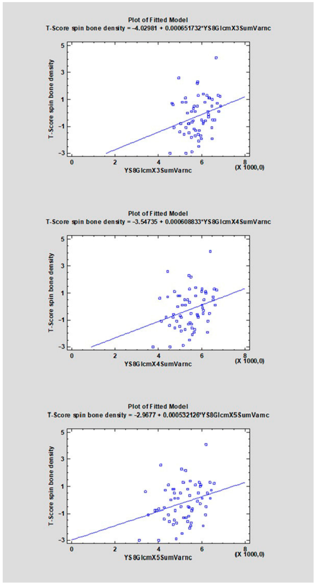
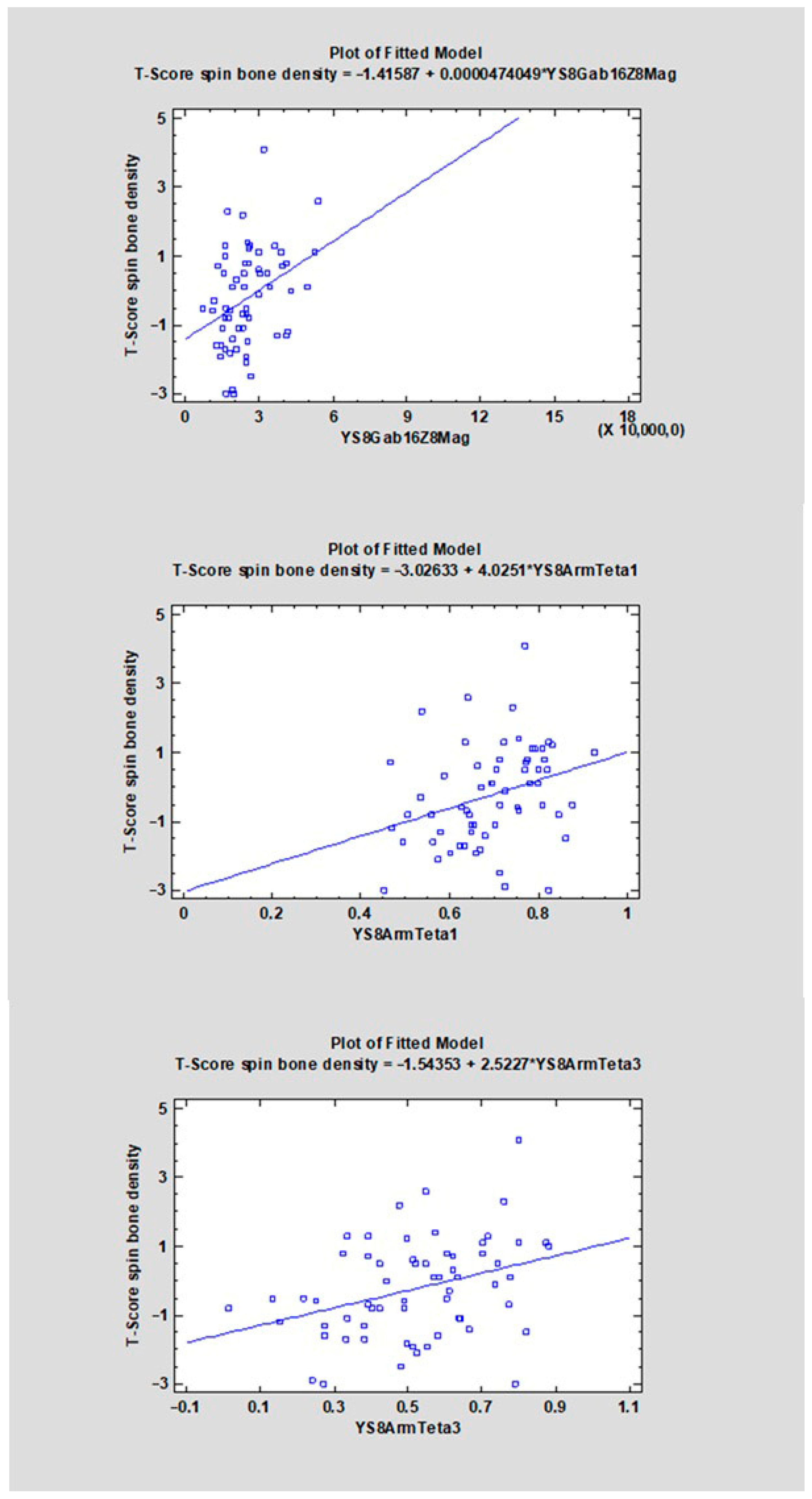
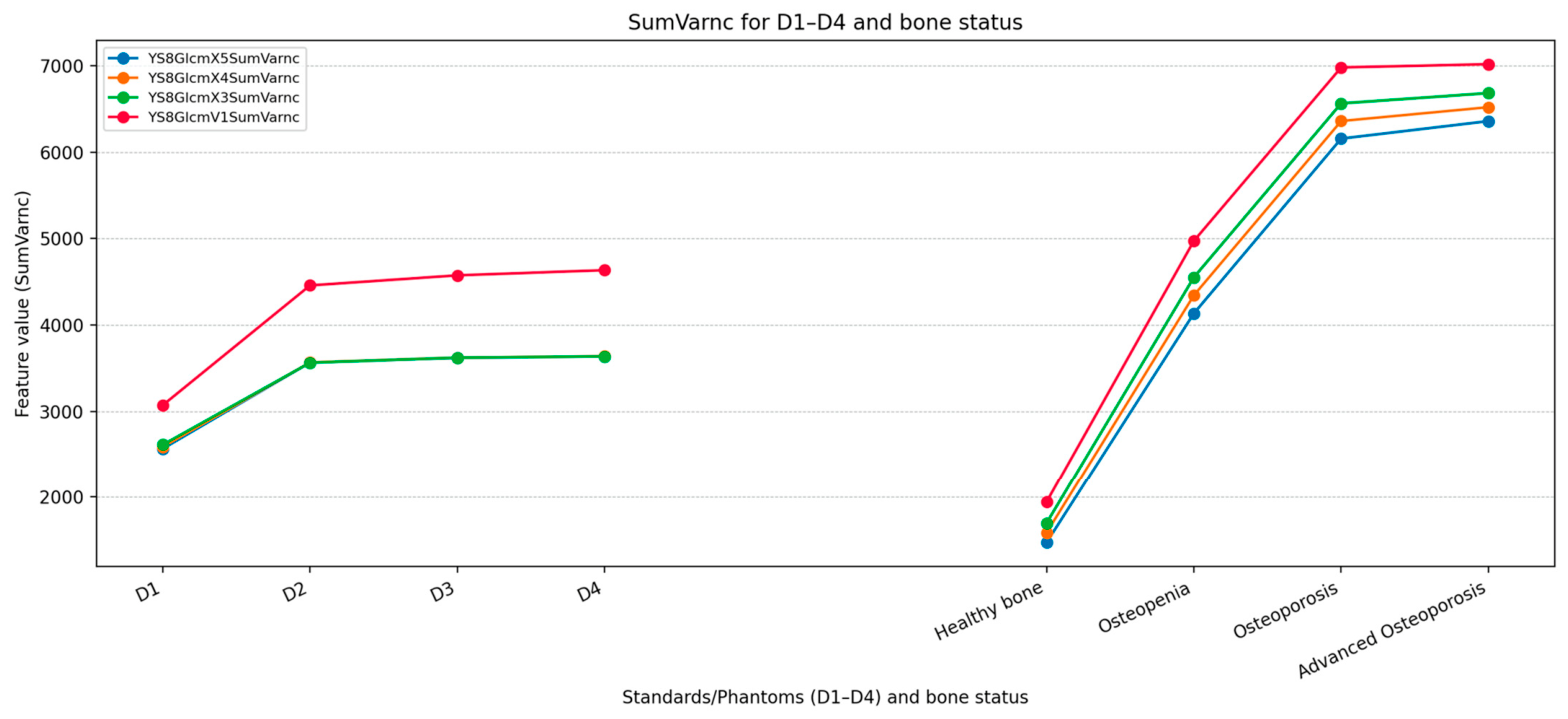
Background: Osteoporosis is a prevalent condition characterized by low bone mass and altered microarchitecture, increasing fracture risk. Early detection remains challenging, as conventional methods such as DXA are limited to specialized settings and often detect disease only after a fracture. Radiomics and three-dimensional (3D) imaging techniques, such as CBCT, may provide novel approaches for assessing bone quality. Methods: This pilot study analyzed 68 CBCT scans from adult patients (41 females, 27 males; mean age 57 years). Three-dimensional regions of interest (ROIs) were delineated in seven maxillofacial and mandibular sites (total 309 ROIs). Radiomic texture features were extracted and compared with corresponding T-scores from DXA measurements. Additionally, synthetic 3D reference phantoms with controlled variations in density, trabecular connectivity, and structural anisotropy were generated to evaluate the sensitivity of texture features to microarchitectural changes. Results: Several radiomic features, including GLCM-, ARM-, and Gradient-derived parameters, demonstrated consistent monotonic trends correlating with bone density and microstructural deterioration. Differences in feature values were observed across healthy, osteopenic, osteoporotic, and advanced osteoporotic states. Reference phantoms confirmed that the observed trends were attributable to structural differences rather than imaging variability. Features such as Sum Variance and Correlation exhibited potential as early indicators of microarchitectural degradation. Conclusions: Three-dimensional CBCT texture analysis may provide a non-invasive, supplementary tool for assessing bone quality and detecting early osteopenic changes. Further studies with larger cohorts are warranted to validate radiomic markers and develop predictive indices for osteoporosis screening.
Keywords: 3D texture analysis; densitometry; CBCT; CT; bone diagnosis; bone structure; osteoporosis; osteopenia; healthy bone 3D texture analysis; densitometry; CBCT; CT; bone diagnosis; bone structure; osteoporosis; osteopenia; healthy bone