Interesting Images

Papillary Thyroid Carcinoma, Bilateral Macronodular Adrenal Cortical Disease-Related Cortisol Excess, and Femoral Enchondroma: A Novel Phenotype–Genotype Based on Next-Generation Sequencing (Variants of APC, MSH6, and CACNA1S Genes)


1
Department of Endocrinology, “Carol Davila” University of Medicine and Pharmacy, 020021 Bucharest, Romania
2
Department of Clinical Endocrinology V, “C.I. Parhon” National Institute of Endocrinology, 011863 Bucharest, Romania
3
Department of Research, “C.I. Parhon” National Institute of Endocrinology, 011863 Bucharest, Romania
4
Department of Radiology and Medical Imaging, “C.I. Parhon” National Institute of Endocrinology, 011863 Bucharest, Romania
5
PhD School of “Carol Davila” University of Medicine and Pharmacy, 020021 Bucharest, Romania
6
Department of Radiology and Medical Imaging, “Dr. Carol Davila” Central Military University Emergency Hospital, 010825 Bucharest, Romania
7
Department of Radiology, “Carol Davila” University of Medicine and Pharmacy, 050474 Bucharest, Romania
8
Department of Radiology and Medical Imaging, “Foisor” Clinical Hospital of Orthopaedics, Traumatology and Osteoarticular TB, 021382 Bucharest, Romania
9
Department of Pathology, “C.I. Parhon” National Institute of Endocrinology, 011863 Bucharest, Romania
10
Oncoteam Diagnostics, 012244 Bucharest, Romania
*
Authors to whom correspondence should be addressed.
Diagnostics 2026, 16(8), 1185; https://doi.org/10.3390/diagnostics16081185

Submission received: 9 March 2026 / Revised: 14 April 2026 / Accepted: 15 April 2026 / Published: 16 April 2026
(This article belongs to the Special Issue State of the Art in the Diagnosis and Management of Endocrine Tumors)

Abstract

This case highlights a novel genotype–phenotype correlation in the field of endocrinology. Specific endocrine and imaging assessment, in addition to next-generation sequencing (NGS), was performed on the Illumina MiSeq platform, using a TruSight One Sequencing Panel kit for genomic analysis of coding regions of 4813 genes. A 54-year-old female was confirmed with a papillary thyroid carcinoma after total thyroidectomy and underwent radioiodine ablative therapy. Three years later, a left femoral enchondroma of almost 3 cm was identified at computed tomography (CT) scan and magnetic resonance imaging (MRI). She experienced hypertension (in addition to obesity, dyslipidaemia and impaired glucose tolerance) and was later confirmed with ACTH-independent cortisol excess [lack of cortisol suppression at 1 mg dexamethasone testing of 13.9 (normal < 1.8 µg/dL)], noting bilateral adrenal tumors, of 4.7 cm (right), respectively, and of 1.6 cm (left) at CT. Right laparoscopic adrenalectomy was performed with post-operative adrenal insufficiency, requiring glucocorticoid replacement and stopping the anti-hypertensive medication. Pathology report confirmed an adrenocortical adenoma (a Ki67 proliferation index of 2%). Noting the unusual association of the mentioned conditions, NGS was performed in the peripheral blood and identified a heterozygote missense variant of the APC gene (c.5759G>A, p.Arg1920Gln), a heterozygote missense variant of the MSH6 gene (c.2092C>G, p.Gln698Glu), and an incidental additional finding: a heterozygote stop gain pathogenic variant of the CACNA1S gene (c.2707C>T, p.Arg903*). The first two are currently classified as variants of uncertain significance. Whether the co-presence of a triple mutation may change the clinical picture and the life-long outcomes across reciprocal influence is still an open matter. Further research will point out the clinical implications of this genotype–phenotype association, which, to our best knowledge, has not been previously reported.

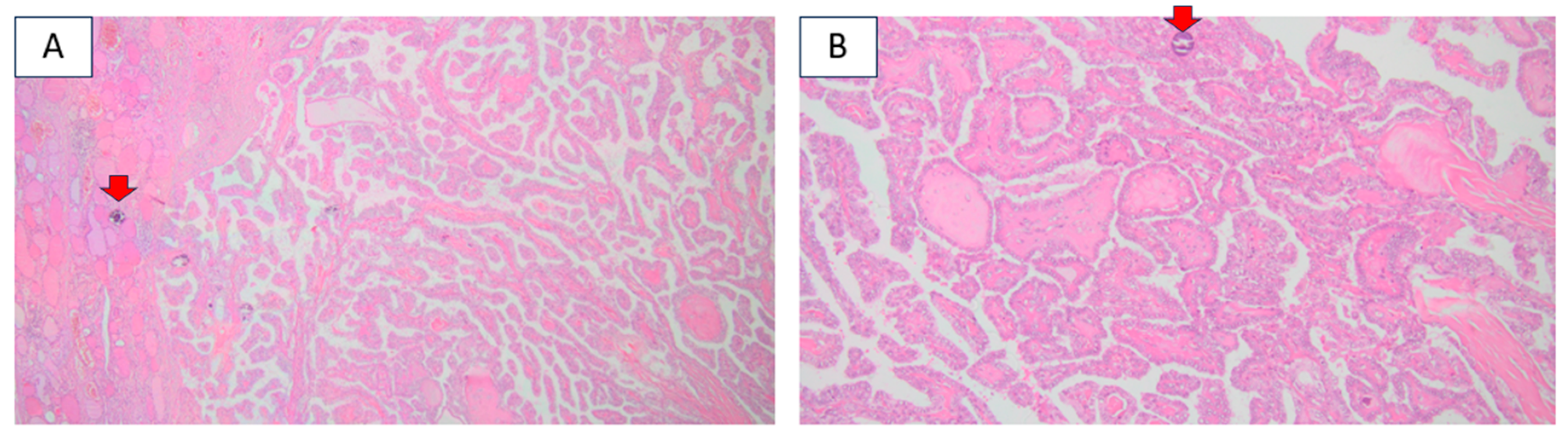
Figure 1. This is a 54-year-old female who was accidentally identified with a thyroid nodule of 3.6 cm in diameter at anterior neck ultrasound. She underwent total thyroidectomy with confirmation of a differentiated thyroid carcinoma. [Histological report (hematoxylin–eosin) confirmed a papillary, sclerosant, multifocal carcinoma with several foci under 1 cm (centimeter), the largest of 0.7 by 0.6 cm, respectively, of 0.6 by 0.6 cm, with psammoma bodies (red arrows). (A) Magnification 4×; (B) magnification 10×.] Radioiodine therapy (100 mCI) was offered to the patient in addition to life-long Thyroid-Stimulating Hormone (TSH)-suppressive levothyroxine therapy, which achieved undetectable serum thyroglobulin levels within the following year.
Diagnostics 16 01185 g001
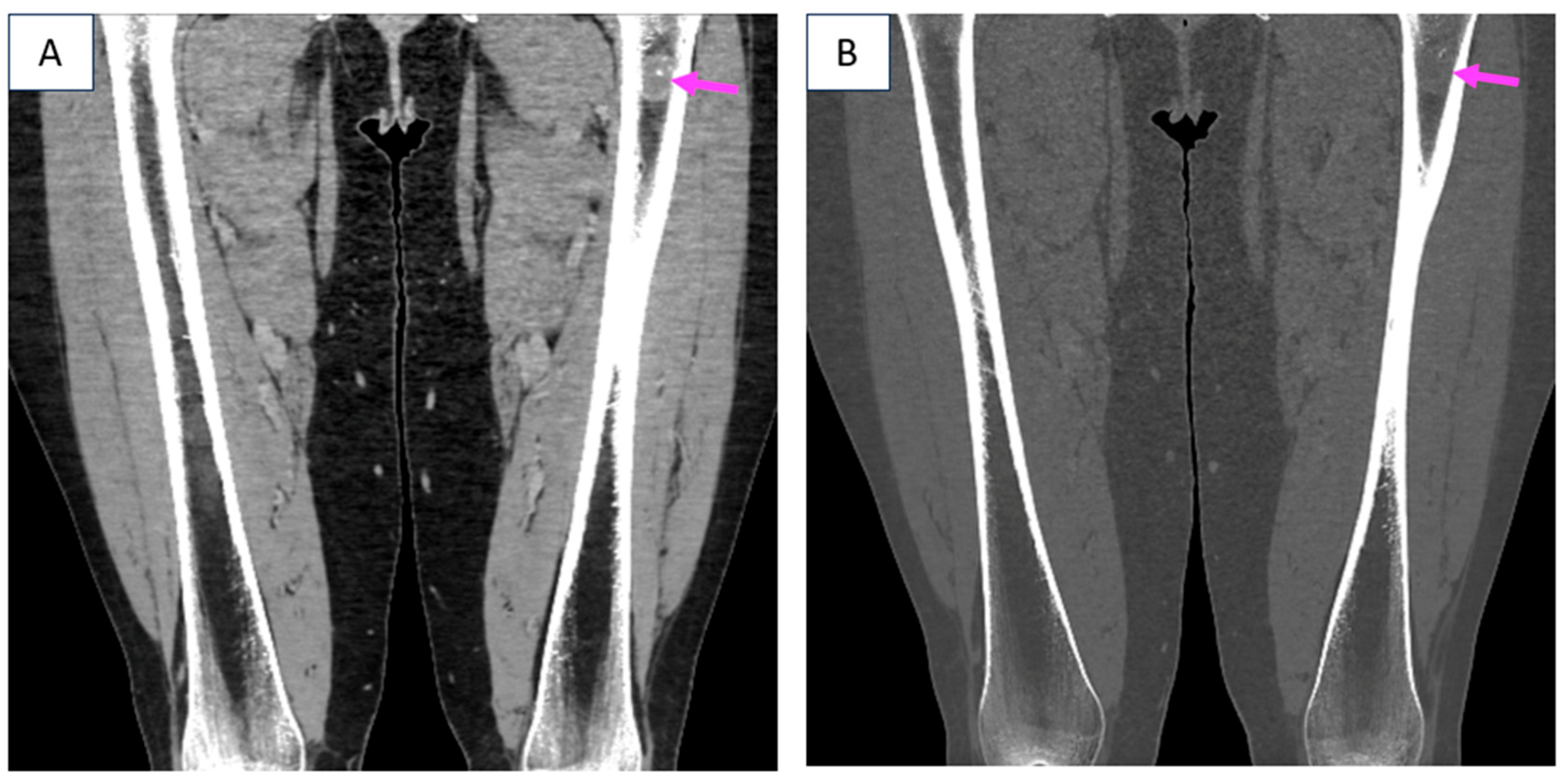
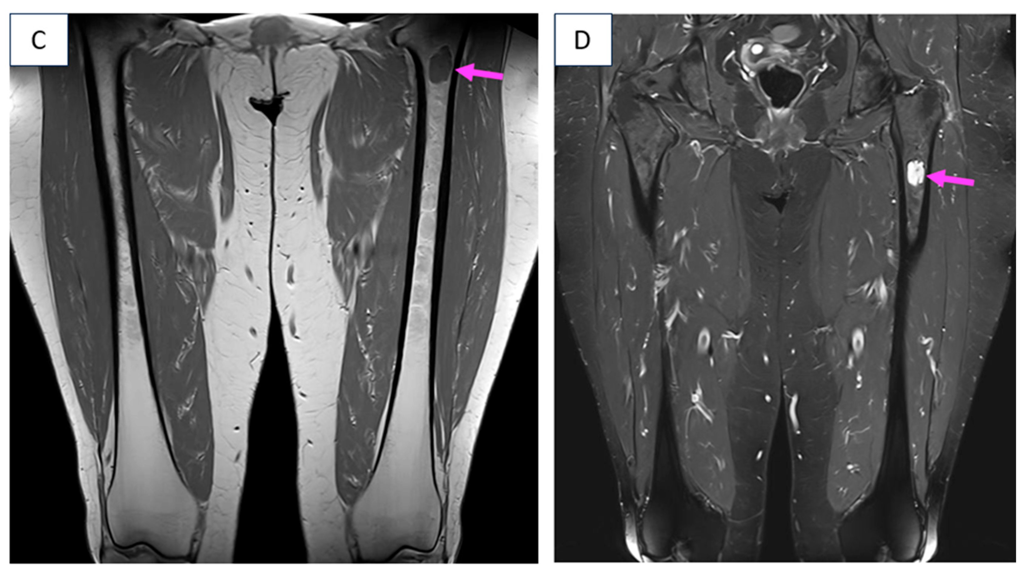
Figure 2. Later on, at the age of 57, the patient experienced non-specific, mild, persistent bone and joint pain and further investigations lead to the identification, not only of incipient arthrosis, but, also, of a left femoral tumor with cartilaginous structure and benign appearance (an enchondroma) at computed tomography (CT) and magnetic resonance imaging (MRI) [(A,B) CT scan (native phase): bone tumor in the diaphysis of the left femur of 1.9 by 2.5 cm, without cortical involvement (pink arrow); (A) coronal plane; (B) coronal plane with bone window. (C,D) Contrast-enhanced MRI scan: bone tumor in the diaphysis of the left femur of 1.9 by 1.8 by 2.9 cm with polycyclic margins, with T1 hypo-signal and T2 hypersignal, without cortical thinning, suggestive of enchondroma (pink arrow); (C) T1 WI HSE, coronal plane; (D) T2 FSE, coronal plane].
Diagnostics 16 01185 g002aDiagnostics 16 01185 g002b
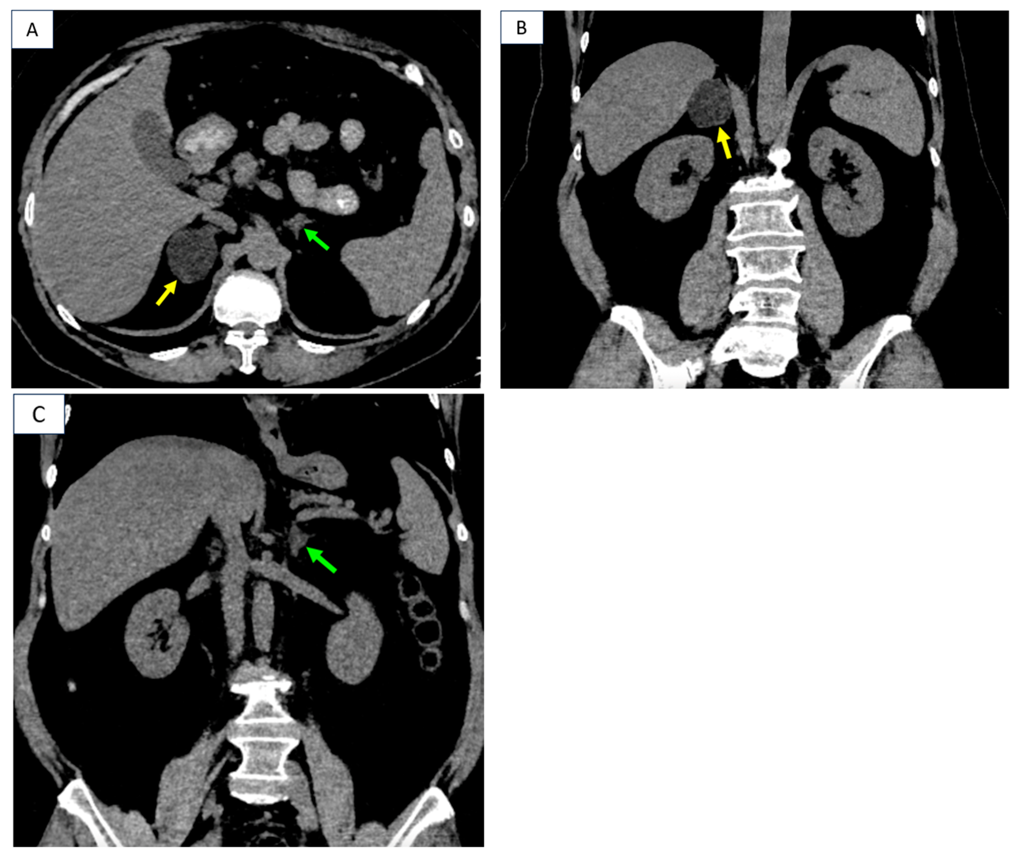
Figure 3. In the meantime, since the age of 56, she was confirmed with arterial hypertension, requiring re-assessment at a tertiary center of endocrinology. The clinical aspects, while not being highly suggestive for a typical Cushing’s syndrome, pointed out metabolic complications in terms of obesity (a body mass index of 36 kg/sqm); dyslipidemia [increased triglycerides of 205 (normal < 200) mg/dL, and total cholesterol of 248 (normal < 200) mg/dL]; impaired glucose tolerance based on a 2 h glycemia during 75 g oral glucose tolerance test of 147 mg/dL, mildly elevated glycated hemoglobin A1c of 6.4 (normal: 4.8–5.9) %, and insulin resistance according to a HOMA-IR (Homeostasis Model Assessment Model of Insulin Resistance) level of 5.3 (insulin resistance at HOMA-IR above the cutoff 2). Hormonal panel was conclusive for Adrenocorticotropic Hormone (ACTH)-independent (autonomous) cortisol excess based on: suppressed baseline morning ACTH of 2.7 (normal: 7.2–63.3) pg/mL, normal morning plasma cortisol of 13.1 (normal: 4.82–19.5) µg/dL without suppression at 1 mg overnight dexamethasone testing according to the second-day plasma cortisol level of 13.9 (normal < 1.8) µg/dL. The central Dual-Energy X-Ray Absorptiometry (DXA) scan excluded osteoporosis/osteopenia (lowest T-score of +0.8 SD, and bone mineral density of 1.098 g/sqcm at total hip), and the trabecular bone score of 1.388 was normal. CT showed bilateral adrenal tumors [3. CT scan (native phase): right adrenal tumor (yellow arrow) of 3.7 by 4.4 by 4.7 cm with negative density, heterogeneous, in close contact with the inferior vena cava, the upper pole of the right kidney, the fifth hepatic segment and the right diaphragmatic crus and left adrenal tumor of 1.1 by 1.6 cm (green arrow); (A) axial plane; (B) coronal plane; (C) coronal plane].
Diagnostics 16 01185 g003
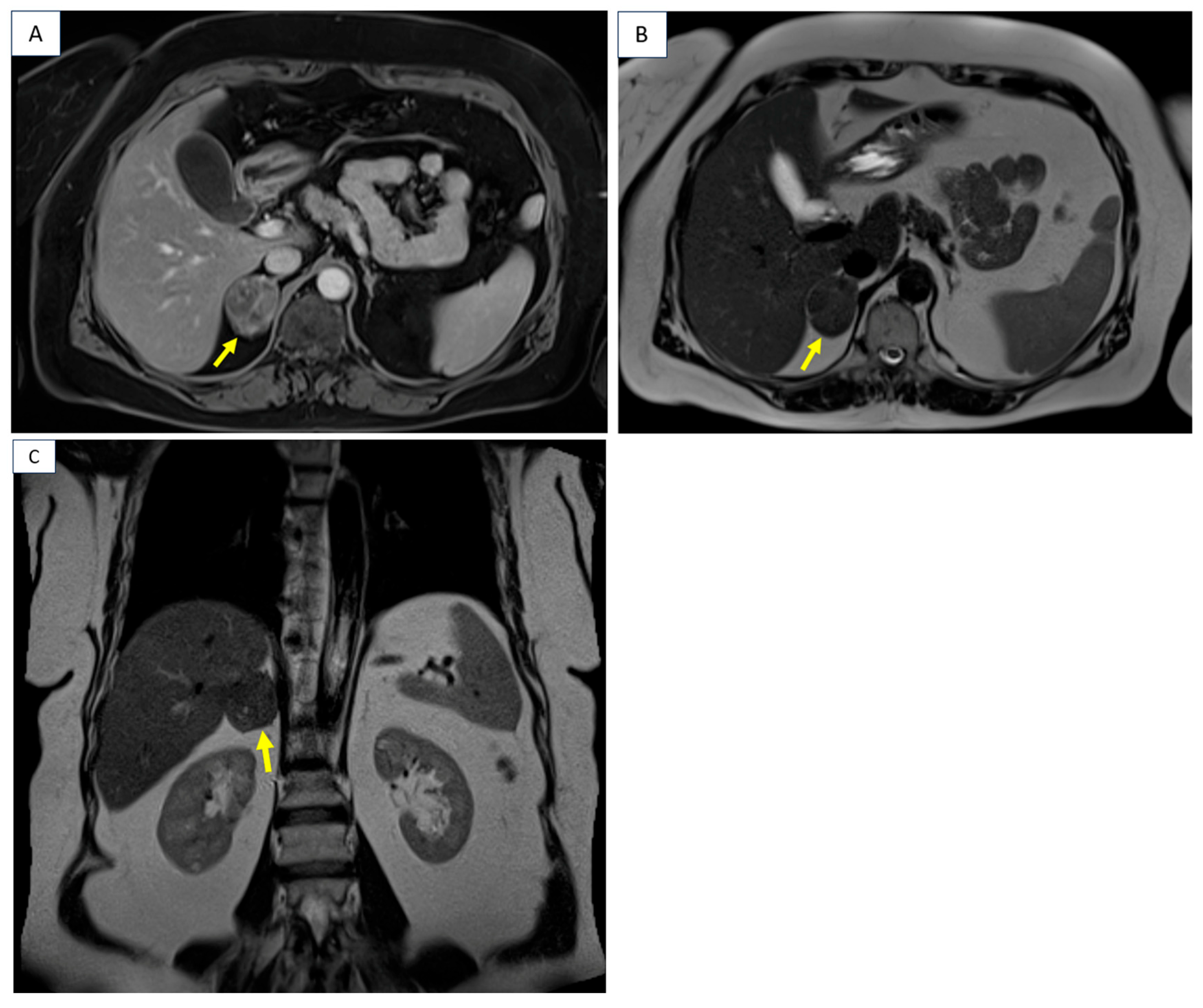
Figure 4. MRI confirmed similar features of the adrenal tumors. [4. Contrast-enhanced MRI scan: right adrenal tumor of 4.2 by 3.3 by 4.5 cm with intracellular lipid content and no restricted diffusion on diffusion-weighted imaging (DWI) (yellow arrow); (A) T1 VIBE Dixon, axial plane; (B) T2 HASTE, axial plane; (C) T2 HASTE, coronal plane].
Diagnostics 16 01185 g004
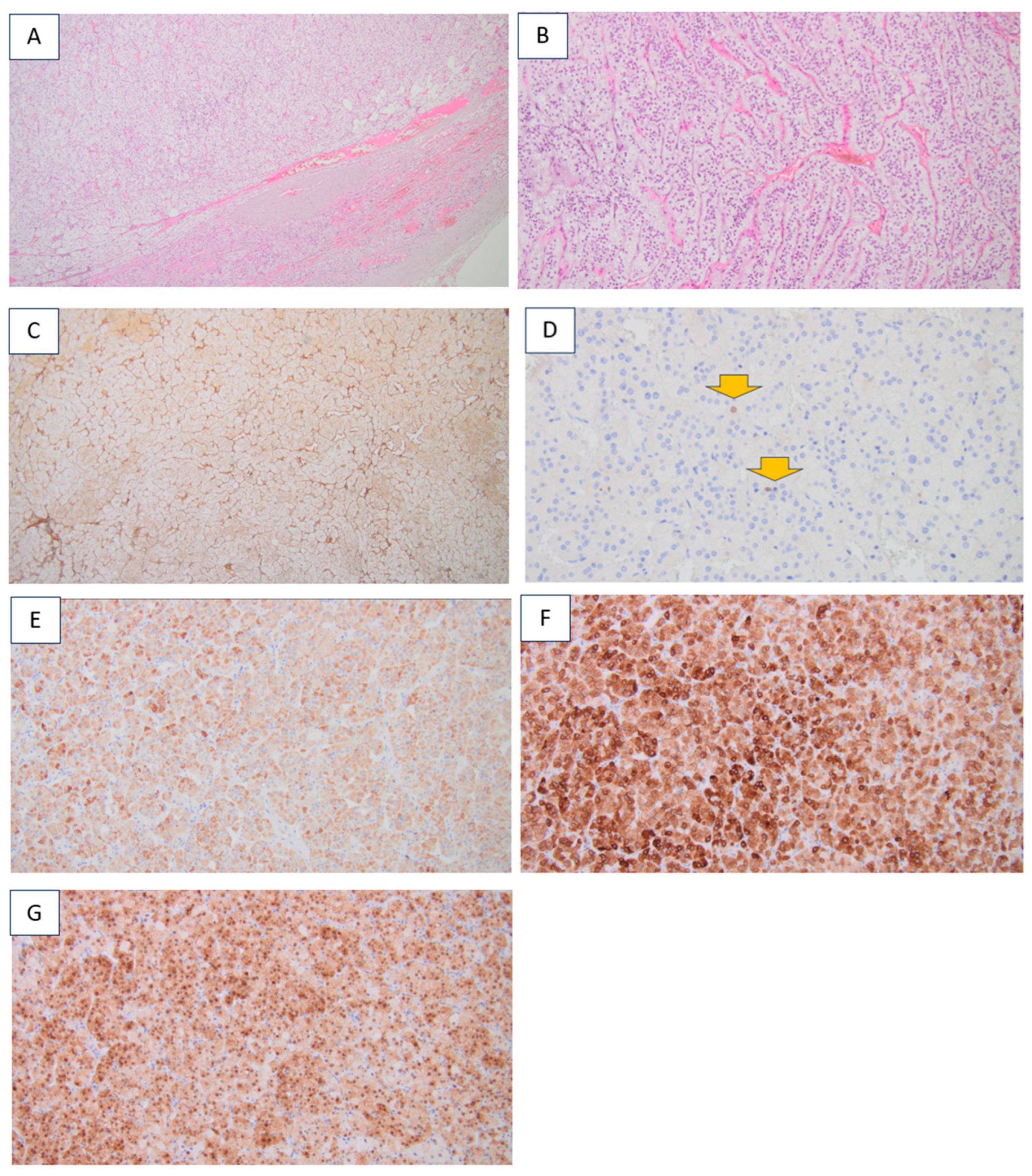
Figure 5. Due to the cortisol over-production with bilateral adrenal gland involvement, but with one tumor significantly larger than the other (>3 cm difference), and the fact that the largest diameter was above the cutoff of 4 cm in the right tumor mass, unilateral laparoscopic adrenalectomy was recommended and performed with a good outcome. Post-operative adrenal insufficiency developed, requiring glucocorticoid replacement for a few months in addition to an improvement in the high blood pressure, so that the anti-hypertensive drugs were no longer necessary. Pathology report confirmed an adrenocortical adenoma [(AC). Histological analysis: (A) hematoxylin–eosin (magnification 4×); (B) hematoxylin–eosin (magnification 10×); (C) intact reticulin network (magnification 4×); (DG) Immunohistochemistry analysis (Weiss score of 0); (D) Ki67 proliferation index of 2% (orange arrows) (magnification 20×); (E) medium-level staining of melan A in the cytoplasm of tumor cells (magnification 10×); (F) strong cytoplasmic staining of inhibin in tumor cells (magnification 10×); (G) medium to high cytoplasmic and strong nuclear staining of calretinin in tumor cells (magnification 10×)].
Diagnostics 16 01185 g005
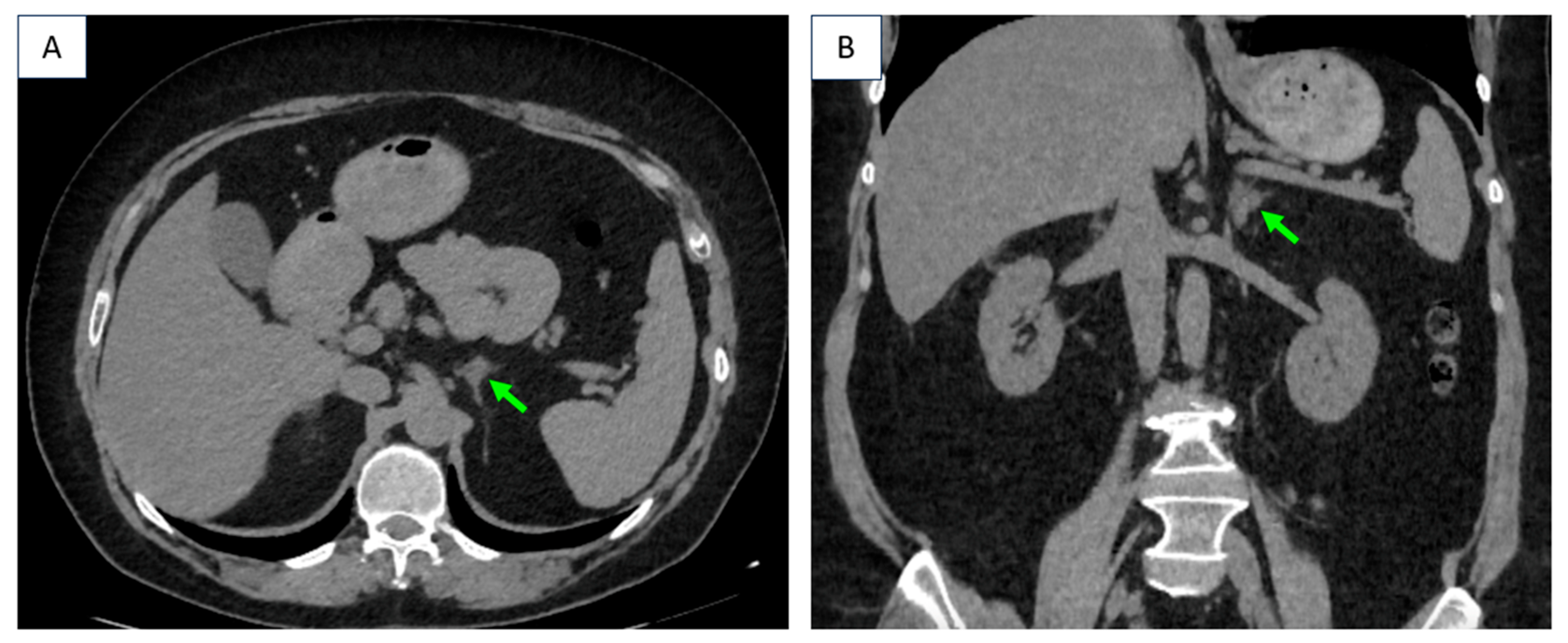
Figure 6. Six months after unilateral adrenalectomy, CT showed stationary left adrenal tumor (of 1.2 by 1.5 cm) [CT scan native phase highlights the tumor (green arrow): (A) axial plane; (B) coronal plane].
Diagnostics 16 01185 g006
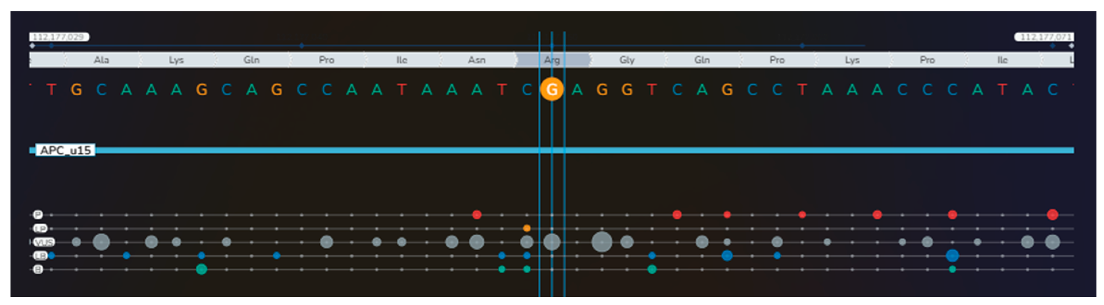
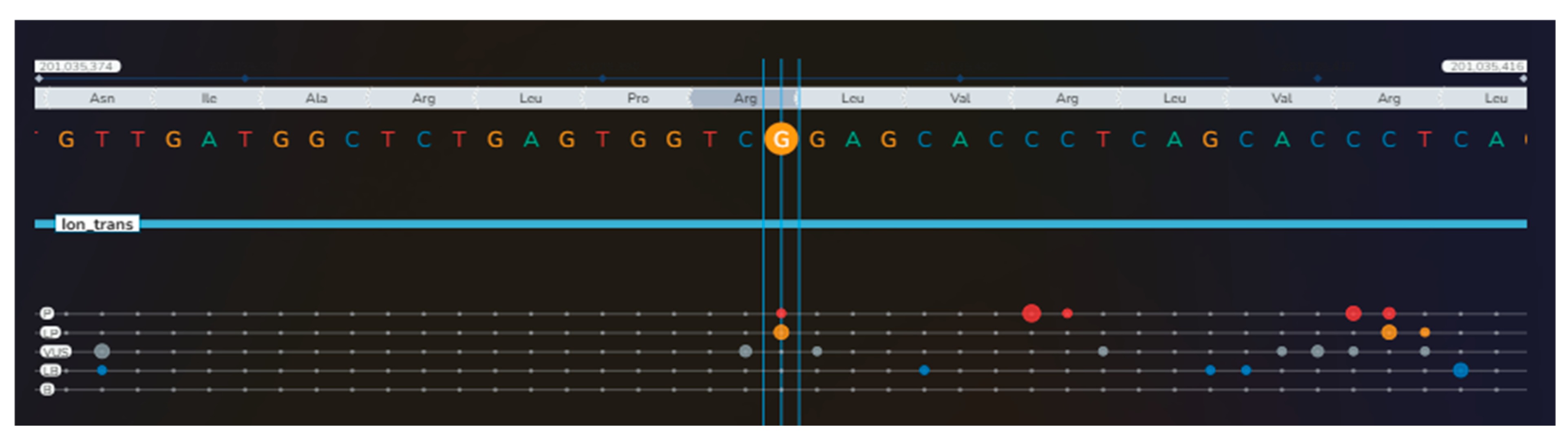
Figure 7. Noting the unusual association of the mentioned conditions, next-generation sequencing (NGS) was performed in the peripheral blood on the Illumina MiSeq platform, using TruSight TruSeq One Sequencing Panel kit (Illumina, San Diego, CA, USA) for genomic analysis of coding regions of 4813 genes. Alignment and Variant Calling were performed using BWA and GATK in Local Run Manager (Illumina). Variants were classified using the American College of Medical Genetics and Genomics (ACMG) criteria and databases, including ClinVar, HGMD, and gnomAD. For tertiary analysis, we used Geneyx Analysis software v6.2 (Geneyx Genomex Ltd., Wilmington, DE, USA), which guided prioritization of genes related to this phenotype (papillary thyroid carcinoma, bilateral macronodular adrenal cortical disease and femoral enchondroma). We identified a heterozygote missense variant of the APC gene (c.5759G>A, p.Arg1920Gln) [P = pathogenic (red bullets); LP = likely pathogenic (orange bullets); VUS = variant of uncertain significance (gray bullets); LB = likely benign (blue bullets); B = benign (green bullets); the dimension of the bullets depends on the number of submissions with the same classification in ClinVar for that specific variant] [1] (Table S1).
Diagnostics 16 01185 g007
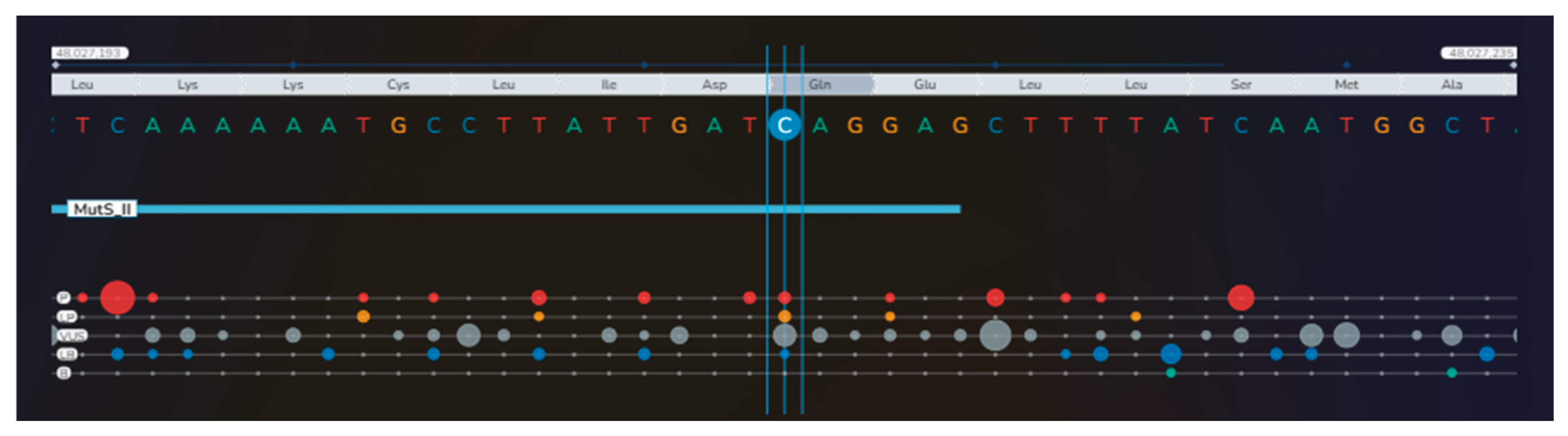
Figure 8. Moreover, a heterozygote missense variant of the MSH6 gene (c.2092C>G, p.Gln698Glu) was also identified: P = pathogenic (red bullets); LP = likely pathogenic (orange bullets); VUS = variant of uncertain significance (gray bullets); LB = likely benign (blue bullets); B = benign (green bullets); the dimension of the bullets depends on the number of submissions with the same classification in ClinVar for that specific variant] [1] (Table S2).
Diagnostics 16 01185 g008
Figure 9. Also, an incidental additional finding included a heterozygote stop gain pathogenic variant of the CACNA1S gene (c.2707C>T, p.Arg903*) [P = pathogenic (red bullets); LP = likely pathogenic (orange bullets); VUS = variant of uncertain significance (gray bullets); LB = likely benign (blue bullets); B = benign (green bullets); the dimension of the bullets depends on the number of submissions with the same classification in ClinVar for that specific variant] [1]. Particular aspects of this case involve the unusual association of endocrine and non-endocrine tumors/malignancy that, to our best knowledge, has not been reported prior, especially in relation to a novel mutational landscape across NGS. Although there are no cases with all three tumors in one individual, the association of any two tumors has been identified across distinct genetic backgrounds from the current one. For instance, the association between enchondromas and adrenal tumors was found in Maffucci syndrome. The underlying genetic defects are still an open matter (probably involving IDH1 and IDH2 genes) [2,3,4,5]. Moreover, enchondromas, especially pulmonary enchondromas, and adrenal tumors are part of the Carney triad [6]. Soft tissue tumors and bone tumors (e.g., osteomas) might be associated with familial adenomatous polyposis underlying APC pathogenic variants [7,8], noting that, traditionally, the tumor-suppressor gene (located on chromosome 5) is linked with this hereditary (autosomal dominant) syndrome [9]. In this case, the gastroenterological evaluation showed no pathological findings, nor was the patient’s family known to have a specific hereditary syndrome. Notably, a recent report of a synchronous thyroid malignancy was connected to a novel APC pathogenic variant (c.2929delG frameshift deletion) [10], while non-medullary (particularly, cribriform-morular variant of the papillary type) thyroid cancer was found to be associated with the syndrome in exceptional population subgroups [11,12,13]. Ongoing genetic research is mandatory to further pinpoint the role of the APC gene in the development of differentiated thyroid cancer. However, in the present case, the p.R1920Q variant of APC gene is currently classified as variant of uncertain significance (VUS) based on extremely low frequency in gnomAD population databases (gnomAD maximal non-founder subpopulations frequency of 0.003%), so it cannot be considered a common polymorphism and the causal relationship with the phenotype on point has not been established so far. Bioinformatic prediction tools show conflicting evidence for functional effects of this missense variant (REVEL: benign, SIFT: benign, Mutation Taster: disease-causing, Polyphen2: deleterious, GenoCanyon: deleterious), and aggregated in silico prediction suggests an uncertain effect. This variant has not been reported in the literature in individuals affected by APC-related conditions, but it is cited in ClinVar by five reputable databases/clinical laboratories and reported as VUS [14]. Interestingly, malignant bone tumors have been rarely associated with MSH6 pathogenic variants. Yet, in this adult female, the imaging assessment was suggestive of a benign tumor (further investigations were declined by the patient) [15]. Pathogenic variants of the MSH6 gene, a DNA mismatch repair gene, are described in Lynch syndrome, an autosomal dominant hereditary cancer syndrome, typically involving colorectal and endometrial carcinoma, but a variable prevalence of gastric, hepatobiliary, pancreatic, and ovarian malignancies has been co-reported, too [16,17,18]. Moreover, some MSH6 variants have been described in a subgroup of individuals showing both intra-adrenal and extra-adrenal paraganglioma [19], while the oncocytic variant of the adrenocortical carcinoma was diagnosed in one patient with Lynch syndrome [20]. MSH6 gene might also be involved in thyroid pathology, as suggested by a retrospective analysis which identified MSH6 variants in 36% of patients with Lynch syndrome in addition to thyroid nodules, underlying adenomas and carcinomas [21]. In this instance, the p.Q1920E variant of the MSH6 gene is also classified as VUS based on extremely low frequency in gnomAD population databases (0.002%) and in silico predictors with an aggregated score suggesting an uncertain effect on protein. This variant has twelve submissions in ClinVar, eleven of them with VUS annotation, and it has been reported in several populations diagnosed with Lynch syndrome: French [22], Danish [23], and Cypriot [24], noting that currently, it remains classified as VUS [25]. Anomalies of the calcium receptor may be rarely involved in the adrenal tumorigenesis, as shown by studies that linked pathogenic variants of genes encoding the T-type calcium channel, such as CACNA1H, to primary hyperaldosteronism and paraganglioma [26,27]. On the other hand, the CACNA1S gene encodes the Cav1.1 L-type calcium channel, having the highest expression in muscle cells, and being typically involved in hypokalaemic periodic paralysis, malignant hyperthermia and congenital myopathy [28,29,30,31]. In addition, some authors identified variants in the CACNA1S gene in patients with endocrine ailments. For instance, a unilateral adrenal nodule was confirmed in a 43-year-old male patient with hypokalaemic periodic paralysis, harboring a missense variant of the CACNA1S gene, as similarly found in the other five adults (family members) [32]. A connection between thyrotoxic periodic paralysis and CACNA1S polymorphisms was hypothesized to increase the calcium channel susceptibility to the thyroid hormone excess [33]. Previous data also linked the CACNA1S gene to hyperthyroidism in children [34]. Following ACMG recommendations [35], we tested a panel of 84 genes for incidental findings, and we identified a pathogenic variant in the CACNA1S gene (c.2707C>T, p.Arg903*), a variant which was previously linked to malignant hyperthermia susceptibility, hypokalemic periodic paralysis or congenital myopathy [36,37]. None of these conditions were presented in this patient, and the exact clinical consequence of this CACNA1S variant remains an open issue, especially in relation to the co-presence of the other two VUS of the APC and MSH6 genes. On a final note, the wide clinical spectrum (thyroid–adrenal–bone) seems to be a consequence of these genetic disturbances, which, currently, are more or less understood. Yet, globally, papillary thyroid cancer remains the most common endocrine malignancy and an incidental diagnosis cannot be entirely ruled out at this point. Moreover, 1% to 5% of the adults aged 50 or older might suffer from (ACTH-independent) autonomous cortisol secretion due to adrenal tumors, a prevalence which is even higher in the population subgroup showing metabolic complications (e.g., dyslipidemia, obesity, diabetes, and hypertension). The genetic background represents a cutting-edge exploration nowadays, especially in bilateral macronodular adrenal cortical disease [38]. On a practical level, long-term TSH suppressive therapy might pose additional cardiovascular risks across the life span, while the contra-lateral adrenal tumor may prove secretory, as well. Whether the co-presence of a triple mutation potentially changes the clinical picture and the life-long outcomes across reciprocal influence is still an open matter. Further research will point out the clinical implications of this novel genotype–phenotype association and potential costs involving NGS testing in such patients with further protocols implementation amid daily endocrine practice. Overall, this vignette is meant to raise awareness among practitioners with multidisciplinary backgrounds in the field of endocrine pathology and genetic connections.
Diagnostics 16 01185 g009

Supplementary Materials

The following supporting information can be downloaded at: https://www.mdpi.com/article/10.3390/diagnostics16081185/s1, Table S1: In-Silico Analysis Scores for APC gene: c.5759G>A, chr5-112177050 G>A, p.Arg1920Gln, rs587780599. Table S2: In-Silico Analysis Scores for MSH6 gene: c.2092C>G, chr2-48027214 C>G, p.Gln698Glu, rs63750832.

Author Contributions

Conceptualization, M.C. (Mara Carsote), S.V.S., A.D., A.-M.G., O.-C.S., D.M., M.C. (Mihai Costachescu), A.M., E.M.P. and D.T.; methodology, M.C. (Mara Carsote), S.V.S., A.D., A.-M.G., O.-C.S., D.M., M.C. (Mihai Costachescu), A.M., E.M.P. and D.T.; software, M.C. (Mara Carsote), S.V.S., A.D., A.-M.G., O.-C.S., D.M., M.C. (Mihai Costachescu), A.M., E.M.P. and D.T.; validation, M.C. (Mara Carsote), S.V.S., A.D., A.-M.G., O.-C.S., D.M., M.C. (Mihai Costachescu), A.M., E.M.P. and D.T.; formal analysis, M.C. (Mara Carsote), S.V.S., A.D., A.-M.G., O.-C.S., D.M., M.C. (Mihai Costachescu), A.M., E.M.P. and D.T.; investigation, M.C. (Mara Carsote), S.V.S., A.D., A.-M.G., O.-C.S., D.M., M.C. (Mihai Costachescu), A.M., E.M.P. and D.T.; resources, M.C. (Mara Carsote), S.V.S., A.D., A.-M.G., O.-C.S., D.M., M.C. (Mihai Costachescu), A.M., E.M.P. and D.T.; data curation, M.C. (Mara Carsote), S.V.S., A.D., A.-M.G., O.-C.S., D.M., M.C. (Mihai Costachescu), A.M., E.M.P. and D.T.; writing—original draft preparation, M.C. (Mara Carsote), S.V.S., A.-M.G., M.C. (Mihai Costachescu) and D.T.; writing—review and editing, M.C. (Mara Carsote); visualization, M.C. (Mara Carsote); supervision, M.C. (Mara Carsote), S.V.S., A.D., A.-M.G., O.-C.S., D.M., M.C. (Mihai Costachescu), A.M., E.M.P. and D.T.; project administration, M.C. (Mara Carsote); funding acquisition, M.C. (Mara Carsote). The endocrine evaluation was provided by M.C. (Mara Carsote), A.-M.G. and O.-C.S., who first provided the index of suspicion of this phenotype for further genetic evaluation. Genetic analysis and the identification of the novel genotype were performed by S.V.S., A.M. and D.M. Imagistic assessment was performed by A.D., M.C. (Mihai Costachescu) and E.M.P. Histological and immunohistochemistry report was performed by D.T. All authors have read and agreed to the published version of the manuscript.

Funding

This research received no external funding.

Institutional Review Board Statement

The study was conducted in accordance with the Declaration of Helsinki, and approved by the Ethical Board of the “C.I. Parhon” National Institute of Endocrinology, Bucharest, Romania (no. 37 from 26 November 2024).

Informed Consent Statement

Written informed consent has been obtained from the patient to publish this paper.

Data Availability Statement

The original contributions presented in this study are included in the article/Supplementary Material. Further inquiries can be directed to the corresponding author.

Acknowledgments

This research is part of the Internal Project from “C.I. Parhon” National Institute of Endocrinology, Bucharest, Romania, entitled “Biomarkers in endocrine tumors—limits of the clinical decision” (ethical approval no. 37 from 26 November 2024). This is also part of a collaboration amid PhD research no. 27859 from 30 September 2024 (ethical approval from “Carol Davila” University of Medicine and Pharmacy, Bucharest, Romania, no. 29336 from 17 October 2025).

Conflicts of Interest

The authors declare no conflicts of interest.

Abbreviations

The following abbreviations are used in this manuscript:
ACTHAdrenocorticotropic Hormone
ACMGAmerican College of Medical Genetics and Genomics
cmCentimeter
CTComputed Tomography
DWIDiffusion-Weighted Imaging
HOMA-IRHomeostasis Model Assessment Model of Insulin Resistance
MRIMagnetic Resonance Imaging
NGSNext-Generation Sequencing
TSHThyroid-Stimulating Hormone
VUSVariant of Uncertain Significance

References

  1. Available online: https://franklin.genoox.com (accessed on 9 March 2026).
  2. Dibas, J.; Eid, A.; Kiswani, S.A.; Sawaftah, Z.; Sarhan, N.; Nofal, A.; Sawafta, O.; Khamaysa, J. A journey with Maffucci syndrome: From skull base chondrosarcoma to multiorgan management: A case report and literature review. Radiol. Case Rep. 2025, 20, 3515–3520. [Google Scholar] [CrossRef] [PubMed]
  3. Tricò, D.; Battaglia, E.; Bernini, G. Maffucci Syndrome Associated With Adrenocorticotropic Hormone-Independent Bilateral Macronodular Adrenal Hyperplasia. J. Endocr. Soc. 2017, 1, 51–56. [Google Scholar] [CrossRef][Green Version]
  4. Dumitrascu, T.; Preda, E.; Ionescu, M. Emphysematous cystitis: An unreported complication after pancreaticoduodenectomy. Med.-Surg. J. 2015, 119, 166–169. [Google Scholar] [PubMed]
  5. Nistor, C.E.; Bugala, N.M.; Daguci, C.; Daguci, L.; Diaconu, O.A.; Rica, A.M. Multiple endocrine neoplasia type 2 syndrome and osteoporosis. Aging Clin. Exp. Res. 2023, 35, S387. [Google Scholar]
  6. Kito, Y.; Kawashima, K.; Saigo, C.; Hasegawa, M.; Nomura, S.; Mikamo, T.; Hanamatsu, Y.; Matsuo, Y.; Takeuchi, T. Thorahcic SMARCA4-deficient undifferentiated tumors with ganglioneuroma and enchondroma: Implications for SLC7A11 and ARID1A expression: A case report. Diagn. Pathol. 2022, 17, 29. [Google Scholar] [CrossRef]
  7. Dinarvand, P.; Davaro, E.P.; Doan, J.V.; Ising, M.E.; Evans, N.R.; Phillips, N.J.; Lai, J.; Guzman, M.A. Familial Adenomatous Polyposis Syndrome: An Update and Review of Extraintestinal Manifestations. Arch. Pathol. Lab. Med. 2019, 143, 1382–1398. [Google Scholar] [CrossRef] [PubMed]
  8. Antohi, C.; Haba, D.; Caba, L.; Ciofu, M.L.; Drug, V.L.; Bărboi, O.B.; Dobrovăț, B.I.; Pânzaru, M.C.; Gorduza, N.C.; Lupu, V.V.; et al. Novel Mutation in APC Gene Associated with Multiple Osteomas in a Family and Review of Genotype-Phenotype Correlations of Extracolonic Manifestations in Gardner Syndrome. Diagnostics 2021, 11, 1560. [Google Scholar] [CrossRef]
  9. Ditonno, I.; Novielli, D.; Celiberto, F.; Rizzi, S.; Rendina, M.; Ierardi, E.; Di Leo, A.; Losurdo, G. Molecular Pathways of Carcinogenesis in Familial Adenomatous Polyposis. Int. J. Mol. Sci. 2023, 24, 5687. [Google Scholar] [CrossRef]
  10. Xu, M.; Zheng, Y.; Zuo, Z.; Zhou, Q.; Deng, Q.; Wang, J.; Wang, D. De novo familial adenomatous polyposis associated thyroid cancer with a c.2929delG frameshift deletion mutation in APC: A case report and literature review. World J. Surg. Oncol. 2023, 21, 73. [Google Scholar] [CrossRef]
  11. Cetta, F. FAP Associated Papillary Thyroid Carcinoma: A Peculiar Subtype of Familial Nonmedullary Thyroid Cancer. Pathol. Res. Int. 2015, 2015, 309348. [Google Scholar] [CrossRef] [PubMed]
  12. Aydemirli, M.D.; van der Tuin, K.; Hes, F.J.; van den Ouweland, A.M.W.; van Wezel, T.; Kapiteijn, E.; Morreau, H. A unique case of two somatic APC mutations in an early onset cribriform-morular variant of papillary thyroid carcinoma and overview of the literature. Fam. Cancer 2020, 19, 15–21. [Google Scholar] [CrossRef] [PubMed]
  13. Cameselle-García, S.; Abdulkader-Nallib, I.; Sánchez-Ares, M.; Cameselle-Teijeiro, J.M. Cribriform morular thyroid carcinoma: Clinicopathological and molecular basis for both a preventive and therapeutic approach for a rare tumor. Oncol. Rep. 2024, 52, 119. [Google Scholar] [CrossRef]
  14. Available online: https://www.ncbi.nlm.nih.gov/clinvar/variation/185443/ (accessed on 9 March 2026).
  15. Shinojima, N.; Ozono, K.; Yamamoto, H.; Abe, S.; Sasaki, R.; Tomita, Y.; Kai, A.; Mori, R.; Yamamoto, T.; Uekawa, K.; et al. Lynch syndrome-associated chordoma with high tumor mutational burden and significant response to immune checkpoint inhibitors. Brain Tumor Pathol. 2023, 40, 185–190. [Google Scholar] [CrossRef]
  16. Abildgaard, A.B.; Nielsen, S.V.; Bernstein, I.; Stein, A.; Lindorff-Larsen, K.; Hartmann-Petersen, R. Lynch syndrome, molecular mechanisms and variant classification. Br. J. Cancer 2023, 128, 726–734. [Google Scholar] [CrossRef]
  17. Maratt, J.K.; Stoffel, E. Identification of Lynch Syndrome. Gastrointest. Endosc. Clin. N. Am. 2022, 32, 45–58. [Google Scholar] [CrossRef]
  18. Williams, M.H.; Hadjinicolaou, A.V.; Norton, B.C.; Kader, R.; Lovat, L.B. Lynch syndrome: From detection to treatment. Front. Oncol. 2023, 13, 1166238. [Google Scholar] [CrossRef]
  19. Ruhle, B.; Kim, N.E.; Ngo, S.; Hughes, E.G.; Sajed, D.P.; Yu, R.; Wu, J.X.; Yeh, M.W.; Livhits, M.J. Genetic Testing Referral Rates for Pheochromocytoma and Paraganglioma in an Academic Tertiary Centre. Clin. Endocrinol. 2025, 103, 147–156. [Google Scholar] [CrossRef]
  20. Kaur, R.J.; Pichurin, P.N.; Hines, J.M.; Singh, R.J.; Grebe, S.K.; Bancos, I. Adrenal Cortical Carcinoma Associated With Lynch Syndrome: A Case Report and Review of Literature. J. Endocr. Soc. 2019, 3, 784–790. [Google Scholar] [CrossRef]
  21. Spinelli, I.; Moffa, S.; Fianchi, F.; Mezza, T.; Cinti, F.; Di Giuseppe, G.; Marmo, C.; Ianiro, G.; Ponziani, F.R.; Tortora, A.; et al. Lynch Syndrome and Thyroid Nodules: A Single Center Experience. Genes 2024, 15, 859. [Google Scholar] [CrossRef]
  22. Wang, Q.; Lasset, C.; Desseigne, F.; Saurin, J.C.; Maugard, C.; Navarro, C.; Ruano, E.; Descos, L.; Trillet-Lenoir, V.; Bosset, J.F.; et al. Prevalence of germline mutations of hMLH1, hMSH2, hPMS1, hPMS2, and hMSH6 genes in 75 French kindreds with nonpolyposis colorectal cancer. Hum. Genet. 1999, 105, 79–85. [Google Scholar] [CrossRef]
  23. Nilbert, M.; Wikman, F.P.; Hansen, T.V.; Krarup, H.B.; Orntoft, T.F.; Nielsen, F.C.; Sunde, L.; Gerdes, A.M.; Cruger, D.; Timshel, S.; et al. Major contribution from recurrent alterations and MSH6 mutations in the Danish Lynch syndrome population. Fam. Cancer 2009, 8, 75–83. [Google Scholar] [CrossRef]
  24. Loizidou, M.A.; Neophytou, I.; Papamichael, D.; Kountourakis, P.; Vassiliou, V.; Marcou, Y.; Kakouri, E.; Ioannidis, G.; Philippou, C.; Spanou, E.; et al. The mutational spectrum of Lynch syndrome in Cyprus. PLoS ONE 2014, 9, e105501. [Google Scholar] [CrossRef][Green Version]
  25. Available online: https://www.ncbi.nlm.nih.gov/clinvar/variation/89251/ (accessed on 9 March 2026).
  26. Weiss, N.; Zamponi, G.W. Genetic T-type calcium channelopathies. J. Med. Genet. 2020, 57, 1–10. [Google Scholar] [CrossRef]
  27. Svahn, F.; Solhusløkk Höse, K.; Stenman, A.; Liu, Y.; Calissendorff, J.; Tham, E.; Végvári, Á.; Zubarev, R.A.; Wang, N.; Korah, R.; et al. Genetic variants and down-regulation of CACNA1H in pheochromocytoma. Endocr.-Relat. Cancer 2024, 31, e230061. [Google Scholar] [CrossRef]
  28. Kessi, M.; Chen, B.; Peng, J.; Yan, F.; Yang, L.; Yin, F. Calcium channelopathies and intellectual disability: A systematic review. Orphanet J. Rare Dis. 2021, 16, 219. [Google Scholar] [CrossRef] [PubMed]
  29. Zhou, W.; Zhao, P.; Gao, J.; Zhang, Y. A novel CACNA1S gene variant in a child with hypokalemic periodic paralysis: A case report and literature review. BMC Pediatr. 2023, 23, 500. [Google Scholar] [CrossRef] [PubMed]
  30. Luo, S.; Zhu, M.; Tan, D.; Qiu, Y.; Zhou, M.; Hong, D. CACNA1S-associated triadopathy presenting with myalgia, muscle weakness, and asymptomatic hyperCKemia. Ther. Adv. Neurol. Disord. 2025, 18, 17562864251317961. [Google Scholar] [CrossRef]
  31. Flucher, B.E. Skeletal muscle CaV1.1 channelopathies. Pflügers Arch. Eur. J. Physiol. 2020, 472, 739–754. [Google Scholar] [CrossRef] [PubMed]
  32. Yang, B.; Yang, Y.; Tu, W.; Shen, Y.; Dong, Q. A rare case of unilateral adrenal hyperplasia accompanied by hypokalaemic periodic paralysis caused by a novel dominant mutation in CACNA1S: Features and prognosis after adrenalectomy. BMC Urol. 2014, 14, 96. [Google Scholar] [CrossRef]
  33. Rasheed, E.; Seheult, J.; Gibney, J.; Boran, G. Does thyrotoxic periodic paralysis have a genetic predisposition? A case report. Ann. Clin. Biochem. 2018, 55, 713–716. [Google Scholar] [CrossRef]
  34. Mao, X.; Tang, L.; Li, H.; Zhang, W.; Liu, L.; Wang, H.; Headar, A. Functional enrichment analysis of mutated genes in children with hyperthyroidism. Front. Endocrinol 2023, 14, 1213465. [Google Scholar] [CrossRef]
  35. Lee, K.; Abul-Husn, N.S.; Amendola, L.M.; Brothers, K.B.; Chung, W.K.; Gollob, M.H.; Gordon, A.S.; Harrison, S.M.; Hershberger, R.E.; Li, M.; et al. ACMG SF v3.3 list for reporting of secondary findings in clinical exome and genome sequencing: A policy statement of the American College of Medical Genetics and Genomics (ACMG). Genet. Med. 2025, 27, 101454. [Google Scholar] [CrossRef]
  36. Hunter, J.M.; Ahearn, M.E.; Balak, C.D.; Liang, W.S.; Kurdoglu, A.; Corneveaux, J.J.; Russell, M.; Huentelman, M.J.; Craig, D.W.; Carpten, J.; et al. Novel pathogenic variants and genes for myopathies identified by whole exome sequencing. Mol. Genet. Genom. Med. 2015, 3, 283–301. [Google Scholar] [CrossRef] [PubMed]
  37. Schartner, V.; Romero, N.B.; Donkervoort, S.; Treves, S.; Munot, P.; Pierson, T.M.; Dabaj, I.; Malfatti, E.; Zaharieva, I.T.; Zorzato, F.; et al. Dihydropyridine receptor (DHPR, CACNA1S) congenital myopathy. Acta Neuropathol. 2017, 133, 517–533. [Google Scholar] [CrossRef] [PubMed]
  38. Mete, O.; Erickson, L.A.; Juhlin, C.C.; de Krijger, R.R.; Sasano, H.; Volante, M.; Papotti, M.G. Overview of the 2022 WHO Classification of Adrenal Cortical Tumors. Endocr. Pathol. 2022, 33, 155–196. [Google Scholar] [CrossRef] [PubMed]
Disclaimer/Publisher’s Note: The statements, opinions and data contained in all publications are solely those of the individual author(s) and contributor(s) and not of MDPI and/or the editor(s). MDPI and/or the editor(s) disclaim responsibility for any injury to people or property resulting from any ideas, methods, instructions or products referred to in the content.