MRI and PET Alterations in Adult Skull Base Tumors: A Narrative Review of Proton Versus Photon Radiotherapy

 , and
, and
Abstract
1. Introduction
2. Materials and Methods
2.1. Literature Search Strategy
2.2. Study Selection and Inclusion Criteria
- Target population: Adult patients (aged ≥18 years) treated for benign or malignant tumors of the skull base. Pediatric cohorts were explicitly excluded due to the developing brain’s distinct radiosensitivity, differing neurocognitive endpoints, and unique survivorship considerations.
- Intervention: Treatment involving external beam radiotherapy, specifically focusing on the comparison and characterization of outcomes following proton beam therapy (PBT) versus photon-based techniques (IMRT, VMAT, and SRS).
- Outcomes of interest: Studies reporting quantitative data on the incidence, temporal evolution, or dosimetric predictors of radiation-induced imaging changes (e.g., RICE, RAIC, and necrosis). Additionally, studies evaluating the diagnostic performance of advanced imaging modalities (MRI sequences and PET tracers) in distinguishing radiation effects from tumor recurrence were prioritized. Exclusion criteria included pediatric-only cohorts (aged under 18 years), preclinical studies, and publications without post-radiotherapy neuroimaging outcomes.
2.3. Data Synthesis and Quality Assessment
3. Results
3.1. Terminology and Definitions
3.2. Pathophysiology of Radiation-Induced Brain Injury
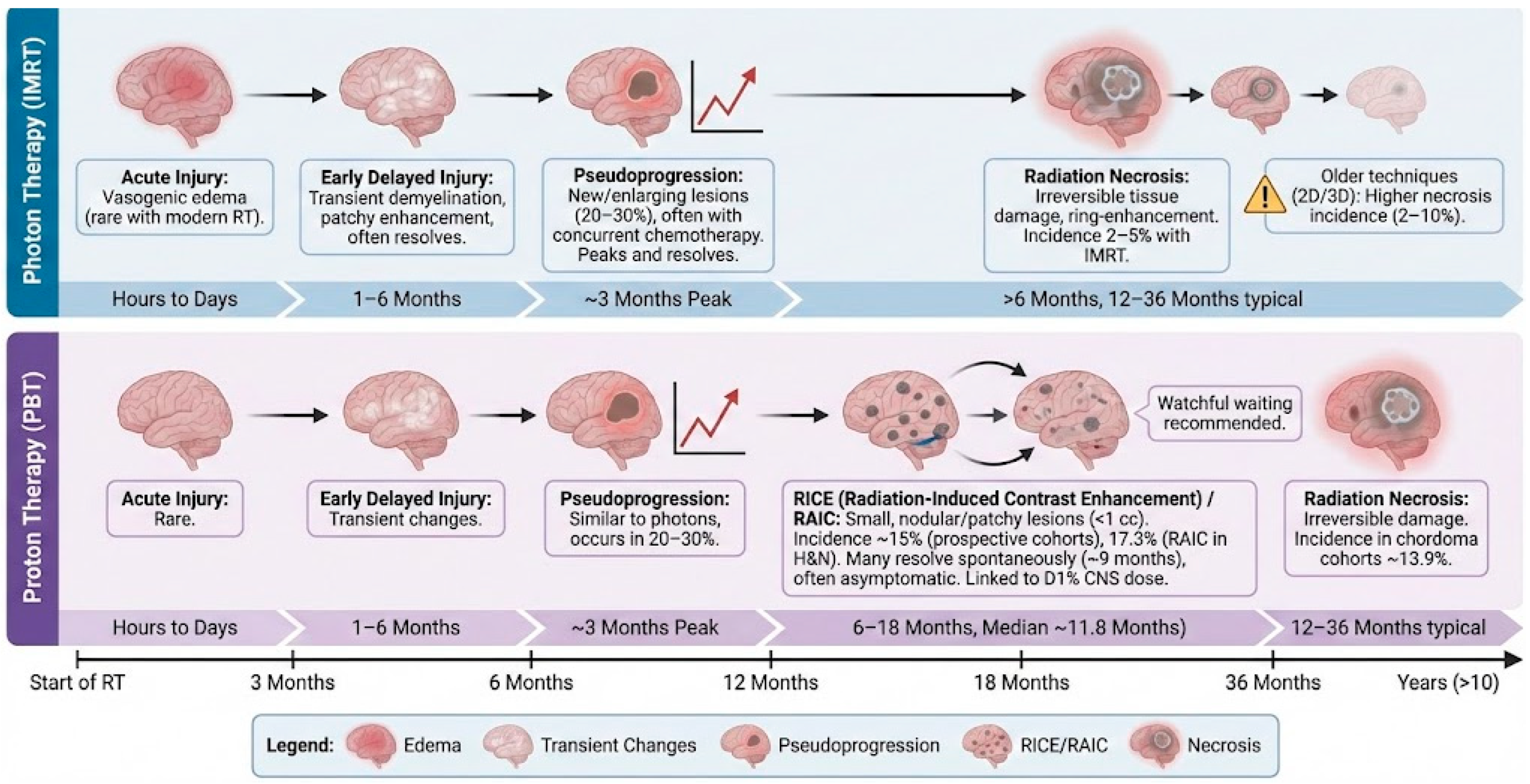
3.3. Categories of Injury
3.3.1. Acute Injury (Within Hours to Days)
3.3.2. Early Delayed Injury (1–6 Months)
3.3.3. Late Delayed Injury (>6 Months)
3.4. MRI Alterations in Skull Base Tumors
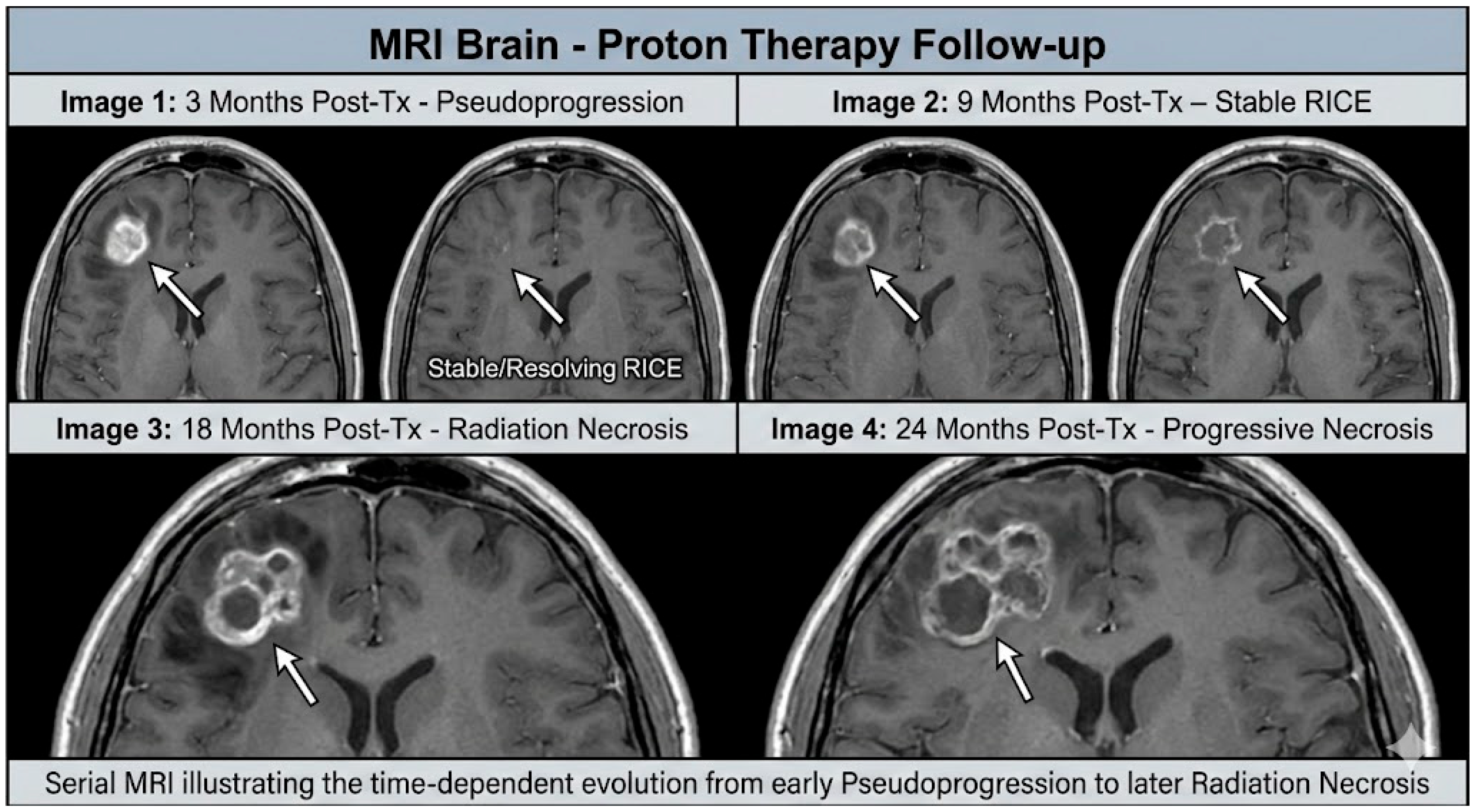
3.4.1. Pseudoprogression
3.4.2. Radiation-Associated Image Changes (RAIC)
3.4.3. Radiation-Induced Contrast Enhancement (RICE)
3.4.4. Radiation Necrosis
3.4.5. Contrast-Enhancing Brain Lesions (CEBL) and RIBI Spectrum
3.5. Tumor-Specific Considerations and Incidence
3.5.1. Glioblastoma vs. Skull Base Tumors: Differentiating Pseudoprogression
3.5.2. Chordoma and Chondrosarcoma
3.5.3. Head and Neck Cancers (Nasopharyngeal/Adenoid Cystic Carcinoma)
3.5.4. Meningioma
3.5.5. Pituitary Adenoma and Craniopharyngioma
3.5.6. Germinoma and Other Germ Cell Tumors
3.6. Dosimetric Predictors and Quantitative Constraints
3.6.1. Photon Versus Proton Therapy
3.6.2. Validated Dose Constraints
3.6.3. Clinical Risk Factors
3.7. Advanced Imaging Techniques
3.7.1. Diffusion-Weighted Imaging (DWI)
3.7.2. Perfusion-Weighted Imaging (PWI)
3.7.3. Magnetic Resonance Spectroscopy (MRS)
3.7.4. Radiomics and Artificial Intelligence
3.7.5. PET Imaging
3.7.6. Evolution of Lesions over Time
3.8. Clinical Management
3.9. Future Directions

| Term | Acronym | Definition | Typical Onset After RT | Clinical Context/Notes |
|---|---|---|---|---|
| Radiation-Induced Brain Injury | RIBI | Umbrella term encompassing the full spectrum of radiation-related effects on normal brain parenchyma, including reversible imaging changes and irreversible necrosis. | Variable (months to years) | General category; includes RICE/RAIC, pseudoprogression, and radiation necrosis. |
| Radiation-Induced Contrast Enhancement | RICE | Focal nodular or patchy contrast-enhancing lesions within irradiated brain tissue, often asymptomatic and potentially reversible. | 6–18 months (median ≈ 12 months) | Frequently reported in proton therapy series; often self-limited. |
| Radiation-Associated Image Changes | RAIC | Imaging-defined post-radiation abnormalities, often considered synonymous with RICE in head and neck literature; may include contrast enhancement and/or T2/FLAIR hyperintensity. | 6–24 months | Commonly described in skull base and head and neck proton therapy cohorts. |
| Pseudoprogression | PsP | Transient treatment-related increase in enhancement and/or edema mimicking tumor progression without true tumor growth. | <6 months (early) | Most commonly described in high-grade glioma after chemoradiotherapy; rare in benign skull base tumors. |
| Radiation Necrosis | RN | Irreversible radiation-induced tissue necrosis characterized by blood–brain barrier breakdown, mass effect, and potential neurological symptoms. | >12 months (may occur years later) | Late toxicity; may require corticosteroids, bevacizumab, or surgery. |
| Contrast-Enhancing Brain Lesion | CEBL | Purely descriptive radiologic term referring to an enhancing lesion of uncertain etiology (tumor vs radiation effect). | Not specific | Neutral imaging descriptor used prior to etiologic clarification. |
| Study | Cohort Characteristics | Tumor Types | Modality | Endpoint Definition | Incidence Rate | Key Dosimetric and Clinical Findings |
|---|---|---|---|---|---|---|
| Lütgendorf-Caucig et al. (2024) [18] | N = 421 (Prospective) | CNS and Skull Base (Mixed) | Proton (PBS) | RICE (New contrast enhancement) | 15% (Total) 4.5% (Symptomatic) | D1% > 57.6 Gy (RBE) to healthy CNS is a significant predictor (HR 3.73). Diabetes and prior RT are independent risk factors. |
| Engeseth et al. (2020) [20] | N = 127 (Retrospective) | Skull Base H&N (NPC, Sinonasal) | Proton (IMPT) | RAIC (Enhancement + T2 changes) | 17.3% (Crude) 14.3% (3-yr Actuarial) | V67Gy (RBE) ≥ 0.17 cc associated with 63% risk of RAIC. 100% of lesions were asymptomatic; 45.5% resolved spontaneously. |
| Mattke et al. (2022) [46] | N = 147 (Prospective) | Skull Base Chordoma | Proton vs. Carbon Ion | Necrosis and Reaction | 13.9% (Necrosis) 21.5% (Reaction) | Necrosis rates comparable between Protons and Carbon Ions. Risk correlates with Dmax and V70Gy volumes. |
| Brandes et al. (2008) [23] | N = 103 (Prospective) | Glioblastoma (GBM) | Photon + Temozolomide | Pseudoprogression | ~30% (specifically in methylated MGMT) | Incidence driven by concurrent chemotherapy and tumor biology; Distinct from standard skull base radiation effects. |
| Lesion Type | Tumor Population | Radiotherapy Modality | Cohort Size (n) | Incidence (%) | Key Dose–Volume Predictor(s) | Median Latency/Follow-Up | Reference |
|---|---|---|---|---|---|---|---|
| Pseudoprogression | Skull base tumors (mixed CNS/skull base prospective cohort) | Proton therapy | 104 | Early transient enhancement reported; lower than RICE rates (exact incidence variably reported) | Higher focal dose; concurrent systemic therapy | Within 3–6 months | Lütgendorf-Caucig et al., 2024 [18] |
| RICE | Head and neck skull base cancers | Proton therapy | 127 | 17.3% overall; 3-year actuarial rate 14.3% | V70 GyRBE ≥ 0.17 cc strongly predictive (~63% RAIC when exceeded) | Median 21 months | Engeseth et al., 2020 [20] |
| Symptomatic RICE | Mixed CNS and skull base tumors (prospective) | Proton therapy | 104 | 15% overall; 4.5% symptomatic | D1% CNS ≥ 57.6 GyRBE independently predictive | Median onset 11.8 months | Lütgendorf-Caucig et al., 2024 [18] |
| Temporal Lobe Reaction (subclinical + clinical) | Skull base chordoma and chondrosarcoma | Proton ± carbon ion therapy | 147 | 21.5% temporal lobe reaction | Higher Dmax; V60–70 GyRBE; larger irradiated volume | Median 20 months | Mattke et al., 2022 [46] |
| Radiation Necrosis (Proton-dominant cohort) | Skull base chordoma and chondrosarcoma | Proton ± carbon ion therapy | 147 | 13.9% necrosis | Higher Dmax; V70 GyRBE; older age | Median 20 months | Mattke et al., 2022 [46] |
| Radiation Necrosis (Carbon Ion Therapy) | Head and neck and skull base tumors | Carbon ion therapy | 95 | Up to 64% | Higher biological dose (RBE effect) | Variable | Miyawaki et al., 2009 [26] |
| Temporal Lobe Necrosis (Photon IMRT era) | Nasopharyngeal carcinoma involving skull base | Photon IMRT | 616 | 2–5% | Total dose >70 Gy; temporal lobe high-dose exposure | 2–5 years | Peng et al., 2012 [47]; Zhou et al., 2013 [48] |
| Skull Base Target/Subsite | Adjacent CNS Structures at Risk | Typical Distribution of RICE/RAIC or Necrosis | Common Imaging Pitfall | Planning/Modality Notes |
|---|---|---|---|---|
| Clivus/petroclival region (e.g., chordoma, chondrosarcoma) | Medial temporal lobes; brainstem; cranial nerves VI–XII | Inferomedial/anterior temporal lobe (often ipsilateral to highest dose); occasionally brainstem | Focal temporal lobe enhancement may mimic progression in adjacent surgical bed; consider time course and dose distribution | High prescriptions (≈70–74 Gy [RBE]) increase risk; avoid systematic distal edge placement in temporal lobe; robust optimization and verification imaging in PBT |
| Cavernous sinus/Meckel’s cave (e.g., meningioma, perineural ACC) | Anterior temporal lobe; frontal lobe; optic pathway; cranial nerves III–VI | Anterior temporal lobe and/or inferior frontal lobe adjacent to cavernous sinus high-dose region | Perineural tumor spread vs. treatment-related cranial nerve/meningeal enhancement; correlate with pre-RT extent and serial evolution | In PBT, consider LET hotspots at end-of-range near temporal pole; distribute distal fall-off across beams when feasible |
| Sellar/suprasellar region (pituitary adenoma, craniopharyngioma) | Optic nerves/chiasm; hypothalamus; frontal lobes | Optic apparatus enhancement; less commonly adjacent frontal lobe changes | Optic nerve or chiasmal enhancement can mimic recurrence or compressive change; temporal lobe injury is uncommon at conventional doses unless re-irradiation | Typically lower dose (≈45–54 Gy) reduces necrosis risk; in re-irradiation, prioritize optic pathway constraints and avoid focal hotspots |
| Sinonasal cavity/paranasal sinuses with skull base extension | Inferior frontal lobes; temporal poles; olfactory groove | Inferior frontal lobe and anterior temporal poles depending on target and beam arrangement | Post-operative/inflammatory sinonasal enhancement vs. residual tumor; cortical enhancement may represent RICE rather than intracranial extension | For anterior proton beams, variable sinus filling can shift range; robust optimization and adaptive assessment are important |
| Nasopharynx/skull base invasion (UCNT/nasopharyngeal carcinoma) | Temporal lobes (inferomedial); brainstem | Inferomedial temporal lobes (often bilateral in large fields) | Temporal lobe necrosis vs. tumor recurrence at skull base; PET and perfusion MRI may help when MRI is equivocal | Historically higher risk with photon techniques; IMRT and PBT reduce high-dose volumes but hotspots remain relevant |
| Jugular foramen/petrous temporal bone | Cerebellum; lower cranial nerves; brainstem | Cerebellar hemispheres or pontomedullary region depending on target | Enhancement along lower cranial nerves vs. post-treatment change; assess diffusion/perfusion and clinical correlation | Beam paths near posterior fossa warrant strict brainstem constraints; consider artifact from hardware/skull base air–bone interfaces |
4. Discussion
4.1. Clinical Implications
4.2. Limitations of Current Evidence
4.3. Future Research
5. Conclusions
Author Contributions
Funding
Institutional Review Board Statement
Informed Consent Statement
Data Availability Statement
Acknowledgments
Conflicts of Interest
Abbreviations
References
- Forst, D.A.; Jones, P.S. Skull Base Tumors. Continuum 2023, 29, 1752–1778. [Google Scholar] [CrossRef]
- Kockro, R.A.; Schwandt, E.; Ringel, F.; Eisenring, C.V.; Nowinski, W.L. Operative Anatomy of the Skull Base: 3D Exploration with a Highly Detailed Interactive Atlas. J. Neurol. Surg. B Skull Base 2022, 83, e298–e305. [Google Scholar] [CrossRef]
- Stacchiotti, S.; Sommer, J. Chordoma Global Consensus Group Building a Global Consensus Approach to Chordoma: A Position Paper from the Medical and Patient Community. Lancet Oncol. 2015, 16, e71–e83. [Google Scholar] [CrossRef] [PubMed]
- Teoh, M.; Clark, C.H.; Wood, K.; Whitaker, S.; Nisbet, A. Volumetric Modulated Arc Therapy: A Review of Current Literature and Clinical Use in Practice. Br. J. Radiol. 2011, 84, 967–996. [Google Scholar] [CrossRef]
- Holtzman, A.L.; Mohammadi, H.; Furutani, K.M.; Koffler, D.M.; McGee, L.A.; Lester, S.C.; Gamez, M.E.; Routman, D.M.; Beltran, C.J.; Liang, X. Impact of Relative Biologic Effectiveness for Proton Therapy for Head and Neck and Skull-Base Tumors: A Technical and Clinical Review. Cancers 2024, 16, 1947. [Google Scholar] [CrossRef]
- Nie, M.; Chen, L.; Zhang, J.; Qiu, X. Pure Proton Therapy for Skull Base Chordomas and Chondrosarcomas: A Systematic Review of Clinical Experience. Front. Oncol. 2022, 12, 1016857. [Google Scholar] [CrossRef] [PubMed]
- Wang, Y.; Zheng, J.; Liu, L.; Wang, Y.; Long, T.; Liu, M.; Gao, J.; Lu, H.-M. Dosimetric Comparison of Intensity-Modulated Proton Therapy and Photon-Based Radiotherapy for Skull Base Tumors. J. Appl. Clin. Med. Phys. 2025, 26, e70268. [Google Scholar] [CrossRef]
- Rampinelli, V.; Testa, G.; Arosio, A.D.; Piazza, C. Skull Base Osteoradionecrosis: From Pathogenesis to Treatment. Curr. Opin. Otolaryngol. Head Neck Surg. 2025, 33, 65–73. [Google Scholar] [CrossRef] [PubMed]
- Lauwens, L.; Ribeiro, M.F.; Zegers, C.M.L.; Høyer, M.; Alapetite, C.; Blomstrand, M.; Calugaru, V.; Di Perri, D.; Iannalfi, A.; Lütgendorf-Caucig, C.; et al. Systematic Review of MRI Alterations in the Brain Following Proton and Photon Radiation Therapy: Towards a Uniform European Particle Therapy Network (EPTN) Definition. Radiother. Oncol. 2025, 208, 110936. [Google Scholar] [CrossRef]
- Katsura, M.; Sato, J.; Akahane, M.; Furuta, T.; Mori, H.; Abe, O. Recognizing Radiation-Induced Changes in the Central Nervous System: Where to Look and What to Look For. Radiographics 2021, 41, 224–248. [Google Scholar] [CrossRef]
- Lo Greco, M.C.; Milazzotto, R.; Liardo, R.L.E.; Acquaviva, G.; La Rocca, M.; Altieri, R.; Certo, F.; Barbagallo, G.M.; Basile, A.; Foti, P.V.; et al. Relapsing High-Grade Glioma from Peritumoral Zone: Critical Review of Radiotherapy Treatment Options. Brain Sci. 2022, 12, 416. [Google Scholar] [CrossRef]
- Kim, J.; Park, Y.-K.; Sharp, G.; Busse, P.; Winey, B. Water Equivalent Path Length Calculations Using Scatter-Corrected Head and Neck CBCT Images to Evaluate Patients for Adaptive Proton Therapy. Phys. Med. Biol. 2017, 62, 59–72. [Google Scholar] [CrossRef] [PubMed]
- Sørensen, B.S.; Pawelke, J.; Bauer, J.; Burnet, N.G.; Dasu, A.; Høyer, M.; Karger, C.P.; Krause, M.; Schwarz, M.; Underwood, T.S.A.; et al. Does the Uncertainty in Relative Biological Effectiveness Affect Patient Treatment in Proton Therapy? Radiother. Oncol. 2021, 163, 177–184. [Google Scholar] [CrossRef]
- Leeman, J.E.; Romesser, P.B.; Zhou, Y.; McBride, S.; Riaz, N.; Sherman, E.; Cohen, M.A.; Cahlon, O.; Lee, N. Proton Therapy for Head and Neck Cancer: Expanding the Therapeutic Window. Lancet Oncol. 2017, 18, e254–e265. [Google Scholar] [CrossRef]
- van de Water, S.; Albertini, F.; Weber, D.C.; Heijmen, B.J.M.; Hoogeman, M.S.; Lomax, A.J. Anatomical Robust Optimization to Account for Nasal Cavity Filling Variation during Intensity-Modulated Proton Therapy: A Comparison with Conventional and Adaptive Planning Strategies. Phys. Med. Biol. 2018, 63, 025020. [Google Scholar] [CrossRef]
- Kiselev, V.A.; Chernyaev, A.P.; Belikhin, M.A.; Zamaltdinov, R.R.; Vladimirkin, S.A.; Gritsenko, S.E.; Nechesnyuk, A.V. Benefits of Robust Optimization in Comparison with PTV-Based Planning Approach to Overcome Beam Positioning Uncertainties in Spot-Scanning Proton Therapy. J. Appl. Clin. Med. Phys. 2025, 26, e70355. [Google Scholar] [CrossRef] [PubMed]
- Liu, W.; Zhang, X.; Li, Y.; Mohan, R. Robust Optimization of Intensity Modulated Proton Therapy. Med. Phys. 2012, 39, 1079–1091. [Google Scholar] [CrossRef] [PubMed]
- Lütgendorf-Caucig, C.; Pelak, M.; Hug, E.; Flechl, B.; Surböck, B.; Marosi, C.; Mock, U.; Zach, L.; Mardor, Y.; Furman, O.; et al. Prospective Analysis of Radiation-Induced Contrast Enhancement and Health-Related Quality of Life After Proton Therapy for Central Nervous System and Skull Base Tumors. Int. J. Radiat. Oncol. Biol. Phys. 2024, 118, 1206–1216. [Google Scholar] [CrossRef]
- Gal, O.; Yu, J.; Mehta, M.P.; Hall, M.D.; Press, R.H.; Odia, Y.; McDermott, M.W.; Fellows, Z.; Wroe, A.; Gutierrez, A.N.; et al. Contrast-Enhancing Lesions Induced by Central Nervous System-Directed Intensity Modulated Proton Therapy: Distribution Patterns, Kinetics, Risk Factors, and Outcomes. Int. J. Radiat. Oncol. Biol. Phys. 2025, 122, 621–630. [Google Scholar] [CrossRef]
- Engeseth, G.M.; Stieb, S.; Mohamed, A.S.R.; He, R.; Stokkevåg, C.H.; Brydøy, M.; Fuller, C.D.; Garden, A.S.; Rosenthal, D.I.; Phan, J.; et al. Outcomes and Patterns of Radiation Associated Brain Image Changes after Proton Therapy for Head and Neck Skull Base Cancers. Radiother. Oncol. 2020, 151, 119–125. [Google Scholar] [CrossRef]
- Balentova, S.; Adamkov, M. Molecular, Cellular and Functional Effects of Radiation-Induced Brain Injury: A Review. Int. J. Mol. Sci. 2015, 16, 27796–27815. [Google Scholar] [CrossRef]
- Barisano, G.; Bergamaschi, S.; Acharya, J.; Rajamohan, A.; Gibbs, W.; Kim, P.; Zada, G.; Chang, E.; Law, M. Complications of Radiotherapy and Radiosurgery in the Brain and Spine. Neurographics 2018, 8, 167–187. [Google Scholar] [CrossRef] [PubMed]
- Brandes, A.A.; Franceschi, E.; Tosoni, A.; Blatt, V.; Pession, A.; Tallini, G.; Bertorelle, R.; Bartolini, S.; Calbucci, F.; Andreoli, A.; et al. MGMT Promoter Methylation Status Can Predict the Incidence and Outcome of Pseudoprogression after Concomitant Radiochemotherapy in Newly Diagnosed Glioblastoma Patients. J. Clin. Oncol. 2008, 26, 2192–2197. [Google Scholar] [CrossRef] [PubMed]
- Smith, E.J.; Naik, A.; Shaffer, A.; Goel, M.; Krist, D.T.; Liang, E.; Furey, C.G.; Miller, W.K.; Lawton, M.T.; Barnett, D.H.; et al. Differentiating Radiation Necrosis from Tumor Recurrence: A Systematic Review and Diagnostic Meta-Analysis Comparing Imaging Modalities. J. Neurooncol. 2023, 162, 15–23. [Google Scholar] [CrossRef]
- Mattke, M.; Ohlinger, M.; Bougatf, N.; Wolf, R.; Welzel, T.; Roeder, F.; Gerum, S.; Fussl, C.; Annon-Eberharter, N.; Ellerbrock, M.; et al. Patterns of Temporal Lobe Reaction and Radiation Necrosis after Particle Radiotherapy in Patients with Skull Base Chordoma and Chondrosarcoma—A Single-Center Experience. Cancers 2024, 16, 718. [Google Scholar] [CrossRef] [PubMed]
- Miyawaki, D.; Murakami, M.; Demizu, Y.; Sasaki, R.; Niwa, Y.; Terashima, K.; Nishimura, H.; Hishikawa, Y.; Sugimura, K. Brain Injury after Proton Therapy or Carbon Ion Therapy for Head-and-Neck Cancer and Skull Base Tumors. Int. J. Radiat. Oncol. Biol. Phys. 2009, 75, 378–384. [Google Scholar] [CrossRef]
- Santoni, R.; Liebsch, N.; Finkelstein, D.M.; Hug, E.; Hanssens, P.; Goitein, M.; Smith, A.R.; O’Farrell, D.; Efird, J.T.; Fullerton, B.; et al. Temporal Lobe (TL) Damage Following Surgery and High-Dose Photon and Proton Irradiation in 96 Patients Affected by Chordomas and Chondrosarcomas of the Base of the Skull. Int. J. Radiat. Oncol. Biol. Phys. 1998, 41, 59–68. [Google Scholar] [CrossRef]
- Nakamura, M.; Mizumoto, M.; Saito, T.; Shimizu, S.; Li, Y.; Oshiro, Y.; Inaba, M.; Hosaka, S.; Fukushima, H.; Suzuki, R.; et al. A Systematic Review and Meta-Analysis of Radiotherapy and Particle Beam Therapy for Skull Base Chondrosarcoma: TRP-Chondrosarcoma 2024. Front. Oncol. 2024, 14, 1380716. [Google Scholar] [CrossRef]
- Kong, D.-S.; Kim, S.T.; Kim, E.-H.; Lim, D.H.; Kim, W.S.; Suh, Y.-L.; Lee, J.-I.; Park, K.; Kim, J.H.; Nam, D.-H. Diagnostic Dilemma of Pseudoprogression in the Treatment of Newly Diagnosed Glioblastomas: The Role of Assessing Relative Cerebral Blood Flow Volume and Oxygen-6-Methylguanine-DNA Methyltransferase Promoter Methylation Status. AJNR Am. J. Neuroradiol. 2011, 32, 382–387. [Google Scholar] [CrossRef]
- McIntyre, M.; Wilson, P.; Gorayski, P.; Bezak, E. A Systematic Review of LET-Guided Treatment Plan Optimisation in Proton Therapy: Identifying the Current State and Future Needs. Cancers 2023, 15, 4268. [Google Scholar] [CrossRef]
- Vestergaard, A.; Kallehauge, J.F.; Muhic, A.; Carlsen, J.F.; Dahlrot, R.H.; Lukacova, S.; Haslund, C.A.; Lassen-Ramshad, Y.; Worawongsakul, R.; Høyer, M. Mixed Effect Model Confirms Increased Risk of Image Changes with Increasing Linear Energy Transfer in Proton Therapy of Gliomas. Radiother. Oncol. 2025, 204, 110716. [Google Scholar] [CrossRef]
- Tas, K.T.; Sheirieh, A.; Lishewski, P.; Sheikhzadeh, F.F.; Smalc, E.; Fragkandrea-Nixon, I.; Elsayad, K.; Zink, K.; Vorwerk, H.; Adeberg, S.; et al. Evaluating of Radiation-Induced Cerebral Contrast Enhancements in Brain Metastases: Analysis of Incidence and Risk Predictors. Clin. Exp. Metastasis 2025, 42, 54. [Google Scholar] [CrossRef] [PubMed]
- Niemierko, A.; Schuemann, J.; Niyazi, M.; Giantsoudi, D.; Maquilan, G.; Shih, H.A.; Paganetti, H. Brain Necrosis in Adult Patients After Proton Therapy: Is There Evidence for Dependency on Linear Energy Transfer? Int. J. Radiat. Oncol. Biol. Phys. 2021, 109, 109–119. [Google Scholar] [CrossRef]
- Bryjova, K.; Mackeprang, P.-H.; Leiser, D.; Weber, D.C. Quality of Life and Clinical Outcomes in Patients with Skull Base Chordoma and Chondrosarcoma Treated with Pencil-Beam Scanning Proton Therapy. Cancers 2025, 17, 3651. [Google Scholar] [CrossRef]
- Ruben, J.D.; Dally, M.; Bailey, M.; Smith, R.; McLean, C.A.; Fedele, P. Cerebral Radiation Necrosis: Incidence, Outcomes, and Risk Factors with Emphasis on Radiation Parameters and Chemotherapy. Int. J. Radiat. Oncol. Biol. Phys. 2006, 65, 499–508. [Google Scholar] [CrossRef] [PubMed]
- Viselner, G.; Farina, L.; Lucev, F.; Turpini, E.; Lungarotti, L.; Bacila, A.; Iannalfi, A.; D’Ippolito, E.; Vischioni, B.; Ronchi, S.; et al. Brain MR Findings in Patients Treated with Particle Therapy for Skull Base Tumors. Insights Imaging 2019, 10, 94. [Google Scholar] [CrossRef] [PubMed]
- Asao, C.; Korogi, Y.; Kitajima, M.; Hirai, T.; Baba, Y.; Makino, K.; Kochi, M.; Morishita, S.; Yamashita, Y. Diffusion-Weighted Imaging of Radiation-Induced Brain Injury for Differentiation from Tumor Recurrence. AJNR Am. J. Neuroradiol. 2005, 26, 1455–1460. [Google Scholar]
- Wang, Y.-L.; Chen, S.; Xiao, H.-F.; Li, Y.; Wang, Y.; Liu, G.; Lou, X.; Ma, L. Differentiation between Radiation-Induced Brain Injury and Glioma Recurrence Using 3D pCASL and Dynamic Susceptibility Contrast-Enhanced Perfusion-Weighted Imaging. Radiother. Oncol. 2018, 129, 68–74. [Google Scholar] [CrossRef]
- Weybright, P.; Sundgren, P.C.; Maly, P.; Hassan, D.G.; Nan, B.; Rohrer, S.; Junck, L. Differentiation between Brain Tumor Recurrence and Radiation Injury Using MR Spectroscopy. AJR Am. J. Roentgenol. 2005, 185, 1471–1476. [Google Scholar] [CrossRef]
- Zheng, Z.; Wang, B.; Zhao, Q.; Zhang, Y.; Wei, J.; Meng, L.; Xin, Y.; Jiang, X. Research Progress on Mechanism and Imaging of Temporal Lobe Injury Induced by Radiotherapy for Head and Neck Cancer. Eur. Radiol. 2022, 32, 319–330. [Google Scholar] [CrossRef]
- Pyka, T.; Hiob, D.; Preibisch, C.; Gempt, J.; Wiestler, B.; Schlegel, J.; Straube, C.; Zimmer, C. Diagnosis of Glioma Recurrence Using Multiparametric Dynamic 18F-Fluoroethyl-Tyrosine PET-MRI. Eur. J. Radiol. 2018, 103, 32–37. [Google Scholar] [CrossRef]
- Galldiks, N.; Tonn, J.C.; Preusser, M.; Albert, N.L. Update to the RANO Working Group and EANO Recommendations for the Clinical Use of PET Imaging in Gliomas. Neuro Oncol. 2025, 27, 2492–2493. [Google Scholar] [CrossRef]
- Ghaith, A.K.; Nguyen, R.; El-Hajj, V.G.; Montaser, A.; De Biase, G.; Ravindran, K.; Perez-Vega, C.; Lee, S.J.; Dominari, A.; Battistin, U.; et al. Proton versus Photon Adjuvant Radiotherapy: A Multicenter Comparative Evaluation of Recurrence Following Spinal Chordoma Resection. Neurosurg. Focus 2024, 56, E9. [Google Scholar] [CrossRef]
- Kirste, S.; Treier, M.; Wehrle, S.J.; Becker, G.; Abdel-Tawab, M.; Gerbeth, K.; Hug, M.J.; Lubrich, B.; Grosu, A.-L.; Momm, F. Boswellia Serrata Acts on Cerebral Edema in Patients Irradiated for Brain Tumors: A Prospective, Randomized, Placebo-Controlled, Double-Blind Pilot Trial. Cancer 2011, 117, 3788–3795. [Google Scholar] [CrossRef]
- Cai, X.; Li, P.; Zhao, J.; Wang, W.; Cheng, J.; Zhang, G.; Cai, S.; Zhang, Z.; Jiang, G.; Zhang, Q.; et al. Definitive Carbon Ion Re-Irradiation with Pencil Beam Scanning in the Treatment of Unresectable Locally Recurrent Rectal Cancer. J. Radiat. Res. 2023, 64, 933–939. [Google Scholar] [CrossRef]
- Mattke, M.; Ohlinger, M.; Bougatf, N.; Harrabi, S.; Wolf, R.J.; Seidensaal, K.; Welzel, T.; Röder, F.; Gerum, S.; Ellerbrock, M.; et al. Proton and Carbon Ion Beam Treatment with Active Raster Scanning Method in 147 Patients with Skull Base Chordoma at the Heidelberg Ion Beam Therapy Center—A Single-Center Experience. Strahlenther. Und Onkol. 2022, 199, 160–168. [Google Scholar] [CrossRef] [PubMed]
- Peng, G.; Wang, T.; Yang, K.-Y.; Zhang, S.; Zhang, T.; Li, Q.; Han, J.; Wu, G. A Prospective, Randomized Study Comparing Outcomes and Toxicities of Intensity-Modulated Radiotherapy vs. Conventional Two-Dimensional Radiotherapy for the Treatment of Nasopharyngeal Carcinoma. Radiother. Oncol. 2012, 104, 286–293. [Google Scholar] [CrossRef] [PubMed]
- Zhou, G.-Q.; Yu, X.-L.; Chen, M.; Guo, R.; Lei, Y.; Sun, Y.; Mao, Y.-P.; Liu, L.-Z.; Li, L.; Lin, A.-H.; et al. Radiation-Induced Temporal Lobe Injury for Nasopharyngeal Carcinoma: A Comparison of Intensity-Modulated Radiotherapy and Conventional Two-Dimensional Radiotherapy. PLoS ONE 2013, 8, e67488. [Google Scholar] [CrossRef] [PubMed]
- Tsao, M.N.; Li, Y.Q.; Lu, G.; Xu, Y.; Wong, C.S. Upregulation of Vascular Endothelial Growth Factor Is Associated with Radiation-Induced Blood-Spinal Cord Barrier Breakdown. J. Neuropathol. Exp. Neurol. 1999, 58, 1051–1060. [Google Scholar] [CrossRef]
- Brandsma, D.; Stalpers, L.; Taal, W.; Sminia, P.; van den Bent, M.J. Clinical Features, Mechanisms, and Management of Pseudoprogression in Malignant Gliomas. Lancet Oncol. 2008, 9, 453–461. [Google Scholar] [CrossRef]
- Quigg, M.; Yen, C.-P.; Chatman, M.; Quigg, A.H.; McNeill, I.T.; Przybylowski, C.J.; Yan, G.; Sheehan, J.P. Risks of History of Diabetes Mellitus, Hypertension, and Other Factors Related to Radiation-Induced Changes Following Gamma Knife Surgery for Cerebral Arteriovenous Malformations. J. Neurosurg. 2012, 117, 144–149. [Google Scholar] [CrossRef] [PubMed]
- Eichkorn, T.; Lischalk, J.W.; Sandrini, E.; Meixner, E.; Regnery, S.; Held, T.; Bauer, J.; Bahn, E.; Harrabi, S.; Hörner-Rieber, J.; et al. Iatrogenic Influence on Prognosis of Radiation-Induced Contrast Enhancements in Patients with Glioma WHO 1-3 Following Photon and Proton Radiotherapy. Radiother. Oncol. 2022, 175, 133–143. [Google Scholar] [CrossRef]
- Hou, J.; He, Y.; Li, H.; Lu, Q.; Lin, H.; Zeng, B.; Xie, C.; Yu, X. MRI-Based Radiomics Models Predict Cystic Brain Radionecrosis of Nasopharyngeal Carcinoma after Intensity Modulated Radiotherapy. Front. Neurol. 2024, 15, 1344324. [Google Scholar] [CrossRef]
- Zhang, Z.; Yang, J.; Ho, A.; Jiang, W.; Logan, J.; Wang, X.; Brown, P.D.; McGovern, S.L.; Guha-Thakurta, N.; Ferguson, S.D.; et al. A Predictive Model for Distinguishing Radiation Necrosis from Tumour Progression after Gamma Knife Radiosurgery Based on Radiomic Features from MR Images. Eur. Radiol. 2018, 28, 2255–2263. [Google Scholar] [CrossRef] [PubMed]
- Lo Greco, M.C.; Milazzotto, R.; Liardo, R.L.E.; Foti, P.V.; Palmucci, S.; Basile, A.; Pergolizzi, S.; Spatola, C. The Role of Reirradiation in Childhood Progressive Diffuse Intrinsic Pontine Glioma (DIPG): An Ongoing Challenge beyond Radiobiology. Brain Sci. 2023, 13, 1449. [Google Scholar] [CrossRef] [PubMed]


Disclaimer/Publisher’s Note: The statements, opinions and data contained in all publications are solely those of the individual author(s) and contributor(s) and not of MDPI and/or the editor(s). MDPI and/or the editor(s) disclaim responsibility for any injury to people or property resulting from any ideas, methods, instructions or products referred to in the content. |
© 2026 by the authors. Licensee MDPI, Basel, Switzerland. This article is an open access article distributed under the terms and conditions of the Creative Commons Attribution (CC BY) license.
Share and Cite
Loganadane, G.; Calugaru, V.; Anzellini, D.; Nicaise, B.; Mezghani, S.; Nguyen, N.P.; Page, B.R. MRI and PET Alterations in Adult Skull Base Tumors: A Narrative Review of Proton Versus Photon Radiotherapy. Diagnostics 2026, 16, 1166. https://doi.org/10.3390/diagnostics16081166
Loganadane G, Calugaru V, Anzellini D, Nicaise B, Mezghani S, Nguyen NP, Page BR. MRI and PET Alterations in Adult Skull Base Tumors: A Narrative Review of Proton Versus Photon Radiotherapy. Diagnostics. 2026; 16(8):1166. https://doi.org/10.3390/diagnostics16081166
Chicago/Turabian StyleLoganadane, Gokoulakrichenane, Valentin Calugaru, Dimitri Anzellini, Benjamin Nicaise, Sarah Mezghani, Nam P. Nguyen, and Brandi R. Page. 2026. "MRI and PET Alterations in Adult Skull Base Tumors: A Narrative Review of Proton Versus Photon Radiotherapy" Diagnostics 16, no. 8: 1166. https://doi.org/10.3390/diagnostics16081166
APA StyleLoganadane, G., Calugaru, V., Anzellini, D., Nicaise, B., Mezghani, S., Nguyen, N. P., & Page, B. R. (2026). MRI and PET Alterations in Adult Skull Base Tumors: A Narrative Review of Proton Versus Photon Radiotherapy. Diagnostics, 16(8), 1166. https://doi.org/10.3390/diagnostics16081166