Article

Investigation of the Prevalence of Associated Genetic Mutations (Co-Mutations) in Patients with Actionable Driver Mutations in Lung Cancer: A Retrospective Study


1
The Ruth and Bruce Rappaport Faculty of Medicine, Technion-Israel Institute of Technology, 1 Efron Street, Haifa 3109601, Israel
2
Department of Oncology, Bnai-Zion Medical Center, 47 Eliyahu Golomb Avenue, Haifa 3339419, Israel
3
The Legacy Heritage Cancer Center, Dr. Larry Norton Institute, Soroka Medical Center, 151 Yitzhack Rager Avenue, Beer Sheva 8457108, Israel
4
Faculty of Health Sciences, Ben Gurion University of the Negev, 1 Ben Gurion Avenue, Beer Sheva 8410501, Israel
5
Pathology Institute, Lady Davis Carmel Medical Center, 7 Michal Street, Haifa 3436212, Israel
6
Institute of Endocrinology, Bnai-Zion Medical Center, 47 Eliyahu Golomb Avenue, Haifa 3339419, Israel
7
The Joseph Fishman Oncology Center, Rambam Medical Center, 6 Efron Street, Haifa 3525409, Israel
8
Department of Cardiothoracic Surgery, Lady Davis Carmel Medical Center, 7 Michal Street, Haifa 3436212, Israel
*
Author to whom correspondence should be addressed.
These authors contributed equally to this work.
Diagnostics 2026, 16(7), 1106; https://doi.org/10.3390/diagnostics16071106

Submission received: 6 February 2026 / Revised: 18 March 2026 / Accepted: 1 April 2026 / Published: 7 April 2026
(This article belongs to the Special Issue Advancements and Innovations in the Diagnosis of Lung Cancer)

Abstract

Background/Objectives: Lung cancer remains the leading cause of cancer-related mortality globally. Approximately 45% of these tumors harbor oncogenic mutations that drive carcinogenesis and are amenable to targeted therapies. Other predictive biomarkers—e.g., PD-L1, TMB, and MSI—play a crucial role in patients’ management. This study aims to investigate the existence of mutation clusters (co-mutations) and evaluate the correlation of these clusters with various clinical and laboratory parameters. Methods: A retrospective study was conducted utilizing pathological samples from lung cancer patients harboring mutations in EGFR, KRAS, ALK, BRAF, MET, HER2, ROS1, NTRK, and NRG1. Data were collected from the Institute of Pathology at Carmel Medical Center between the years 2022 and 2024. Patients were stratified using a Two-Step Cluster Analysis algorithm based on actionable mutations and co-mutations. Heatmaps and dendrograms were generated to assess the correlation between these genomic clusters, clinical metrics, and predictive biomarkers. Results: The study cohort included 129 patients with actionable mutations. Five distinct clusters were identified: Clusters 1, 2, and 3 exhibited a high expression of STK11 and TP53 co-mutations alongside KRAS drivers (n = 38, n = 12, and n = 23, respectively). Clusters 4 and 5 demonstrated high expression of ALK alterations and tumor suppressor gene mutations (n = 31 and n = 25, respectively). Cluster comparisons demonstrated statistically significant differences between clusters regarding age, gender, PD-L1 expression, and tumor mutational burden. No significant associations were found regarding ethnicity or microsatellite instability status. Conclusions: By constructing clusters based on the aggregate of genomic alterations in patients with actionable mutations, it is possible to predict associations with distinct demographic and clinical characteristics. Future research should apply this analytical approach to larger cohorts to further characterize these subgroups and investigate potential correlations with therapeutic efficacy.

1. Introduction

Lung cancer is a primary cause of cancer mortality worldwide, resulting in high death rates among both men and women [1,2,3]. Despite increasing awareness regarding early diagnosis, lung cancer is frequently detected at advanced stages where therapeutic options are limited, resulting in a poor prognosis [1]. Consequently, frequent screening of high-risk individuals is essential to facilitate early detection and significantly improve survival rates. The accurate and early diagnosis of pulmonary lesions is critical to tailoring the most appropriate therapeutic regimen and improving patient outcomes [1].
Lung cancer is defined as the uncontrolled growth of abnormal cells originating in the lungs, leading to tumor formation and compromised pulmonary function. It originates in the epithelium of the large and small bronchi and may metastasize via the lymphatic system or hematogenous spread, with common metastatic sites including the liver, adrenal glands, bone, and the central nervous system [1,4,5].
The incidence of lung cancer is steadily rising. In 2020, it was the second most diagnosed cancer (following breast cancer), accounting for 11.4% of 19.3 million cases. By 2022, it became the most diagnosed malignancy, with nearly 2.5 million new cases, representing 12.4% of all global cancer cases (one in eight new cancers). Regarding mortality, lung cancer is the leading cause of cancer death, rising from 18% in 2020 to 18.7% in 2022 [2,3]. The primary risk factor is tobacco smoking, though non-smokers exposed to second-hand smoke are also at risk. Additional risk factors include occupational exposure to carcinogens (e.g., asbestos, radon), air pollution, chronic lung diseases (e.g., COPD), and hereditary cancer syndromes [1].
Lung malignancies are classified into two main histological groups: Non-Small Cell Lung Carcinoma (NSCLC) and Small Cell Lung Carcinoma (SCLC). NSCLC accounts for 80% of cases, while SCLC comprises nearly 20%. This study focuses on NSCLC, which is further subdivided into histological subtypes, primarily Squamous Cell Carcinoma (SCC) and Adenocarcinoma (ADC), each accounting for 30–40% of cases. Large Cell Carcinoma (LCC) is a less common subtype, representing just under 10% of cases [5].
Current diagnostic and therapeutic strategies for lung cancer rely on histological diagnosis and molecular profiling [6]. Advanced next-generation sequencing (NGS) allows for the high-quality detection of a broad spectrum of mutations [7]. A significant proportion of NSCLC tumors harbor actionable mutations—genetic alterations treatable with targeted biological therapies rather than cytotoxic chemotherapy. The most prevalent actionable mutations discussed herein include Epidermal Growth Factor Receptor (EGFR), Kirsten rat sarcoma virus (KRAS) G12C, and ALK [8,9,10,11].
The KRAS G12C mutation is the most common alteration in the KRAS gene, which encodes a key protein in cell proliferation pathways. KRAS mutations are present in 15–20% of NSCLC cases. This specific mutation is targetable by two biological agents, sotorasib and adagrasib, which selectively bind covalently to the KRAS protein, inhibiting its activity [12,13,14].
EGFR (Epidermal Growth Factor Receptor) mutations occur in approximately 15% of NSCLC cases in Western Europe and the USA and are frequently associated with non-smokers. EGFR encodes a transmembrane tyrosine kinase receptor involved in cell proliferation cascades. These mutations are treated with Tyrosine Kinase Inhibitors (TKIs) such as Osimertinib [8,10,11,14,15].
ALK (Anaplastic Lymphoma Kinase) gene rearrangements result from translocation, typically with EML4. Found in 3–7% of NSCLC cases, this translocation leads to the overexpression of the ALK tyrosine kinase receptor, driving cell growth. This alteration is managed with TKIs such as crizotinib, ceritinib, lorlatinib, and alectinib [16]. Additional, less common actionable mutations include BRAF, MET, HER2, RET, ROS1, NTRK, and NRG1 [11,17].
Beyond molecular profiling, immunotherapeutic biomarkers such as PD-L1 expression, tumor mutational burden (TMB), and microsatellite instability (MSI) aid in treatment selection for NSCLC [18,19,20]. PD-L1 (Programmed Death-Ligand 1) expression on tumor cells suppresses the immune response by interacting with PD-1 on T-cells, inhibiting effector T-cells and promoting regulatory T-cells. Immune Checkpoint Inhibitors (ICIs) block this pathway to restore anti-tumor immunity [18]. TMB quantifies mutations within tumor DNA; high TMB correlates with better response to immunotherapy [19]. MSI refers to hypermutable DNA microsatellites due to defective mismatch repair (MMR) mechanisms and serves as a predictive marker for immunotherapy response [20].
This study utilized statistical analysis to determine whether specific clusters of co-mutations accompany actionable driver mutations. We specifically examined significant co-mutations including TP53, KRAS, STK11, KEAP1, and CDKN2A, alongside others of unclear significance. The existence of such clusters may influence prognosis and the efficacy of targeted therapies. We hypothesized that such clusters exist and aimed to identify them to optimize future therapeutic strategies. Additionally, we assessed the correlation of these clusters with biomarkers (PD-L1, TMB, MSI) and demographic parameters (age, gender, ethnicity) to identify patterns that may inform future studies on clinical efficacy and toxicity.

2. Materials and Methods

2.1. Study Design and Population

This retrospective, non-interventional study included 129 patient samples archived at the Institute of Pathology database at Carmel Medical Center, Haifa. The cohort consisted of patients diagnosed with NSCLC who possessed a complete molecular profile containing at least one of the following actionable mutations: EGFR, KRAS, ALK, BRAF, MET, HER2, ROS1, NTRK, or NRG1. Lung tumor biopsy samples collected between 2022 and 2024 were included. Patients were screened based on the presence of specific actionable mutations defined in the inclusion criteria.

2.2. Data Collection

Following the initial screening of 129 patients, relevant data were extracted using pathological reports. Subsequently, statistical analysis and result interpretation were performed. All samples were sourced from the existing repository at the Institute of Pathology, Carmel Medical Center. Following data collection and screening, statistical analysis was performed on an anonymized dataset (identified only by serial numbers).

2.3. Molecular Profiling

All molecular profiling was performed on formalin-fixed paraffin-embedded tumor specimens as part of routine clinical testing at Carmel Medical Center Pathology Department laboratories. Next-generation sequencing was performed using the Oncomine™ Comprehensive Assay v3 (Thermo Fisher Scientific, Waltham, MA, USA), which covers 161 genes encompassing major cancer drivers and tumor suppressors. The Oncomine Dx Target Test detects 46 cancer driver gene variants, including EGFR mutations (including L858R, T790M, and exon 19 deletions); BRAF, KRAS, ERBB2, and MET exon 14 skipping mutations; and ALK, ROS1, RET, and NTRK1/2/3 fusions. The assay is validated to detect single-nucleotide variants and small insertions/deletions, and to detect gene fusions and copy number alterations.
Tumor mutational burden (TMB) was defined as the number of somatic mutations per megabase (mut/Mb) within the panel’s reportable coding territory.
Microsatellite instability (MSI) was assessed using MSI status, reported as MSI-high versus microsatellite-stable (MSS).

2.4. Ethics

The study was approved by the Helsinki Committee of Carmel Medical Center protocol CMC-0073-24 on 23 September 2024.

2.5. Statistical Analysis

Cluster analysis was performed based on actionable mutations, co-mutations, and copy number variations (CNVs) using a Two-Step Cluster Analysis algorithm. This hybrid method is designed for categorical and/or continuous data and operates in two phases.

2.5.1. Pre-Clustering

The algorithm scans data to create small sub-clusters using a log-likelihood distance measure suitable for categorical (binary) variables. Since our variables are binary (presence or absence of a mutation), a log-likelihood distance metric estimates similarity based on the likelihood that data points will appear in the same cluster.

2.5.2. Hierarchical Clustering

Sub-clusters are merged into final clusters based on model fit criteria (BIC or AIC). The algorithm determines the optimal number of clusters automatically, though manual specification is possible.
Five clusters were generated for all patients. Heatmaps were created for visualization using ArrayGen (Undri, Pune, Maharashtra, India). Heatmaps visually represent the relationships between mutation types across patients. The color gradient ranges from light yellow (0; absence/low presence of a mutation) to dark brown (+2; high intensity/prevalence of the mutation). It is important to note that in binary data clustering, color intensity reflects the prevalence or significance of a mutation within the context of the cluster. Heatmaps depict binary alteration status per patient (0 = absent; 1 = present). Color intensity is used solely as a visual encoding of binary status and does not represent continuous measurements. If aggregated heatmaps are displayed (by cluster), the color scale reflects the proportion of cases within the cluster harboring the alteration (0–100%).

2.5.3. Dendrograms

A dendrogram was constructed for each heatmap to visualize the hierarchical structure. It illustrates the similarity between patient mutation profiles or the co-occurrence of specific mutations. Branch height indicates the degree of dissimilarity; shorter distances imply high similarity.

2.5.4. Comparative Statistics

Clusters were compared based on demographics (age, gender, ethnicity) and biomarkers (PD-L1 through immunohistochemistry, TMB, and MSI/MSS via NGS). Continuous variables were tested for normality using the Kolmogorov–Smirnov test and analyzed using Student’s t-test or Mann–Whitney U test as appropriate. Bonferroni correction was applied to address multiple hypothesis testing. Categorical variables were analyzed using Chi-square or Fisher’s exact tests. A two-tailed p-value of ≤0.05 was considered statistically significant. Analysis was performed using IBM SPSS version 26 (IBM, Armonk, NY, USA).
Clustering was performed on a binary matrix indicating the presence (1) or absence (0) of each genomic alteration per case. The clustering inputs included (1) actionable driver alterations (EGFR, KRAS, ALK, BRAF, MET, HER2, ROS1, NTRK, NRG1) and (2) co-occurring alterations and copy number changes selected a priori based on biological relevance and/or prevalence in the cohort (e.g., TP53, STK11, KEAP1, CDKN2A, ARID1A).
Two-step clustering was performed using the log-likelihood distance for categorial/binary inputs with the two-step procedure’s standard pre-clustering stage followed by hierarchical agglomeration. The optimal number of clusters was selected by BIC/AIC as implemented in the software, and we report the resulting cluster quality using the silhouette coefficient (average silhouette width). To assess stability, we performed a sensitivity analysis by reporting the concordance of cluster assignments across resamples using the adjusted Rand index, and confirming that the major cluster-defining patterns were preserved.
Continuous variables (e.g., age, PD-L1 percentage, TMB, mut/Mb) were summarized as mean ± SD or median (IQR), depending on distribution; categorial variables were summarized as counts and percentage. For comparisons across the five clusters, we used an omnibus test first (one-way ANOVA for approximately normal variables or Kruskal–Wallis for non-normal variables), followed by post hoc pairwise comparisons only when the omnibus test was significant. We controlled multiplicity using Bonferroni correction within each prespecified family of pairwise comparisons and report both unadjusted and Bonferroni-adjusted p-values. Alongside p-values, we report effect sizes with 95% confidence intervals for continuous outcomes (Cohen’s d) and for categorical outcomes odds ratios/risk differences as applicable.

3. Results

To analyze binary categorical data (presence/absence of mutations), a Two-Step Cluster Analysis was performed on 129 patients’ samples. This unsupervised algorithm grouped patients based on mathematical proximity. Five clusters were generated (cluster 1, n = 38; cluster 2, n = 12; cluster 3, n = 23; cluster 4, n = 31; and cluster 5, n = 25).
Each cluster was visualized using a dendrogram and a heatmap. In the heatmaps, mutations (x-axis) were color-coded to distinguish between primary mutations, co-mutations, and CNVs. Patients (y-axis) were listed by serial number. AI tools (Cloud, ChatGPT 5.2 deep research) were utilized to help identify mutation patterns within the algorithmic clusters.

3.1. Clustering of Actionable Mutation and Co-Mutation Cluster Patterns

3.1.1. Cluster 1 (n = 38)

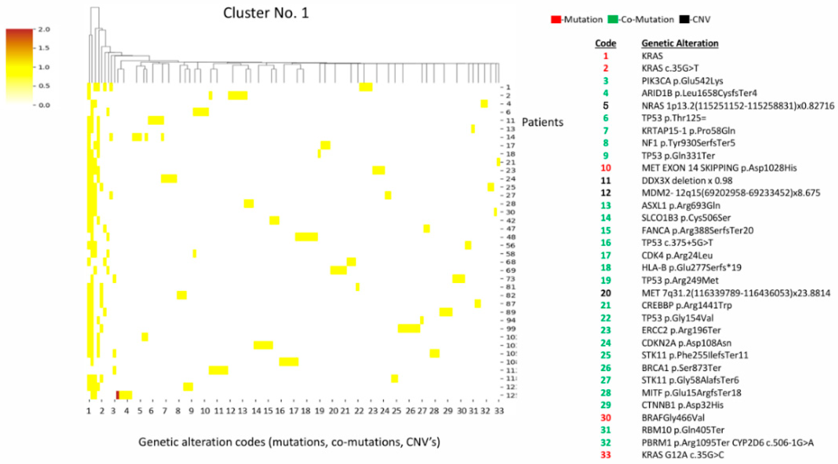
Cluster 1 is characterized by KRAS mutations (various subtypes) alongside STK11 co-mutations, a high frequency of TP53 mutations, and a high concentration of MET alterations (exon 14 skipping + amplification) (Figure 1).

3.1.2. Cluster 2 (n = 12)

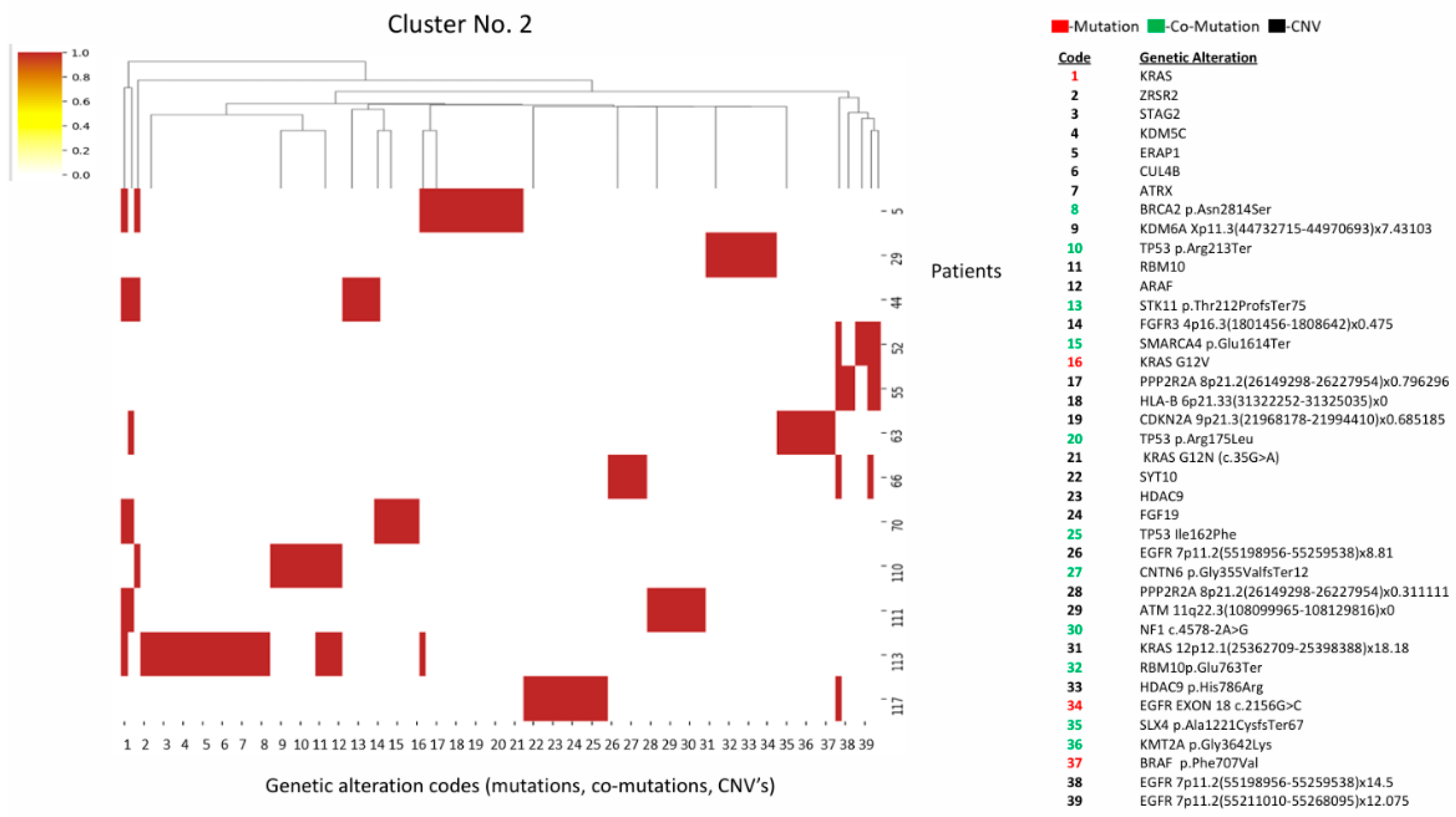
Cluster two has a similar KRAS/STK11/TP53 triad to cluster 1, but is distinguished by a higher frequency of EGFR CNV amplifications and mutations in chromatin remodeling genes (KDM6A, ATRX, SMARCA4) (Figure 2).

3.1.3. Cluster 3 (n = 23)

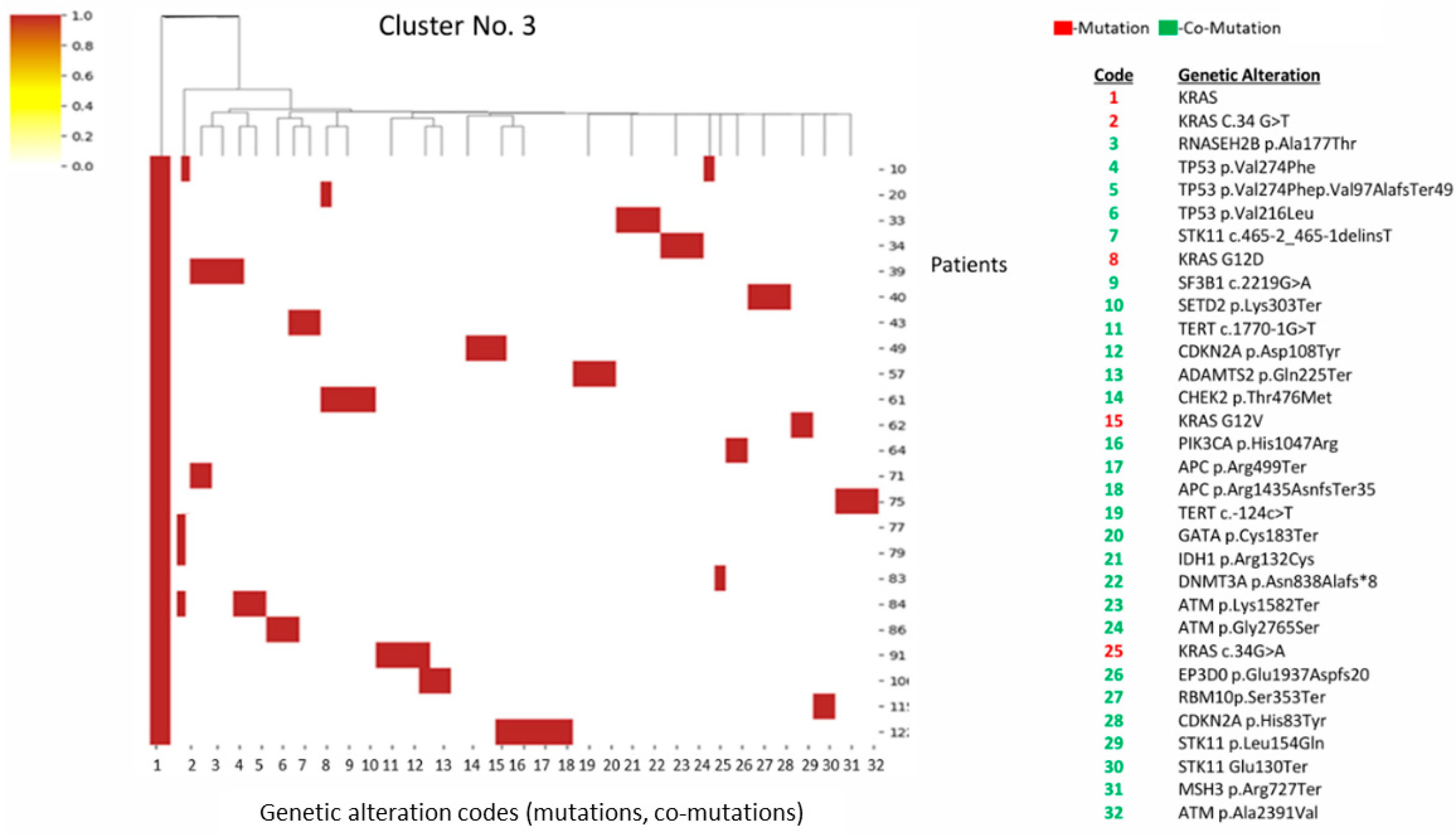
Cluster 3 has a high presence of KRAS and STK11 co-mutations, but notably lower TP53 frequency compared to clusters 1 and 2. Contains mutations in DNA repair pathways (Figure 3).

3.1.4. Cluster 4 (n = 31)

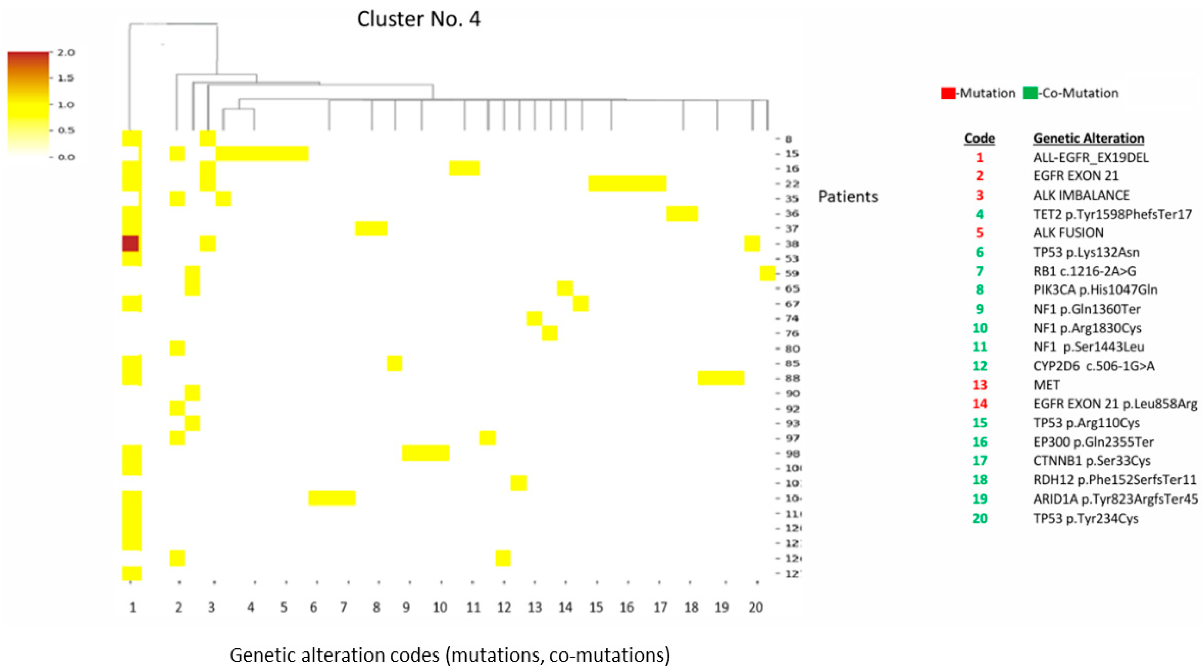
Cluster 4 is defined by EGFR-activating mutations co-occurring with ALK fusions. This represents an oncogene-driven profile with less genomic instability (Figure 4). Within cluster 4, ALK fusions were identified by NGS, performed as part of routine diagnostics. The biopsy timing for all cases was obtained at initial diagnosis prior to exposure to EGFR- or ALK-targeted therapy. These details are provided to support interpretation given that EGFR and ALK alterations are typically mutually exclusive on treatment-naïve NSCLC, although rare concomitant cases have been reported.

3.1.5. Cluster 5 (n = 25)

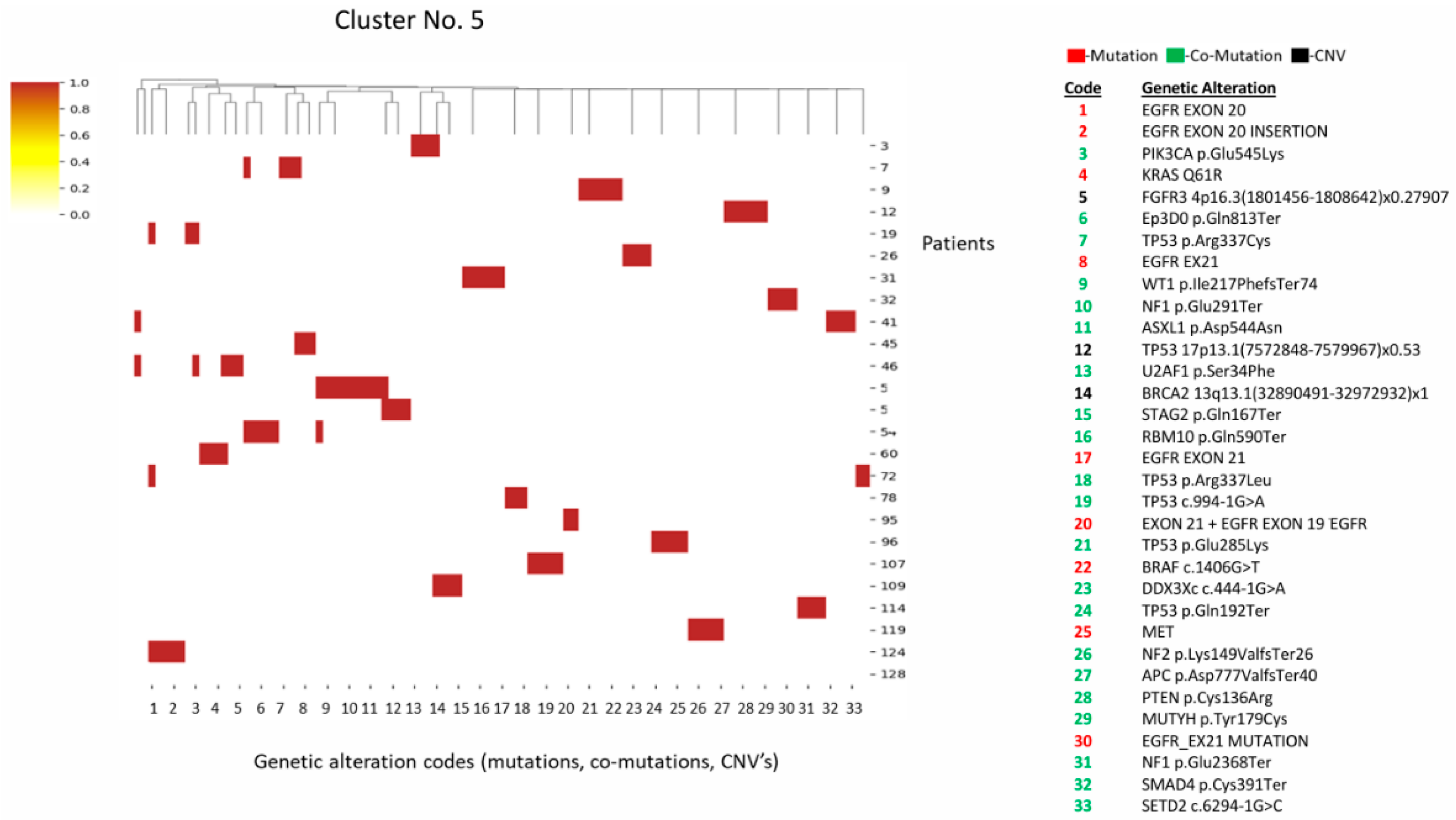
Cluster 5 is characterized by EGFR exon 20 mutations/insertions. Unlike cluster 4, these tend to co-occur with tumor suppressor loss (e.g., NF1, TP53, PTEN) (Figure 5).

3.2. Immunotherapeutic Biomarker Analysis

3.2.1. PD-L1 Expression

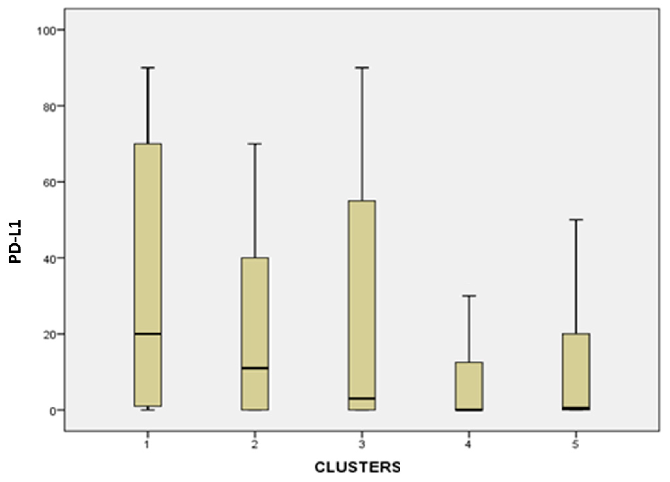
A comparison of clusters according to the ordinal categorical variable showed that the mean PD-L1 expression was lowest in cluster 4 samples (Figure 6). Cluster 4 had significantly lower PD-L1 expression compared to cluster 1 (p = 0.001) and cluster 3 (p = 0.032). Between cluster 1 and 5, there was a difference with a significance level of 0.011. Between cluster 3 and 4, there was a difference with a significance level of 0.032 (Figure 6).

3.2.2. Tumor Mutational Burden (TMB)

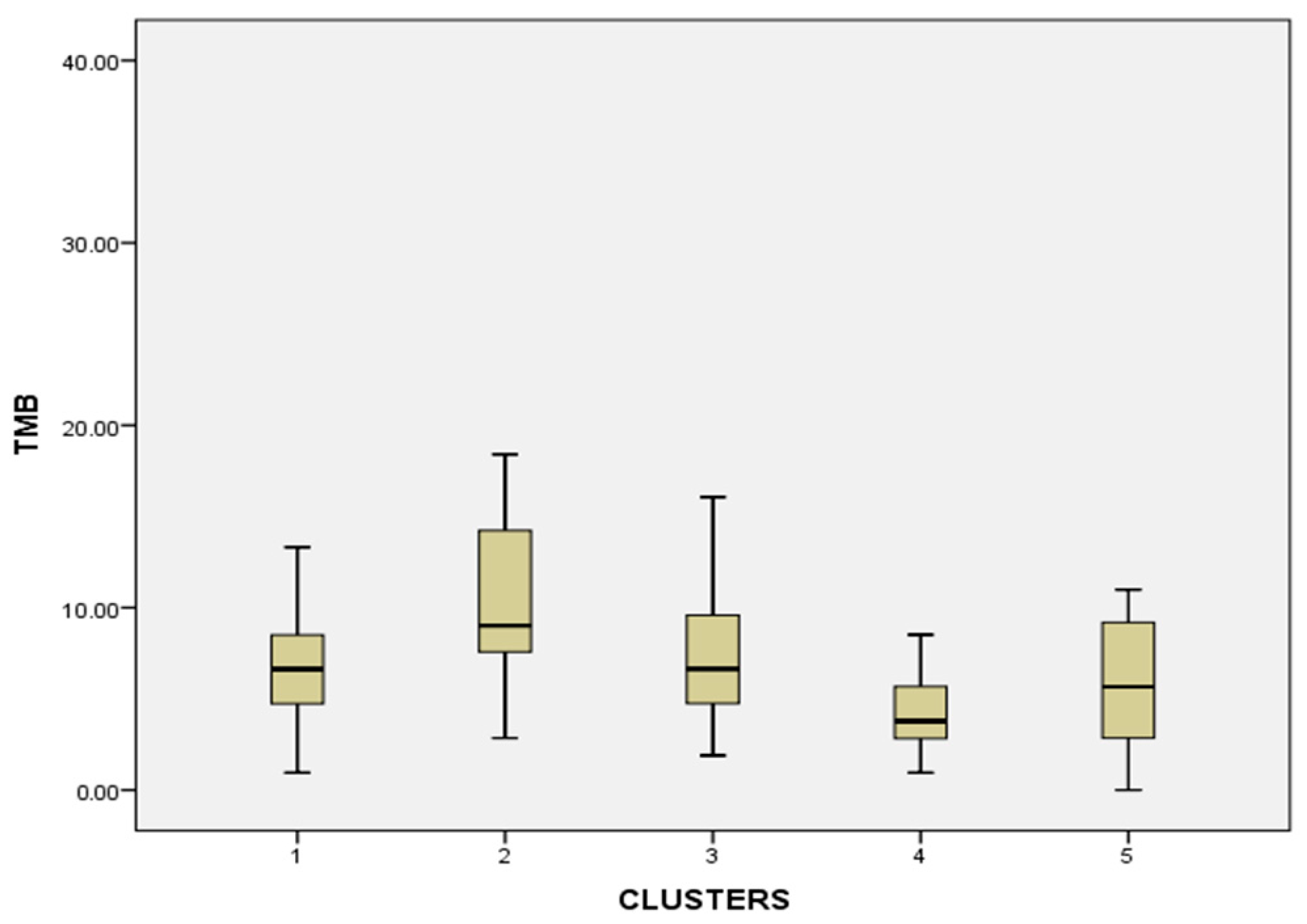
TMB statistical significance: Cluster 4 had significantly lower TMB than clusters 1 (p = 0.012), 2 (p = 0.001), and 3 (p = 0.009). Cluster 2 had significantly higher TMB than clusters 1 (p = 0.013), 4 (p = 0.001), and 5 (p = 0.007) (Figure 7).

3.2.3. MSI/MSS

No statistically significant differences were found between clusters in terms of their microsatellite instability; all evaluable patients were microsatellite-stable (MSS).

3.3. Demographic Analysis

3.3.1. Age

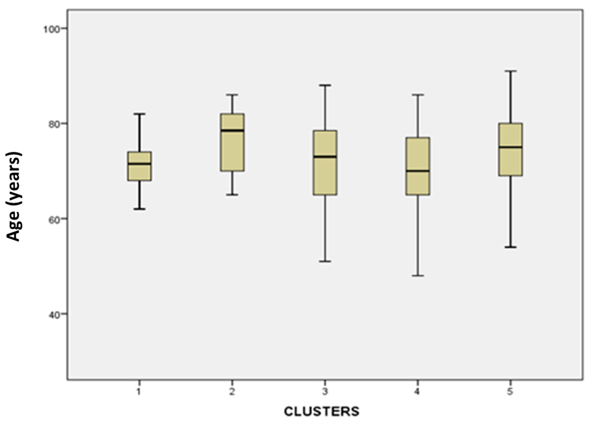
Age of patients analysis (Figure 8) yielded significant differences were between cluster 1 vs. 2 (p = 0.026), cluster 1 vs. 5 (p = 0.031), cluster 2 vs. 4 (p = 0.039), and cluster 4 vs. 5 (p = 0.044). Clusters 1 and 4 represented a younger demographic population.

3.3.2. Gender

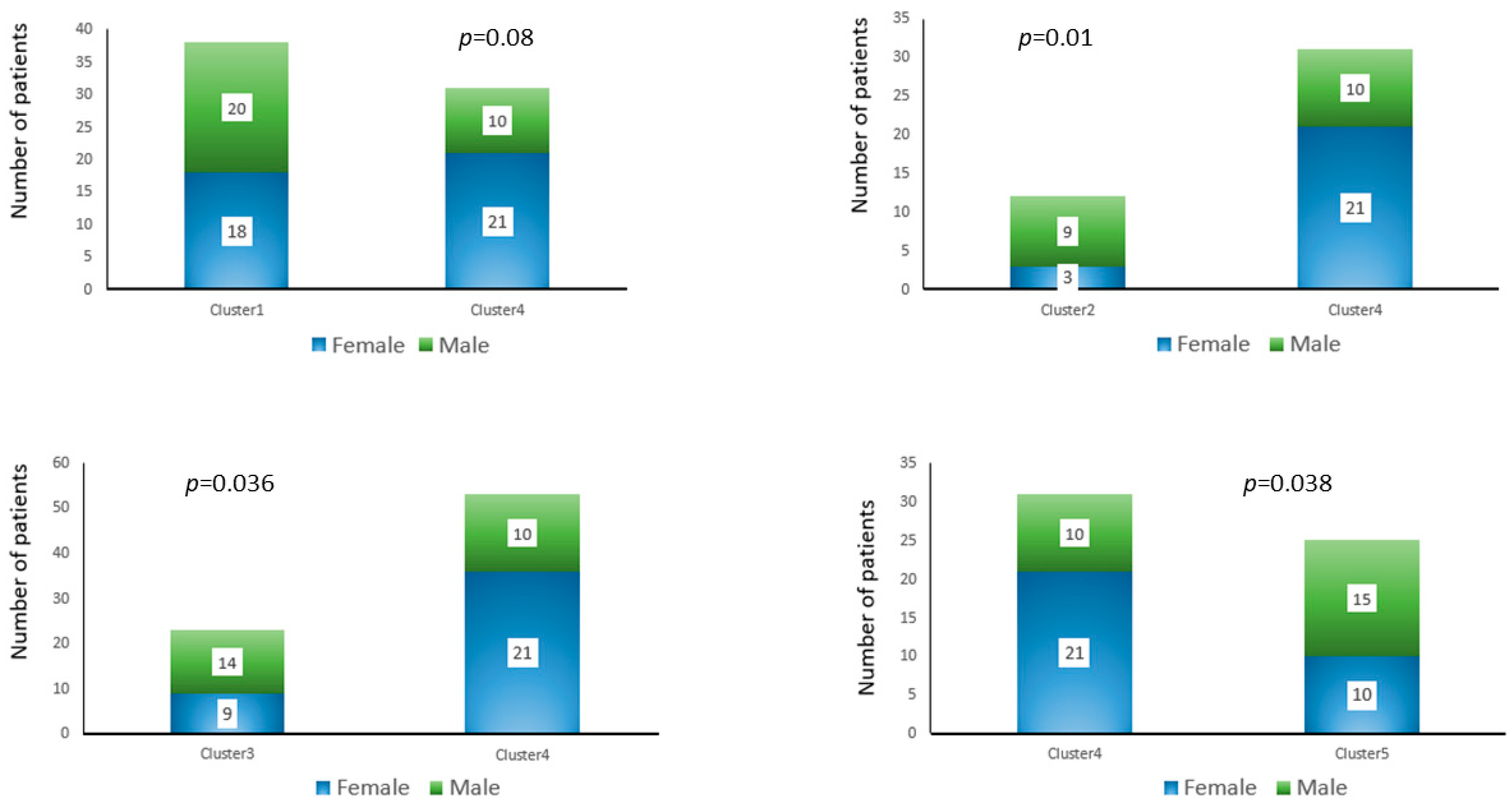
Figure 9 presents the comparison between genders among the clusters. Cluster 4 comprised significantly more women than men, while clusters 1, 2, 3, and 5 had fewer women than men. The distribution of men and women between cluster 1 and cluster 4 was not statistically significant. The findings of the analysis showed a statistically significant difference between cluster 2 and cluster 4 (p = 0.01), as well as between cluster 3 and cluster 4 (p = 0.036) and between cluster 4 and cluster 5 (p = 0.036).

3.3.3. Ethnic

No significant differences were observed between Jewish and Arab patients’ ethnicity across clusters.

4. Discussion

This study sought to identify prevalent associations between co-mutations and actionable driver mutations in lung cancer patients, and to examine their correlations with immunotherapeutic biomarkers and demographics. Our goal was to establish a foundation for future research investigating the clinical implications of these genomic subgroups to enhance precision medicine in lung cancer and to contribute to improve the prognosis of lung cancer patients.
The cluster analysis revealed distinct genomic patterns. KRAS-driven tumors (clusters 1 and 3) frequently co-occurred with STK11. This finding aligns with recent publications on the biomarker STK11’s co-mutation with KRAS and the implication as per therapeutic outcome with neoadjuvant treatment using ICIs (immune checkpoint inhibitors) for NSCLC patients [21,22,23]. Furthermore, clusters 1 and 3 exhibited varying degrees of TP53 involvement, which is in line with Schabath’s 2015 study [24]. Skoulidis et al. [25] observed that, in lung adenocarcinoma, PD-L1 expression plays a role in the response of STK11 and TP53 co-mutations with KRAS. In the current study, cluster 1 displayed high PD-L1 expression, suggesting potential immunogenicity. Suzawa et al.’s [26] findings indicated that KRAS mutation is common in NSCLC tumors harboring MET alteration. Similarly, in the present study, cluster 1 was associated with high levels of MET alterations. These co-existing genomic changes could each be targeted by specific agents; however, due to reported therapeutic resistance [26], it is important to know the molecular profiling of each tumor when proposing a treatment plan.
In contrast, EGFR-driven tumors (clusters 4 and 5) exhibited distinct co-mutation profiles. NSCLC patients with both mutated EGFR and ALK fusions were described in a review by Kemper [27]. In the current study, cluster 4 was characterized by EGFR and ALK fusions, and demonstrated significantly lower PD-L1, which is in line with a previous publication by Gainor et al. [28]. Moreover, cluster 4 had the lowest TMB levels compared to other clusters. Cluster 4’s composition is indicative of an “oncogene-addicted” phenotype driven by signaling pathways (i.e., EGFR) rather than genomic instability. Cluster 5, involving EGFR exon 20 insertions, which are relatively rare [29,30], was associated with tumor suppressor loss [31,32]. Cluster 5 patients bearing low TMB are predicted to have shorter survival than those with high TMB (i.e., cluster 2) [33].
Statistically, cluster 4 (EGFR/ALK) presented with significantly lower biomarkers (PD-L1, TMB) compared to the KRAS-dominant clusters (clusters 1, 2, and 3). Clusters 1, 2, and 3 include mutations KRAS, STK11, and TP53, which may affect the immunogenicity of the patient by increasing PD-L1 expression [34]. KRAS, STK11, and TP53 mutations may also affect the presence of accompanying mutations related to genes involved in stabilizing the chromatin (cluster 2) and DNA repairs pathways (cluster 3), as similarly found in pancreatic cancer genomic mutations [35]. This aligns with the hypothesis that KRAS, STK11, and TP53 mutations may drive immunogenicity and genomic instability (higher TMB), whereas EGFR/ALK alterations (clusters 4 and 5) rely on oncogenic signaling, resulting in “colder” tumors immunologically (with decreased PD-L1 and TMB expression). This means that lung cancer tumors harboring EGFR/ALK mutations rely less on genomic instability.
Hence, future research would deepen the investigation on this topic, i.e., as PD-L1 and TMB have clinical significance, and explore the creation of clusters that are in correlation with PD-L1 and TMB. Further studies should assess the effect of immune-therapy-related biomarkers on the clustering in these patients and check the clinical significance by outcomes of the treatments such as efficiency and toxicity.
Frille et al. 2024 [36] presented interactions between four co-mutations in NSCLC: KRAS, STK11, TP53 and KEAP1. They reported that combined KRAS, G12C and TP53 co-occurrence may be targeted by immunotherapy administered to patients diagnosed with advanced tumor stage in stage IV.
The current study does not claim prognostic effects because outcomes were not analyzed. The cluster patterns were contextualized with existing literature that examines KRAS-STK11-KEAP1 biology in relation to immunotherapy, and EGFR-TP53 co-mutation as a clinically relevant subgroup in EGFR-mutated NSCLC.
Demographically, cluster 4 contained a significantly higher proportion of women and younger patients compared to other clusters, aligning with known epidemiological data regarding EGFR mutations [37]. Genetic factors presented in never-smokers were determined in lung cancer arising from oncogenic mutations. Ha et al. studied oncogenic driver mutations in 198 Asian females diagnosed with lung adenocarcinoma [38]. Their analysis of 26 lung-cancer-related genes harboring 214 mutations along with fusion genes found that EGFR was the dominant driver gene in 63% of patients. The mutation of ALK was detected in 7% of cases. Although cluster 4 consisted of more women than other clusters, it is difficult to draw a clear clinically significant conclusion; it is plausible that the differences were arbitrary or as a result of other variables that were not assessed during the present study.
Chang et al. [39] reported that low-dose computed tomography used for lung cancer screening detected a higher prevalence of subsolid nodules in Asian females than in males. As subsolid nodules may lead to overdiagnosis, it is proposed that this method should be integrated with a multiomics approach. Moreover, Chen et al. [40] suggest that an interdisciplinary strategy should be applied in the clinical setting to optimize precision medicine. Combining factors such as screening protocols and radiologic and genetic data could enhance accuracy, along with polygenic risk scores and molecular biomarkers to refine the stratification of patients.
Further research should be conducted with a bigger sample, and the analysis of clinical results could determine whether there is an effect of these characteristics on the mutation profile in the different clusters.
In addition, ARID1A alterations—identified as genomic events—have been investigated in other tumor types as candidate synthetic lethal contexts, including preclinical evidence supporting vulnerability to EZH2 inhibition in ARID1A-mutant cells, and broader reviews summarizing multiple synthetic lethal strategies under investigation [41]. These observations are not evidence of efficacy in NSCLC, but support a rationale for documenting ARID1A alterations as potential trial-relevant features in future studies.
This study has several limitations: its retrospective design and a modest sample size of 129 patients, as well as heterogeneity in cluster sizes (e.g., n = 12 in cluster 2 vs. n = 38 in cluster 1), which may affect statistical power. Moreover, the tumor stage was not part of the present study, although different stages (early vs. late stage) may be related to multiple coexisting mutations. Additionally, the lack of behavioral data (smoking status or drinking alcohol) in the database limits the ability to control for environmental confounders while creating a more clinically accurate cluster. The use of an unsupervised algorithm, while mathematically robust, groups purely by proximity without inherent clinical weighting. The pathology repository used for the present study did not contain longitudinal outcome data (e.g., overall survival, progression-free survival, line of therapy, or response assessments) precluding valid hazard ratios/Kaplan–Meier curves in the current dataset. Therefore, the present study analysis was conducted without comparing “driver only” versus “driver + co-mutation” subgroups. This study is also limited by the absence of clinical cofounders (e.g., prior systemic therapy) that may influence mutation patterns.
Future prospective studies are recommended with larger cohorts to validate these clusters and assess their clinical utility regarding treatment efficacy and toxicity. Specifically, investigating the impact of these co-mutation clusters on survival outcomes and resistance mechanisms is warranted.

5. Conclusions

In this retrospective cohort, the unsupervised clustering of binary genomic alterations identified reproducible patterns of co-occurring mutations among NSCLC cases with actionable drivers and demonstrated differences in biomarker distribution (PD-L1 and TMB) across clusters. These findings generate testable hypotheses regarding biologically distinct molecular subgroups, but prospective validation in larger cohorts with comprehensive clinical covariates and treatment outcomes is required before clinical prediction claims can be made.

Author Contributions

Conceptualization, A.A., W.S., M.S.-A. and D.L.F.; methodology, Y.S. and E.S.; software, Y.S. and E.S.; validation, A.A., W.S., M.S.-A. and D.L.F.; formal analysis, Y.S. and E.S.; investigation, Y.S. and E.S.; resources, A.A., W.S., M.S.-A., D.L.F., L.S., Y.S., H.N., K.M., S.M. and E.S.; data curation, Y.S. and E.S.; writing—original draft preparation, Y.S.; writing—review and editing, A.A., W.S., M.S.-A., D.L.F., L.S., H.N., K.M., S.M. and E.S.; visualization, Y.S.; supervision, A.A., W.S., M.S.-A. and D.L.F.; project administration, A.A., W.S., M.S.-A. and D.L.F. All authors have read and agreed to the published version of the manuscript.

Funding

This research received no external funding.

Institutional Review Board Statement

The study was conducted in accordance with the Declaration of Helsinki, and approved by the Institutional Review Board Ethics Committee of Carmel Medical Center protocol code CMC-0073-24 on 23 September 2024.

Informed Consent Statement

Patient consent was waived due to the retrospective nature of the study.

Data Availability Statement

The raw data supporting the conclusions of this article will be made available by the authors on request.

Acknowledgments

During the preparation of this manuscript/study, the authors used the AI tools Cloud and ChatGPT 5.2 deep research to help identify mutation patterns within algorithmic clusters. The authors have reviewed and edited the output and take full responsibility for the content of this publication. During manuscript preparation, no patient-level, identifiable, or case-level genomic data were entered into the AI system. All clustering, statistical analyses, and interpretation of genomic patterns were derived from the statistical software outputs and the underlying dataset.

Conflicts of Interest

The authors declare no conflicts of interest.

Abbreviations

The following abbreviations are used in this manuscript:
MSIMicrosatellite Instability
PD-L1Programmed Death-Ligand 1
TMBTumor Mutational Burden
COPDChronic Obstructive Pulmonary Disease
NSCLCNon-Small Cell Lung Carcinoma
SCLCSmall Cell Lung Carcinoma
ADCAdenocarcinoma
SCCSquamous Cell Carcinoma
NGSNext-Generation Sequencing
LCCLarge Cell Carcinoma
EGFREpidermal Growth Factor Receptor
TKITyrosine Kinase Inhibitors
ALKAnaplastic Lymphoma Kinase
ICIImmune Checkpoint Inhibitors
CNVCopy Number Variation
KRASKirsten Rat Sarcoma Virus
STK11Serine Threonine Kinase 11
TP53Tumor Protein 53

References

  1. World Health Organization. Lung Cancer. Available online: https://www.who.int/news-room/fact-sheets/detail/lung-cancer (accessed on 10 January 2026).
  2. Sung, H.; Ferlay, J.; Siegel, R.L.; Laversanne, M.; Soerjomataram, I.; Jemal, A.; Bray, F. Global Cancer Statistics 2020: GLOBOCAN Estimates of Incidence and Mortality Worldwide for 36 Cancers in 185 Countries. CA Cancer J. Clin. 2021, 71, 209–249. [Google Scholar] [CrossRef]
  3. Bray, F.; Laversanne, M.; Sung, H.; Ferlay, J.; Siegel, R.L.; Soerjomataram, I.; Jemal, A. Global cancer statistics 2022: GLOBOCAN estimates of incidence and mortality worldwide for 36 cancers in 185 countries. CA Cancer J. Clin. 2024, 74, 229–263. [Google Scholar] [CrossRef]
  4. Milovanovic, I.S.; Stjepanovic, M.; Mitrovic, D. Distribution patterns of the metastases of the lung carcinoma in relation to histological type of the primary tumor: An autopsy study. Ann. Thorac. Med. 2017, 12, 191–198. [Google Scholar] [CrossRef] [PubMed]
  5. Kitamura, H.; Yazawa, T.; Okudela, K.; Shimoyamada, H.; Sato, H. Molecular and Genetic Pathogenesis of Lung Cancer: Differences Between Small-Cell and Non-Small-Cell Carcinomas. Open Pathol. J. 2008, 2, 106–114. [Google Scholar] [CrossRef][Green Version]
  6. Liu, S.; Xu, T.; Cao, X.; Li, H.; Jin, R. Histological transformation in lung cancer: Mechanisms, clinical characteristics, and therapeutic approaches. Biochim. Biophys. Acta (BBA) Rev. Cancer 2025, 1880, 189413. [Google Scholar] [CrossRef] [PubMed]
  7. Satam, H.; Joshi, K.; Mangrolia, U.; Waghoo, S.; Zaidi, G.; Rawool, S.; Thakare, R.P.; Banday, S.; Mishra, A.K.; Das, G.; et al. Next-Generation Sequencing Technology: Current Trends and Advancements. Biology 2023, 12, 997. [Google Scholar] [CrossRef]
  8. Raphael, A.; Dudnik, E.; Hershkovitz, D.; Jain, S.; Olsen, S.; Soussan-Gutman, L.; Ben-Shitrit, T.; Dvir, A.; Nechushtan, H.; Peled, N.; et al. FGFR Fusions as an Acquired Resistance Mechanism Following Treatment with Epidermal Growth Factor Receptor Tyrosine Kinase Inhibitors (EGFR TKIs) and a Suggested Novel Target in Advanced Non-Small Cell Lung Cancer (aNSCLC). J. Clin. Med. 2022, 11, 2475. [Google Scholar] [CrossRef]
  9. Raphael, A.; Onn, A.; Holtzman, L.; Dudnik, J.; Urban, D.; Kian, W.; Cohen, A.Y.; Moskovitz, M.; Zer, A.; Bar, J.; et al. The Impact of Comprehensive Genomic Profiling (CGP) on the Decision-Making Process in the Treatment of ALK-Rearranged Advanced Non-Small Cell Lung Cancer (aNSCLC) After Failure of 2nd/3rd-Generation ALK Tyrosine Kinase Inhibitors (TKIs). Front. Oncol. 2022, 12, 874712. [Google Scholar] [CrossRef]
  10. Mountzios, G.; Planchard, D.; Metro, G.; Tsiouda, D.; Prelaj, A.; Lampaki, S.; Shalata, W.; Riudavets, M.; Christopoulos, P.; Girard, N.; et al. Molecular Epidemiology and Treatment Patterns of Patients With EGFR Exon 20-Mutant NSCLC in the Precision Oncology Era: The European EXOTIC Registry. JTO Clin. Res. Rep. 2022, 4, 100433. [Google Scholar] [CrossRef]
  11. Le, X.; Elamin, Y.Y.; Zhang, J. New Actions on Actionable Mutations in Lung Cancers. Cancers 2023, 15, 2917. [Google Scholar] [CrossRef]
  12. Vijayalakshmi, R.; Krishnamurthy, A. Targetable “driver” mutations in non small cell lung cancer. Indian J. Surg. Oncol. 2011, 2, 178–188. [Google Scholar] [CrossRef] [PubMed]
  13. El Osta, B. KRAS G12C mutation: From undruggable target to potentially agnostic biomarker. Transl. Lung Cancer Res. 2023, 12, 1147–1151. [Google Scholar] [CrossRef]
  14. Wu, J.; Lin, Z. Non-Small Cell Lung Cancer Targeted Therapy: Drugs and Mechanisms of Drug Resistance. Int. J. Mol. Sci. 2022, 23, 15056. [Google Scholar] [CrossRef]
  15. Yoneda, K.; Imanishi, N.; Ichiki, Y.; Tanaka, F. Treatment of Non-small Cell Lung Cancer with EGFR-mutations. J. UOEH 2019, 41, 153–163. [Google Scholar] [CrossRef]
  16. Liao, B.C.; Lin, C.C.; Shih, J.Y.; Yang, J.C. Treating patients with ALK-positive non-small cell lung cancer: Latest evidence and management strategy. Ther. Adv. Med. Oncol. 2015, 7, 274–290. [Google Scholar] [CrossRef]
  17. Olmedo, M.E.; Cervera, R.; Cabezon-Gutierrez, L.; Lage, Y.; de la Fuente, E.C.; Rueda, A.G.; Mielgo-Rubio, X.; Trujillo, J.C.; Couñago, F. New horizons for uncommon mutations in non-small cell lung cancer: BRAF, KRAS, RET, MET, NTRK, HER2. World J. Clin. Oncol. 2022, 13, 276–286. [Google Scholar] [CrossRef]
  18. Lin, X.; Kang, K.; Chen, P.; Zeng, Z.; Li, G.; Xiong, W.; Yi, M.; Xiang, B. Regulatory mechanisms of PD-1/PD-L1 in cancers. Mol. Cancer 2024, 23, 108. [Google Scholar] [CrossRef]
  19. Strickler, J.H.; Hanks, B.A.; Khasraw, M. Tumor Mutational Burden as a Predictor of Immunotherapy Response: Is More Always Better? Clin. Cancer Res. 2021, 27, 1236–1241. [Google Scholar] [CrossRef]
  20. Li, K.; Luo, H.; Huang, L.; Luo, H.; Zhu, X. Microsatellite instability: A review of what the oncologist should know. Cancer Cell Int. 2020, 20, 16. [Google Scholar] [CrossRef] [PubMed]
  21. Sisca, L.; Cascetta, P.; Aijaz, A.; Catania, C.; Facchinetti, F.; Naqash, A.R.; Ricciuti, B.; Cortellini, A. KRAS and STK11 co-mutations in resectable non-small cell lung cancer: Enduring prognostic value and impaired immunotherapy response. Transl. Lung Cancer Res. 2025, 14, 2374–2382. [Google Scholar] [CrossRef] [PubMed]
  22. Rosner, S.; Connor, S.; Sanber, K.; Zahurak, M.; Zhang, T.; Gurumurthy, I.; Zeng, Z.; Presson, B.; Singh, D.; Rayes, R.; et al. Divergent Clinical and Immunologic Outcomes Based on STK11 Co-mutation Status in Resectable KRAS-Mutant Lung Cancers Following Neoadjuvant Immune Checkpoint Blockade. Clin. Cancer Res. 2025, 31, 339–351. [Google Scholar] [CrossRef] [PubMed]
  23. Manolakos, P.; Ward, L.D. A Critical Review of the Prognostic and Predictive Implications of KRAS and STK11 Mutations and Co-Mutations in Metastatic Non-Small Lung Cancer. J. Pers. Med. 2023, 13, 1010. [Google Scholar] [CrossRef] [PubMed]
  24. Schabath, M.B.; Welsh, E.A.; Fulp, W.J.; Chen, L.; Teer, J.K.; Thompson, Z.J.; Engel, B.E.; Xie, M.; Berglund, A.E.; Creelan, B.C.; et al. Differential association of STK11 and TP53 with KRAS mutation-associated gene expression, proliferation and immune surveillance in lung adenocarcinoma. Oncogene 2016, 35, 3209–3216. [Google Scholar] [CrossRef]
  25. Skoulidis, F.; Goldberg, M.E.; Greenawalt, D.M.; Hellmann, M.D.; Awad, M.M.; Gainor, J.F.; Schrock, A.B.; Hartmaier, R.J.; Trabucco, S.E.; Gay, L.; et al. STK11/LKB1 Mutations and PD-1 Inhibitor Resistance in KRAS-Mutant Lung Adenocarcinoma. Cancer Discov. 2018, 8, 822–835. [Google Scholar] [CrossRef]
  26. Suzawa, K.; Offin, M.; Lu, D.; Kurzatkowski, C.; Vojnic, M.; Smith, R.S.; Sabari, J.K.; Tai, H.; Mattar, M.; Khodos, I.; et al. Activation of KRAS Mediates Resistance to Targeted Therapy in MET Exon 14-Mutant Non-Small Cell Lung Cancer. Clin. Cancer Res. 2019, 25, 1248–1260. [Google Scholar] [CrossRef]
  27. Kemper, M.; Elges, S.; Kies, P.; Wiebe, K.; Lenz, G.; Bleckmann, A.; Evers, G. What do we know about the role of neoadjuvant targeted therapy in early-stage EGFR-mutant and ALK-fused non-small cell lung cancer?—A narrative review of the current literature. Transl. Lung Cancer Res. 2024, 13, 2813–2827. [Google Scholar] [CrossRef]
  28. Gainor, J.F.; Shaw, A.T.; Sequist, L.V.; Fu, X.; Azzoli, C.G.; Piotrowska, Z.; Huynh, T.G.; Zhao, L.; Fulton, L.; Schultz, K.R.; et al. EGFR Mutations and ALK Rearrangements Are Associated with Low Response Rates to PD-1 Pathway Blockade in Non-Small Cell Lung Cancer: A Retrospective Analysis. Clin. Cancer Res. 2016, 22, 4585–4593. [Google Scholar] [CrossRef]
  29. Garzón-Ibáñez, M.; Reyes, R.; Molina-Vila, M.Á.; Sullivan, I.G. Landscape and clinical implications of EGFR exon 20 insertions in non-small cell lung cancer patients. Clin. Transl. Oncol. 2025, 27, 3559–3569. [Google Scholar] [CrossRef]
  30. Seo, D.; Lim, J.H. Targeted Therapies for EGFR Exon 20 Insertion Mutation in Non-Small-Cell Lung Cancer. Int. J. Mol. Sci. 2024, 25, 5917. [Google Scholar] [CrossRef] [PubMed]
  31. Canale, M.; Andrikou, K.; Priano, I.; Cravero, P.; Pasini, L.; Urbini, M.; Delmonte, A.; Crinò, L.; Bronte, G.; Ulivi, P. The Role of TP53 Mutations in EGFR-Mutated Non-Small-Cell Lung Cancer: Clinical Significance and Implications for Therapy. Cancers 2022, 14, 1143. [Google Scholar] [CrossRef]
  32. Christopoulus, P.; Kluck, K.; Kichner, M.; Lüders, H.; Roeper, J.; Falkenstern-Ge, R.-F.; Szewczyk, M.; Sticht, F.; Saalfeld, F.C.; Wesseler, C.; et al. The impact of TP53 co-mutations and immunologic microenvironment on outcome of lung cancer with EGFR exon 20 insertions. Eur. J. Cancer 2022, 170, 106–118. [Google Scholar] [CrossRef]
  33. Dong, Y.; Khan, L.; Yao, Y. Immunological features of EGFR-mutant non-small cell lung cancer and clinical practice: A narrative review. J. Natl. Cancer Cent. 2024, 4, 289–298. [Google Scholar] [CrossRef]
  34. Zhang, F.; Wang, J.; Xu, Y.; Cai, S.; Li, T.; Wang, G.; Li, C.; Zhao, L.; Hu, Y. Co-occurring genomic alterations and immunotherapy efficacy in NSCLC. NPJ Precis. Oncol. 2022, 6, 4. [Google Scholar] [CrossRef]
  35. Abraham, S.; Zhang, J.Y.; Walther, Z.; Parekh, J.; Lacy, J.; Cecchini, M.; Hu, Y. Relations between mutant KRAS and TP53 subtypes and other co-mutations in pancreatic cancer. J. Clin Oncol. 2023, 41, e16294. [Google Scholar] [CrossRef]
  36. Frille, A.; Boeschen, M.; Wirtz, H.; Stiller, M.; Blaker, H.; von Laffert, M. TP53 co-mutations n advanced lung adenocarcinoma: Comparative bioinformatic analyses suggest ambivalent character on overall survival alongside KRAS, STK11 and KEAP1 mutations. Front. Oncol. 2024, 14, 1357583. [Google Scholar] [CrossRef]
  37. Bell, D.W.; Brannigan, B.W.; Matsuo, K.; Finkelstein, D.M.; Sordella, R.; Settleman, J.; Mitsudomi, T.; Haber, D.A. Increased prevalence of EGFR-mutant lung cancer in women and in East Asian populations: Analysis of estrogen-related polymorphisms. Clin. Cancer Res. 2008, 14, 4079–4084. [Google Scholar] [CrossRef] [PubMed]
  38. Ha, S.Y.; Choi, S.J.; Cho, J.H.; Choi, H.J.; Lee, J.; Jung, K.; Irwin, D.; Liu, X.; Lira, M.E.; Mao, M.; et al. Lung cancer in never-smoker Asian females is driven by oncogenic mutations, most often involving EGFR. Oncotarget 2015, 6, 5465–5474. [Google Scholar] [CrossRef]
  39. Chang, Y.C.; Hung, Y.C.; Wu, Y.J.; Tang, E.K.; Wu, F.Z. Understanding East-West differences in subsolid nodules: Prevalence and overdiagnosis implications in lung cancer screening. Ann. Med. 2025, 57, 2478321. [Google Scholar] [CrossRef] [PubMed]
  40. Chen, H.-H.; Wu, Y.-J.; Wu, F.-Z. Precision Medicine in Lung Cancer Screening: A Paradigm Shift in Early Detection—Precision Screening for Lung Cancer. Diagnostics 2025, 15, 1562. [Google Scholar] [CrossRef]
  41. Kearns, B.; McKell, A.; Steveson, I.; Worley, P.; Barton, B.; Bennett, J.; Anderson, D.; Harris, J.; Christensen, J.; Barrott, J.J. ARID1A and Its Impact Across the Hallmarks of Cancer. Int. J. Mol. Sci. 2025, 26, 4644. [Google Scholar] [CrossRef] [PubMed]
Figure 1. Heatmap and cluster 1 pattern. Key alterations: KRAS mutations (multiple, including G12A, G12A, G12D, G12V); STK11 mutations; TP53 mutations (several loss-of-function); MET exon 14 skipping + MET amplification; MDM2 amplification; NF1 and PIK3CA mutations. Pattern: high frequency of TP53 mutations; frequent KRAS/STK11 co-mutations; MET alterations (exon 14 skipping + amplification).
Diagnostics 16 01106 g001
Figure 2. Heatmap and cluster 2 pattern. Key alterations: KRAS mutations (G12V, G12N); STK11 mutations; EGFR amplification (CNV), STK11, TP53 mutations, multiple chromatin modifiers: KDM6A, ATRX, SMARCA4; HLA-B deletion. Pattern: KRAS/STK11/TP53 triad similar to cluster 1, but with more CNVs; EGFR CNV amplifications; chromatin remodeling gene alterations.
Diagnostics 16 01106 g002
Figure 3. Heatmap and cluster 3 pattern. Key alterations: KRAS mutations (G12D, G12V, c.34G>A); co-occurring STK11 mutations; TP53, CDKN2A, ATM, TERT, SETD2, and APC. Pattern: Strong presence of KRAS + STK11 mutations, but less TP53 activity than cluster 1; presence of DNA repair pathway genes: ATM, CHEK2, DNMT3A, IDH1.
Diagnostics 16 01106 g003
Figure 4. Heatmap and cluster 4 pattern. Key alterations: EGFR-activating mutations (EX19DEL, EX21 L858R); ALK fusions; TP53, ARID1A, PIK3CA. Pattern: EGFR/ALK oncogene-driven tumors; less genomic instability, more oncogene-addicted.
Diagnostics 16 01106 g004
Figure 5. Heatmap and cluster 5 pattern. Key alterations: EGFR exon 20 mutations/insertions; NF1, NF2, TP53, PTEN, SETD2; PIK3CA, BRAF, KRAS (Q61R); similar to cluster 4, but with more diverse tumor suppressor loss. Pattern: EGFR exon 20 mutations; tumor suppressor mutations.
Diagnostics 16 01106 g005
Figure 6. Boxplot graph of PD-L1 expression across the different clusters. The analysis was performed on a continuous variable with values ranging from 0 to 100. Between cluster 1 and 4, there is a difference with a significance level of p = 0.001. Between cluster 1 and 5, there is a difference with a significance level of p = 0.011. Between cluster 3 and 4, there is a difference with a significance level of p = 0.032. Between cluster 2 and 4, there is a non-significant difference, p = 0.061.
Diagnostics 16 01106 g006
Figure 7. Boxplot graph of tumor mutational burden (TMB) expression across the different clusters. The analysis was conducted on a continuous variable.
Diagnostics 16 01106 g007
Figure 8. Boxplot graph of the age (years) variable across the different clusters.
Diagnostics 16 01106 g008
Figure 9. Comparison between genders among the clusters.
Diagnostics 16 01106 g009
Disclaimer/Publisher’s Note: The statements, opinions and data contained in all publications are solely those of the individual author(s) and contributor(s) and not of MDPI and/or the editor(s). MDPI and/or the editor(s) disclaim responsibility for any injury to people or property resulting from any ideas, methods, instructions or products referred to in the content.