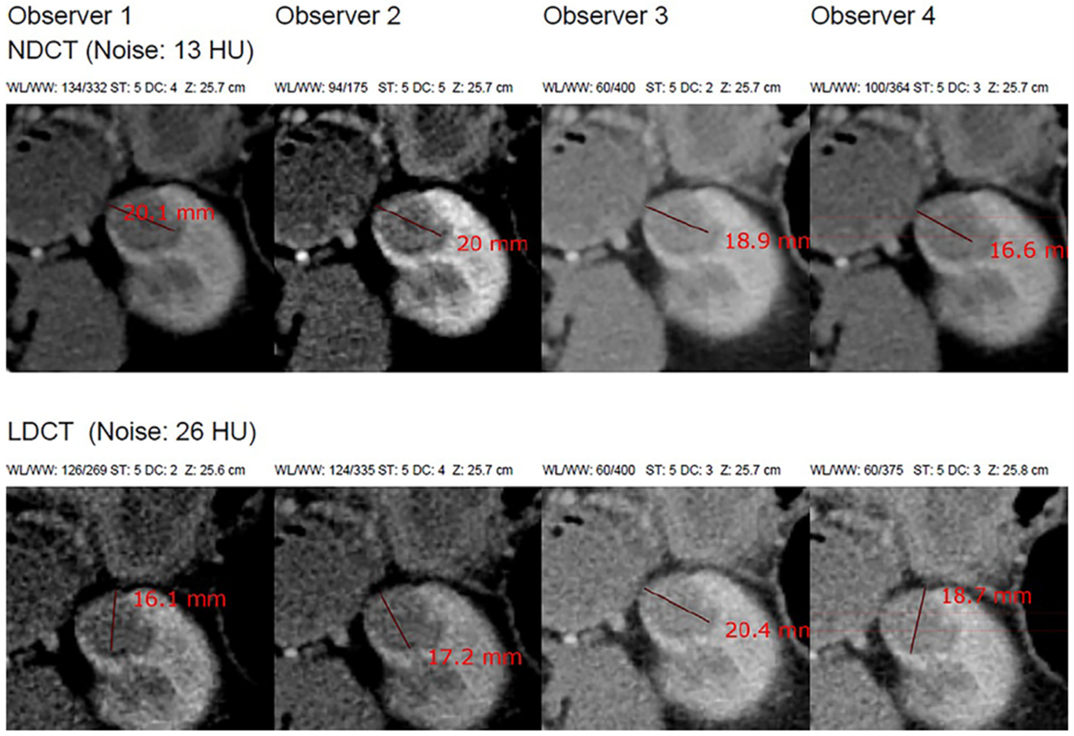
Uncertainty-Aware Framework for CT Radiation Dose Optimization in the Active Surveillance of Small Renal Masses: Clinical and Radiological Considerations

 , , , and
Abstract
1. Introduction
2. Materials and Methods
2.1. Dataset Description
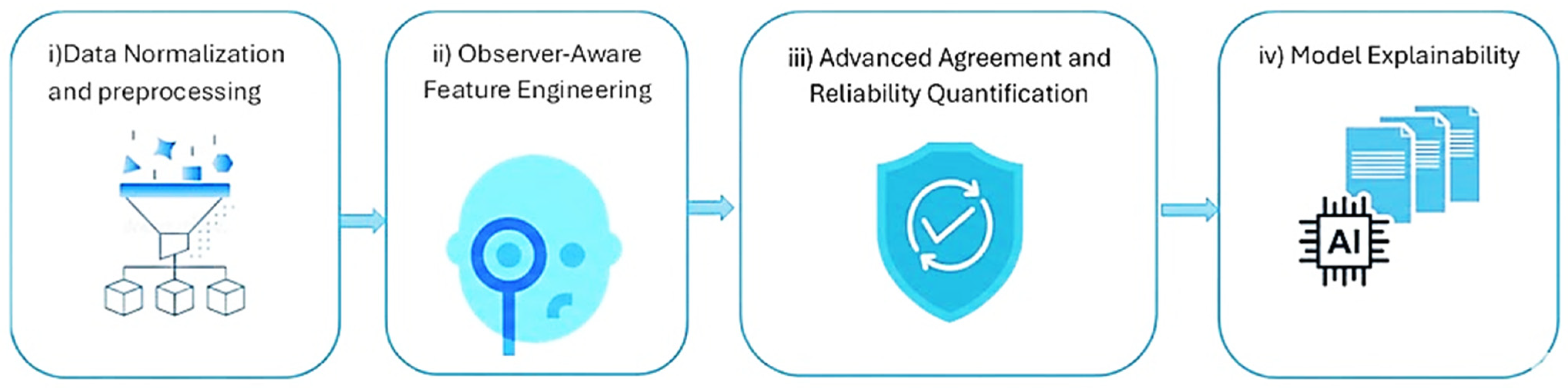
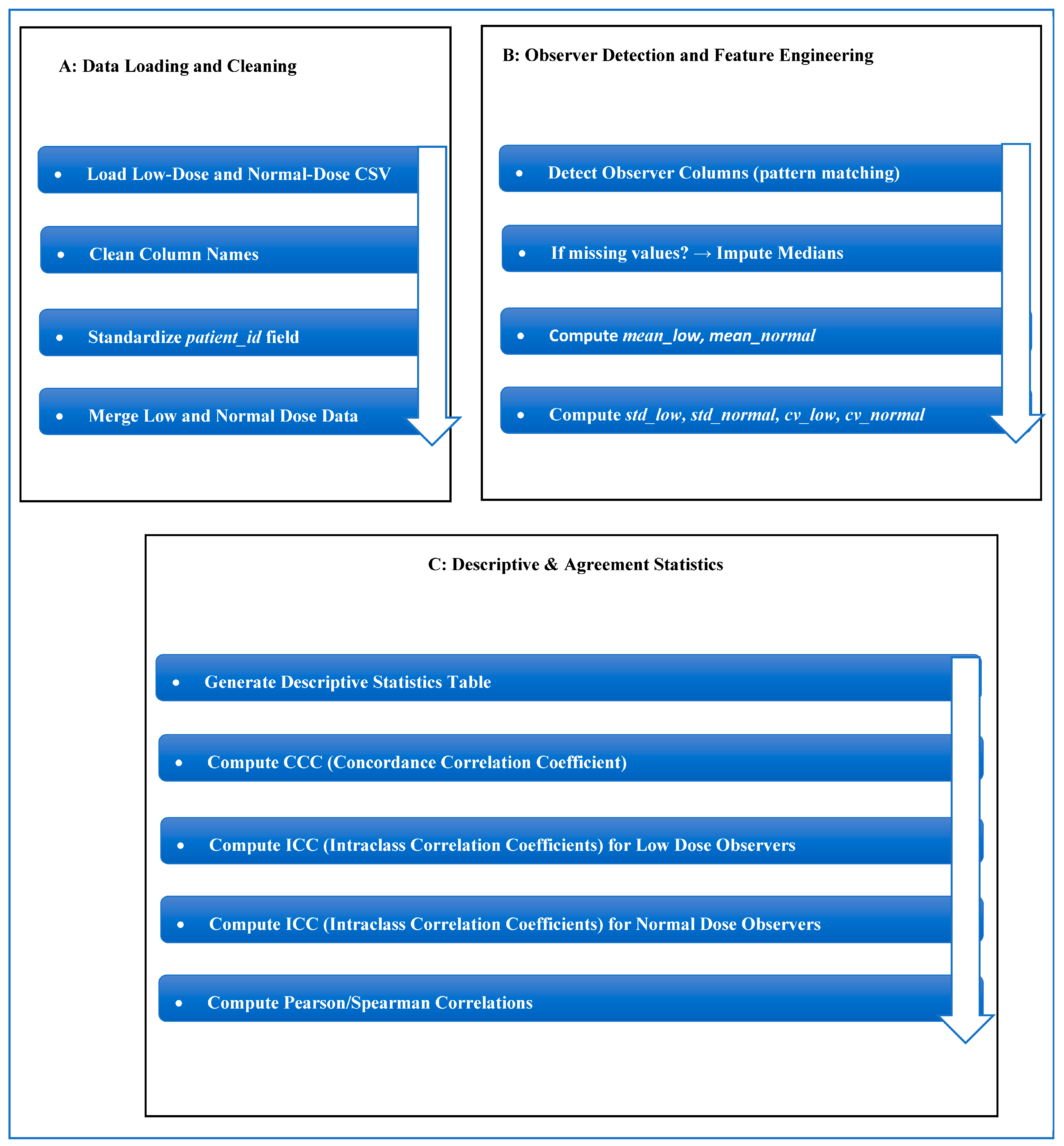
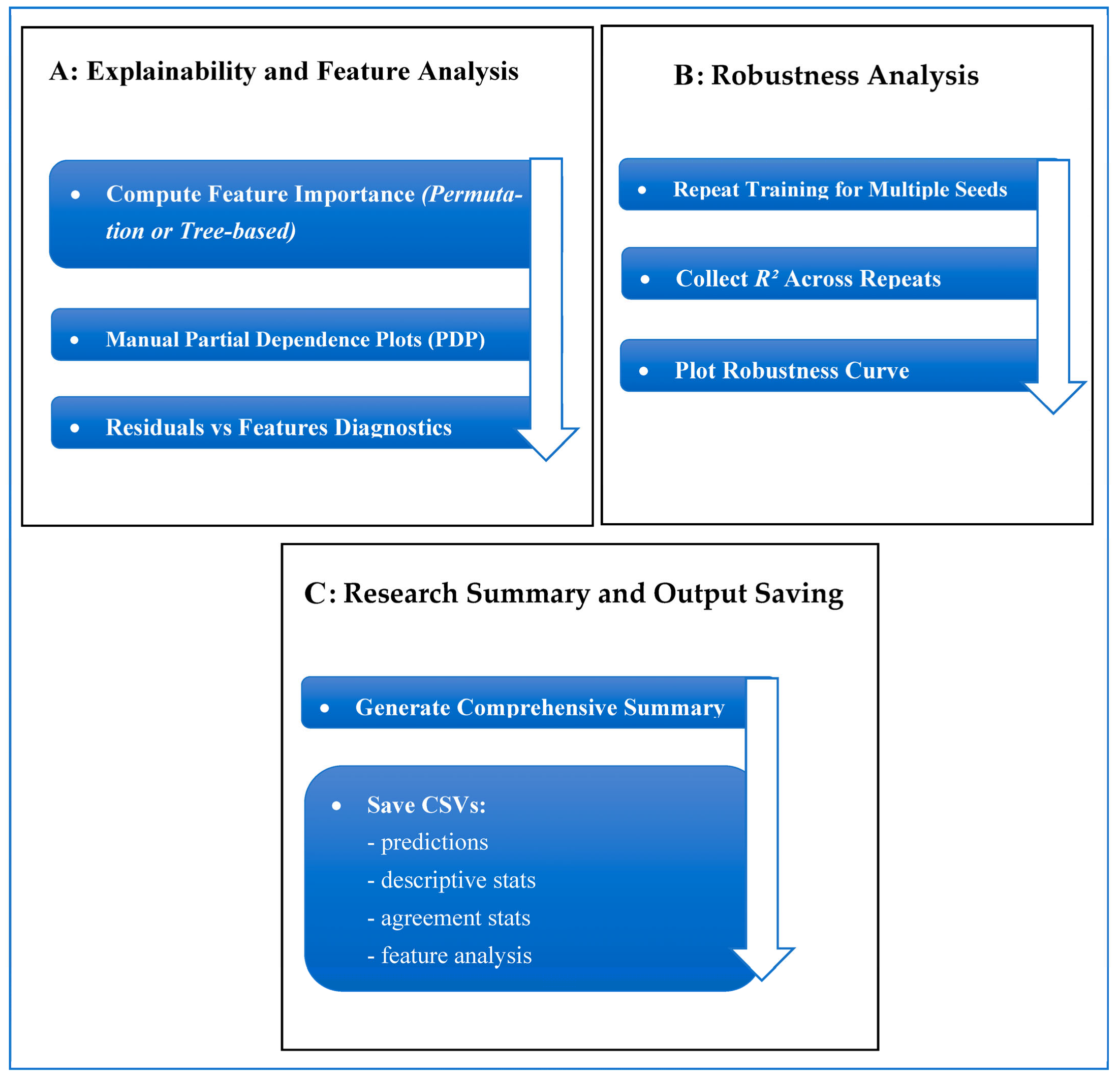
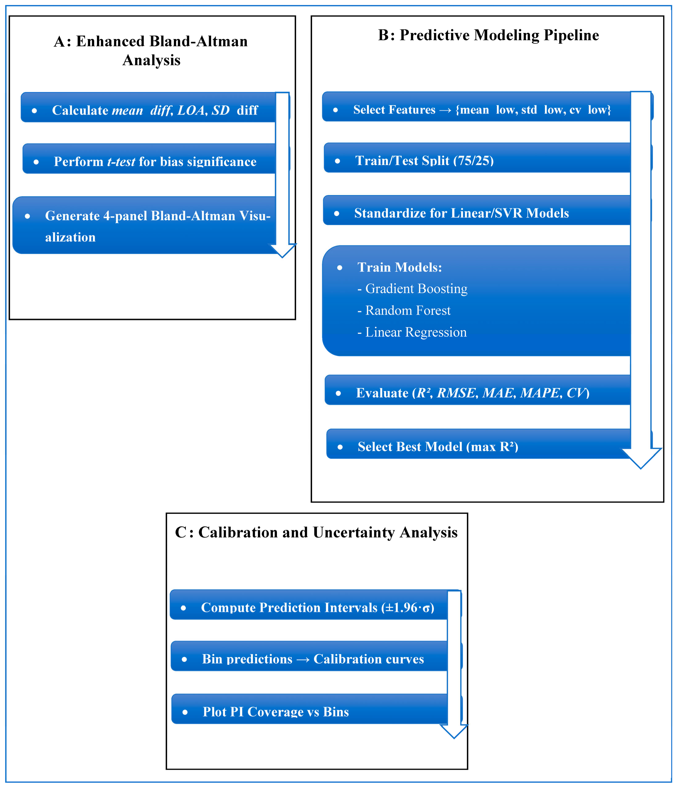
2.2. Proposed Work Framework
2.3. Machine Learning Algorithms and Architectural Innovation
2.4. Architectural Innovations & Equations
2.5. Quantification of Uncertainty and Clinical Interpretation
3. Results
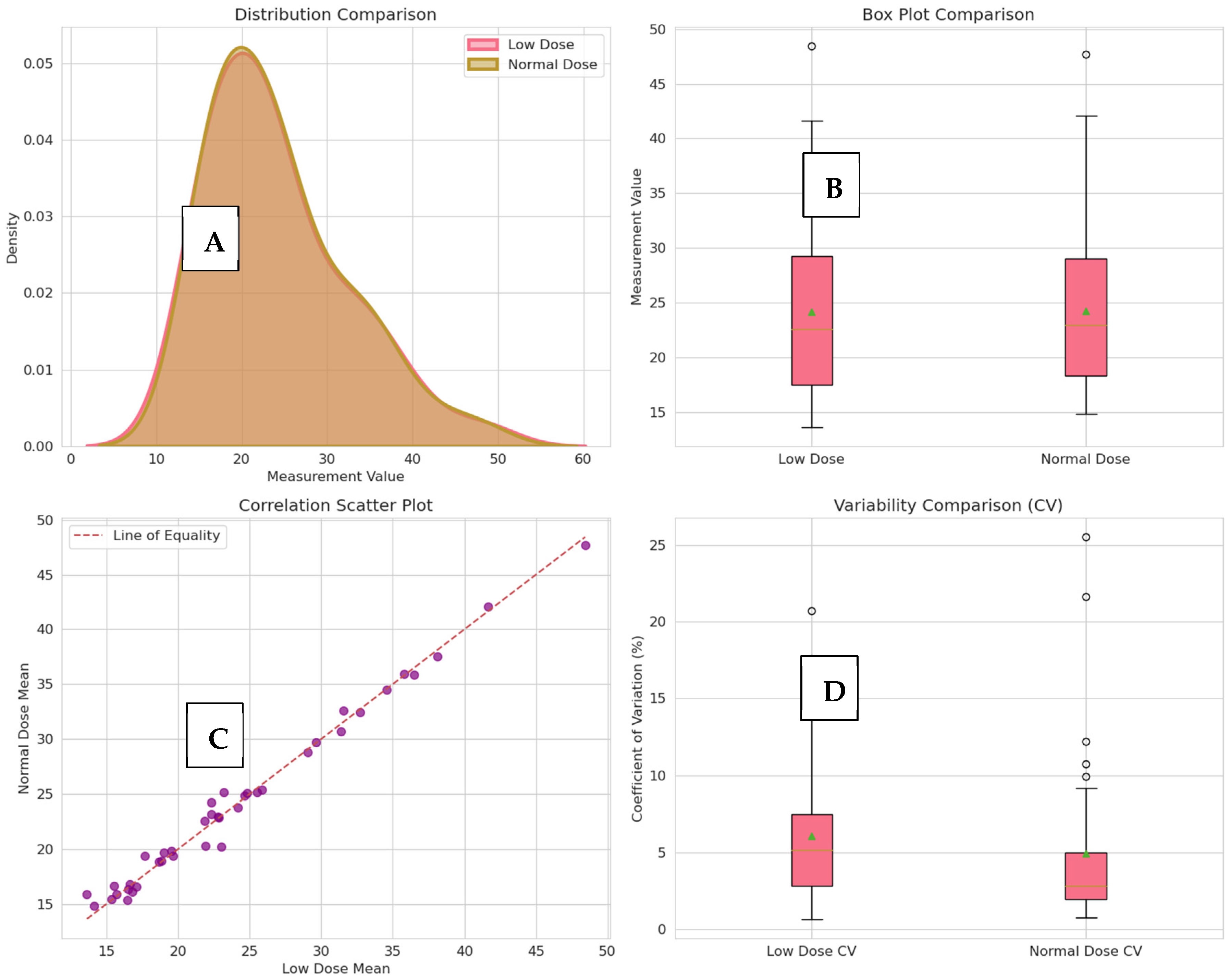
3.1. Descriptive Statistics
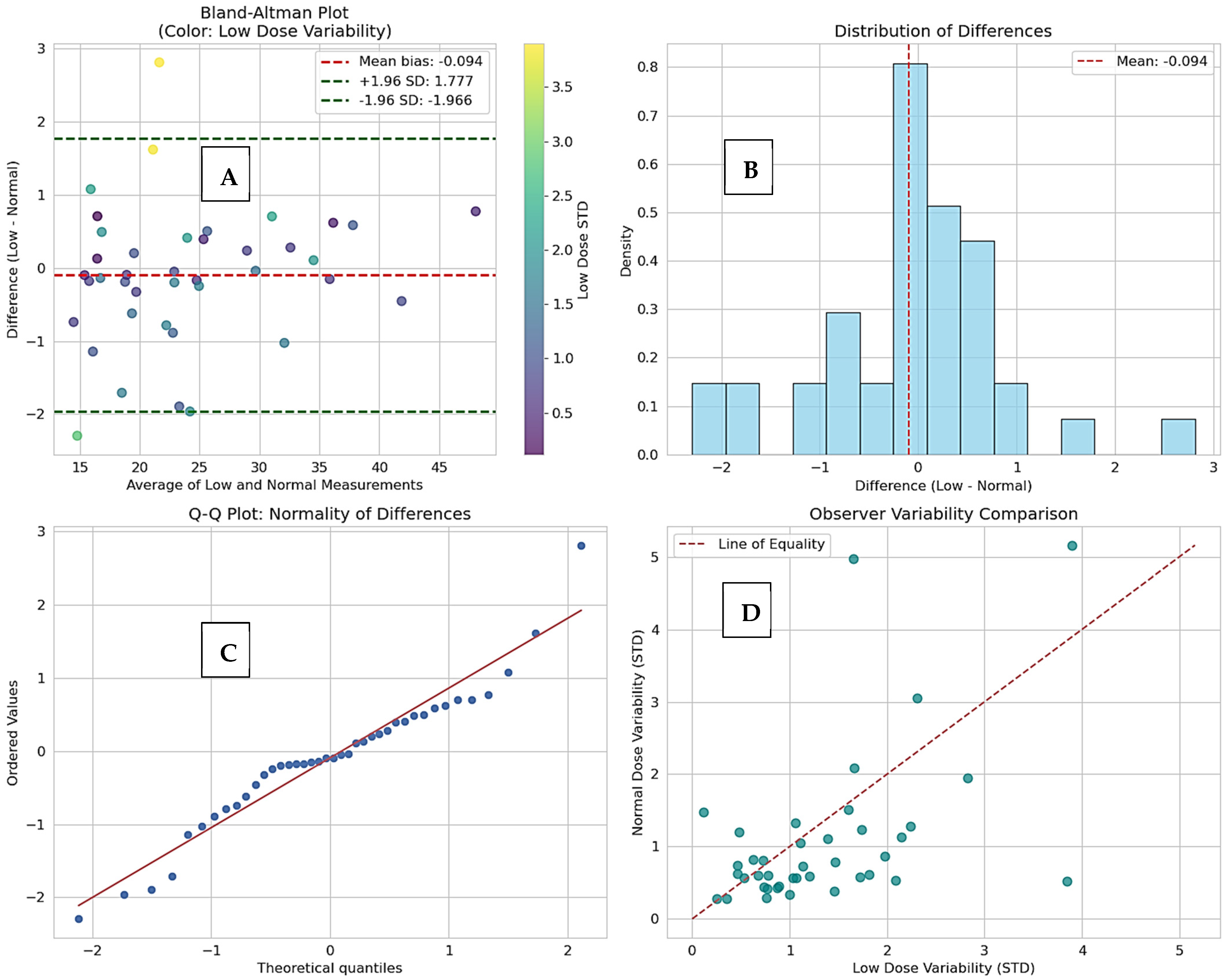
3.2. Inter-Protocol Agreement Analysis
3.3. Model Performance
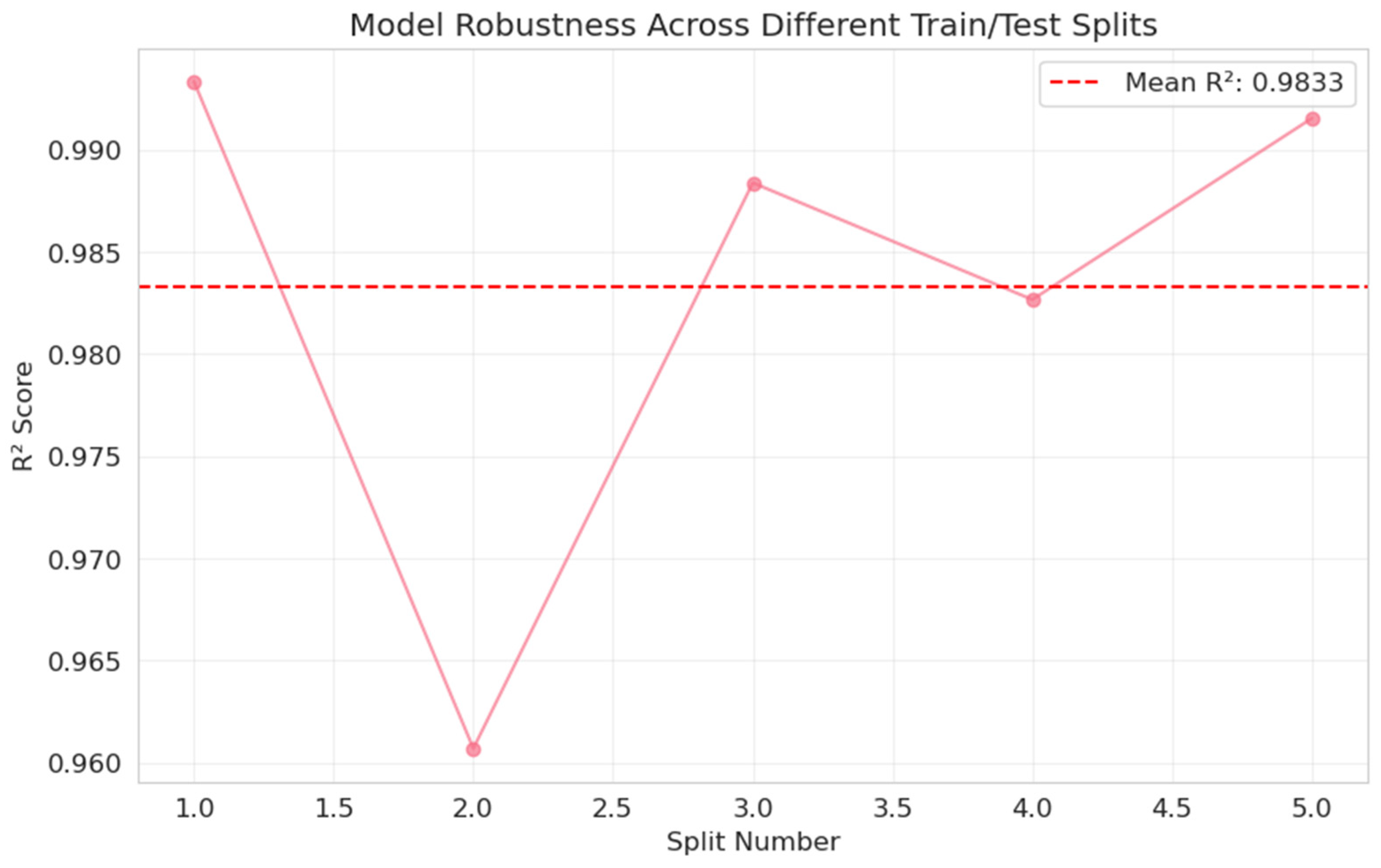
3.4. Feature Importance and Robustness
4. Discussion
4.1. Uncertainty and Clinical Decision-Making
4.2. Clinical and Radiological Considerations
4.3. Limitations and Future Work
5. Conclusions
Author Contributions
Funding
Institutional Review Board Statement
Informed Consent Statement
Data Availability Statement
Conflicts of Interest
References
- Borgbjerg, J.; Breen, B.S.; Kristiansen, C.H.; Larsen, N.E.; Medrud, L.; Mikalone, R.; Müller, S.; Naujokaite, G.; Negård, A.; Nielsen, T.K.; et al. Agreement between Routine-Dose and Lower-Dose CT with and without Deep Learning–based Denoising for Active Surveillance of Solid Small Renal Masses: A Multiobserver Study. Radiol. Imaging Cancer 2025, 7, e240250. [Google Scholar] [CrossRef]
- Borgbjerg, J.; Larsen, N.E.; Salte, I.M.; Grønli, N.R.; Klæstrup, E.; Negård, A. Radiation dose in CT-based active surveillance of small renal masses may be reduced by 75%: A retrospective exploratory multiobserver study. Res. Diagn. Interv. Imaging 2023, 5, 100019. [Google Scholar] [CrossRef]
- Sebastià, C.; Corominas, D.; Musquera, M.; Paño, B.; Ajami, T.; Nicolau, C. Active surveillance of small renal masses. Insights Imaging 2020, 11, 63. [Google Scholar] [CrossRef] [PubMed]
- Saber, A.; Hassan, E.; Elbedwehy, S.; Awad, W.A.; Emara, T.Z. Leveraging ensemble convolutional neural networks and metaheuristic strategies for advanced kidney disease screening and classification. Sci. Rep. 2025, 15, 12487. [Google Scholar] [CrossRef] [PubMed]
- Campbell, S.C.; Clark, P.E.; Chang, S.S.; Karam, J.A.; Souter, L.; Uzzo, R.G. Renal mass and localized renal cancer: Evaluation, management, and follow-up: AUA guideline: Part I. J. Urol. 2021, 206, 199–208. [Google Scholar] [CrossRef]
- Kalra, M.K.; Maher, M.M.; Toth, T.L.; Hamberg, L.M.; Blake, M.A.; Shepard, J.-A.; Saini, S. Strategies for CT radiation dose optimization. Radiology 2004, 230, 619–628. [Google Scholar] [CrossRef] [PubMed]
- Rebez, G.; Pavan, N.; Mir, M.C. Available active surveillance follow-up protocols for small renal mass: A systematic review. World J. Urol. 2021, 39, 2875–2882. [Google Scholar] [CrossRef] [PubMed]
- Elbedwehy, S.; Hassan, E.; Saber, A.; Elmonier, R. Integrating neural networks with advanced optimization techniques for accurate kidney disease diagnosis. Sci. Rep. 2024, 14, 21740. [Google Scholar] [CrossRef]
- Saber, A.; Emara, T.; Elbedwehy, S.; Hassan, E. A novel approach for breast cancer detection using a Nesterov accelerated adam optimizer with an attention mechanism. Sci. Rep. 2025, 15, 27065. [Google Scholar] [CrossRef]
- Filippi, L.; Bianconi, F.; Marongiu, A.; Spanu, A.; Palumbo, B. SPECT-based radiomics in oncology: Current evidence, technical challenges, and future directions. Clin. Transl. Imaging 2026, 1–9. [Google Scholar] [CrossRef]
- Shin, Y.J.; Chang, W.; Ye, J.C.; Kang, E.; Oh, D.Y.; Lee, Y.J.; Park, J.H.; Kim, Y.H. Low-Dose Abdominal CT Using a Deep Learning-Based Denoising Algorithm: A Comparison with CT Reconstructed with Filtered Back Projection or Iterative Reconstruction Algorithm. KJR 2020, 21, 356–364. [Google Scholar] [CrossRef]
- Hassan, E.; Saber, A.; Emara, T.; Elbedwehy, S. Enhanced Monkeypox Skin Lesion Classification Using Pooling-Based Vision Transformer Architecture. In The Role of Artificial Intelligence in Advancing Applied Life Sciences; IGI Global Scientific Publishing: Hershey, PA, USA, 2025; pp. 291–306. [Google Scholar] [CrossRef]
- Borgbjerg, J.; Larsen, N.E.; Salte, I.M.; Grønli, N.R.; Klæstrup, E.; Negård, A. Dataset on renal tumor diameter assessment by multiple observers in normal-dose and low-dose CT. Data Brief 2023, 51, 109672. [Google Scholar] [CrossRef]
- Hassan, E.; Talaat, A.S.; Elsabagh, M.A. Intelligent text similarity assessment using Roberta with integrated chaotic perturbation optimization techniques. J. Big Data 2025, 12, 164. [Google Scholar] [CrossRef]
- Ottilinger, T.; Martini, K.; Baessler, B.; Sartoretti, T.; Bauer, R.W.; Leschka, S.; Sartoretti, E.; Walter, J.E.; Frauenfelder, T.; Wildermuth, S.; et al. Semi-automated volumetry of pulmonary nodules: Intra-individual comparison of standard dose and chest X-ray equivalent ultralow dose chest CT scans. Eur. J. Radiol. 2022, 156, 110549. [Google Scholar] [CrossRef] [PubMed]
- Gierada, D.S.; Rydzak, C.E.; Zei, M.; Rhea, L. Improved Interobserver Agreement on Lung-RADS Classification of Solid Nodules Using Semiautomated CT Volumetry. Radiology 2020, 297, 675–684. [Google Scholar] [CrossRef] [PubMed] [PubMed Central]
- Hassan, E.; Alqahtani, F.; Elbedwehy, S.; Talaat, A.S. Automated detection of pinworm parasite eggs using YOLO convolutional block attention module for enhanced microscopic image analysis. Front. Bioeng. Biotechnol. 2025, 13, 1559987. [Google Scholar] [CrossRef]
- Cacioppa, L.M.; Cellina, M.; Agliata, G.; Mariotti, F.; Rossini, N.; Valeri, T.; Francavilla, G.; Felicioli, A.; Bruno, A.; Rosati, M.; et al. Spectral Computed Tomography Angiography in Visceral Artery Aneurysms: Technical Principles and Clinical Applications. Tomography 2026, 12, 22. [Google Scholar] [CrossRef]
- Park, J.; Joo, I.; Jeon, S.K.; Kim, J.M.; Park, S.J.; Yoon, S.H. Automated abdominal organ segmentation algorithms for non-enhanced CT for volumetry and 3D radiomics analysis. Abdom. Radiol. 2025, 50, 1448–1456. [Google Scholar] [CrossRef]
- Han, D.; Cai, J.; Heus, A.; Heuvelmans, M.; Imkamp, K.; Dorrius, M.; Pelgrim, G.-J.; de Jonge, G.; Oudkerk, M.; van den Berge, M.; et al. Detection and size quantification of pulmonary nodules in ultralow-dose versus regular-dose CT: A comparative study in COPD patients. Br. J. Radiol. 2023, 96, 20220709. [Google Scholar] [CrossRef] [PubMed]
- Wang, H.; Li, L.-L.; Shang, J.; Song, J.; Liu, B. Application of deep learning image reconstruction in low-dose chest CT scan. Br. J. Radiol. 2022, 95, 20210380. [Google Scholar] [CrossRef] [PubMed]
- Zhang, M.; Gu, S.; Shi, Y. The use of deep learning methods in low-dose computed tomography image reconstruction: A systematic review. Complex Intell. Syst. 2022, 8, 5545–5561. [Google Scholar] [CrossRef]
- Bril, S.I.; Wendrich, A.W.; Swartz, J.E.; Wegner, I.; Pameijer, F.; Smid, E.J.; Bol, G.H.; Pothen, A.J.; de Bree, R. Interobserver agreement of skeletal muscle mass measurement on head and neck CT imaging at the level of the third cervical vertebra. Eur. Arch. Otorhinolaryngol. 2019, 276, 1175–1182. [Google Scholar] [CrossRef]
- Ye, J.; Cai, W.; Huang, F.; zhen Xu, X. Dose optimization of stereotactic radiosurgery by 18F-FHBG PET/CT tracking of HSV-TK gene vectors in recurrent glioblastoma: A prospective controlled study. J. Radiat. Res. Appl. Sci. 2026, 19, 102127. [Google Scholar] [CrossRef]
- Liu, Y.; Mo, C.; Dong, Y.; Tang, P.; Li, H.; Xu, Z.; Zhao, Y.; Chen, H.; Hu, K. Development of new CAIX-targeted radioligands as theranostic pairs for clear cell renal cell carcinoma. Chem. Eng. J. 2026, 532, 174381. [Google Scholar] [CrossRef]
- Adika, A.; Zibelman, M. Perioperative Immune-Checkpoint Inhibition in Renal Cell Carcinoma: Lessons Learned and Future Directions. Cancers 2026, 18, 527. [Google Scholar] [CrossRef]
- García-Figueiras, R.; Baleato-González, S.; Busquets-Carrera, O.; Fraga-Piñeiro, C.; Luna, A. Quantitative Dual-Energy CT in Abdominal Imaging: Technical Considerations and Emerging Clinical Applications. RadioGraphics 2026, 46, e250074. [Google Scholar] [CrossRef] [PubMed]
- Shams, M.Y.; Hassan, E.; Gamil, S.; Ibrahim, A.; Gabr, E.; Gamal, S.; Ibrahim, E.; Abbas, F.; Mohammed, A.; Khamis, A.; et al. Skin Disease Classification: A Comparison of ResNet50, MobileNet, and Efficient-B0. J. Curr. Multidiscip. Res. 2025, 1, 1–7. [Google Scholar] [CrossRef]
- Bhatnagar, R.; Hassan, E.; Saber, A.; Shams, M.Y.; Elbedwehy, S. Enhanced Vision Transformer with Lion Optimizer for Accurate Classification of Monkeypox Skin Images. In Proceedings of the 2024 4th International Conference on Technological Advancements in Computational Sciences (ICTACS); IEEE: New York, NY, USA, 2024; pp. 1897–1902. [Google Scholar] [CrossRef]
- Hu, Q.-J.; Liu, Y.-W.; Chen, C.; Kang, S.-C.; Sun, Z.-Y.; Wang, Y.-J.; Xiang, M.; Xia, L.-M.; Guan, H.-X. Prospective study of low- and standard-dose chest CT for pulmonary nodule detection: A comparison of image quality, size measurements and radiation exposure. Curr. Med. Sci. 2021, 41, 966–973. [Google Scholar] [CrossRef]
- Pan, Z.; Zhang, Y.; Zhang, L.; Wang, L.; Zhao, K.; Li, Q.; Wang, A.; Hu, Y.; Xie, X. Detection, measurement, and diagnosis of lung nodules by ultra-low-dose CT in lung cancer screening: A systematic review. BJR Open 2025, 6, tzae041. [Google Scholar] [CrossRef]
- Li, E.L.; Ma, A.L.; Wang, T.; Fu, Y.F.; Liu, H.Y.; Li, G.C. Low-dose versus standard-dose CT-guided biopsy for pulmonary nodules: A randomized controlled trial. J. Cardiothorac. Surg. 2023, 18, 86. [Google Scholar] [CrossRef]






| Parameter | Value |
|---|---|
| Patient ID (KiTS19) | KiTS-00019 |
| Age (years) | 58 |
| Sex | Male |
| BMI (kg/m2) | 36.4 |
| Tumor Size (cm) | 1.2 |
| Tumor Histology | Oncocytoma |
| CT System | GE MEDICAL SYSTEMS LightSpeed VCT |
| Reconstruction Technique (Kernel) | STANDARD |
| Tube Voltage (kVp) | 140 |
| Slice Thickness (mm) | 3.75 |
| Scan Range (cm) | 31.1 |
| Variable | N | Mean (SD) | 95% CI | Median [Q1, Q3] | Min–Max | Skewness | Kurtosis |
|---|---|---|---|---|---|---|---|
| mean_low | 40 | 24.1543 (8.2095) | [21.6102, 26.6985] | 22.5774 [17.5154, 29.2217] | 13.6433–48.4367 | 1.0102 | 0.4978 |
| mean_normal | 40 | 24.2487 (8.0336) | [21.7591, 26.7384] | 22.9537 [18.3572, 29.0546] | 14.8568–47.6634 | 1.0116 | 0.4388 |
| Metric | Value | Interpretation |
|---|---|---|
| Concordance Correlation Coefficient (CCC) | 0.9930 | Almost perfect agreement |
| ICC: Low-dose observers | 0.9642 | Excellent reliability |
| ICC: Normal-dose observers | 0.9654 | Excellent reliability |
| Pearson Correlation | 0.9933 | Very strong relationship |
| Spearman Correlation | 0.9827 | Monotonic ranking preserved |
| Mean Absolute Difference | 0.6760 mm | Minimal systematic error |
| Root Mean Square Difference | 0.9474 mm | Very low dispersion |
| Model | R2 | MAE (mm) | RMSE (mm) | MAPE | CV R2 (Mean ± SD) | 95% PI Coverage |
|---|---|---|---|---|---|---|
| Linear Regression | 0.9933 | 0.4239 | 0.4775 | 2.07% | 0.9632 ± 0.0274 | 100% |
| Random Forest | 0.9766 | 0.6657 | 0.8951 | 2.87% | 0.9034 ± 0.0780 | 90% |
| Gradient Boosting | 0.9737 | 0.8149 | 0.9485 | 3.72% | 0.9146 ± 0.0744 | 90% |
| SVR | 0.7201 | 2.2208 | 3.0943 | 10.06% | 0.4415 ± 0.2253 | 90% |
| Bin | Mean Predicted | Mean Actual | N | Calibration Error |
|---|---|---|---|---|
| 1 | 16.0616 | 16.6700 | 1 | +0.6084 |
| 2 | 17.0402 | 16.3910 | 1 | −0.6492 |
| 3 | 19.2817 | 18.9639 | 1 | −0.3177 |
| 4 | 19.2984 | 19.6698 | 1 | +0.3713 |
| 5 | 19.9186 | 19.8773 | 1 | −0.0414 |
| 6 | 19.9293 | 19.4318 | 1 | −0.4975 |
| 7 | 22.4583 | 23.2181 | 1 | +0.7598 |
| 8 | 23.8622 | 23.7645 | 1 | −0.0977 |
| 9 | 25.9144 | 25.3878 | 1 | −0.5266 |
| Feature | Importance Score | Corr with Target | Mean | Std |
|---|---|---|---|---|
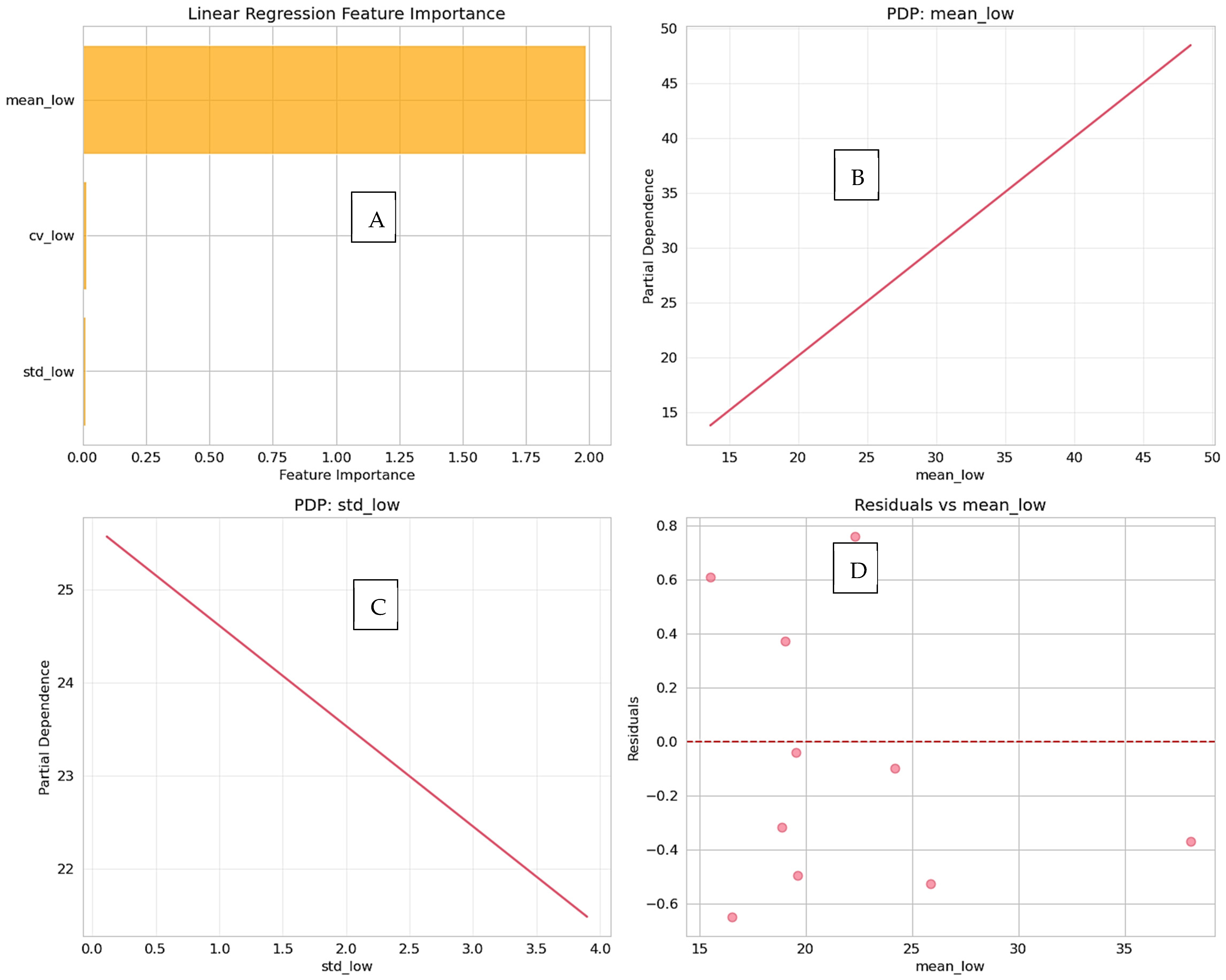
| mean_low | 1.9868 | 0.9933 | 24.1543 | 8.2095 |
| cv_low | 0.0130 | −0.4235 | 6.1048 | 4.6161 |
| std_low | 0.0099 | −0.1557 | 1.3232 | 0.8680 |
| Split | R2 Score |
|---|---|
| 1 | 0.9933 |
| 2 | 0.9607 |
| 3 | 0.9884 |
| 4 | 0.9827 |
| 5 | 0.9915 |
| Metric | Value |
|---|---|
| Mean R2 | 0.9833 |
| Std | 0.0119 |
| Min | 0.9607 |
| Max | 0.9933 |
| Feature | Standard-Dose CT | Low-Dose CT | Clinical Implications |
|---|---|---|---|
| Radiation Dose | ~10–12 mSv | ~1–3 mSv | Low-dose CT significantly reduces patient radiation exposure, which is important for repeated surveillance and younger patients. |
| Image Quality | High spatial resolution, low noise | Slightly increased noise; sufficient for accurate diameter measurement | Low-dose CT maintains diagnostic quality; AI denoising can further enhance image clarity. |
| Measurement Accuracy | Reference standard | Near-perfect agreement with standard-dose (CCC ~0.993) | Low-dose CT reliably reproduces tumor size measurements, clinically interchangeable with standard-dose. |
| Scan Time | Standard | Comparable | No additional workflow burden: low-dose protocols integrate easily into existing practice. |
| Typical Clinical Use | Initial diagnosis, pre-surgical planning, follow-up | Routine follow-up, active surveillance, dose-sensitive populations | Low-dose CT is preferable for repeated imaging or patients at risk from cumulative radiation exposure. |
| Tumor ID | Standard-Dose CT (mm) | Low-Dose CT (mm) | Difference (Low − Standard, mm) |
|---|---|---|---|
| 1 | 25.4 | 25.3 | −0.1 |
| 2 | 18.7 | 18.6 | −0.1 |
| 3 | 32.0 | 32.1 | +0.1 |
| 4 | 12.5 | 12.4 | −0.1 |
| 5 | 40.2 | 40.1 | −0.1 |
| 6 | 28.6 | 28.7 | +0.1 |
| 7 | 15.0 | 14.9 | −0.1 |
| 8 | 21.3 | 21.2 | −0.1 |
| 9 | 36.7 | 36.8 | +0.1 |
| 10 | 10.8 | 10.8 | 0.0 |
Disclaimer/Publisher’s Note: The statements, opinions and data contained in all publications are solely those of the individual author(s) and contributor(s) and not of MDPI and/or the editor(s). MDPI and/or the editor(s) disclaim responsibility for any injury to people or property resulting from any ideas, methods, instructions or products referred to in the content. |
© 2026 by the authors. Licensee MDPI, Basel, Switzerland. This article is an open access article distributed under the terms and conditions of the Creative Commons Attribution (CC BY) license.
Share and Cite
Elsabagh, M.A.; Samy Talaat, A.; Elwi, D.; Hassan, S.M.; Alqassimi, S.; Hassan, E. Uncertainty-Aware Framework for CT Radiation Dose Optimization in the Active Surveillance of Small Renal Masses: Clinical and Radiological Considerations. Diagnostics 2026, 16, 943. https://doi.org/10.3390/diagnostics16060943
Elsabagh MA, Samy Talaat A, Elwi D, Hassan SM, Alqassimi S, Hassan E. Uncertainty-Aware Framework for CT Radiation Dose Optimization in the Active Surveillance of Small Renal Masses: Clinical and Radiological Considerations. Diagnostics. 2026; 16(6):943. https://doi.org/10.3390/diagnostics16060943
Chicago/Turabian StyleElsabagh, M. A., Amira Samy Talaat, Dalia Elwi, Shaimaa M. Hassan, Sameer Alqassimi, and Esraa Hassan. 2026. "Uncertainty-Aware Framework for CT Radiation Dose Optimization in the Active Surveillance of Small Renal Masses: Clinical and Radiological Considerations" Diagnostics 16, no. 6: 943. https://doi.org/10.3390/diagnostics16060943
APA StyleElsabagh, M. A., Samy Talaat, A., Elwi, D., Hassan, S. M., Alqassimi, S., & Hassan, E. (2026). Uncertainty-Aware Framework for CT Radiation Dose Optimization in the Active Surveillance of Small Renal Masses: Clinical and Radiological Considerations. Diagnostics, 16(6), 943. https://doi.org/10.3390/diagnostics16060943