Association Between Calcaneal Inclination Angle and Spinal and Lower Limb Alignment: A Retrospective Radiographic Analysis

 , , ,
, , ,  ,
,  and
and
Abstract
1. Introduction
2. Materials and Methods
2.1. Selection of Research Subjects
2.1.1. Inclusion Criteria
- (a)
- Adult men and women aged 20–60 years.
- (b)
- Patients who underwent weight-bearing lateral radiographs of both feet, whole-spine anteroposterior (AP) and lateral views, and full-length standing AP scanograms of the lower legs.
2.1.2. Exclusion Criteria
- (a)
- History of trauma or surgery that could cause structural problems in the lumbar spine (e.g., fracture, osteoporosis, and lumbar surgery).
- (b)
- History of diseases causing deformity of the lumbar structure (e.g., congenital malformation of the lumbar spine, ankylosing spondylitis, idiopathic scoliosis, tumors, and spina bifida).
- (c)
- Patients deemed unsuitable for participation in this study by the principal investigator, including those with (1) severe musculoskeletal pain that limited standing or walking posture during imaging, (2) poor radiographic image quality due to motion artifacts or improper positioning, or (3) any comorbid neurological or systemic disease that could affect spinal or lower-limb alignment.
2.2. Study Design
2.3. Body Alignment Parameters
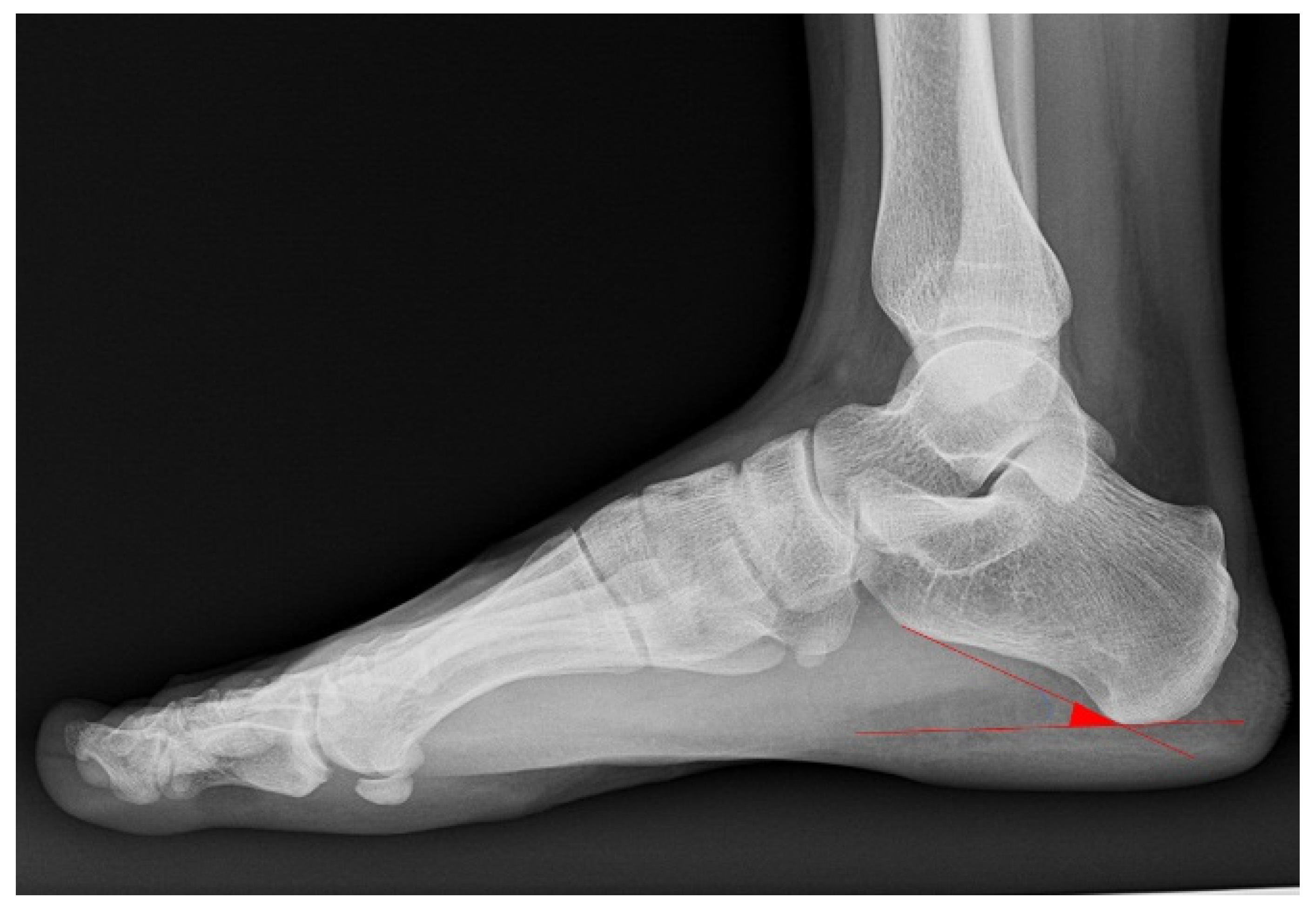
2.3.1. CIA
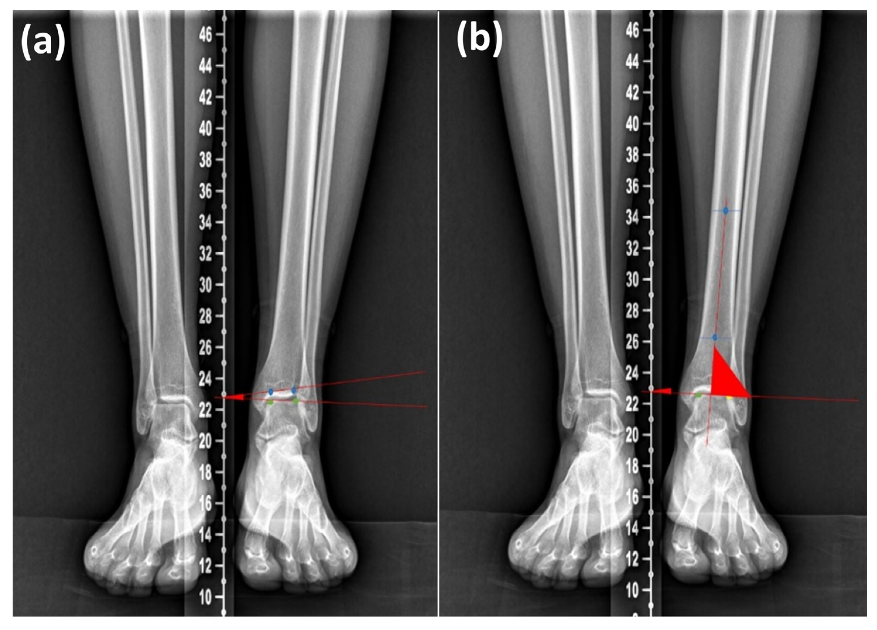
2.3.2. Alignment Parameter of the Ankle
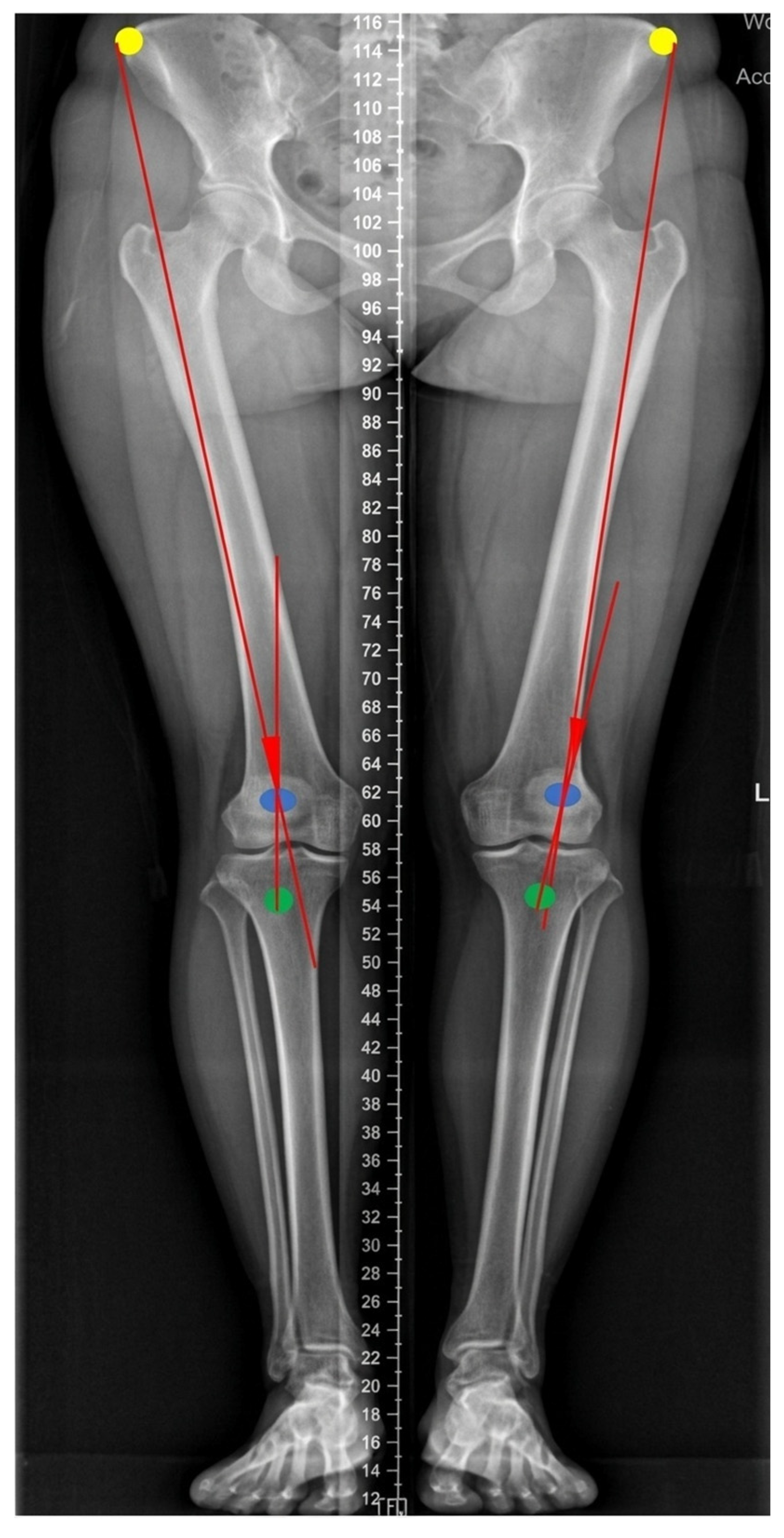
2.3.3. Alignment Parameter of the Knee
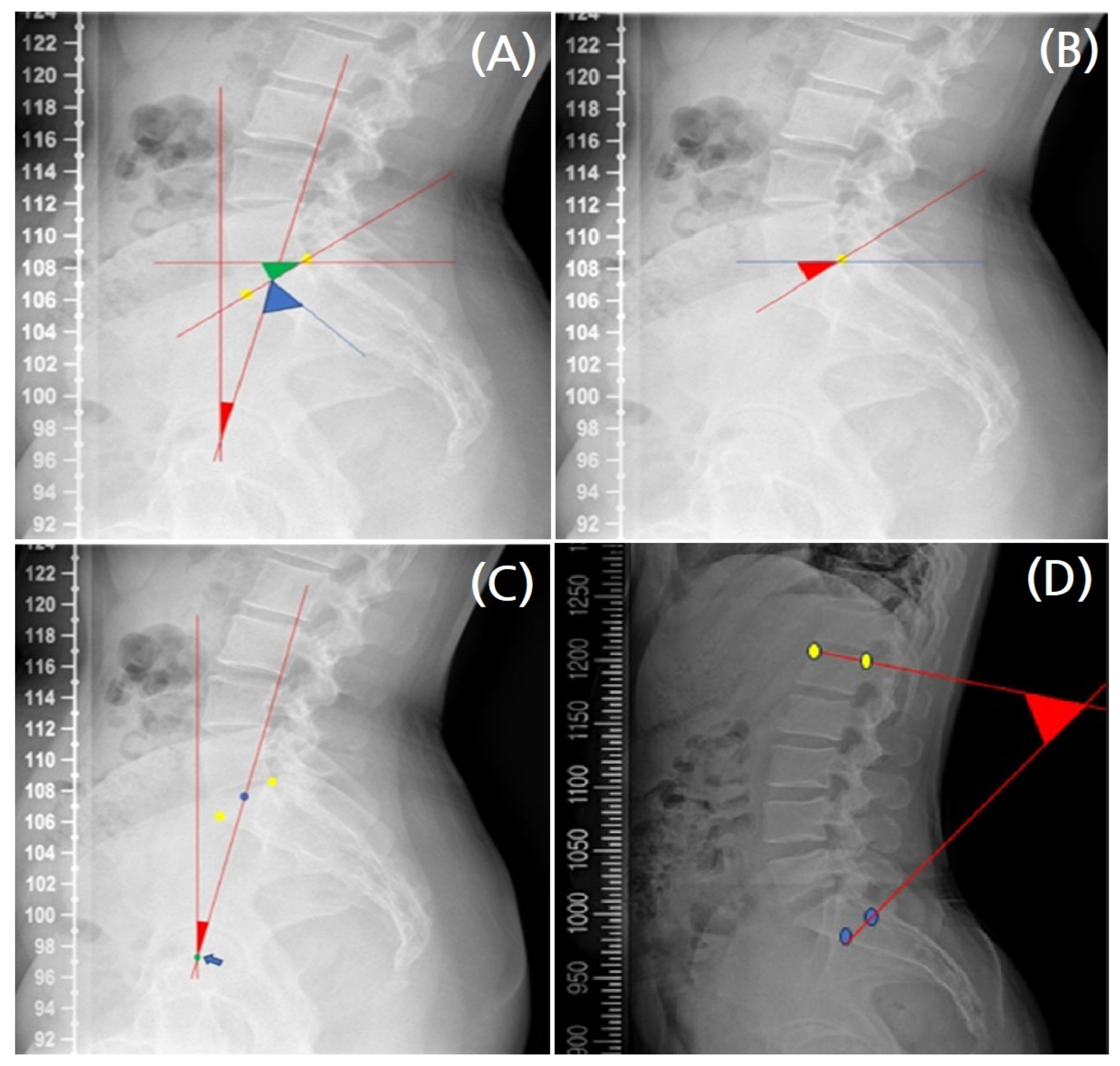
2.3.4. Alignment Parameter of Pelvis
2.3.5. Alignment Parameter of the Lumbosacral Region
2.4. Data Extraction
2.5. Statistical Analysis
3. Results
3.1. Demographic Data of Participants
3.2. Analysis of Participants’ CIA Measurements
3.3. Body Alignment Parameter Measurements and ANOVA Analysis of Mean Parameters According to Foot Structure
4. Discussion
5. Conclusions
Author Contributions
Funding
Institutional Review Board Statement
Informed Consent Statement
Data Availability Statement
Acknowledgments
Conflicts of Interest
Abbreviations
| CMM | Chuna Manual Medicine |
| CIA | Calcaneal Inclination Angle |
| TTA | Tibiotalar Tilt Angle |
| TA | Tibiotalar Angle |
| Q-angle | Quadriceps Angle |
| PI | Pelvic Incidence |
| PT | Pelvic Tilt |
| SS | Sacral Slope |
| LL | L1–S1 Lordosis |
| AP | Anteroposterior |
| PACS | Picture Archiving and Communication System |
| SD | Standard Deviation |
| ANOVA | Analysis of Variance |
References
- Xue, X.; Wang, Y.; Xu, X.; Li, H.; Li, Q.; Na, Y.; Tao, W. Postural control deficits during static single-leg stance in chronic ankle instability: A systematic review and meta-analysis. Am. J. Sports Med. 2024, 52, 142–153. [Google Scholar] [CrossRef] [PubMed]
- Kim, J.S.; Lee, M.Y. The effect of short foot exercise using visual feedback on the balance and accuracy of knee joint movement in subjects with flexible flatfoot. Medicine 2020, 99, e19260. [Google Scholar] [CrossRef] [PubMed]
- Kakad, M.; Chandanwale, A.; Kabre, D.R.; Mangwalkar, V.A.; N.K, S. Study of association of back pain with foot disorders like calcaneal spur, foot span, foot pain, and foot posture. Indian J. Orthop. Surg. 2025, 11, 122–129. [Google Scholar] [CrossRef]
- Nair, P.; Deland, J.; Ellis, S.J. Current concepts in adult acquired flatfoot deformity. Curr. Orthop. Pract. 2015, 26, 160–168. [Google Scholar] [CrossRef]
- Abousayed, M.M.; Alley, M.C.; Shakked, R.; Rosenbaum, A.J. Adult-acquired flatfoot deformity: Etiology, diagnosis, and management. JBJS Rev. 2017, 5, e7. [Google Scholar] [CrossRef] [PubMed]
- Lee, J.-H.; Woo, H.; Jang, J.-S.; Kim, J.I.; Na, Y.C.; Kim, K.-R.; Cho, E.; Lee, J.-H.; Park, T.-Y. Comparison of concordance between Chuna manual therapy diagnostic methods (palpation, X-ray, artificial intelligence program) in lumbar spine: An exploratory, cross-sectional clinical study. Diagnostics 2022, 12, 2732. [Google Scholar] [CrossRef]
- Lee, J.-S.; Park, K.-W.; Kim, H.-T.; Park, S.-Y.; Shin, B.-C. Systematic review of the diagnosis of pelvic deviation for Chuna manual therapy. J. Korean Med. Rehabil. 2022, 32, 83–94. [Google Scholar] [CrossRef]
- Khamis, S.; Dar, G.; Peretz, C.; Yizhar, Z. The relationship between foot and pelvic alignment while standing. J. Hum. Kinet. 2015, 45, 39–46. [Google Scholar] [CrossRef]
- Ghasemi, M.S.; Koohpayehzadeh, J.; Kadkhodaei, H.; Ehsani, A.A. The effect of foot hyperpronation on spine alignment in standing position. Med. J. Islam. Repub. Iran. 2016, 30, 466. [Google Scholar]
- Korean Society of Chuna Manual Medicine for Spine & Nerve. Chuna Manual Medicine, 2.5th ed.; Korean Society of Chuna Manual Medicine for Spine & Nerve: Seoul, Republic of Korea, 2017. [Google Scholar]
- Lamm, B.M.; Stasko, P.A.; Gesheff, M.G.; Bhave, A. Normal foot and ankle radiographic angles, measurements, and reference points. J. Foot Ankle Surg. 2016, 55, 991–998. [Google Scholar] [CrossRef]
- Yaka, H.; Kesik, K.; Başbuğ, V. Is medial or lateral localization of osteochondral lesions of talus related to foot angles? J. Orthop. Surg. Res. 2023, 18, 96. [Google Scholar] [CrossRef]
- Koca, T.; Göğebakan, H.; Koçyiğit, B.; Nacitarhan, V. Foot functions in ankylosing spondylitis. Clin. Rheumatol. 2019, 38, 979–985. [Google Scholar] [CrossRef]
- Burgener, F.A.; Kormano, M.; Pudas, T. Differential Diagnosis in Conventional Radiology, 3rd ed.; Thieme Medical Publishers: New York, NY, USA, 2007. [Google Scholar]
- Gao, F.; Ma, J.; Sun, W.; Guo, W.; Li, Z.; Wang, W. The influence of knee malalignment on the ankle alignment in varus and valgus gonarthrosis based on radiographic measurement. Eur. J. Radiol. 2016, 85, 228–232. [Google Scholar] [CrossRef]
- Levangie, P.K.; Norkin, C.C.; Lewek, M.D. Joint Structure and Function: A Comprehensive Analysis, 6th ed.; F.A. Davis Company: Philadelphia, PA, USA, 2018. [Google Scholar]
- Blom, A.; Warwick, D.; Whitehouse, M. Apley & Solomon’s System of Orthopaedics and Trauma, 10th ed.; CRC Press: Boca Raton, FL, USA, 2017. [Google Scholar]
- Oatis, C.A. Kinesiology: The Mechanics and Pathomechanics of Human Movement, 3rd ed.; Lippincott Williams & Wilkins: Philadelphia, PA, USA, 2017. [Google Scholar]
- Ramadan, A.G.; Elhafez, K.A.A.; Negm, M.A. Assessment of pelvic parameters before and after postero-lateral interbody fusion surgery in patients with lumbo-sacral instability. Egypt. J. Hosp. Med. 2018, 73, 6830–6837. [Google Scholar]
- Ohashi, M.; Hasegawa, K.; Hatsushikano, S.; Imai, N.; Tashi, H.; Makino, T.; Minato, K.; Sato, M.; Watanabe, K. Anatomical pelvic parameters using the anterior pelvic plane: Normative values and estimation of the standing sagittal alignment in healthy volunteers. Spine Surg. Relat. Res. 2023, 8, 39–47. [Google Scholar] [CrossRef] [PubMed]
- Celestre, P.C.; Dimar, J.R., 2nd; Glassman, S.D. Spinopelvic parameters: Lumbar lordosis, pelvic incidence, pelvic tilt, and sacral slope: What does a spine surgeon need to know to plan a lumbar deformity correction? Neurosurg. Clin. N. Am. 2018, 29, 323–329. [Google Scholar] [CrossRef] [PubMed]
- Ryu, S.M.; Lee, T.K.; Lee, S.H. Prevalence of flatfoot among young Korean males and the correlation among flatfoot angles measured in weight-bearing lateral radiographs. Medicine 2022, 101, e29720. [Google Scholar] [CrossRef]
- Menz, H.B.; Dufour, A.B.; Riskowski, J.L.; Hillstrom, H.J.; Hannan, M.T. Foot posture, foot function and low back pain: The Framingham Foot Study. Rheumatology 2013, 52, 2275–2282. [Google Scholar] [CrossRef]
- Umunnah, J.; Ogbueche, C.; Uchenwoke, C. Association of tibiofemoral angle, quadriceps angle and body mass index in a selected adolescent population. Afr. Health Sci. 2020, 20, 1274–1282. [Google Scholar] [CrossRef]
- Kondratiev, A.; Smetankina, N.; Staude, V. Biomechanical analysis of stress–strain distribution in the lumbar spine–sacrum–pelvis system with emphasis on sacroiliac joint dysfunction. Prosthesis 2024, 7, 4. [Google Scholar] [CrossRef]
- Tim, S.; Mazur-Bialy, A. The most common functional disorders and factors affecting female pelvic floor. Life 2021, 11, 1397. [Google Scholar] [CrossRef]
- Teraguchi, M.; Hashizume, H.; Asai, Y.; Oka, H.; Nagata, K.; Ishimoto, Y.; Iwasaki, H.; Tsutsui, S.; Takami, M.; Tanaka, S.; et al. Association between Modic changes, disc degeneration, and pelvic incidence-lumbar lordosis mismatch in a large population-based cohort: The Wakayama Spine Study. Eur. Spine J. 2023; Epub ahead of printing. [Google Scholar] [CrossRef]
- Koçyiğit, B.; Nacitarhan, V.; Koca, T.; Berk, E. Lumbosacral alignment in lumbar disc herniation. Ann. Med. Res. 2019, 26, 2069–2073. [Google Scholar] [CrossRef]
- Wang, J.; Li, Y.; Yang, G.; Jin, K. Age-related dysfunction in balance: A comprehensive review of causes, consequences, and interventions. Aging Dis. 2024, 15, 225–240. [Google Scholar] [CrossRef] [PubMed]
- Korean Society of Chuna Manual Medicine for Spine & Nerve. Chuna Manual Medicine Academy Basic Training Course Workbook; Korean Society of Chuna Manual Medicine for Spine & Nerve: Seoul, Republic of Korea, 2024; Volume 1. [Google Scholar]
- Khoury, A.; Hatem, M.; Bowler, J. Hip–spine syndrome: Rationale for ischiofemoral impingement, femoroacetabular impingement and abnormal femoral torsion leading to low back pain. J. Hip Preserv. Surg. 2020, 7, 390–400. [Google Scholar] [CrossRef] [PubMed]
- Davids, J.R.; Gibson, T.W.; Pugh, L.I. Quantitative segmental analysis of weight-bearing radiographs of the foot and ankle for children: Normal alignment. J. Pediatr. Orthop. 2005, 25, 769–776. [Google Scholar] [CrossRef] [PubMed]
- Lee, K.T.; Kim, K.C.; Park, Y.U.; Kim, T.W.; Lee, Y.K. Radiographic evaluation of foot structure following fifth metatarsal stress fracture. Foot Ankle Int. 2011, 32, 796–801. [Google Scholar] [CrossRef]
- Murley, G.S.; Menz, H.B.; Landorf, K.B. A protocol for classifying normal- and flat-arched foot posture for research studies using clinical and radiographic measurements. J. Foot Ankle Res. 2009, 2, 22. [Google Scholar] [CrossRef]
- Bai, T.; Yang, X.; Yao, Z.; Zhou, X.; Wu, H.; Li, B.; Wang, J.; Sun, Y. Convolutional network for plantar pressure parsing. In Proceedings of the 2021 14th International Congress on Image and Signal Processing, BioMedical Engineering and Informatics (CISP-BMEI), Shanghai, China, 23–25 October 2021; IEEE: Piscataway, NJ, USA, 2021. [Google Scholar]
- Wang, M.; Gu, Y.; Baker, J. Analysis of foot kinematics wearing high heels using the Oxford foot model. Technol. Health Care 2018, 26, 125–133. [Google Scholar] [CrossRef]
- Herchenröder, M.; Wilfling, D.; Steinhäuser, J. Evidence for foot orthoses for adults with flatfoot: A systematic review. J. Foot Ankle Res. 2021, 14, 57. [Google Scholar] [CrossRef]
- Maynou, C.; Szymanski, C.; Thiounn, A. The adult cavus foot. EFORT Open Rev. 2017, 2, 221–229. [Google Scholar] [CrossRef]
- Sanpera, I.; Villafranca-Solano, S.; Muñoz-Lopez, C.; Sanpera-Iglesias, J. How to manage pes cavus in children and adolescents? EFORT Open Rev. 2021, 6, 466–474. [Google Scholar] [CrossRef]
- Lau, B.; Allahabadi, S.; Palanca, A. Understanding radiographic measurements used in foot and ankle surgery. J. Am. Acad. Orthop. Surg. 2022, 30, 81–91. [Google Scholar] [CrossRef] [PubMed]
- Lee, Y.-C.; Lin, G.; Wang, M.-J.J. Comparing 3D foot scanning with conventional measurement methods. J. Foot Ankle Res. 2014, 7, 44. [Google Scholar] [CrossRef] [PubMed]
- Barton, C.J.; Levinger, P.; Crossley, K.M.; Webster, K.E.; Menz, H.B. The relationship between rearfoot, tibial, and hip kinematics in individuals with patellofemoral pain syndrome. Clin. Biomech. 2012, 27, 702–705. [Google Scholar] [CrossRef] [PubMed]
- Lee, J.H.; Kim, C.G.; Jo, D.C.; Moon, S.J.; Park, T.Y.; Ko, Y.S.; Nam, H.W.; Lee, J.H. Diagnostic X-ray from the perspective of Chuna manual medicine, based on the listing system of spinal and pelvic subluxation. J. Korea Churna Man. Med. Spine Nerve 2014, 9, 1–14. [Google Scholar]



| Variable | (n = 100) | |
|---|---|---|
| Sex | Male (n) | 36 |
| Female (n) | 64 | |
| Age (years; mean [SD]) | 32.5 (11.3) | |
| Blood pressure | Systolic (mmHg; mean [SD]) | 120.6 (10.5) |
| Diastolic (mmHg; mean [SD]) | 75.6 (8.5) | |
| Heart rate (bpm; mean [SD]) | 78.9 (10.6) | |
| Body temperature (°C; mean [SD]) | 36.7 (0.3) | |
| Height (cm; mean [SD]) | 165.0 (8.2) | |
| Weight (kg; mean [SD]) | 62.8 (10.8) | |
| Body mass index (kg/m2; mean [SD]) | 22.9 (2.7) | |
| Variable | N | Mean ± SD (°) | Sub-Variable | N | Mean ± SD (°) |
|---|---|---|---|---|---|
| CIA (Rt.) | 100 | 24.01 ± 4.81 | Pes planus | 5 | 15.06 ± 2.51 |
| Normal | 57 | 21.50 ± 2.38 | |||
| Pes cavus | 38 | 28.97 ± 2.68 | |||
| CIA (Lt.) | 100 | 24.29 ± 4.75 | Pes planus | 5 | 15.60 ± 1.52 |
| Normal | 58 | 21.82 ± 2.08 | |||
| Pes cavus | 37 | 29.34 ± 2.90 |
| Variable | Sub-Variable | N | Mean ± SD (°) | F | p-Value | Variable | Sub-Variable | N | Mean ± SD (°) | F | p-Value |
|---|---|---|---|---|---|---|---|---|---|---|---|
| TTA—Rt. foot | All | 100 | 2.01 ± 3.09 | 0.37 | >0.05 | TTA—Lt. foot | All | 100 | 1.56 ± 1.35 | 1.09 | >0.05 |
| Pes planus | 5 | 1.47 ± 0.87 | Pes planus | 5 | 1.45 ± 0.93 | ||||||
| Normal | 57 | 1.84 ± 1.34 | Normal | 58 | 1.69 ± 1.15 | ||||||
| Pes cavus | 38 | 2.34 ± 4.74 | Pes cavus | 37 | 2.60 ± 4.84 | ||||||
| TA—Rt. foot | All | 100 | 91.68 ± 3.69 | 0.41 | >0.05 | TA—Lt. foot | All | 100 | 91.50 ± 3.64 | 0.79 | >0.05 |
| Pes planus | 5 | 90.88 ± 2.10 | Pes planus | 5 | 91.32 ± 0.65 | ||||||
| Normal | 57 | 91.29 ± 0.49 | Normal | 58 | 91.32 ± 0.55 | ||||||
| Pes cavus | 38 | 91.91 ± 0.55 | Pes cavus | 37 | 92.29 ± 0.50 | ||||||
| Q-angle—Rt. foot | All | 100 | 10.08 ± 6.29 | 0.28 | >0.05 | Q-angle—Lt. foot | All | 100 | 10.35 ± 6.46 | 2.01 | >0.05 |
| Pes planus | 5 | 11.87 ± 10.08 | Pes planus | 5 | 10.87 ± 5.00 | ||||||
| Normal | 57 | 10.19 ± 6.10 | Normal | 58 | 10.55 ± 5.24 | ||||||
| Pes cavus | 38 | 9.68 ± 6.18 | Pes cavus | 37 | 10.80 ± 6.79 | ||||||
| PI—Rt. foot | All | 100 | 53.79 ± 14.68 | 2.09 | >0.05 | PI—Lt. foot | All | 100 | 53.79 ± 14.68 | 0.61 | >0.05 |
| Pes planus | 5 | 56.66 ± 8.81 | Pes planus | 5 | 60.52 ± 10.19 | ||||||
| Normal | 57 | 56.07 ± 15.10 | Normal | 58 | 53.87 ± 14.90 | ||||||
| Pes cavus | 38 | 50.00 ± 14.13 | Pes cavus | 37 | 52.77 ± 14.92 | ||||||
| PT—Rt. foot | All | 100 | 13.18 ± 8.02 | 3.30 | 0.041 | PT—Lt. foot | All | 100 | 13.18 ± 8.02 | 0.36 | >0.05 |
| Pes planus | 5 | 12.70 ± 1.43 | Pes planus | 5 | 15.99 ± 1.43 | ||||||
| Normal | 57 | 14.90 ± 8.79 | Normal | 58 | 13.24 ± 1.12 | ||||||
| Pes cavus | 38 | 10.68 ± 6.62 | Pes cavus | 37 | 12.73 ± 1.26 | ||||||
| SS—Rt. foot | All | 100 | 40.61 ± 10.82 | 0.58 | >0.05 | SS—Lt. foot | All | 100 | 40.61 ± 10.82 | 0.37 | >0.05 |
| Pes planus | 5 | 43.95 ± 8.77 | Pes planus | 5 | 44.53 ± 9.57 | ||||||
| Normal | 57 | 41.18 ± 11.21 | Normal | 58 | 40.63 ± 11.08 | ||||||
| Pes cavus | 38 | 39.32 ± 39.32 | Pes cavus | 37 | 40.05 ± 10.73 | ||||||
| LL—Rt. foot | All | 100 | 58.02 ± 10.78 | 0.19 | >0.05 | LL—Lt. foot | All | 100 | 58.02 ± 10.78 | 0.23 | >0.05 |
| Pes planus | 5 | 59.20 ± 9.46 | Pes planus | 5 | 60.64 ± 9.21 | ||||||
| Normal | 57 | 58.47 ± 10.81 | Normal | 58 | 58.21 ± 10.99 | ||||||
| Pes cavus | 38 | 57.18 ± 11.10 | Pes cavus | 37 | 57.35 ± 10.85 |
| Variable | Sub-Variable | N | Pearson r | p-Value | Variable | Sub-Variable | N | Pearson r | p-Value |
|---|---|---|---|---|---|---|---|---|---|
| TTA—Rt. foot | Pes planus | 5 | 0.4135 | >0.05 | TTA—Lt. foot | Pes planus | 5 | −0.1639 | >0.05 |
| Normal | 57 | 0.1760 | >0.05 | Normal | 58 | −0.1376 | >0.05 | ||
| Pes cavus | 38 | −0.1525 | >0.05 | Pes cavus | 37 | −0.0476 | >0.05 | ||
| TA—Rt. foot | Pes planus | 5 | −0.0291 | >0.05 | TA—Lt. foot | Pes planus | 5 | −0.3247 | >0.05 |
| Normal | 57 | −0.0519 | >0.05 | Normal | 58 | −0.0939 | >0.05 | ||
| Pes cavus | 38 | 0.2218 | >0.05 | Pes cavus | 37 | 0.0711 | >0.05 | ||
| Q-angle—Rt. foot | Pes planus | 5 | 0.2611 | >0.05 | Q-angle—Lt. foot | Pes planus | 5 | −0.1630 | >0.05 |
| Normal | 57 | −0.0474 | >0.05 | Normal | 58 | −0.1088 | >0.05 | ||
| Pes cavus | 38 | −0.0831 | >0.05 | Pes cavus | 37 | −0.3623 | 0.0275 | ||
| PI—Rt. Foot | Pes planus | 5 | −0.6009 | >0.05 | PI—Lt. foot | Pes planus | 5 | −0.5802 | >0.05 |
| Normal | 57 | −0.1758 | >0.05 | Normal | 58 | −0.3308 | 0.0112 | ||
| Pes cavus | 38 | −0.5677 | 0.0002 | Pes cavus | 37 | −0.1048 | >0.05 | ||
| PT—Rt. foot | Pes planus | 5 | 0.15180 | >0.05 | PT—Lt. foot | Pes planus | 5 | 0.4326 | >0.05 |
| Normal | 57 | −0.05155 | >0.05 | Normal | 58 | −0.1120 | >0.05 | ||
| Pes cavus | 38 | 0.13980 | >0.05 | Pes cavus | 37 | −0.0452 | >0.05 | ||
| SS—Rt. foot | Pes planus | 5 | −0.6289 | >0.05 | SS—Lt. foot | Pes planus | 5 | −0.7628 | >0.05 |
| Normal | 57 | −0.1962 | >0.05 | Normal | 58 | −0.3587 | 0.0057 | ||
| Pes cavus | 38 | −0.1255 | >0.05 | Pes cavus | 37 | −0.1134 | >0.05 | ||
| LL—Rt. foot | Pes planus | 5 | −0.02871 | >0.05 | LL—Lt. foot | Pes planus | 5 | −0.1631 | >0.05 |
| Normal | 57 | −0.09353 | >0.05 | Normal | 58 | −0.1691 | >0.05 | ||
| Pes cavus | 38 | −0.56770 | 0.0002 | Pes cavus | 37 | −0.3355 | 0.0423 |
Disclaimer/Publisher’s Note: The statements, opinions and data contained in all publications are solely those of the individual author(s) and contributor(s) and not of MDPI and/or the editor(s). MDPI and/or the editor(s) disclaim responsibility for any injury to people or property resulting from any ideas, methods, instructions or products referred to in the content. |
© 2026 by the authors. Licensee MDPI, Basel, Switzerland. This article is an open access article distributed under the terms and conditions of the Creative Commons Attribution (CC BY) license.
Share and Cite
Han, Y.; Jeon, S.; Woo, H.; Ha, W.; Park, T.-Y.; Lee, J.-H.; Lee, J. Association Between Calcaneal Inclination Angle and Spinal and Lower Limb Alignment: A Retrospective Radiographic Analysis. Diagnostics 2026, 16, 921. https://doi.org/10.3390/diagnostics16060921
Han Y, Jeon S, Woo H, Ha W, Park T-Y, Lee J-H, Lee J. Association Between Calcaneal Inclination Angle and Spinal and Lower Limb Alignment: A Retrospective Radiographic Analysis. Diagnostics. 2026; 16(6):921. https://doi.org/10.3390/diagnostics16060921
Chicago/Turabian StyleHan, Yunhee, Seojae Jeon, Hyeonjun Woo, Wonbae Ha, Tae-Yong Park, Jin-Hyun Lee, and Junghan Lee. 2026. "Association Between Calcaneal Inclination Angle and Spinal and Lower Limb Alignment: A Retrospective Radiographic Analysis" Diagnostics 16, no. 6: 921. https://doi.org/10.3390/diagnostics16060921
APA StyleHan, Y., Jeon, S., Woo, H., Ha, W., Park, T.-Y., Lee, J.-H., & Lee, J. (2026). Association Between Calcaneal Inclination Angle and Spinal and Lower Limb Alignment: A Retrospective Radiographic Analysis. Diagnostics, 16(6), 921. https://doi.org/10.3390/diagnostics16060921