Systematic Review

Clinical Implications of the Localization and Morphological Variability of the Mental Foramen—A Systematic Review


by
Mariola Krzykawska-Krupska
1,
Janusz Pach
1,*,
Piotr Regulski
1,
Jacek Tomczyk
2,
Izabela Strużycka
3,
Kazimierz Szopiński
4,
Katarzyna Osipowicz
5,6 and
Anna Pogorzelska
4
1
Digital Imaging and Virtual Reality Laboratory, Department of Dental and Maxillofacial Radiology, Medical University of Warsaw, ul. Binieckiego St. 6, 02-097 Warsaw, Poland
2
Institute of Biological Sciences, Cardinal Stefan Wyszyński University, Wóycickiego St. 1/3, 01-938 Warsaw, Poland
3
Department of Comprehensive Dentistry, Medical University of Warsaw, Binieckiego St. 6, 02-097 Warsaw, Poland
4
Department of Dentomaxillofacial Radiology, Faculty of Dental Medicine, Medical University of Warsaw, Binieckiego St. 6, 02-097 Warsaw, Poland
5
Department of Dermatology and Venereology, Infant Jesus Clinical Hospital, Koszykowa St. 82a, 02-008 Warsaw, Poland
6
Faculty of Health Sciences, University of Kalisz, Nowy Świat St. 4, 62-800 Kalisz, Poland
*
Author to whom correspondence should be addressed.
Diagnostics 2026, 16(5), 779; https://doi.org/10.3390/diagnostics16050779

Submission received: 10 January 2026 / Revised: 22 February 2026 / Accepted: 23 February 2026 / Published: 5 March 2026
(This article belongs to the Special Issue Recent Achievements in Oral and Maxillofacial Radiology)

Abstract

Background: The mental foramen is a key anatomical structure of the mandible through which the mental nerve and accompanying vessels emerge. Accurate knowledge of its location and morphology is essential for safe dental and surgical procedures in the anterior mandible. Objective: This study was conducted as a systematic review to summarize current evidence on the morphology, localization, and anatomical variants of the mental foramen and their clinical relevance. Methods: The PubMed and Google Scholar databases were searched for studies published between 2015 and 2025 in accordance with current systematic review guidelines. Cone-beam computed tomography (CBCT) studies and anthropological investigations assessing the position, dimensions, and anatomical variants of the mental foramen were included. Results: Thirty-five studies (30 CBCT-based and 5 anthropological) comprising a total of 6240 mandibles or patients were analyzed qualitatively. Considerable variability was observed in the horizontal and vertical position of the mental foramen in relation to mandibular borders and dental landmarks. Anatomical variations included differences in size and shape, the presence of unilateral or bilateral accessory mental foramina, and rare cases of unilateral or bilateral absence of the foramen. Conclusions: The synthesis of recent CBCT and anthropological data across diverse populations highlights clinically relevant patterns of variability. This study identifies key positional patterns and variants of the mental foramen, which can inform clinical planning and help reduce the risk of mental nerve injury.

1. Introduction

The mental foramen (foramen mentale) is a key anatomical structure of the mandibular body [1]. It constitutes the opening through which the mental nerve and accompanying vessels emerge, supplying sensation to the lower lip, oral mucosa, and gingiva. Understanding its localization, morphology, and potential anatomical variations is crucial in oral surgery, implantology, endodontics, and radiological diagnostics [2]. Precise identification of the mental foramen is indispensable for safe planning of invasive procedures in the anterior mandible, helping minimize neurological complications arising from mental nerve injury, such as paresthesia [3,4,5,6,7].
Individual and population-based variability in the location and number of mental foramina has been reported in numerous radiological and clinical studies [3,4]. Previous reviews often focused on single populations or lacked the most recent CBCT data published after 2019, limiting the ability to formulate generalized clinical conclusions. Existing systematic reviews and meta-analyses typically address only selected aspects (e.g., CBCT imaging or the prevalence of accessory foramina) and cover literature only up to approximately 2020, further justifying the need for an updated and expanded analysis.
The present study is based on the latest international literature and highlights the importance of considering the position and potential duplications or absence of the mental foramen during surgical procedures in the anterior mandible. Morphometric data from imaging and anthropological research are integrated with their practical implications for clinical decision-making across various dental specialties.
The aim of this review was to summarize current knowledge regarding mental foramen morphology, its location in relation to key anatomical landmarks, its associated anomalies, and the clinical relevance of these features for dental and surgical practice. The study was conducted as a literature review in accordance with PRISMA 2020 guidelines [5].
Compared to recent systematic reviews published between 2019 and 2020, and incorporating the most recent studies published from 2020 to 2025, this review provides several novel contributions. First, it includes CBCT-based studies from a broader range of populations worldwide, rather than focusing on single populations or limited regions. Second, it systematically compiles data on rare anatomical variants, such as accessory mental foramina, multiple foramina, and cases of absence of the mental foramen, which were not comprehensively addressed in earlier reviews. Third, it integrates both vertical and horizontal positional data of the mental foramen relative to teeth and mandibular borders, enhancing clinical applicability. Finally, the review translates these morphometric findings into practical guidance for implant placement, endodontic procedures, and surgical planning, highlighting “safety zones” for individualized patient care. Collectively, by including the most up-to-date evidence and a wider global perspective, these aspects fill important gaps in the literature and provide a more comprehensive understanding of mental foramen anatomy across diverse populations.

2. Materials and Methods

2.1. Search Strategy and Data Extraction

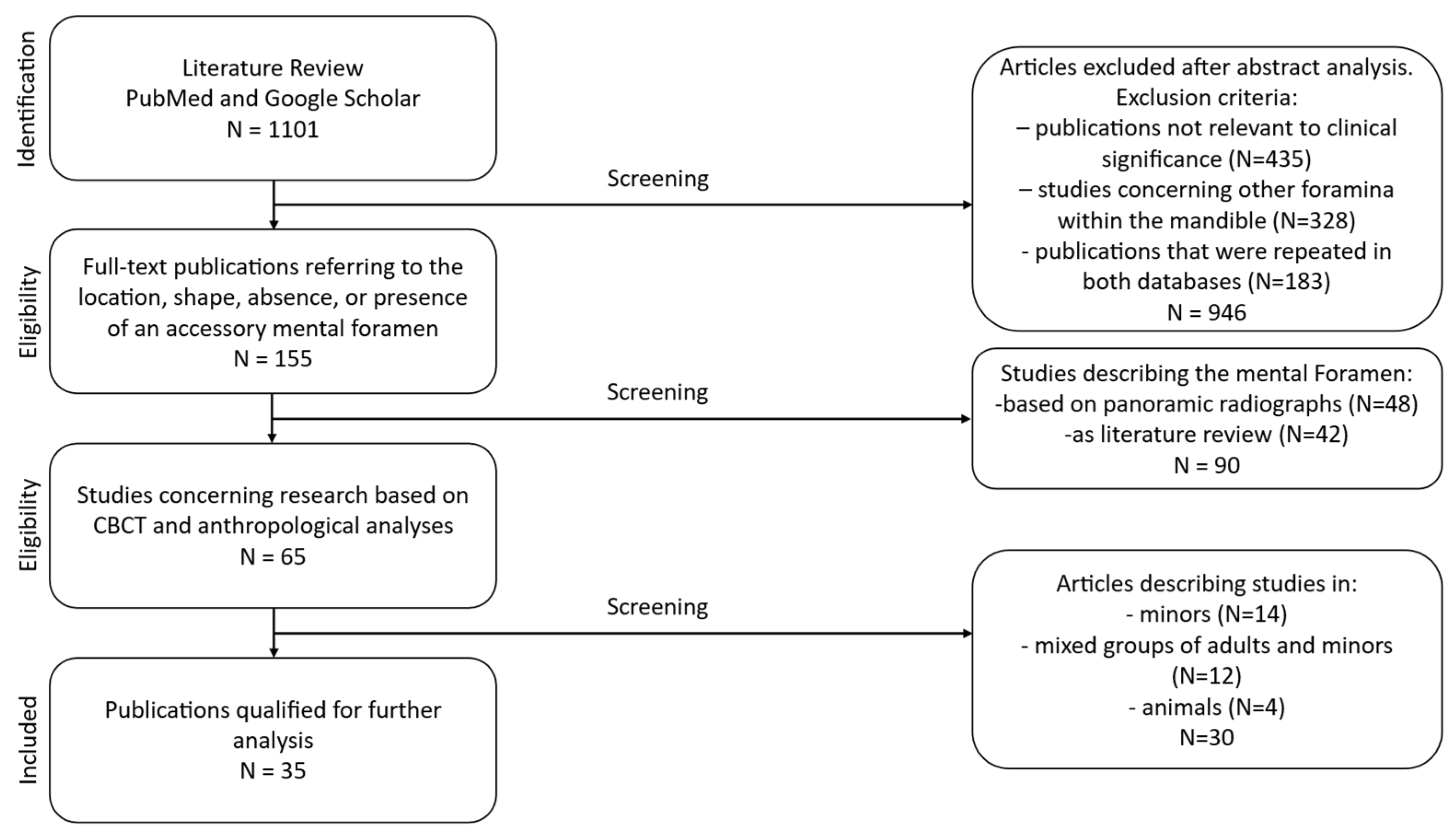
The PubMed and Google Scholar databases were searched for studies published from 2015 to 2025 using the keywords “foramen mentale” and “radiology” and the filter “full text”. As a consequence, grey literature (theses, conference proceedings, and other non-indexed reports) was not systematically searched. A total of 1101 publications were identified, of which 946 were excluded after abstract screening. Studies not directly related to clinical investigations and those examining foramina other than the mental foramen were excluded. Publications that were repeated in both databases were excluded too. The remaining 155 full-text articles were assessed for information concerning localization, shape, and the presence or absence of accessory foramina.
Study selection and data extraction were performed independently by two reviewers to ensure methodological rigor and reduce potential bias. Each reviewer screened titles, abstracts, and full texts according to the predefined eligibility criteria. Any discrepancies between the two reviewers were resolved through discussion, and when consensus could not be reached, a third reviewer was consulted. This approach ensured consistency and reliability in the inclusion of studies and the extraction of relevant data.

2.2. Eligibility Criteria

Inclusion criteria comprised CBCT-based studies and anthropological investigations. Original observational studies on adult patients or skeletal specimens, regardless of sex or ethnicity, were included. Excluded were panoramic radiograph–based studies and literature reviews. Of the 155 full-text articles assessed, 90 were excluded at the full-text stage because they did not meet the predefined inclusion criteria. Sixty-five publications met the initial criteria, and after excluding studies involving minors, studies involving mixed-age populations without separable adult data, and preclinical studies describing the mental foramen in animal models rather than in humans, 35 papers were retained for final analysis. Articles in English and Polish were evaluated. The review followed the PRISMA 2020 framework, including title/abstract screening and full-text assessment. The PRISMA flowchart [Figure 1] presents the selection process, the number of records identified, duplicate removal, screening exclusions, and full-text rejections. Ultimately, 35 studies (30 CBCT and 5 anthropological) involving 6240 mandibles/patients from various ethnic groups were included.

2.3. Risk of Bias and Data Synthesis

Due to substantial heterogeneity in measurement protocols and reported parameters, which precluded meaningful data harmonization, a quantitative meta-analysis was not performed; therefore, a qualitative synthesis was conducted. The PRISMA 2020 checklist accompanies this review in the Supplementary Materials. The methodological quality and risk of bias of the included studies were assessed using the Joanna Briggs Institute (JBI) Critical Appraisal Tools (2025 version), applying the checklist for analytical cross-sectional studies and, where appropriate, the checklist for case reports. The results of the risk of bias assessment are presented in Supplementary Tables S1 and S2. Overall, most studies were judged to be of low to moderate risk of bias.

3. Results

The distribution of studies by methodology, population characteristics, and key morphometric parameters is presented in Table 1. Morphometric values for the analyzed variables are summarized in Table 2.
Across the included studies, the mental foramen showed variation in horizontal and vertical position, morphology, and the presence of anatomical variants, as summarized in Table 1 and Table 2.
The mental foramen was most frequently located horizontally between the first and second premolars (position III according to the Tebo and Telford classification), with alignment with the second premolar (position IV) reported more often in selected populations, including Indian, Chilean, and Sudanese cohorts. Vertically, the mental foramen was predominantly positioned below the root apex line across most CBCT and anthropological studies, regardless of population. Morphologically, an oval shape was reported more commonly than round or irregular forms, while anatomical variants such as accessory mental foramina were uncommon and typically reported in a minority of cases. Rare findings, including multiple or absent mental foramina, were limited to isolated case reports.
Although a formal meta-analysis was not feasible due to substantial heterogeneity in measurement protocols, reporting formats, and units of analysis (patients, mandibles, or sides), an alternative quantitative synthesis was performed. Specifically, for studies reporting extractable prevalence data, horizontal mental foramen positions were mapped to the Tebo and Telford classification, and study-level prevalences were summarized using medians (Table 3). This approach allows quantitative comparison of the most commonly reported positions, such as position III and position IV.
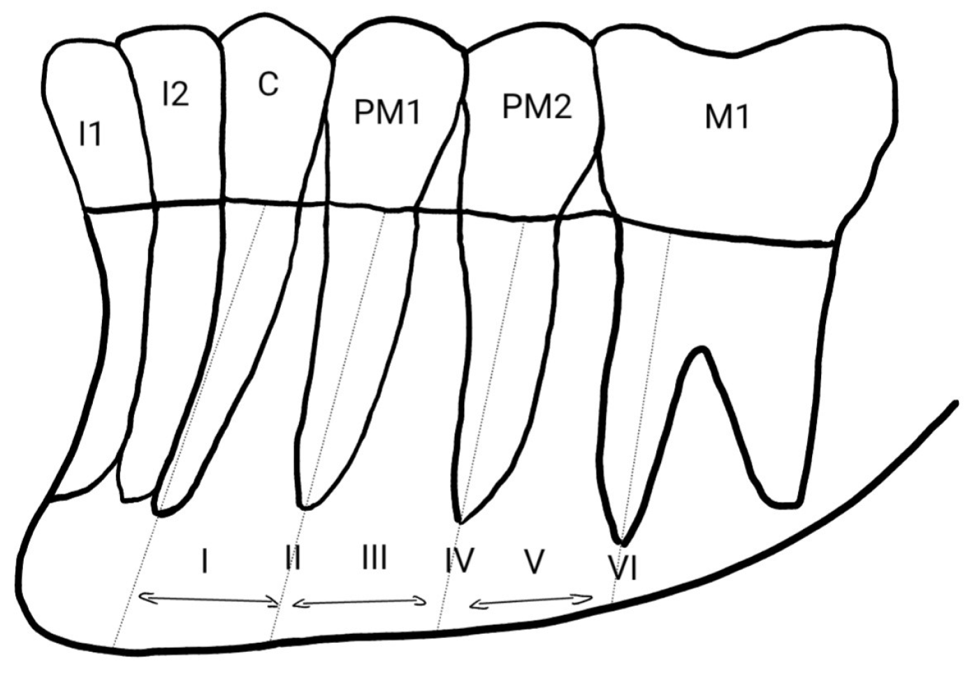
To determine the horizontal position of the mental foramen relative to the mandibular dentition, the Tebo and Telford classification is commonly employed. According to this system, the foramen may be located in six positions: mesial to the first premolar (I); in line with the first premolar apex (II); between the first and second premolar roots (III); in line with the second premolar roots (IV); between the second premolar and first molar roots (V); or in line with the mesial root of the first molar (VI) [Figure 2]. Most researchers found the foramen in position III—between the first and second premolars [4,9,10,11,12].
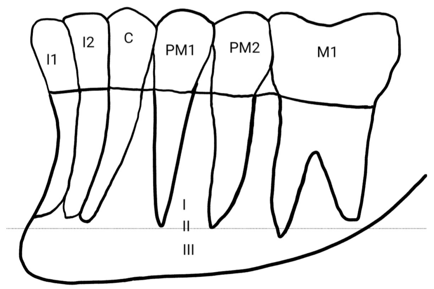
The vertical position classification proposed by Zmysłowska-Polakowska et al. identifies three positions: above the apical line (I), in line with the apices (II), and below the apical line (III) [Figure 3]. This latter location was most frequently reported in the other publications analyzed [8,18].
Knowledge of the precise location of the mental foramen is also crucial for effective local anesthesia delivery, influencing treatment success and minimizing complications during procedures such as endodontic therapy, apical surgery, periodontal operations, and extractions [8,19]. CBCT provides highly accurate localization [7], whereas panoramic radiographs may fail to reveal of this anatomical structure [16].
One anomaly is the presence of an accessory mental foramen, which typically appears as a small opening near the main foramen, usually located distally and connected to the mandibular canal [11]. A foramen not connected to the canal is referred to as a nutrient foramen [20]. Accessory foramina may occur unilaterally [21] or bilaterally [20]. Cases reporting two accessory foramina on the same side have also been documented [16]. Those connected to the mandibular canal contain neurovascular bundles, and their injury can cause complications such as lower lip numbness [1].
A rare (ranged from 0.02–0.47%) anomaly involves unilateral [22] or bilateral absence of the mental foramen [17]. Cases combining absence on one side and duplication on the other have also been reported [22].
Authors also observed differences in size and shape related to sex and ethnicity, describing the foramen as round, oval, or irregular [3,10,11,23,24].

4. Discussion

4.1. Position of the Mental Foramen in Relation to the Superior and Inferior Border of the Mandible

Studies conducted on Palestinian, Nepalese, Iranian and Vietnamese populations have shown that the mean distance from the mental foramen to the inferior border of the mandible is greater than its distance to the alveolar crest [6,7,19,25]. In contrast, Ahmed et al., examining a Saudi Arabian population, demonstrated that the mental foramen was located closer to the inferior border of the mandible in both women and men. Their study also showed that the greatest distance between the mental foramen and the inferior border occurred in individuals over 56 years of age, whereas the greatest distance from the alveolar crest was found in participants aged 26–40 years [24].
These discrepancies may reflect a combination of biological and methodological factors. Age-related alveolar bone resorption and tooth loss lead to progressive reduction in the distance between the mental foramen and the superior mandibular border, whereas the distance to the inferior border remains relatively stable. However, population-specific craniofacial morphology, differences in dental status of study samples, and variations in CBCT acquisition protocols and landmark definitions may also contribute substantially to the observed variability. This suggests that normative reference values derived from one population may not be directly transferable to others, underscoring the importance of individualized radiological assessment.

4.2. Position of the Mental Foramen in Relation to Tooth Groups

Many authors assess the position of the mental foramen in relation to tooth groups using the classification proposed by Tebo and Telford [4,8,18]. Other researchers report its location with reference to adjacent tooth groups [6,9,25,26]. Studies carried out in Polish [8], Brazilian [27], Syrian [18], Italian [26] and Saudi [3,28,29] populations most commonly found the mental foramen located between the first and second premolars. Similar results were obtained by Reda et al. in another Italian population [13]. Comparable results were also reported by Oliveira et al. in their study of the Brazilian population [30]. This location was consistent on both sides of the mandible.
Different conclusions were reported by researchers from Chile [12], India [9,10], Jordan [4], Sudan [25] and Palestine [6], who most frequently observed the foramen aligned with the long axis of the second premolar. In these studies, the frequency of this location was similar on the left and right sides.
Studies in a Romanian population showed that the mental foramen was most commonly located below the second premolar on the left side, while on the right side it was most often positioned between the premolars [14]. Subramanian et al. demonstrated in their study of the Zambian population that the position of the mental foramen, on both the right and left sides, was most frequently located between the second premolar and the first molar [31].
These interpopulation differences likely reflect complex interactions between genetic background, mandibular growth patterns, dental arch morphology and functional loading related to diet and mastication. In addition, methodological variation—particularly differences in reference points, classification systems and CBCT acquisition protocols—may partially explain inconsistent findings between studies. Together, these factors highlight the need for population-specific datasets and standardized reporting frameworks in future research.

4.3. Vertical Position of the Mental Foramen

According to the classification proposed by Zmysłowska-Polakowska et al., describing the vertical position of the mental foramen, all authors investigating the location of this structure found that it most commonly appears in position III, i.e., below the root apex line [8,18,26].
This relative uniformity suggests that, despite substantial horizontal positional variability, the vertical relationship of the mental foramen to adjacent tooth apices may be more conserved across populations. Nevertheless, advanced alveolar bone resorption, particularly in partially edentulous and edentulous patients, may significantly alter this relationship and should always be evaluated using CBCT.

4.4. Shape of the Mental Foramen

Most studies assessing the shape of the mental foramen reported that it is predominantly oval [3,15,21,26]. However, Sheth et al. found that its shape was round in the majority of their study population [9].
These discrepancies may reflect true population-specific morphological variation or differences in image resolution, slice thickness and observer interpretation. From a clinical perspective, shape variability may influence detectability of the foramen and the apparent width of the neurovascular bundle, reinforcing the importance of high-quality three-dimensional imaging in surgical planning.

4.5. Presence of Accessory Mental Foramina, Absence of the Mental Foramen and Its Rare Variation

An accessory mental foramen is usually a small opening in the mandibular body that communicates with the mandibular canal and is typically located near the main mental foramen [11]. The reviewed literature includes cases describing unilateral accessory foramina [6,21,26,32], while other authors reported accessory foramina present bilaterally [11,20]. Thomaidi et al. documented a case of two accessory mental foramina on one side [1]. Yovchev et al. described an instance of an accessory foramen on the right side with simultaneous absence of the main foramen on the left [22]. Khaled et al. reported a patient with unilateral absence of the mental foramen on the left side while demonstrating normal sensory function of the lips, gingiva and chin [33]. Lauhr et al. reported a bilateral absence of the mental foramen in the mandible of a Caucasian male [17]. Borghesi et al. described a case of a patient with the presence of five mental foramina [34]. Predoiu et al. reported that in a 28-year-old European female, three mental foramina were present on the right side. On the left side, a single mental foramen was observed, located between the apices of the premolar roots [35].
A particularly rare case involving the mental foramen was described by Tallada et al. In their examination of the mandible of a 31-year-old woman, they identified the presence of an accessory mental foramen on the right side, located lingually. Both foramina (the buccal and the lingual) were anatomically connected [36].
This configuration challenges the conventional assumption of exclusively buccal emergence of the mental nerve and underscores the necessity of multiplanar CBCT evaluation rather than reliance on two-dimensional imaging alone.
These findings emphasize that reliance on expected anatomical patterns alone may lead to iatrogenic nerve injury, particularly during implant placement, apical surgery or flap elevation. Comprehensive CBCT assessment is therefore essential to identify accessory canals, bifurcations and atypical exit points of the mental nerve.
The present review enabled the compilation of data from various regions of the world, revealing both recurrent topographic patterns of the mental foramen and significant population-specific differences. This represents added value compared with earlier studies focused on single populations or older specimen series.
Specifically, unlike earlier narrative reviews or single-population analyses, the present study integrates CBCT-based morphometric data from multiple geographic regions, systematically summarizes rare anatomical variants, and explicitly links anatomical variability with clinical risk management strategies. In doing so, it addresses gaps in the literature regarding the global variability of mental foramen topography and its relevance to contemporary implantology and oral surgery.

4.6. Limitations

This review has several limitations. The included studies differed substantially in terms of CBCT protocols, exposure parameters, definitions of anatomical landmarks, and population characteristics, making a quantitative meta-analysis impossible. Not all studies reported sampling strategies, calibration procedures, or measurement conditions in sufficient detail, which increases the risk of systematic bias.
Panoramic radiograph studies were deliberately excluded to obtain the most precise and comparable morphometric data regarding the mental foramen. CBCT allows three-dimensional assessment of mandibular structures without geometric distortions or superimposition of tissues, which are inherent limitations of panoramic imaging. However, we recognize that panoramic studies constitute a substantial portion of the literature and may provide useful comparative information. Their exclusion may introduce selection bias and affect the overall representativeness of the review.
Limiting the review to full-text articles published in English and Polish may also have resulted in language bias, excluding relevant research published in other languages. Additionally, publication bias should be considered, as studies reporting rare anatomical variants or statistically significant findings may be overrepresented. Finally, applying a full-text availability restriction and not systematically searching grey literature (theses, conference proceedings, and other non-indexed reports) may have led to missing relevant evidence.
These factors should be kept in mind when interpreting the results and generalizing the conclusions. Despite these limitations, we believe the review provides a comprehensive synthesis of CBCT-based morphometric and topographic data on the mental foramen, including rare anatomical variants, which is valuable for both clinical and anthropological applications.

4.7. Clinical Implications

The collected data confirm that in most studied populations, the mental foramen is most commonly located between and below the apices of the first and second premolars or below the second premolar, below the root apex line. This information is essential when planning implant placement, root apex resections and mucoperiosteal flap design in the anterior mandible. Considering anatomical variations—such as the presence of an anterior loop, accessory foramina or positioning closer to the lower mandibular border—allows for establishing individualized “safety zones” for each patient based on CBCT assessment, thus minimizing the risk of injury to the mental nerve.
However, the substantial interindividual and interpopulation variability observed across studies suggests that fixed “safe distances” based solely on average anatomical values may be insufficient. Instead, individualized CBCT-based risk assessment should be used to define patient-specific safety margins.
Based on the analyzed studies, several parameters should be routinely included in CBCT reports: vertical and horizontal position of the mental foramen, distance to adjacent root apices, relationship to the mandibular borders, and presence of anatomical variants (accessory foramina, anterior loop). Standardizing these descriptions would facilitate comparison between centers and improve surgical and implantological safety.

4.8. Future Research Directions

The findings indicate several areas requiring further investigation.
First, there is a lack of large, prospective CBCT studies that simultaneously account for age, sex, degree of alveolar bone resorption, dental status and ethnic background as potential determinants of mental foramen position.
Second, studies combining morphometric analysis of the mental foramen with treatment outcomes (e.g., rates of neurological complications after implant placement or apical surgery) are needed to verify the clinical value of proposed “safety zones”.
Another promising direction is the use of machine learning and automated segmentation in CBCT imaging to detect mental foramina and their anatomical variants. Developing and validating such tools could support treatment planning, particularly in patients with atypical morphology or advanced bone loss.
Finally, large, well-characterized cranial collections from diverse geographic regions should be included in anthropological studies to better understand population and evolutionary determinants of mental foramen variability and their relevance to contemporary clinical practice.

5. Conclusions

This systematic review provides a comprehensive summary of the anatomical variations in the mental foramen across different populations. Our findings confirm that the mental foramen may vary in shape (oval, round, or irregular), vertical position (closer to the alveolar crest or inferior border), and horizontal location (ranging from mesial to the first premolar to the mesial root of the first molar). Rare anomalies, such as unilateral or bilateral accessory foramina or absence of the foramen, were also identified.
The MF most commonly corresponded to position III (between the first and second premolars), with a median prevalence of 56% and a wide interpopulation range of 26–92%. Position IV was also frequently reported (median 51.3%). Vertically, the MF was predominantly located below the root apex line, with frequencies reaching 48–92% in CBCT-based studies.
Morphometric data showed that the distance between the MF and the inferior mandibular border ranged from 10 to 16 mm, while the anterior loop length reached up to 7.5 mm. Accessory mental foramina were observed in 1–12% of cases, whereas complete absence of the MF was rare (approximately 0.02–0.47%).
The compiled data offer practical guidance for clinicians in oral surgery, implantology, orthodontics, and endodontics, emphasizing the importance of assessing the position of the mental foramen to minimize the risk of mental nerve injury and related post-operative complications. This review serves as a basis for standardizing CBCT reporting and developing population-specific recommendations, supporting individualized clinical planning for implant placement, endodontic procedures, and surgical interventions in the anterior mandible.

Supplementary Materials

The following supporting information can be downloaded at: https://www.mdpi.com/article/10.3390/diagnostics16050779/s1, Table S1: Risk of bias appraisal of included observational studies using the JBI Critical Appraisal Checklist for Analytical Cross-Sectional Studies. Table S2: Risk of bias appraisal of included case reports using the JBI Critical Appraisal Checklist for Case Reports. Table S3: PRISMA 2020 Checklist.

Author Contributions

Conceptualization, M.K.-K. and J.P.; methodology, M.K.-K., J.P. and P.R.; software, P.R.; validation, M.K.-K., J.T., I.S. and P.R.; formal analysis, M.K.-K.; investigation, M.K.-K., J.P., A.P. and K.O.; resources, M.K.-K. and K.S.; data curation, M.K.-K. and J.P.; writing—original draft preparation, M.K.-K. and J.P.; writing—review and editing, M.K.-K., K.S., J.T. and I.S.; visualization, M.K.-K. and P.R.; supervision, K.S., J.T., I.S. and P.R. All authors have read and agreed to the published version of the manuscript.

Funding

This research received no external funding.

Institutional Review Board Statement

Not applicable.

Informed Consent Statement

Not applicable.

Data Availability Statement

No new data were created or analyzed in this study.

Conflicts of Interest

The authors declare no conflicts of interest.

References

  1. Thomaidi, Z.M.; Tsatsarelis, C.; Papadopoulos, V. Accessory Mental Foramina in Dry Mandibles: An Observational Study Along with Systematic Review and Meta-Analysis. Dent. J. 2025, 13, 94. [Google Scholar] [CrossRef] [Scilit]
  2. Mahmoudinezhad, S.S.; AryanKia, A.; Shooshtari, S.S.; Moradi, K. The Effect of Mandibular Angulation on Preoperative Assessment of Dental Implant Insertion at Premolar Region: CBCT Study. BioMed Res. Int. 2022, 2022, 7879239. [Google Scholar] [CrossRef] [Scilit] [PubMed] [PubMed Central]
  3. AlQahtani, N.A. Assessment of the position and level of mental nerve for placement of implants using cone-beam computed tomography & panoramic radiograph in the Saudi population. Saudi Dent. J. 2022, 34, 315–320. [Google Scholar] [CrossRef] [Scilit] [PubMed] [PubMed Central]
  4. Al Dalalah, A.; Kana’an, N.; Ersheidat, A.; Momani, M.; Altantawi, H. Evaluating the Anterior Loop of the Mental Nerve Using Cone Beam CT Scans in the Jordanian Population. Cureus 2024, 16, e58519. [Google Scholar] [CrossRef] [Scilit] [PubMed] [PubMed Central]
  5. Page, M.J.; McKenzie, J.E.; Bossuyt, P.M.; Boutron, I.; Hoffmann, T.C.; Mulrow, C.D.; Shamseer, L.; Tetzlaff, J.M.; Akl, E.A.; Brennan, S.E.; et al. The PRISMA 2020 statement: An updated guideline for reporting systematic reviews. BMJ 2021, 372, n71. [Google Scholar] [CrossRef] [Scilit]
  6. Abu-Ta’a, M.F.; Qubain, K.J.; Beshtawi, K.R. The mental foramen, anatomical parameters through a radiographic approach to aid in dental implantology: A retrospective analysis in a sample of a Palestinian population. Heliyon 2023, 9, e13886. [Google Scholar] [CrossRef] [Scilit] [PubMed] [PubMed Central]
  7. Mallahi, E.; Abesi, F.; Rajaei-Rad, F.; Gholinia, H. Anatomical characteristics of mental foramen and canal: A cone-beam computed tomography analysis. J. Clin. Exp. Dent. 2024, 16, 1004–1011. [Google Scholar] [CrossRef] [PubMed] [PubMed Central]
  8. Zmyslowska-Polakowska, E.; Radwanski, M.; Ledzion, S.; Leski, M.; Zmyslowska, A.; Lukomska-Szymanska, M. Evaluation of Size and Location of a Mental Foramen in the Polish Population Using Cone-Beam Computed Tomography. BioMed Res. Int. 2019, 2019, 1659476. [Google Scholar] [CrossRef] [Scilit] [PubMed] [PubMed Central]
  9. Sheth, K.; Banga, K.S.; Pawar, A.M.; Gutmann, J.L.; Kim, H.C. Shape and anatomical relationship of the mental foramen to the mandibular premolars in an Indian sub-population: A retrospective CBCT analysis. Restor. Dent. Endod. 2021, 47, e1. [Google Scholar] [CrossRef] [Scilit] [PubMed] [PubMed Central]
  10. Srivastava, S.; Sethi, K.K.; Sinha, A.; Srivastava, A.; Mishra, A.; Khan, N.; Pandey, S.; Sethi, K. Assessing the Prevalence and Length of the Anterior Loop of Inferior Alveolar Nerve and the Position and Dimension of Mental Foramen Using Cone Beam Computed Tomography. Cureus 2024, 16, e68535. [Google Scholar] [CrossRef] [Scilit]
  11. Barbosa, D.A.F.; Maferano, E.F.E.; Teixeira, R.C.; Kurita, L.M.; Pimenta, A.V.M.; Silva, P.G.B.; Chaves, F.N.; Gurgel, M.L.; Costa, F.W.G. Epidemiological and Radiomorphometric Aspects of the Accessory Mental Foramen in Brazilian Individuals: An Analysis by Cone Beam Computed Tomography. J. Clin. Exp. Dent. 2024, 16, 1379–1385. [Google Scholar] [CrossRef] [Scilit] [PubMed] [PubMed Central]
  12. Guzmán, J.; Abarca, J.; Navarro, P.; Garay, I.; Arnabat-Domínguez, J.; Betancourt, P. Morphometric Analysis of the Mandibular Canal and Its Anatomical Variants in a Chilean Subpopulation: Cone Beam Computed Tomography Study. Diagnostics 2024, 14, 1914. [Google Scholar] [CrossRef] [Scilit] [PubMed] [PubMed Central]
  13. Reda, R.; Zanza, A.; Bhandi, S.; Biase, A.; Testarelli, L.; Miccoli, G. Surgical-anatomical evaluation of mandibular premolars by CBCT among the Italian population. Dent. Med. Probl. 2022, 59, 209–216. [Google Scholar] [CrossRef] [Scilit] [PubMed]
  14. Gherghiţă, O.R.; Csiki, I.E.; Bordea, E.N.; Pellegrini, A.; Cismaş, S.C.; Motaş, N.; Nimigean, V.R.; Nimigean, V. Morphometric study for determining the anteroposterior position of the mental foramen in dentate human subjects. Rom. J. Morphol. Embryol. 2021, 62, 517–523. [Google Scholar] [CrossRef] [Scilit] [PubMed] [PubMed Central]
  15. Shashidhar, K.; Sridevi, N.S. Anthropometric analysis of mental foramen in dry human mandibles in Karnataka. Natl. J. Clin. Anat. 2019, 8, 148–152. [Google Scholar] [CrossRef] [Scilit]
  16. Mohebiniya, M.; Kamani, M. Multiple Mental Foramina: A Rare Anatomical Variation Detected by Cone-Beam Computed Tomography (CBCT). Cureus 2024, 16, e63524. [Google Scholar] [CrossRef] [Scilit] [PubMed] [PubMed Central]
  17. Lauhr, G.; Coutant, J.C.; Normand, E.; Laurenjoye, M.; Ella, B. Bilateral absence of mental foramen in a living human subject. Surg. Radiol. Anat. 2015, 37, 403–405. [Google Scholar] [CrossRef] [Scilit] [PubMed]
  18. Haffaf, R.A.; Younes, M.M.; Shqera, A.Y.; Assad, M.; Hasan, A.K. Assessment of Mental Foramen Characteristics in Syrian Population Using Cone Beam Computed Tomography: A Pilot Study. Cureus 2024, 16, e58549. [Google Scholar] [CrossRef] [Scilit] [PubMed] [PubMed Central]
  19. Singh, P.; Adhikari, B.; Koju, S.; Gupta, S.; Pradhan, M.; Gurung, D.; Joshi, U. Mean Distance of Mental Foramen from Inferior Border of Mandible among Patients Visiting the Outpatient Dental Department in a Tertiary Care Centre. JNMA J. Nepal Med. Assoc. 2023, 61, 787–790. [Google Scholar] [CrossRef] [Scilit] [PubMed] [PubMed Central]
  20. Karabıyık, Z.; Kıranatlı, M. A Rare Anatomical Finding: Bilateral Accessory Mental Foramen. Case Rep. Dent. 2021, 2021, 6020515. [Google Scholar] [CrossRef] [Scilit] [PubMed] [PubMed Central]
  21. Tiwari, N. Left Accessory Mental Foramen in Dry Mandibles in Department of Anatomy in a Medical College: A Descriptive Cross-sectional Study. JNMA J. Nepal Med. Assoc. 2022, 60, 805–807. [Google Scholar] [CrossRef] [Scilit] [PubMed] [PubMed Central]
  22. Yovchev, D.; Mihaylova, H.; Kirilova, J.; Deliverska, E. Rare anatomic variations: Agenesis of left mental foramen coexisting with two right mental foramina. J. IMAB 2024, 30, 5645–5647. [Google Scholar] [CrossRef] [Scilit]
  23. Alam, B.F.; Yusof, A.; Shah, S.A.; Abdullah, J.Y.; Nawi, M.A.A. Morphometric Analysis of the Dimension and Distance to Anatomical Structures From the Mental Foramen Using Cone Beam Computed Tomography. Cureus 2024, 16, e71496. [Google Scholar] [CrossRef] [Scilit]
  24. Ahmed, A.A.; Ahmed, R.M.; Jamleh, A.; Spagnuolo, G. Morphometric Analysis of the Mandibular Canal, Anterior Loop, and Mental Foramen: A Cone-Beam Computed Tomography Evaluation. Int. J. Environ. Res. Public Health 2021, 18, 3365. [Google Scholar] [CrossRef] [Scilit] [PubMed] [PubMed Central]
  25. Le, L.N.; Do, T.T.; Truong, L.T.; Truong, M.H.; Ngoc, D.K.H.; Nguyen, L.M.; Nguyen, L., Jr. Cone Beam CT Assessment of Mandibular Foramen and Mental Foramen Positions as Essential Anatomical Landmarks: A Retrospective Study in Vietnam. Cureus 2024, 16, e59337. [Google Scholar] [CrossRef] [Scilit]
  26. Taschieri, S.; Vitelli, C.; Albano, D.; Sconfienza, L.; Del Fabbro, M.; Francetti, L.; Parrini, M.; Corbella, S. Evaluation of mental foramen and inferior alveolar nerve canal and its relationship to adjacent anatomical landmarks using cone-beam computer tomography. J. Biol. Regul. Homeost. Agents 2021, 35, 107–115. [Google Scholar] [CrossRef] [Scilit] [PubMed]
  27. Dall’Azen, G.C.; Morosolli, A.R.C.; Hartmann, R.C.; Rockenbach, M.I.B. Measurement of the mandibular canal and mandibular incisive canal using cone beam tomography in adults from South Brazil. J. Oral Sci. 2024, 66, 169–175. [Google Scholar] [CrossRef] [Scilit] [PubMed]
  28. Alazemi, A.; Muhairi, E.A.; Alharbi, N.; Abdunabi, F.; Mashyakhy, M.; Chaudhry, J.; Moharamzadeh, K.; Abed, R.E.; Jamal, M. Evaluation of Mental Foramen and Mandibular Canal Course and Location in an Emirati Subpopulation. Int. Dent. J. 2024, 74, 1142–1150. [Google Scholar] [CrossRef] [Scilit] [PubMed] [PubMed Central]
  29. Ghandourah, A.O.; Badaoud, M.B.; Dahlawi, A.; Alghamdi, A.; Alhazmi, F.; Sembawa, S.N.; Demyati, A.K. A radiographic analysis of the location of the mental foramen. Saudi Dent. J. 2023, 35, 354–358. [Google Scholar] [CrossRef] [Scilit] [PubMed] [PubMed Central]
  30. Dos Santos Oliveira, R.; Rodrigues Coutinho, M.; Kühl Panzarella, F. Morphometric Analysis of the Mental Foramen Using Cone-Beam Computed Tomography. Int. J. Dent. 2018, 2018, 4571895. [Google Scholar] [CrossRef] [Scilit] [PubMed] [PubMed Central]
  31. Subramanian, B.; Anthony, S.N.; Mubbunu, L.; Hachombwa, C.; Mlawa, M.S.; Majambo, M.M.; Sasi, R.M. Anthropometrics Analysis of Mental Foramen and Accessory Mental Foramen in Zambian Adult Human Mandibles. Sci. World J. 2019, 2019, 9093474. [Google Scholar] [CrossRef] [Scilit] [PubMed] [PubMed Central]
  32. Aljarbou, F.; Riyahi, A.M.; Altamimi, A.; Alabdulsalam, A.; Jabhan, N.; Aldosimani, M.; Alamri, H.M. Anatomy of the accessory mental foramen in a Saudi subpopulation: A multicenter CBCT study. Saudi Dent. J. 2021, 33, 1012–1017. [Google Scholar] [CrossRef] [Scilit] [PubMed] [PubMed Central]
  33. Beshtawi, K.R.; Qirresh, E. A rare case of an anatomical variant of nonexistent mental foramen. Radiol. Case Rep. 2024, 20, 1082–1086. [Google Scholar] [CrossRef] [Scilit] [PubMed] [PubMed Central]
  34. Borghesi, A.; Pezzotti, S.; Nocivelli, G.; Maroldi, R. Five mental foramina in the same mandible: CBCT findings of an unusual anatomical variant. Surg. Radiol. Anat. 2018, 40, 635–640. [Google Scholar] [CrossRef] [Scilit] [PubMed]
  35. Predoiu, M.; Rusu, M.C.; Chiriţă, A.L. A rare anatomic variation: Triple mental foramina. Morphologie 2019, 103, 110–115. [Google Scholar] [CrossRef] [Scilit] [PubMed]
  36. Tallada, A.K.; Ahmed, J.; Sujir, N.; Shenoy, N.; Pawar, S.M.; Muralidharan, A.; Mallya, S. Accessory lingual mental foramen: A case report of a rare anatomic variation. Oral Radiol. 2024, 40, 410–414. [Google Scholar] [CrossRef] [Scilit] [PubMed]
Figure 1. PRISMA flowchart.
Diagnostics 16 00779 g001
Figure 2. Horizontal position of MF according to Zmysłowska-Polakowska.
Diagnostics 16 00779 g002
Figure 3. Vertical position of MF according to Zmysłowska-Polakowska.
Diagnostics 16 00779 g003
Table 1. Characteristics of selected studies included in the review.
Study IDCountry/
Population
Sample Size (Mandibles/Patients)MethodMF Horizontal
Position
MF Vertical PositionVariants ReportedKey Finding
Abu-Ta’a et al., 2023 [6]Palestine106 casesCBCTIII (PM1–PM2)
49%
Below apex
48%
Accessory MF rarePredominant location of MF between the first and second premolars, observed in nearly half of the studied cases
Mallahi et al., 2024 [7]Iran355 casesCBCTII–IIIBelow apexAnterior loopMF most commonly located inferiorly, with frequent identification of an anterior loop in this population
Zmysłowska-
Polakowska et al., 2019 [8]
Poland201 casesCBCTIII
56%
Below apex
54%
Type III horizontal position was the most frequent pattern in the Polish population, exceeding half of the examined cases
Sheth et al., 2021 [9]India475 casesCBCTIII
71%
Below apex
66%
High predominance of type III horizontal position in the Indian population, observed in more than two-thirds of cases
Srivastava et al., 2024 [10]India250 casesCBCTPM2 62%Below apex
62%
Anterior loop (long)Anterior loop length reached up to 6–7 mm, representing a clinically relevant extension of the mental nerve
Barbosa et al., 2024 [11]Brazil250 casesCBCTIII
58%
Below apex
58%
Accessory MFAccessory mental foramina were detected in a notable proportion of cases, indicating a non-negligible prevalence in this population
Guzmán et al., 2024 [12]Chile342 casesCBCTPM2
62%
Below apex
62%
Alignment of the MF with the second premolar represented the dominant horizontal position in this Chilean subpopulation
Reda et al., 2022 [13]Italy492 casesCBCTIII
92%
Below apex
92%
Symmetrical positioning of MF was consistently observed on both sides of the mandible
Gherghiță et al., 2021 [14]Romania40 casesCBCTLeft: PM2/41% Right: PM1–PM2 56%Below apexDistinct side-related differences in horizontal MF position were identified between the left and right mandible
Shashidhar et al., 2019 [15]India180 casesAnthropometry IV
88%
Below apexThe mental foramen was most frequently positioned posterior to the second premolar, with no side-related differences observed
Mohebiniya et al., 2024 [16]
(case report)
Iran1 caseCBCTVariableMultiple MFRare anatomical variation characterized by bilateral duplication of MF identified on CBCT imaging
Lauhr et al., 2015 [17]
(case report)
Caucasus1 caseCBCTExtremely rare anatomical finding of bilateral absence of MF documented in a living subject
Footnote: MF position: II (in line with the first premolar apex), III (between the first and second premolar roots), IV (in line with the second premolar roots); PM1—first premolar, PM2—second premolar.
Table 2. Morphometric values.
ParameterReported RangeReported Mean ± SD (Where Available)Studies Reporting Mean (n)Notes
Distance MF–Inferior Border10–16 mm11.83 ± 1.83 mm,
11.54 mm; 13.52 mm
3Often greater in older patients due to alveolar bone resorption
Distance MF–Alveolar Crest7–12 mmnot reported0Greatly reduced in edentulous mandibles
Horizontal positionMostly position III (PM1–PM2)not reported0With exceptions in Sudanese, Indian, Chilean populations
Vertical positionPredominantly below root apicesnot reported0Type III (Zmysłowska-Polakowska)
MF shapeOval > Round > Irregularnot reported0Shape correlated with sex in some populations
Accessory MF1–12%not reported0Most commonly located distally to main MF
Anterior loop length1–7.5 mmRight: 6.43 ± 2.17 mm; Left: 6.56 ± 1.81 mm1Must be evaluated preoperatively
Footnote: III (MF position between the first and second premolar roots); PM1—first premolar, PM2—second premolar.
Table 3. Reported prevalence (%) of horizontal mental foramen positions according to the Tebo and Telford classification across included studies.
StudyIIIIIIIVVVI
Abu-Ta’a et al., 2023 [6]NRNR49NR6/5NR
Mallahi et al., 2024 [7]NRNRNRNRNRNR
Zmysłowska-Polakowska et al., 2019 [8] (right/left site)0/04.5/5.058.7/55.434.4/31.34.5/7.90/0
Sheth et al., 2021 [9] (right/left site)0/06/1026/1462/716/50/0
Srivastava et al., 2024 [10]01.027.261.69.80.4
Barbosa et al., 2024 [11]NRNRNRNRNRNR
Guzmán et al., 2024 [12]NR9.562.025.53.0NR
Reda et al., 2022 [13]NR892NRNRNR
Gherghiță et al., 2021 [14]NRNR5641NRNR
Shashidhar et al., 2019 [15]NRNRNR88NRNR
Median (%)0.08.856.051.36.40.4
Footnote: MF Position according the Tebo and Telford classification: I—mesial to the first premolar, II—in line with the first premolar apex, III—between the first and second premolar roots, IV—in line with the second premolar roots, V—between the second premolar and first molar roots, VI—in line with the mesial root of the first molar; NR = not reported.
Disclaimer/Publisher’s Note: The statements, opinions and data contained in all publications are solely those of the individual author(s) and contributor(s) and not of MDPI and/or the editor(s). MDPI and/or the editor(s) disclaim responsibility for any injury to people or property resulting from any ideas, methods, instructions or products referred to in the content.