Effect of Lymphatic Invasion on Survival and Recurrence After Liver Transplantation in Patients with Hepatocellular Carcinoma and Its Prognostic Significance
Abstract
1. Introduction
2. Materials and Methods
Statistical Analysis
3. Results
4. Discussion
5. Conclusions
Author Contributions
Funding
Institutional Review Board Statement
Informed Consent Statement
Data Availability Statement
Acknowledgments
Conflicts of Interest
References
- Vogel, A.; Meyer, T.; Sapisochin, G.; Salem, R.; Saborowski, A. Hepatocellular carcinoma. Lancet 2022, 400, 1345–1362. [Google Scholar] [CrossRef] [Scilit]
- Marrero, J.A.; Kulik, L.M.; Sirlin, C.B.; Zhu, A.X.; Finn, R.S.; Abecassis, M.M.; Roberts, L.R.; Heimbach, J.K. Diagnosis, staging, and management of hepatocellular carcinoma: 2018 practice guidance by the American Association for the study of liver diseases. Hepatology 2018, 68, 723–750. [Google Scholar] [CrossRef] [Scilit]
- Moon, H.; Choi, J.E.; Lee, I.J.; Kim, T.H.; Kim, S.H.; Ko, Y.H.; Kim, H.B.; Nam, B.H.; Park, J.-W. All-treatment array of hepatocellular carcinoma from initial diagnosis to death: Observation of cumulative treatments. J. Cancer Res. Clin. Oncol. 2017, 43, 2327–2339. [Google Scholar] [CrossRef] [Scilit]
- Al-Ameri, A.; Yu, X.; Zheng, S. Predictors of post-recurrence survival in hepatocellular carcinoma patients following liver transplantation: Systematic review and meta-analysis. Transplant. Rev. 2022, 36, 100676. [Google Scholar] [CrossRef] [Scilit]
- Degroote, H.; Geerts, A.; Verhelst, X.; Van Vlierberghe, H. Different models to predict the risk of recurrent hepatocellular carcinoma in the setting of liver transplantation. Cancers 2022, 14, 2973. [Google Scholar] [CrossRef] [Scilit]
- Sakamoto, K.; Ogawa, K.; Tohyama, T.; Ueno, Y.; Tamura, K.; Inoue, H.; Nakamura, T.; Watanabe, J.; Takai, A.; Takada, Y. Serosal invasion is a strong prognostic factor for hepatocellular carcinoma after hepatectomy. Hepatol. Res. 2018, 49, 419–431. [Google Scholar] [CrossRef] [Scilit]
- Brandão, A.B.M.; Rodriguez, S.; Marroni, C.A.; Junior, A.M.F.; Fernandes, M.V.; Mucenic, M. Performance of eight predictive models for hepatocellular carcinoma recurrence after liver transplantation: A comparative study. Ann. Hepatol. 2024, 29, 101184. [Google Scholar] [CrossRef] [Scilit] [PubMed]
- Wake, K.; Sato, T. “The sinusoid” in the liver: Lessons learned from the original definition by Charles Sedg-wick Minot (1900). Anat. Rec. 2015, 298, 2071–2080. [Google Scholar] [CrossRef] [Scilit] [PubMed]
- Trutmann, M.; Sasse, D. The lymphatics of the liver. Anat. Embryol. 1994, 190, 201–209. [Google Scholar] [CrossRef] [Scilit] [PubMed]
- Ohtani, O.; Ohtani, Y. Lymph circulation in the liver. Anat. Rec. 2008, 291, 643–652. [Google Scholar] [CrossRef] [Scilit]
- Pupulim, L.F.; Vilgrain, V.; Ronot, M.; Becker, C.D.; Breguet, R.; Terraz, S. Hepatic lymphatics: Anatomy and related diseases. Abdom. Imaging 2015, 40, 1997–2011. [Google Scholar] [CrossRef] [Scilit]
- Baluk, P.; Fuxe, J.; Hashizume, H.; Romano, T.; Lashnits, E.; Butz, S.; Vestweber, D.; Corada, M.; Molendini, C.; Dejana, E.; et al. Functionally specialized junctions between endothelial cells of lymphatic vessels. J. Exp. Med. 2007, 204, 2349–2362. [Google Scholar] [CrossRef] [Scilit]
- Rofstad, E.K.; Galappathi, K.; Mathiesen, B.S. Tumor interstitial fluid pressure-a link between tumor hypoxia, microvascular density, and lymph node metastasis. Neoplasia 2014, 16, 586–594. [Google Scholar] [CrossRef] [Scilit]
- Zhou, H.; Lei, P.J.; Padera, T.P. Progression of Metastasis through Lymphatic System. Cells 2021, 10, 627. [Google Scholar] [CrossRef] [Scilit]
- Roy, S.; Banerjee, P.; Ekser, B.; Bayless, K.; Zawieja, D.; Alpini, G.; Glaser, S.S.; Chakraborty, S. Targeting Lymphangiogenesis and Lymph Node Metastasis in Liver Cancer. Am. J. Pathol. 2021, 191, 2052–2063. [Google Scholar] [CrossRef] [Scilit]
- Yokomori, H.; Oda, M.; Kaneko, F.; Kawachi, S.; Tanabe, M.; Yoshimura, K.; Kitagawa, Y.; Hibi, T. Lymphatic marker podoplanin/D2-40 in human advanced cirrhotic liver re-evaluations of microlymphatic abnormalities. BMC Gastroenterol. 2010, 10, 131. [Google Scholar] [CrossRef] [Scilit]
- Fisher, S.B.; Patel, S.H.; Kooby, D.A.; Weber, S.; Bloomston, M.; Cho, C.; Hatzaras, I.; Schmidt, C.; Winslow, E.; Staley, C.A., III; et al. Lymphovascular and perineural invasion as selection criteria for adjuvant therapy in intrahepatic cholangiocarcinoma: A multi-institution analysis. HPB 2012, 14, 514–522. [Google Scholar] [CrossRef] [Scilit]
- Pinero, F.; Carrihlo, F.J.; Silva, M.O. Predictive models for recurrence risk of hepatocellular carcinoma after liver transplantation: Still an unmet need. Liver Int. 2017, 37, 648–650. [Google Scholar] [CrossRef] [Scilit] [PubMed]
- Kim, D.G.; Yim, S.H.; Min, E.K.; Choi, M.C.; Joo, D.J.; Kim, M.S.; Lee, J.G. Cumulative exposure to tacrolimus during early period after liver transplantation does not affect the recurrence of hepatocellular carcinoma. Sci. Rep. 2023, 13, 20236. [Google Scholar] [CrossRef] [Scilit] [PubMed]
- Todeschini, L.; Cristin, L.; Martinino, A.; Mattia, A.; Agnes, S.; Giovinazzo, F. The Role of mTOR Inhibitors after Liver Transplantation for Hepatocellular Carcinoma. Curr. Oncol. 2023, 30, 5574–5592. [Google Scholar] [CrossRef] [Scilit] [PubMed]
- Williams, C.S.; Leek, R.D.; Robson, A.M.; Banerji, S.; Prevo, R.; Harris, A.L.; Jackson, D.G. Absence of lymphangiogenesis and intratumoural lymph vessels in human metastatic breast cancer. J. Pathol. 2003, 200, 195–206. [Google Scholar] [CrossRef] [Scilit] [PubMed]
- De Ridder, J.A.M.; Knijn, N.; Wiering, B.; De Wilt, J.H.W.; Nagtegaal, I.D. Lymphatic Invasion is an Independent Adverse Prognostic Factor in Patients with Colorectal Liver Metastasis. Ann. Surg. Oncol. 2015, 22, 638–645. [Google Scholar] [CrossRef] [Scilit]
- Lupinacci, R.M.; Mello, E.S.; Pinheiro, R.S.; Marques, G.; Coelho, F.F.; Kruger, J.A.; Perini, M.V.; Herman, P. Intrahepatic Lymphatic Invasion but not Vascular Invasion is a Major Prognostic Factor after Resection of Colorectal Cancer Liver Metastases. World J. Surg. 2014, 38, 2089–2096. [Google Scholar] [CrossRef] [Scilit]
- Cornwell, L.B.; Mcmasters, K.M.; Chagpar, A.B. The Impact of Lymphovascular Invasion on Lymph Node Status in Patients with Breast Cancer. Am. Surg. 2011, 77, 874–877. [Google Scholar] [CrossRef] [Scilit]
- Royston, D.; Jackson, D.G. Mechanisms of lymphatic metastasis in human colorectal adenocarcinoma. J. Pathol. 2009, 217, 608–619. [Google Scholar] [CrossRef] [Scilit]
- Xia, F.; Wu, L.; Lau, W.-Y.; Li, G.; Huan, H.; Qian, C.; Ma, K.; Bie, P. Positive Lymph Node Metastasis Has a Marked Impact on the Long-Term Survival of Patients with Hepatocellular Carcinoma with Extrahepatic Metastasis. PLoS ONE 2014, 9, e95889. [Google Scholar] [CrossRef] [Scilit]
- Hasegawa, K.; Makuuchi, M.; Kokudo, N.; Izumi, N.; Ichida, T.; Kudo, M.; Ku, Y.; Sakamoto, M.; Nakashima, O.; Matsui, O.; et al. Impact of histologically confirmed lymph node metastases on patient survival after surgical resection for hepatocellular carcinoma: Report of a Japanese nationwide survey. Ann. Surg. 2014, 259, 166–170. [Google Scholar] [CrossRef] [Scilit]
- Sugino, T.; Yamaguchi, T.; Hoshi, N.; Kusakabe, T.; Ogura, G.; Goodison, S.; Suzuki, T. Sinusoidal tumor angiogenesis is a key component in hepatocellular carcinoma metastasis. Clin. Exp. Metastasis 2008, 25, 835–841. [Google Scholar] [CrossRef] [Scilit]
- Thelen, A.; Jonas, S.; Benckert, C.; Weichert, W.; Schott, E.; Bötcher, C.; Dietz, E.; Wiedenmann, B.; Neuhaus, P.; Scholz, A. Tumor-Associated Lymphangiogenesis Correlates with Prognosis after Resection of Human Hepatocellular Carcinoma. Ann. Surg. Oncol. 2009, 16, 1222–1230. [Google Scholar] [CrossRef] [Scilit] [PubMed]
- Sotiropoulos, G.; Molmenti, E.; Fouzas, I.; Sgourakis, G.; Radtke, A.; Malagó, M.; Lang, H. Liver Transplantation for Hepatocellular Carcinoma with Intrahepatic Lymphatic Invasion: Case Reports. Transplant. Proc. 2008, 40, 3213–3214. [Google Scholar] [CrossRef] [Scilit] [PubMed]
- Cioca, A.; Ceausu, A.R.; Marin, I.; Raica, M.; Cimpean, A.M. The multifaceted role of podoplanin expression in hepatocellular carcinoma. Eur. J. Histochem. 2017, 61, 2707. [Google Scholar] [CrossRef] [Scilit]
- Paduch, R. The role of lymphangiogenesis and angiogenesis in tumor metastasis. Cell. Oncol. 2016, 39, 397–410. [Google Scholar] [CrossRef] [Scilit]
- Korita, P.V.; Wakai, T.; Shirai, Y.; Sakata, J.; Takizawa, K.; Cruz, P.V.; Ajioka, Y.; Hatakeyama, K. Intrahepatic Lymphatic Invasion Independently Predicts Poor Survival and Recurrences after Hepatectomy in Patients with Colorectal Carcinoma Liver Metastases. Ann. Surg. Oncol. 2007, 14, 3472–3480. [Google Scholar] [CrossRef] [Scilit] [PubMed]
- Sonohara, F.; Nomoto, S.; Inokawa, Y.; Kanda, M.; Yamada, S.; Fujii, T.; Sugimoto, H.; Kodera, Y. Serosal invasion strongly associated with recurrence after curative hepatic resection of hepatocellular carcinoma: A retrospective study of 214 consecutive cases. Medicine 2015, 94, e602. [Google Scholar] [CrossRef] [Scilit] [PubMed]
- Xiaohong, S.; Huikai, L.; Feng, W.; Ti, Z.; Yunlong, C.; Qiang, L. Clinical Significance of Lymph Node Metastasis in Patients Undergoing Partial Hepatectomy for Hepatocellular Carcinoma. World J. Surg. 2010, 34, 1028–1033. [Google Scholar] [CrossRef] [Scilit]
- Liver Cancer Study Group of Japan. General Rules for the Clinical and Pathological Study of Primary Liver Cancer, 6th ed.; Kanehara Co., Ltd.: Tokyo, Japan, 2015; p. 94. [Google Scholar]
- Saif, M.W.; Knost, J.A.; Chiorean, E.G.; Kambhampati, S.R.; Yu, D.; Pytowski, B.; Qin, A.; Kauh, J.S.; O’Neil, B.H. Phase 1 study of the anti-vascular endothelial growth factor receptor 3 monoclonal antibody LY3022856/IMC-3C5 in patients with advanced and refractory solid tumors and advanced colorectal cancer. Cancer Chemother. Pharmacol. 2016, 78, 815–824. [Google Scholar] [CrossRef] [Scilit] [PubMed]
- Alitalo, A.; Detmar, M. Interaction of tumor cells and lymphatic vessels in cancer progression. Oncogene 2012, 31, 4499–4508. [Google Scholar] [CrossRef] [Scilit]



| Lymphatic Invasion (No) | Lymphatic Invasion (Yes) | p | |||||||||
|---|---|---|---|---|---|---|---|---|---|---|---|
| Mean ± sd/n% | Median | Mean ± sd/n% | Median | ||||||||
| Age (year) | 58.3 | ± | 8.2 | 60.0 | 57.2 | ± | 9.7 | 59.0 | 0.601 | m | |
| Gender | Female | 18 | 14.9% | 11 | 19.3% | X2 | |||||
| Male | 103 | 85.1% | 46 | 80.7% | |||||||
| BMI | 26.4 | ± | 3.7 | 26.0 | 26.8 | ± | 3.7 | 27.0 | 0.465 | m | |
| Meld | 12.2 | ± | 6.1 | 10.0 | 12.3 | ± | 5.6 | 11.0 | 0.689 | m | |
| AFP (ng/mL) | 68.0 | ± | 267.7 | 7.5 | 138.5 | ± | 336.8 | 23.0 | 0.001 | m | |
| Tumor Diameter (mm) | 31.1 | ± | 20.0 | 25.0 | 41.1 | ± | 23.4 | 40.0 | 0.003 | m | |
| Number Of HCC Lesions | 2.2 | ± | 2.2 | 1.0 | 3.6 | ± | 3.4 | 3.0 | 0.000 | m | |
| Child–Pugh Score | A | 60 | 49.6% | 22 | 38.6% | 0.047 | X2 | ||||
| B | 57 | 47.1% | 28 | 49.1% | |||||||
| C | 4 | 3.3% | 7 | 12.3% | |||||||
| Locoregional Treatment | (no) | 93 | 76.9% | 31 | 54.4% | 0.002 | X2 | ||||
| (yes) | 28 | 23.1% | 26 | 45.6% | |||||||
| Tumor Differentiation Grade | well | 20 | 16.5% | 14 | 24.6% | 0.000 | X2 | ||||
| moderate | 53 | 43.8% | 37 | 64.9% | |||||||
| advanced | 48 | 39.7% | 6 | 10.5% | |||||||
| Microvascular Invasion | (no) | 87 | 71.9% | 16 | 28.1% | 0.000 | X2 | ||||
| (yes) | 34 | 28.1% | 41 | 71.9% | |||||||
| Diabetes Mellitus | (no) | 92 | 76.0% | 50 | 87.7% | 0.070 | X2 | ||||
| (yes) | 29 | 24.0% | 7 | 12.3% | |||||||
| Tumor Recurrence | (no) | 109 | 90.1% | 39 | 68.4% | 0.000 | X2 | ||||
| (yes) | 12 | 9.9% | 18 | 31.6% | |||||||
| Follow-Up Time (month) | 59.9 | ± | 31.6 | 64.0 | 60.1 | ± | 38.8 | 71.0 | 0.735 | m | |
| Mortality | (no) | 100 | 82.6% | 34 | 59.6% | 0.001 | X2 | ||||
| (yes) | 21 | 17.4% | 23 | 40.4% | |||||||
| Disease-Free Survival | Univariate Model | Multivariate Model | ||||||||
|---|---|---|---|---|---|---|---|---|---|---|
| HR | % 95 GA | p | HR | % 95 GA | p | |||||
| Age | 0.95 | 0.92 | - | 0.99 | 0.014 | |||||
| Gender | 0.63 | 0.27 | - | 1.46 | 0.280 | |||||
| BMI | 0.99 | 0.89 | - | 1.10 | 0.832 | |||||
| Meld | 0.98 | 0.91 | - | 1.04 | 0.465 | |||||
| AFP | 1.00 | 1.00 | - | 1.00 | 0.714 | |||||
| Max. Tumor Size (mm) | 1.03 | 1.02 | - | 1.04 | 0.000 | 1.03 | 1.01 | - | 1.04 | 0.000 |
| Number Of HCC Lesions | 1.19 | 1.10 | - | 1.28 | 0.000 | 1.18 | 1.09 | - | 1.29 | 0.000 |
| Child–Pugh Score | 0.94 | 0.51 | - | 1.73 | 0.849 | |||||
| Locoregional Treatment | 2.32 | 1.13 | - | 4.75 | 0.021 | |||||
| Tumor Differentiation grade | 0.68 | 0.41 | - | 1.13 | 0.135 | |||||
| Microvascular Invasion | 10.40 | 3.63 | - | 29.83 | 0.000 | |||||
| Diabetes Mellitus | 0.83 | 0.32 | - | 2.18 | 0.709 | |||||
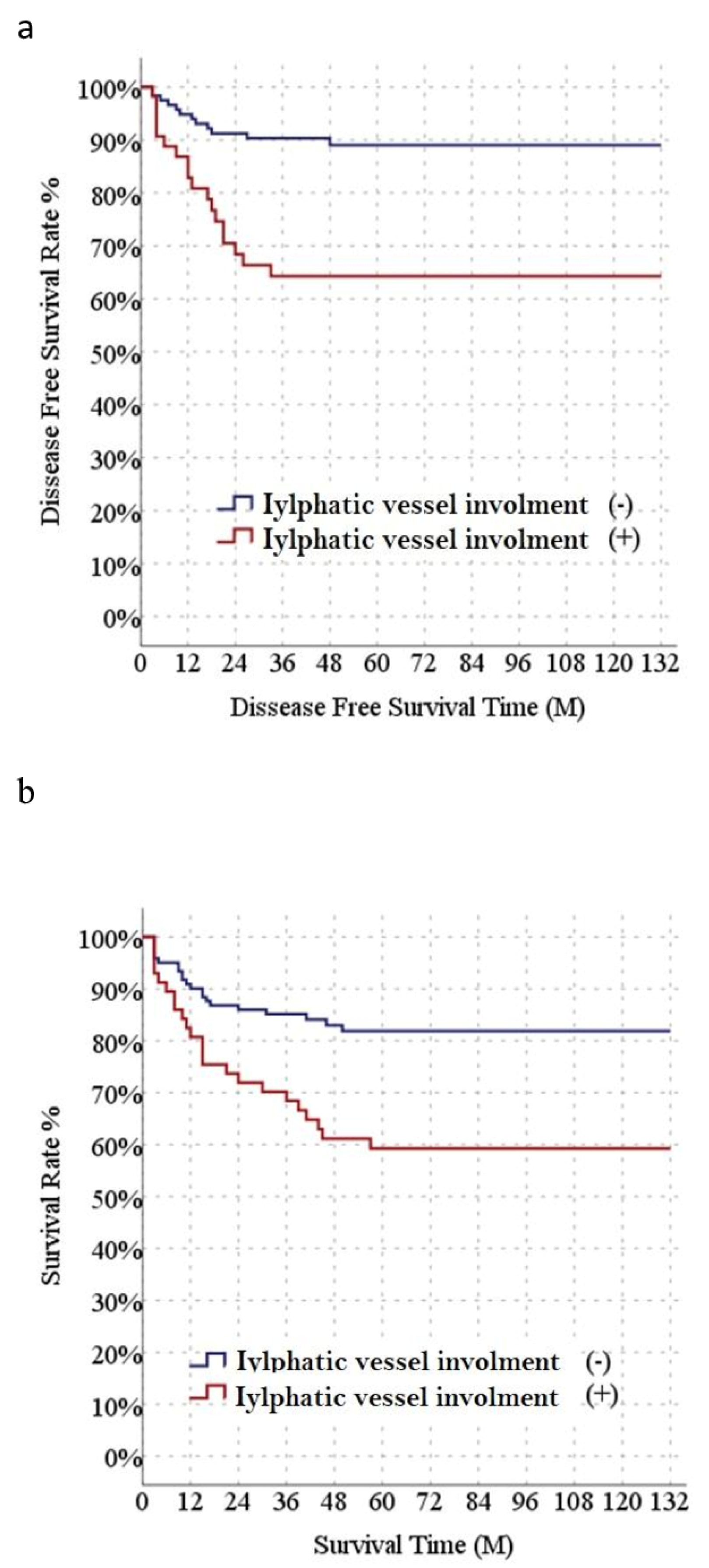
| lymphatic İnvasion | 3.76 | 1.81 | - | 7.81 | 0.000 | 2.39 | 1.11 | - | 5.13 | 0.026 |
| Overall Survival | Univariate Model | Multivariate Model | ||||||||
| HR | % 95 GA | p | HR | % 95 GA | p | |||||
| Age | 0.99 | 0.95 | - | 1.02 | 0.460 | |||||
| Gender | 1.05 | 0.47 | - | 2.35 | 0.908 | |||||
| BMI | 1.01 | 0.93 | - | 1.10 | 0.731 | |||||
| Meld | 1.00 | 0.96 | - | 1.06 | 0.855 | |||||
| AFP | 1.00 | 1.00 | - | 1.00 | 0.812 | |||||
| Tumor Size (mm) | 1.02 | 1.01 | - | 1.03 | 0.003 | 1.01 | 1.00 | 1.03 | 0.021 | |
| Number Of HCC Lesions | 1.10 | 1.01 | - | 1.20 | 0.022 | |||||
| Child–Pugh Score | 1.41 | 0.88 | - | 2.26 | 0.154 | |||||
| Locoregional Treatment | 1.27 | 0.69 | - | 2.35 | 0.444 | |||||
| Tumor Differentiation grade | 0.76 | 0.50 | - | 1.16 | 0.202 | |||||
| Microvascular Invasion | 1.93 | 1.06 | - | 3.51 | 0.031 | |||||
| Diabetes Mellitus | 0.77 | 0.35 | - | 1.74 | 0.535 | |||||
| Lymphatic Invasion | 2.50 | 1.39 | - | 4.52 | 0.002 | 2.19 | 1.20 | 4.01 | 0.011 | |
| Cox Regression | ||||||||||
| Lymphatic Invasion (No) | Lymphatic Invasion (Yes) | p | |||||||||
|---|---|---|---|---|---|---|---|---|---|---|---|
| Mean ± sd/n% | Median | Mean ± sd/n% | Median | ||||||||
| Age | 57.6 | ± | 8.7 | 59.0 | 56.3 | ± | 10.4 | 60.0 | 0.722 | m | |
| Gender | Female | 13 | 14.0% | 7 | 22.6% | 0.259 | X2 | ||||
| Male | 80 | 86.0% | 24 | 77.4% | |||||||
| BMI | 26.2 | ± | 3.8 | 26.0 | 26.9 | ± | 3.6 | 26.0 | 0.338 | m | |
| Meld | 12.8 | ± | 6.3 | 12.0 | 14.4 | ± | 6.2 | 13.0 | 0.131 | m | |
| AFP (ng/mL) | 33.0 | ± | 61.4 | 7.5 | 154.2 | ± | 396.6 | 16.0 | 0.009 | m | |
| Tumor Size (mm) | 31.0 | ± | 20.4 | 25.0 | 37.5 | ± | 24.5 | 32.0 | 0.192 | m | |
| Number Of HCC Lesions | 2.0 | ± | 1.7 | 1.0 | 3.5 | ± | 2.8 | 3.0 | 0.002 | m | |
| Child–Pugh Score | A | 41 | 44.1% | 8 | 25.8% | 0.036 | X2 | ||||
| B | 48 | 51.6% | 18 | 58.1% | |||||||
| C | 4 | 4.3% | 5 | 16.1% | |||||||
| Tumor Differentiation Grade | well | 14 | 15.1% | 8 | 25.8% | 0.025 | X2 | ||||
| moderate | 43 | 46.2% | 19 | 61.3% | |||||||
| advanced | 36 | 38.7% | 4 | 12.9% | |||||||
| Microvascular Invasion | (no) | 69 | 74.2% | 11 | 35.5% | 0.000 | X2 | ||||
| (yes) | 24 | 25.8% | 20 | 64.5% | |||||||
| Diabetes Mellitus | (no) | 73 | 78.5% | 25 | 80.6% | 0.799 | X2 | ||||
| (yes) | 20 | 21.5% | 6 | 19.4% | |||||||
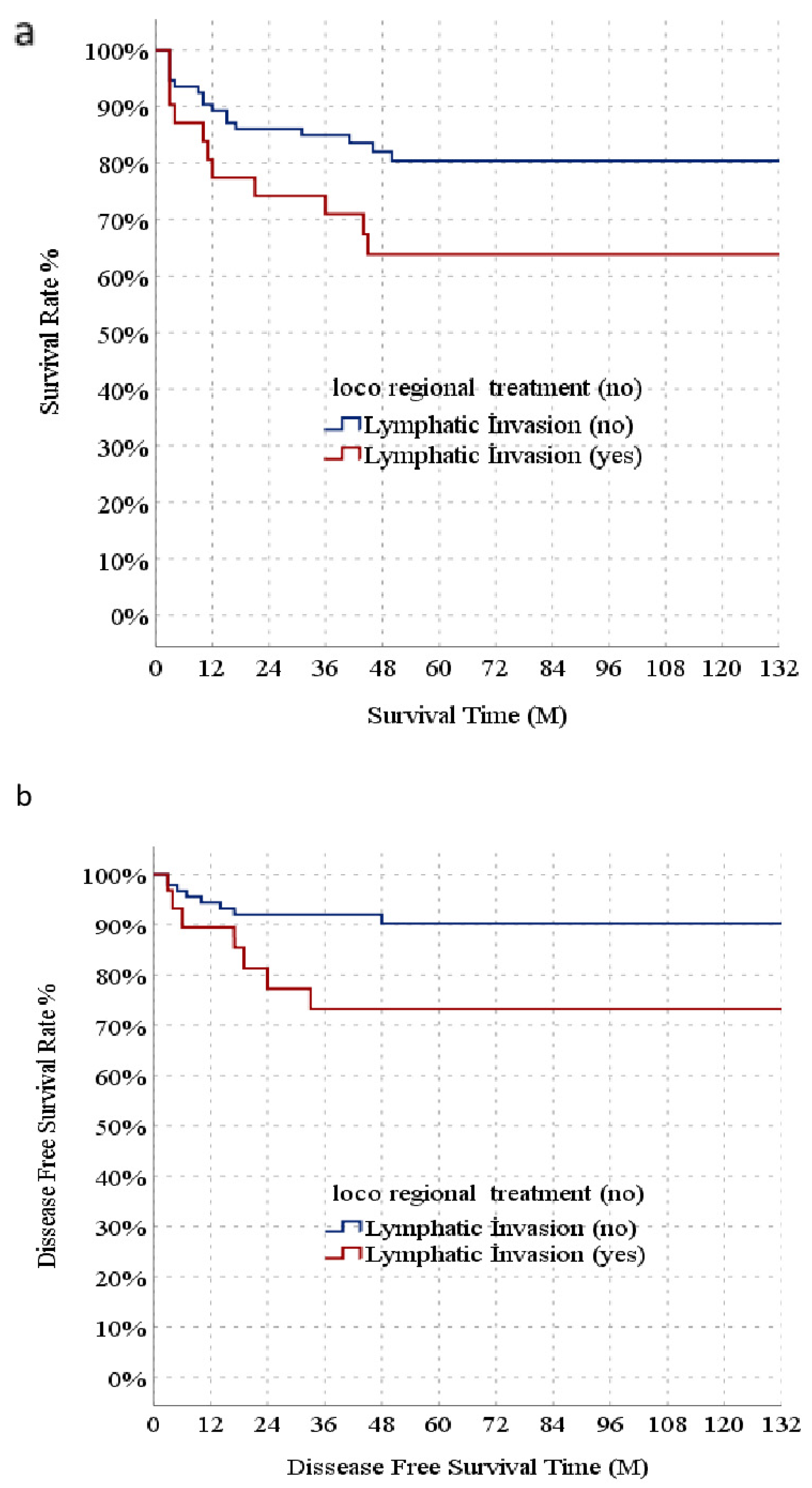
| Tumor recurrence | (no) | 85 | 91.4% | 24 | 77.4% | 0.039 | X2 | ||||
| (yes) | 8 | 8.6% | 7 | 22.6% | |||||||
| Follow-Up Time (month) | 57.5 | ± | 32.4 | 55.0 | 61.7 | ± | 39.7 | 72.0 | 0.596 | t | |
| Mortality | (no) | 76 | 81.7% | 20 | 64.5% | 0.047 | X2 | ||||
| (yes) | 17 | 18.3% | 11 | 35.5% | |||||||
Disclaimer/Publisher’s Note: The statements, opinions and data contained in all publications are solely those of the individual author(s) and contributor(s) and not of MDPI and/or the editor(s). MDPI and/or the editor(s) disclaim responsibility for any injury to people or property resulting from any ideas, methods, instructions or products referred to in the content. |
© 2026 by the authors. Licensee MDPI, Basel, Switzerland. This article is an open access article distributed under the terms and conditions of the Creative Commons Attribution (CC BY) license.
Share and Cite
Tüysüz, U.; Batı, İ.B.; Yılmaz, T.U. Effect of Lymphatic Invasion on Survival and Recurrence After Liver Transplantation in Patients with Hepatocellular Carcinoma and Its Prognostic Significance. Diagnostics 2026, 16, 741. https://doi.org/10.3390/diagnostics16050741
Tüysüz U, Batı İB, Yılmaz TU. Effect of Lymphatic Invasion on Survival and Recurrence After Liver Transplantation in Patients with Hepatocellular Carcinoma and Its Prognostic Significance. Diagnostics. 2026; 16(5):741. https://doi.org/10.3390/diagnostics16050741
Chicago/Turabian StyleTüysüz, Umut, İmam Bakır Batı, and Tonguc Utku Yılmaz. 2026. "Effect of Lymphatic Invasion on Survival and Recurrence After Liver Transplantation in Patients with Hepatocellular Carcinoma and Its Prognostic Significance" Diagnostics 16, no. 5: 741. https://doi.org/10.3390/diagnostics16050741
APA StyleTüysüz, U., Batı, İ. B., & Yılmaz, T. U. (2026). Effect of Lymphatic Invasion on Survival and Recurrence After Liver Transplantation in Patients with Hepatocellular Carcinoma and Its Prognostic Significance. Diagnostics, 16(5), 741. https://doi.org/10.3390/diagnostics16050741