Article

Three-Dimensional Evaluation of TMJ Morphology in Individuals with Maxillary or Mandibular Impacted Canines: A CBCT-Based Retrospective Study


Department of Orthodontics, Faculty of Dentistry, İnönü University, Malatya 44280, Türkiye
*
Author to whom correspondence should be addressed.
Diagnostics 2026, 16(3), 496; https://doi.org/10.3390/diagnostics16030496

Submission received: 21 December 2025 / Revised: 2 February 2026 / Accepted: 4 February 2026 / Published: 6 February 2026
(This article belongs to the Section Medical Imaging and Theranostics)

Abstract

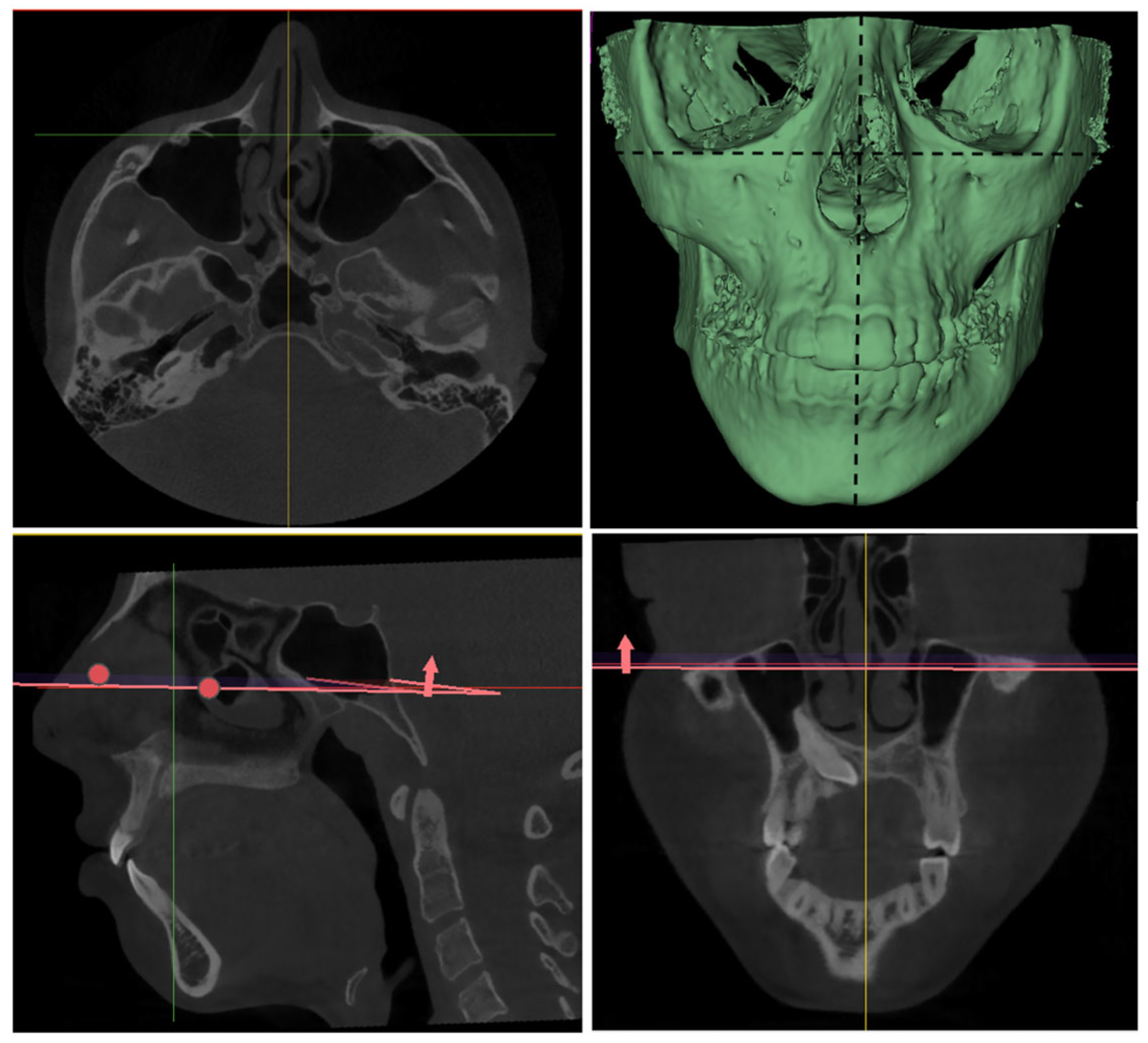
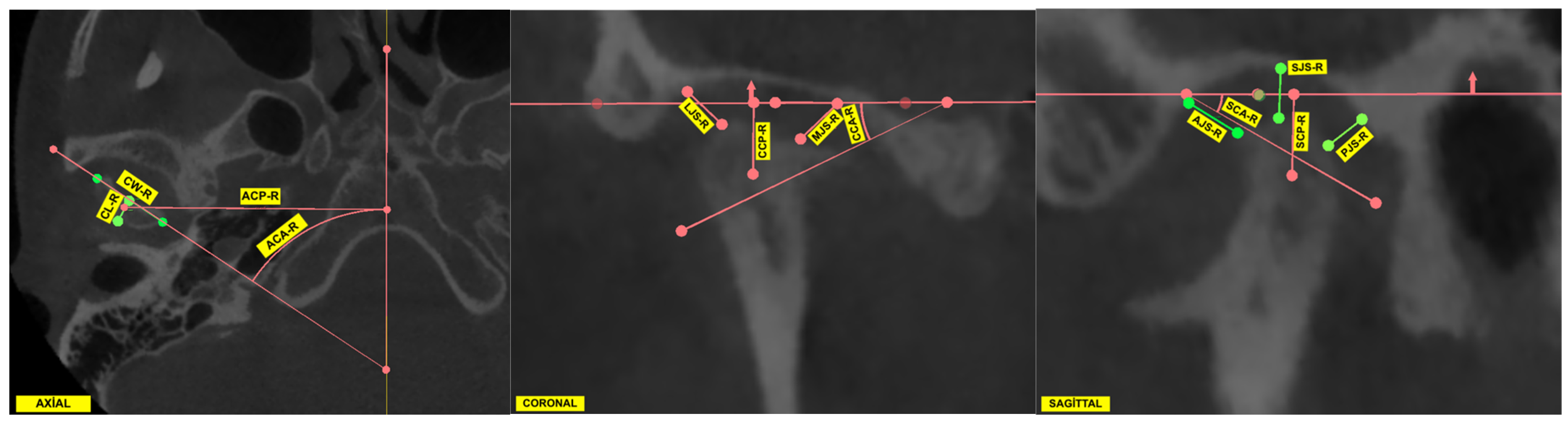
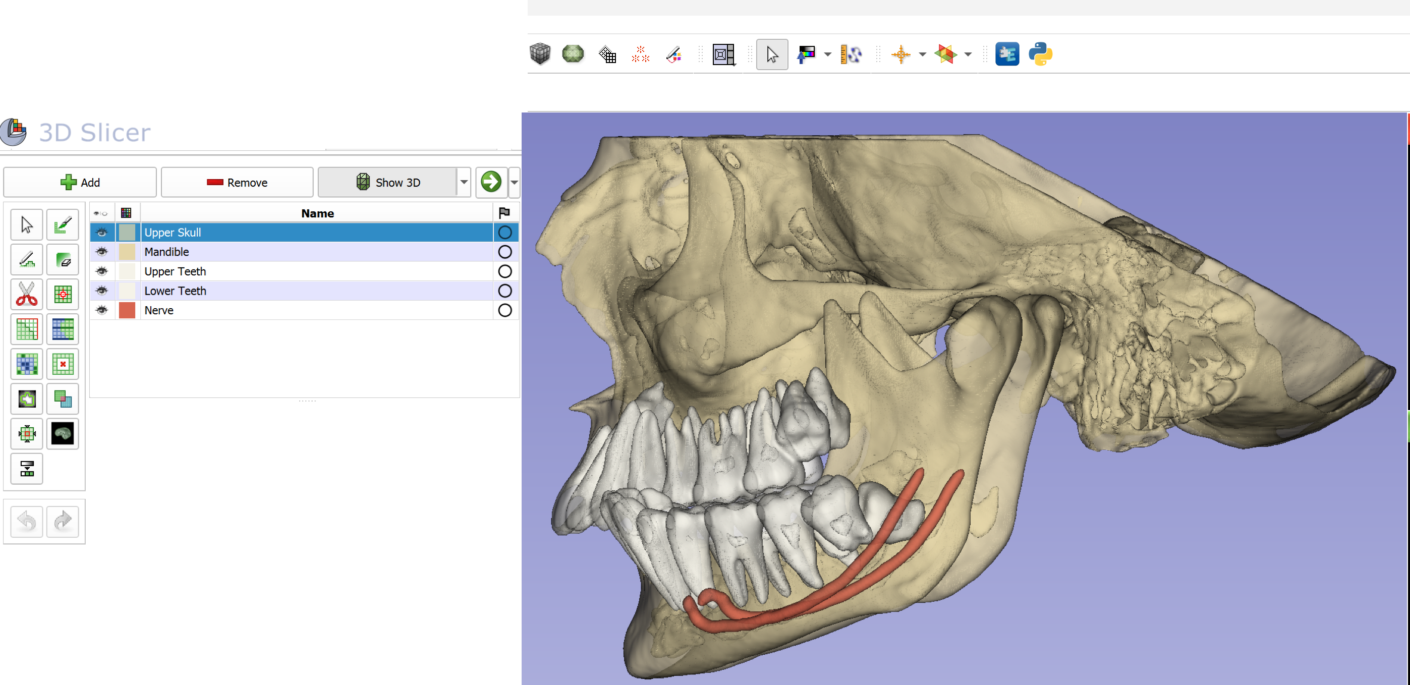
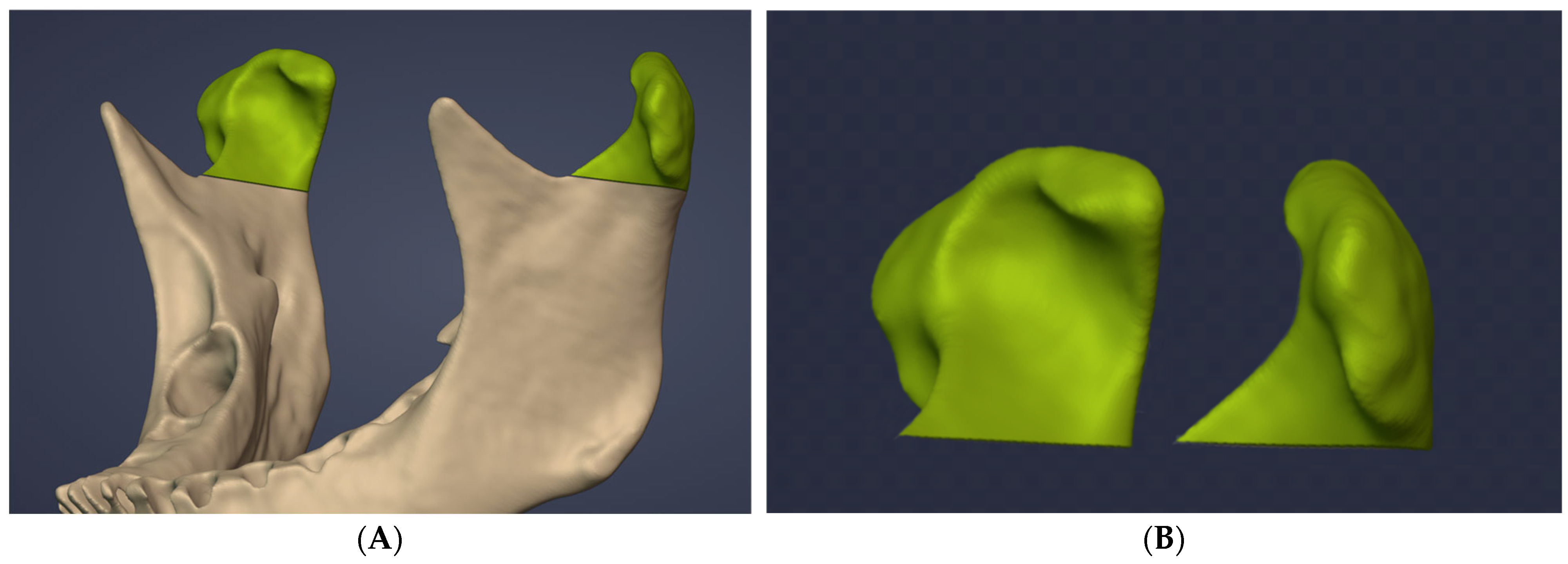
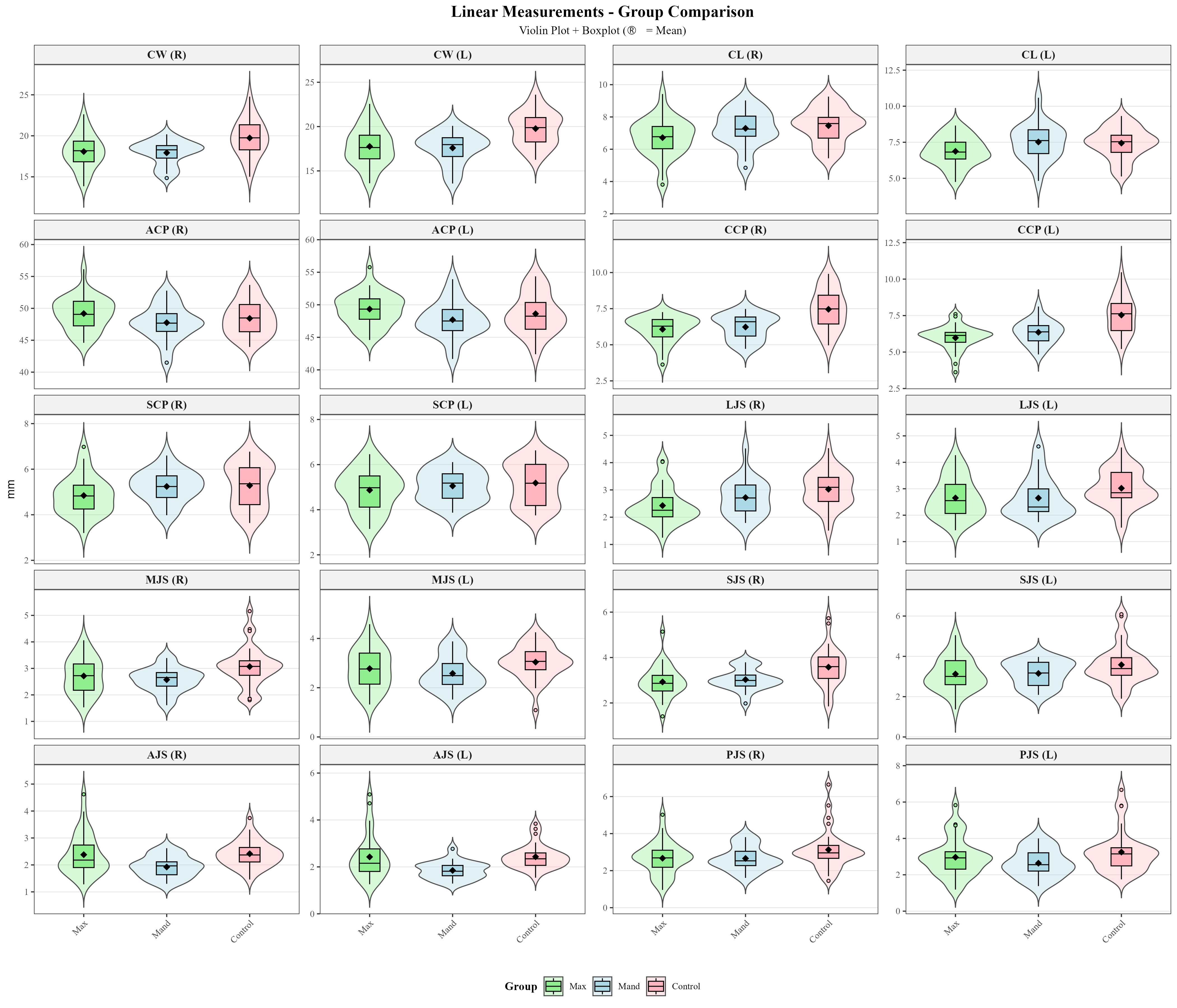
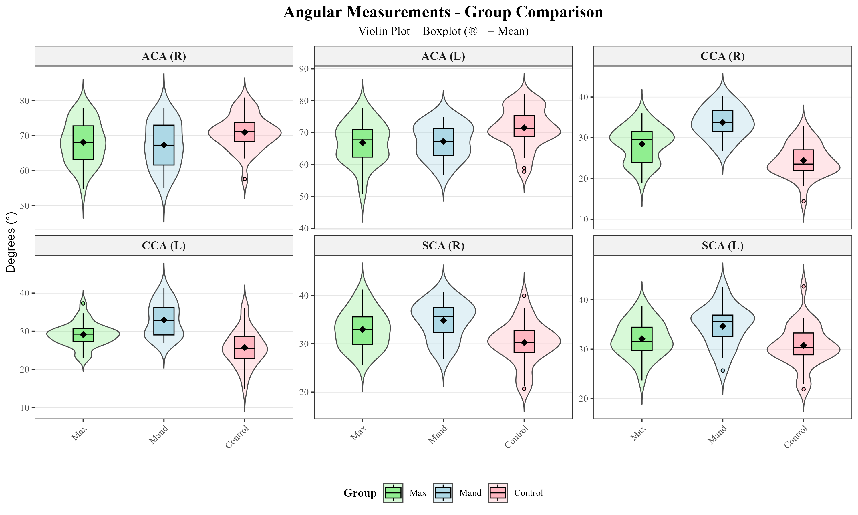
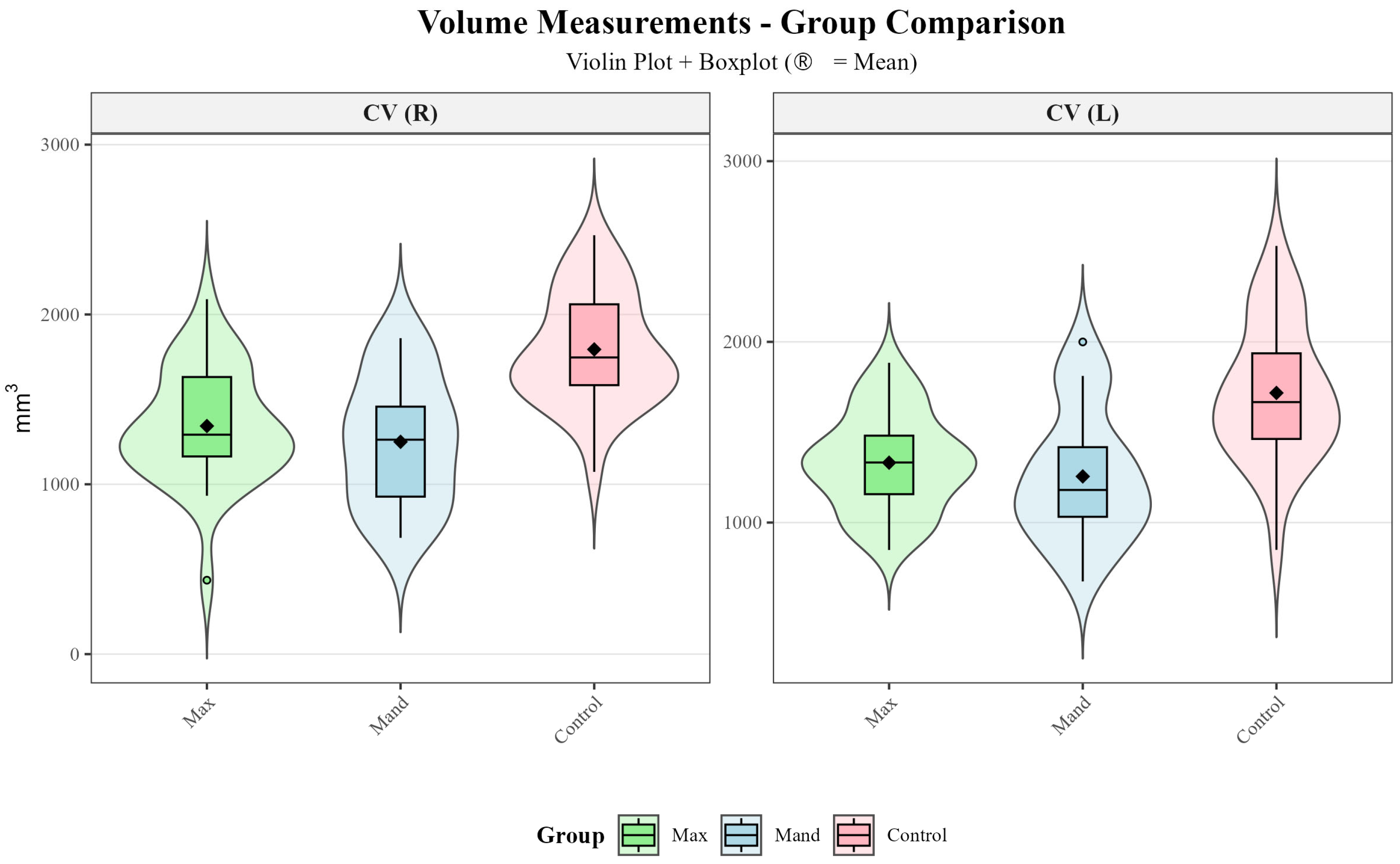
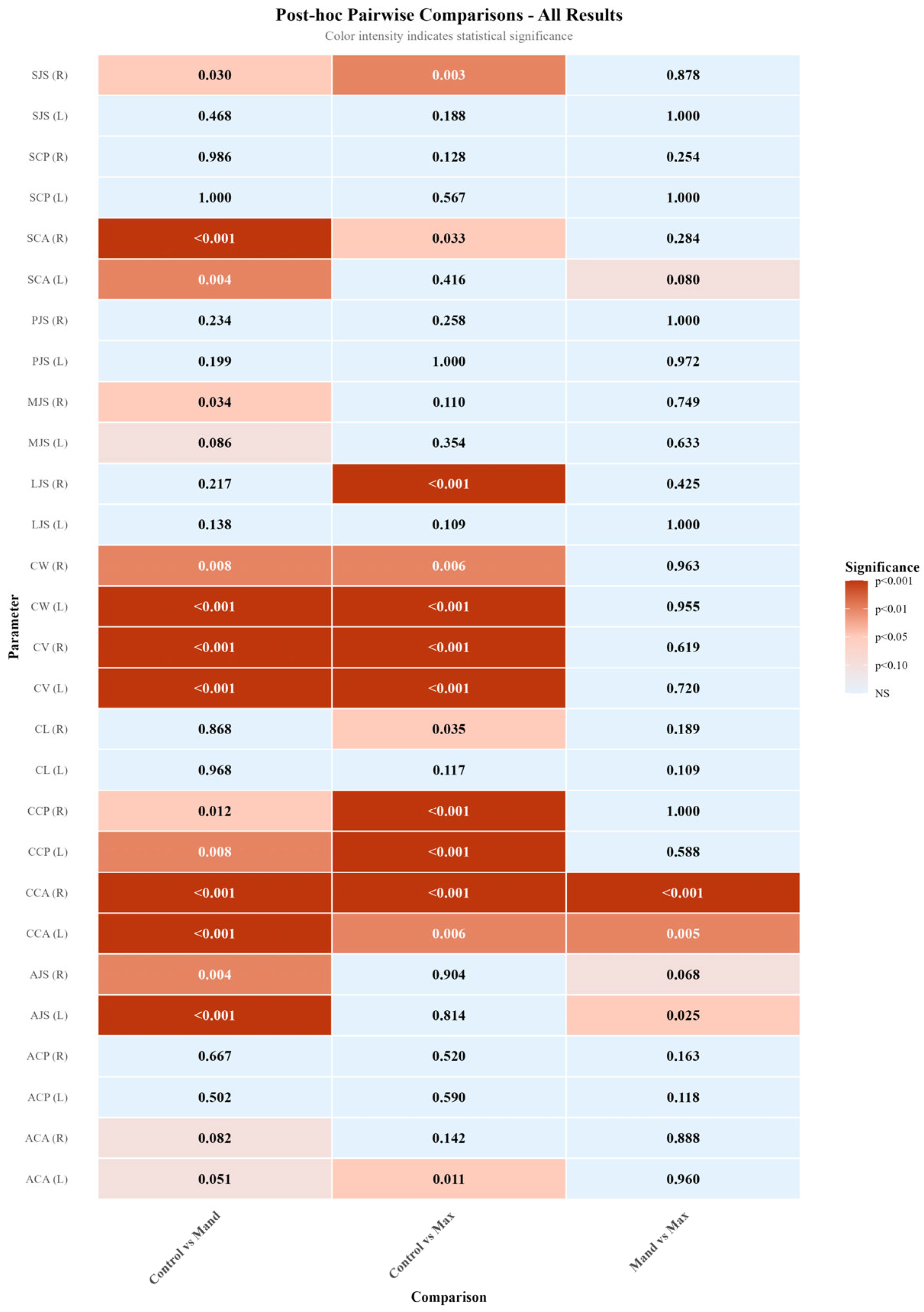
Background/Objectives: This study aimed to evaluate temporomandibular joint (TMJ) morphology in individuals with impacted maxillary and mandibular canine teeth using cone beam computed tomography (CBCT) and to compare the findings with those of a control group without impacted canines. Methods: A total of 80 individuals were included in this retrospective study. Based on CBCT images, participants were divided into three groups: the impacted maxillary canine group (n = 30), impacted mandibular canine group (n = 20), and control group (n = 30). CBCT images were oriented in the 3D Slicer software according to the Frankfurt Horizontal plane and the midsagittal reference line. Condylar width, length, position, angular parameters, joint spaces, and condylar volume were measured. Appropriate parametric and non-parametric statistical tests were used for intergroup comparisons. Results: The control group exhibited significantly higher values of condylar width, coronal condylar position and angle, certain joint spaces, and condylar volume compared with both impacted maxillary and mandibular canine groups (p < 0.05). In particular, significant differences were observed for condylar width (p ≤ 0.002) (Control: 19.76 ± 2.09 mm, Maxillary: 17.92 ± 2.14 mm, Mandibular: 17.76 ± 1.64 mm), coronal condylar position (p < 0.001) (Control: 7.50 ± 1.34 mm, Maxillary: 6.02 ± 0.89 mm, Mandibular: 6.30 ± 0.83 mm), coronal condylar angle (p < 0.001) (Control: 25.09° ± 4.40, Maxillary: 28.80° ± 3.70, Mandibular: 33.37° ± 4.10), and condylar volume (p < 0.001) (Control: 1755.87 ± 357.32 mm3, Maxillary: 1337.18 ± 302.65 mm3, Mandibular: 1252.71 ± 369.24 mm3). No significant differences were found between the impacted maxillary and mandibular canine groups for most parameters (p > 0.05). Right–left side comparisons demonstrated that bilateral symmetry was largely preserved, except for condylar volume (p > 0.05). Conclusions: The presence of impacted canines may influence TMJ morphology, particularly at the level of condylar morphometry and joint spaces. Therefore, considering TMJ morphology in addition to local dental factors when evaluating impacted canines may provide a more comprehensive approach to orthodontic diagnosis and treatment planning.
Keywords: TMJ; impacted maxillary canine; impacted mandibular canine; CBCT TMJ; impacted maxillary canine; impacted mandibular canine; CBCT